Associate editor: B. Teicher
Targeting DNA damage response: Threshold, chromatin landscape and beyond
Stefania Gon
floni
⁎
Department of Biology, University of Rome“Tor Vergata”, via della Ricerca Scientifica, I-00133 Rome, Italy
a b s t r a c t
a r t i c l e i n f o
Keywords: DNA damage Histone modifications DNA repair γ-H2AX Chromatin Tip60 c-Abl ATMCells are continually exposed to DNA assaults from exogenous and endogenous sources. To maintain genomic integrity, cells have evolved a highly conserved mechanism for repairing DNA lesions and, in particular, DNA double strand breaks (DSBs). Emerging evidence indicates that DNA repair/signaling machinery acts in an in-tegrated fashion with chromatin structure at damaged sites. This review focuses on the interplay between histone modifications and the chromatin-mediated response to DNA damage.
© 2013 Elsevier Inc. All rights reserved.
Contents
1. Introduction . . . 46
2. When does DNA damage occur? . . . 47
3. How do cells respond to DNA insults? . . . 47
4. Stepwise response induced by DNA lesions . . . 47
5. Dynamics of histone modifications influences chromatin structure . . . 48
6. Timing and threshold for DDR . . . 48
7. Connections between DNA damage signaling and chromatin landscape . . . 49
8. Concluding remarks . . . 50
Conflict of interest . . . 50
Acknowledgments . . . 50
References . . . 50
1. Introduction
Maintaining genomic integrity in response to DNA assaults is an
essential process for living cells (
Jackson & Bartek, 2009; Ciccia &
Elledge, 2010; Negrini et al., 2010
). To achieve this goal, cells use
dynamic signaling networks that can sense, interpret and respond
to various DNA stressors (
Bekker-Jensen et al., 2010
). Signal duration
and amplitude of stresses evoke very complex patterns of protein
–
protein interactions. Canonical DNA damage kinases trigger several
posttranslational modi
fications of DNA signaling proteins (
Al-Hakim
et al., 2010; Bensimon et al., 2011
). These modi
fications show a
dynamic behavior where signals of phosphorylation are interpreted
through ubiquitin- (or Sumo-) mediated signal decoding (
van Attikum
& Gasser, 2005; Ramaekers & Wouters, 2011
). The consequence is a
timely recruitment (and disassembly) of large complexes near the
dam-aged site (
Panier & Durocher, 2009; van Attikum & Gasser, 2009; Ulrich &
Walden, 2010
). The
first modification induced by DNA damage
affects the histone variant H2AX on S139 to form
γ-H2AX.
S139-Abbreviations: DSBs, double strand breaks;γ-H2AX, phosphorylation of histone variant H2AX in Ser139; SUMO, small ubiquitin modifier; ROS, reactive oxygen species; IR, ionizing radiation; Tip60, Tat-interactive 60; HAT, histone acetyltransferase; HDAC, histone deacetylase; NHEJ, non homologous end joining; HR, homologous recombination; DDR, DNA Damage response; NER, nucleotide excision repair; BER, base excision repair; MMR, mismatch repair; HP1, heteochromatin Protein1; INO80, inositol requiring 80; SWR1, sick with Rat8 ts; FHA, forkhead-associated; BRCT, breast cancer-terminal; UBD, ubiquitin bind-ing domain; SIM, sumo interactbind-ing motif; MDC1, mediator of DNA damage checkpoint 1; BRCA1, breast cancer 1, early onset; FACT, facilitates chromatin transcription; SIRT1, sirtuin (silent mating type information regulation 2 homolog)1 deacetylase; WSTF, Williams– Beuren syndrome transcription factor tyrosine kinase; EYA, eyes absent protein phosphatase; MOF, males absent on thefirst; ATM, ataxia telangiectasia; MRN, Mre11/ Nbs1/Rad50 complex; RNF8, RINGfinger protein 8; UBC13, ubiquitin-conjugating enzyme E2 13.
⁎ Tel.: +39 06 72594319; fax: +39 06 2023500. E-mail address:Stefania.Gonfl[email protected].
0163-7258/$– see front matter © 2013 Elsevier Inc. All rights reserved.
http://dx.doi.org/10.1016/j.pharmthera.2012.12.006
Contents lists available at
SciVerse ScienceDirect
Pharmacology & Therapeutics
phosphorylation induces a massive accumulation of proteins in
γ-H2AX
foci (
Stucki et al., 2005
). The latter are microscopically visible aggregates,
present in large segments of chromatin
flanking the lesions, where the
balance of opposing enzymes drives targeted recruitment,
protein-protein interactions and posttranslational modi
fications (
Huen et al.,
2007; Kolas et al., 2007; Mailand et al., 2007; Wang & Elledge, 2007;
Doil et al., 2009; Stewart et al., 2009
). Most of these enzymes are highly
connected
“hub” proteins interacting in complex regulatory circuits that
allow temporary local clustering and reversibility of the interactions
(
Maiani et al., 2011
). A second layer of complexity of DNA damage
signaling relies on its profound impact on chromatin status (
Gasch
et al., 2001; Rieger & Chu, 2004; Reinhardt et al., 2011
). At
first
glance, the DNA damage response induces a global repression of
transcription (
Vichi et al., 1997; Svejstrup, 2002
) followed by a delayed
transcriptional response, prolonging cell cycle arrest (
Fei & El-Deiry,
2003; Elkon et al., 2005
). In recent years, posttranscriptional control
circuits are emerging as a third level of regulation of DDR signaling
networks, recently reviewed by Boucas and co-workers (
Boucas et al.,
2012
). They involve either RNA-binding proteins (RBPs) or non-coding
RNAs, each of them impacting on protein biosynthesis (
Fan et al., 2002;
Matsuoka et al., 2007; Paulsen et al., 2009; Francia et al., 2012
).
Chromatin structure and histone modi
fications are actively
interconnected elements controlling the mechanisms underlying
genome-integrity maintenance. While it appears intuitive that
chro-matin compaction protects DNA from lesions, chrochro-matin state plays
a central role for local signaling at the break points. For instance,
“open” relaxed chromatin domains influence loading of DNA repair
proteins onto chromatin near the site of the lesion (
Xu & Price,
2011
). Remodeling protein complexes (ATPase and Tip60 acetyl
transferase HAT) can modulate chromatin accessibility and in
combi-nation with histone modi
fications promote subsequent chromatin
ubiquitination (
Lukas, 2010; Shanbhag et al., 2010
). Thus, the
dynam-ic landscape of chromatin, through a sophistdynam-icated combination of
posttranslational modi
fications, may directly influence the choice of
a speci
fic DNA repair pathway adopted by the cells (
Xu & Price,
2011; Chapman et al., 2012; Soria et al., 2012
). In this review, I will
focus speci
fically on the interplay between histone modifications,
centered on H2AX phosphorylation, and the chromatin-mediated
re-sponse to DNA damage.
2. When does DNA damage occur?
The major endogenous sources of DNA damage are reactive oxygen
species (ROS) or unrepaired DNA lesions causing replication fork
collapse in the cell (
Ward & Chen, 2001; Zou & Elledge, 2003;
Kryston et al., 2011; De Zio et al., 2012
). DNA breaks also arise
follow-ing treatment with exogenous genotoxic agents or ionizfollow-ing radiation
(IR) (
Roos & Kaina, 2012
). To counteract these different types of lesions
cells explore multiple DNA repair pathways (
Aziz et al., 2012
). One
type of lesion, the DNA Double strand break (DSB) is particularly
dangerous for cells, as free DNA ends created by the lesion are
susceptible to degradation or re-ligation, and promote genomic
insta-bility. DSBs can be generated by collapse of replication fork, ionizing
ra-diation IR, exposure of speci
fic compounds, and even during the
processing of other lesions (
Aziz et al., 2012
). Under normal
circum-stances, DSBs are even programmed by the cell: in germ cells during
meiotic repair (
Keeney & Neale, 2006
), or in lymphocytes during
rearrangement of immunoglobulin genes (
Dudley et al., 2005
). DSBs
can be repaired by two major pathways, homologous recombination
(HR) and non homologous end-joining (NHEJ). In NHEJ, the broken
ends are directly ligated in an error-prone manner (
Lieber, 2010
),
fre-quently causing small insertions, deletions or substitutions at the
break site. On the contrary, HR requires DNA end processing and a
tem-plate (homologous DNA molecule) for error-free copying and repairing
of the lesion.
3. How do cells respond to DNA insults?
Many studies done in the past on DDR have de
fined a hierarchical
order among the different players, those, depending on their
subcel-lular localization, are described as sensors, transducers, mediators
and effectors (
Bekker-Jensen et al., 2010
). Sensors promptly respond
to signals and are directly bound to chromatin, whereas transducers,
mediators and effectors guide the DNA damage response events
(
Jackson & Bartek, 2009; Ciccia & Elledge, 2010; Negrini et al.,
2010
). Intuitively, a simple activation and regulation of this cascade
occurs when DNA damage is not extensive and can be promptly
repaired. When damage is prolonged, the balance of multi-layered
connected pathways for DNA repairing and cell cycle arrest or
initiat-ing apoptosis (or senescence), leads cells to a decision point between
survival or death (
Ciccia & Elledge, 2010
). DNA repair is essential for
cell viability and normal growth, while irreparable damage de
finitely
leads to a programmed cell death. To counter this, cells explore
sever-al different mechanisms for repairing DNA breaks. According to the
type of damage, they use nucleotide excision repair (NER), base
exci-sion repair (BER), mismatch repair (MMR). In addition, to repair the
most dangerous lesions as DNA double strand breaks (DSB), cells
can use two distinct pathways the (non-homologous end joining)
NHEJ or HR (homologous recombination) repair systems (
Aziz et al.,
2012
). Both compete one with each other and the choice between
them is linked to the cell cycle phase and to the accumulation of
speci
fic markers. Many studies in the past have indicated that NHEJ
pathway mainly occurs during G1-early S phase, even if it can be
used during the whole cell cycle. On the contrary, HR repair is only
used in late S-G2 phase (
Takata et al., 1998; Shrivastav et al., 2008
).
4. Stepwise response induced by DNA lesions
Emerging evidence suggests an active role of chromatin in DNA
damage response (
Bao, 2011; Luijsterburg & van Attikum, 2011;
Lukas et al., 2011; Xu & Price, 2011; Miller & Jackson, 2012; Soria et
al., 2012
). Chromatin is a complex scaffold that compacts and
orga-nizes DNA in eukaryotic cells. Chromatin structure relies on a basic
unit, the nucleosome. The nucleosome is formed by 146 base pair of
DNA wrapped around a core composed by four different histones
H2A, H2B, H3 and H4 (
Campos & Reinberg, 2009
). The central core
of the nucleosome is formed by two H3-H4 dimers, surrounded by
two H2A
–H2B dimers (
Campos & Reinberg, 2009
). Chromatin
structure is quite dynamic and can be modi
fied through different
mechanisms. Diverse classes of enzymes can modulate chromatin
com-paction. One class consists of large multi-protein complexes that need
the energy of ATP hydrolysis to slide the nucleosomes or alter/
exchange histone composition within the chromatin
fibers. Another
class of enzymes mediates covalent modi
fications of histone tails.
Histone tails extend outward from the nucleosome and contain sites
for regulatory modi
fications such as phosphorylation, ubiquitylation,
methylation and acetylation. Histone modi
fications regulate chromatin
functions. For instance, Lysine methylation (K4) of N-terminal tail of
histone H3 (H3K4me) leads to gene activation and euchromatin
forma-tion (
Margueron & Reinberg, 2010
). On the contrary, methylation
of histone H3 on K9 (H3K9me) creates an interaction motif for HP1
(Heterochromatin Protein 1) proteins, and promotes the formation of
heterochromatin, leading to gene silencing (
Bannister et al., 2001;
Grewal & Jia, 2007
). Changes in histone modi
fication, implicated in
the switch from euchromatin to heterochromatin, are regulated by
pair-opposing enzymes that allow the dynamic rewriting of histone
marks as histone acetyl transferases (HATs) and histone deacetylases
(HDACs), HATs/HDACs, and (histone methyltransferase (HTMs) and
histone demethylases (HDMs) HTMs/HDMs, also by DNA
methyl-transferase (
Gallinari et al., 2007
). Histone modi
fications, when present
in speci
fic combinations with other histone marks, can form a specific
‘code’ for recruiting some important effectors required for signaling
ampli
fication and/or chromatin remodeling (
Downs et al., 2004;
Morrison et al., 2004; van Attikum et al., 2004
). Often, some histone
marks compete one with each other or even for the same residue. This
is particularly relevant for lysine residue, which can be alternatively
modi
fied by ubiquitylation, sumoylation, acetylation or methylation.
Posttranslational modi
fications of histone by changing the charge of
amino acid residue can alter the stability of the interaction with DNA
or with other charged protein interfaces (
Cairns, 2005; Kouzarides,
2007; Shahbazian & Grunstein, 2007; Campos & Reinberg, 2009;
Suganuma & Workman, 2011
).
In addition, histone modi
fications alter nucleosome composition,
promoting the release of phospho-H2A histone from damaged
chro-matin (
Ikura et al., 2007
), through a histone variant exchange event.
This event is regulated by an opposing action of two INO80 and
SWR1 remodelers (
Papamichos-Chronakis et al., 2006
). Interestingly,
enrichment of certain histone variants (e.g. H2AX) localizes sparse
speci
fic domains within chromatin fiber. Thus, differential expression
of histone variants throughout the cell cycle and their respective
positioning/localization onto chromatin (
Talbert & Henikoff, 2010;
Boyarchuk et al., 2011; Szenker et al., 2011
) critically in
fluence DNA
damage response. In addition, histone chaperones (in synergy with
ATP-dependent remodeling factors) (
Clapier & Cairns, 2009
) regulate
the exchange with free unlabeled histones acting as
“erasers” for
speci
fic posttranslational histone modifications. In a wide sense,
histone variants (with their posttranslational labels) could be
consid-ered a
“removable/temporary” platform for assembling local
signal-ing circuitry onto chromatin. The amplitude and duration of such
circuitry is likely regulated by feedback control and contribute to
the
fine-tuning of the DDR and to the maintenance of genome
integrity.
5. Dynamics of histone modi
fications influences chromatin structure
Chromatin packaging is variable, typically distinguished in two
states: a condensed (heterochromatin) and a more open (euchromatin)
structure. Euchromatin is an accessible, gene rich and transcriptionally
active region. Histones within euchromatin are highly acetylated and
H3 is methylated on K4 and K36 (H3K4me and H3K36me2) (
Barski et
al., 2007; Guenther et al., 2007
). On the contrary, heterochromatin has
low gene density and instead contains many repetitive sequences
(
de Wit et al., 2007; Peng & Karpen, 2008
). Histones within
heterochro-matin are poorly acetylated and H3 is highly methylated on K9 and K36
(H3K9me3; H3K36me3) (
Pokholok et al., 2005; Vakoc et al., 2006;
Guenther et al., 2007; Peng & Karpen, 2008
). Posttranslational modi
fica-tions of histones (PTM) can act as binding motif for proteins containing
PTM-binding domain (
Yun et al., 2011
). For instance, bromodomains
bind speci
fically to acetylated lysine residues, whereas chromodomains
recognize methylated lysine residues (
Kouzarides, 2007; Ruthenburg et
al., 2007; Shahbazian & Grunstein, 2007
). Other important PTM-binding
domains present in DDR proteins include FHA or BRCT (breast cancer1,
early-onset) domains (
Mohammad & Yaffe, 2009
) that recognize
phosphorylated epitopes on target proteins as well as UBD (ubiquitin
binding domain) domains and SIM (sumo interacting motif) motifs
that bind ubiquitin and SUMO respectively (
Kerscher et al., 2006;
Hofmann, 2009
).
The
first evidence that chromatin is modified at site of DNA break
came from the discovery that the histone variant H2AX is
phosphor-ylated on its C-terminal tail following DNA damage (
Miller &
Jackson, 2012
). Exposure of cells to DNA damaging agents activates
phosphorylation of many target proteins (
Bensimon et al., 2011
).
However, S139-phosphorylation of histone variant H2AX is
consid-ered one of the early markers of the DDR. This modi
fication
modu-lates the H2AX-interaction with DNA and promotes assembly of
signaling complexes onto chromatin. While, there is a little evidence
that
γ-HAX has been involved in DNA repair per se, it plays a central
role for the initiation and ampli
fication of DNA damage signals.
Signaling at DSBs induces
γ-H2AX, eliciting timely engagement of
MDC1 (mediator of DNA damage checkpoint 1), 53BP1 (p53 binding
protein 1) and BRCA1 (breast cancer 1, early onset). Bidirectional
spreading of
γ-H2AX far from DNA lesions further enhances damage
signaling and helps to delineate the chromatin region involved in
DDR (
Yuan et al., 2010
). Silenced chromatin regions are not permissive
for
γ-H2AX spreading (
Kim et al., 2007
) and H2AX dynamics within the
nucleosome are coupled to its various posttranslational modi
fications.
Besides phosphorylation, other modi
fications such as ubiquitylation
and acetylation of H2AX variant promote the recruitment of DDR
pro-teins near the breaks. DNA damage-dependent modi
fications (i.e. poly
ADP-rybosylation) of histone chaperone FACT (Facilitates Chromatin
Transcription) inhibit its interaction with nucleosomes reducing the
H2AX/H2A exchange (
Du et al., 2006; Heo et al., 2008
). On the contrary,
the Tip60 complex (Tat-interactive protein 60), is a chromatin modi
fier
and acetylates H2AX, enhancing its mobility within chromatin (
Ikura et
al., 2007
). Interestingly Tip60-mediated acetylation (on K5) is required
for the subsequent ubiquitination of H2AX (on K119) by RNF8/UBC13
(ubiquitin-conjugating enzymes) (see
Fig. 1
) (
Ikura et al., 2007
). In
mammalian cells, SIRT1 (a protein deacetylase) negatively regulates
Tip60-mediated acetylation of histone H2AX (
Yamagata & Kitabayashi,
2009
). Conversely, in yeast, a remodeling factor INO80 (Inositol
requir-ing 80) retains phospho-H2A within the nucleosome (
Papamichos-Chronakis et al., 2006
). In combination with remodeling factors that
promote
γ-H2AX eviction from chromatin, several protein phosphatases
can also negatively regulate the function of
γ-H2AX by promoting its
de-phosphorylation (
Nazarov et al., 2003; Chowdhury et al., 2005; Keogh et
al., 2006; Chowdhury et al., 2008
). The histone variant H2AX is also
decorated by additional modi
fications which contribute to chromatin
re-sponse to DNA damage (reviewed by
Miller & Jackson, 2012
). H2AX is
phosphorylated on its C-terminal tyrosine residue by WSTF, a non
ca-nonical tyrosine kinase (
Xiao et al., 2009
). Following DNA damage, this
phoshorylation is removed by the phosphatase EYA (
Cook et al., 2009
).
The pair-opposing enzymes WSTF or EYA are both important for an
effective DDR, pointing out the relevance of this tyrosine modi
fication
for H2AX function (
Cook et al., 2009; Xiao et al., 2009
). In sum, the
fine-tuning of H2AX modifications and its dynamics mediated by the
ef-fect of speci
fic action of pair enzymes offers a tunable switch for DNA
damage signaling events.
6. Timing and threshold for DDR
Exposure of cells to genotoxic compounds activates the
phosphatidylinositol-3-kinase-related kinase (PI3KK) family of
ki-nases (ATM, ATR and DNA-PKcs). Although the PI3K related kiki-nases
are considered important players in DDR, an unrelated tyrosine
kinase c-Abl has more recently also associated with the activation of
key upstream event of DDR (
Gon
floni, 2010a; Meltser et al., 2011;
Wang et al., 2011; Maiani et al., 2012
). In response to DNA damage,
PI3K related kinases mediate the phosphorylation of H2AX on S139
(
γ-H2AX). However, other numerous modifications (acetylation and
methylation) occur on core histones in response to DNA damage.
Two key modi
fications occur on H4, K16 acetylation and K20
methyl-ation respectively. H4K16Ac modi
fication is mediated by Tip60 and
MOF (
Murr et al., 2006; Li et al., 2010
). While Tip60 mediates H2A
and H4K16 acetylation at the site of break (
Fig. 1
), MOF seems to
con-trol global level of H4K16ac and does not localize at DSB (
Sharma et
al., 2010
). Interestingly, Tip60 can also be activated by a histone
mark (H3K9me3) associated with heterochromatin (
Sun et al.,
2009; Sun et al., 2010
). Lack of either HAT enzymes (Tip60 and
MOF) causes defective HR and NHEJ repair, suggesting that both are
required for ef
ficient DSB repair. However, the precise mechanism
by which acetylation of H4K16 promotes DNA repair still remains
elu-sive. Interestingly, a combination of histone marks such as H2b-Ub
(ubiquitilated H2B) and H4K16ac induces decompaction of
nucleo-some (
Shogren-Knaak & Peterson, 2006; Fierz et al., 2011
). Thus,
Tip60-mediated acetylation at DSB promotes instability of nucleosomes
near the site of break. This could facilitate a shift of the local chromatin
structure into an open relaxed conformation more permissive for the
ubiquitin-dependent signaling at the damage site.
Posttranslational modi
fications of histones, both in the tails and in
the core region affect the functional landscape of chromatin by
regulat-ing DNA accessibility. A key aspect of the role of histone modi
fications
relies on their dynamic nature (
Krebs, 2007
); the precise timing of
addition and removal of speci
fic marks (or entire histone) determines
a dynamic temporal regulation of chromatin functions (
Krebs, 2007
).
H2AX-phosphorylation on S139 (
γ-H2AX) is an early marker of DDR.
Several studies indicate that
γ-H2AX acts as beacon for proteins with
dedicated phosphor-S/T binding domains (FHA, BRCT), promoting a
sequential assembly of ubiquitin-dependent signaling cascades (see
Fig. 1
). Thus, DNA breaks initially promote repair and also a DNA
sig-naling cascade for assisting repair (
Yuan & Chen, 2010
). At damaged
site, the ef
ficiency of signaling is enhanced by local concentration of
factors. Signaling amplitude and duration are regulated through
dynamic editing and removal of speci
fic marks. This eventually could
arrest the repair process for an alternative path leading to cell death.
Most likely, survival of DNA-damaged cells strictly depends both on
the removal of the lesion coupled with an ef
ficient DNA damage
signal-ing decay. This aspect is particularly crucial in the cellular context of
perinatal oocytes more sensitive to genotoxic stress than somatic
cells. (
Gon
floni, 2010a; Gonfloni, 2010b; Maiani et al., 2012
). In
imma-ture oocytes, pharmacological inhibition of c-Abl tyrosine kinase
atten-uates the toxic effect induced by chemotherapeutic drugs (
Gon
floni et
al., 2009; Maiani et al., 2012
). Our studies indicate that c-Abl inhibition
works on distinct levels of DNA damage signaling both at early time
points reducing on
γ-H2AX phosphorylation and then impinging on a
downstream effector TAp63 (
Gon
floni et al., 2009; Maiani et al.,
2012
). This supports the hypothesis that ampli
fication of DNA damage
signaling cascade leads germ cells towards death, as a default path, if
not attenuated (
Maiani et al., 2011
).
7. Connections between DNA
damage signaling and chromatin landscape
Now we move to the next point, how the H2AX dynamics, regulated
by posttranslational modi
fications, in tandem with histone chaperones
and remodelers contribute to the DDR? Recent evidence supports an
active role of chromatin in DNA damage response. Chromatin
compac-tion protects DNA from lesions. Heterochromatin, compared to
euchromatin, is densely compact, transcriptionally silent and may act
as barrier limiting access to all DDR factors (
Soria et al., 2012
). However,
generation of DNA breaks and the early steps of DNA damage signaling
and repair occur ef
ficiently within the heterochromatin domains
(
Baldeyron et al., 2011; Chiolo et al., 2011; Jakob et al., 2011
). Final
steps of DNA signaling and repair (accumulation of RAD 51 and
γ-H2AX spreading) are instead relocalized and confined in more
accessible environment at the periphery of the heterochromatin region.
The expansion of the heterochromatin facilitates the repositioning of
damaged DNA near the surrounding euchromatin domains (
Chiolo et
al., 2011; Jakob et al., 2011; Baldeyron et al., 2011
) to
finalize late
steps of DNA repair (
Soria et al., 2012
). Interestingly, a similar
repositioning is observed during DNA replication (
Quivy et al., 2004
).
It is possible that cells have evolved such mechanisms to prevent
ectopic recombination between the repetitive sequences within
hetero-chromatin, by restricting the processing of DNA ends at the periphery
of heterochromatin (
Quivy et al., 2004; Peng & Karpen, 2008; Chiolo
et al., 2011
). This could in turn prevent possible chromosomal
rearrangements and genomic instability. How a sophisticated control
on DNA accessibility is linked to DNA repair? ATM is a master regulator
of DNA damage response. ATM mediates the phosphorylation of
γ-H2AX promoting a signaling cascade, which leads to the assembly
of DNA repair machinery and to activation of cell cycle checkpoints.
However, how DNA breaks upregulate the activity of ATM is not
completely clari
fied. Recent biochemical studies indicate that the
auto-phosphorylation of ATM on S1981 is not the primary mechanism for
ATM activation. This autophosphorylation is indeed dispensabile for
Fig. 1. Tip60-mediated acetylation of H2AX promotes Ubiquitin-dependent signaling at damage sites. H2AX phosphorylation by ATM provides a docking site for MCD1 and leads to the recruitment of ubiquitin ligase RNF8 and NuA4 complex at damaged sites. (NuA4 is a large complex form by Tip60, p400 motor ATPase and other subunits). Then, Tip60 mediates acetylation of histone H2AX, in combination with the action p400 ATPase, generates an open relaxed chromatin structure, facilitating ubiquitin-dependent signaling at the damage sites. Ac=acetylation, p=phosphorylation.
ATM function under some conditions (
Lee & Paull, 2005; Dupre et al.,
2006; Pellegrini et al., 2006
). Most likely ATM activation is also
mediat-ed through binding with MRN (complex formmediat-ed by mre11 nuclease,
Rad50 ATPse and Nbs1) DNA binding complex (
Uziel et al., 2003;
Di
filippantonio et al., 2005; Falck et al., 2005; Lee & Paull, 2005;
Cerosaletti et al., 2006
). Deletion of mre11, rad50 or nbs1 elements of
MRN complex signi
ficantly reduces activation of ATM following DNA
damage in vivo (
Uziel et al., 2003; Di
filippantonio et al., 2005; Falck et
al., 2005; Cerosaletti et al., 2006
). Recent studies indicate that Tip60
acetyltransferase is required for ATM activation (
Sun et al., 2005
).
Tip60 and ATM form a complex in which Tip60 interacts with highly
conserved FATC domain of ATM. This interaction in turn facilitates the
acetylation of ATM on K3016 (
Jiang et al., 2006; Sun et al., 2007
).
Tip60's chromodomain recognizes speci
fically H3K9me3 (
Sun et al.,
2009
); and the binding with H3K9me3 increases Tip60 HAT activity
through an allosteric mechanism. Mutations in the chromodomain
binding motif prevent both the interaction between Tip60 and
H3K9me3 and the upregulation of Tip60's HAT activity. The consequence
is a reduction of acetylation and activation of ATM kinase activity
medi-ated by Tip60 (
Sun et al., 2009
). Reduction of global H3K9me3 levels, by
acting on opposing enzymes either on KDM4D demethylases (
Whetstine
et al., 2006
) or Suv39 h1 and Suv39hu2 methyltransferase (
Peters et al.,
2001
), signi
ficantly decreases Tip60 activation following DNA damage
(
Sun et al., 2009
). In addition, cells with low level of H3K9me3 show
an increased sensitivity to IR and genomic instability (
Sun et al., 2009
).
Taken together, these observations suggest that the chromodomain
functions as sensor for Tip60 activation, modulating both recruitment
and HAT activity at DNA damage sites. Thus, a direct interaction between
methylated histones and Tip60's chromodomain indicates that
chroma-tin structure plays a role in DNA repair (
Sun et al., 2010
). Tip60 is stably
associated with ATM in cells, and both proteins are recruited at DSBs
(
Fig. 2
), most likely in an inactive state (
Sun et al., 2005; Jiang et al.,
2006
). Recent evidence indicates that loss of functional MRN complex
delayed the recruitment and activation of Tip60 after DNA damage
(
Sun et al., 2009
). However the requirement of both MNR complex and
Tip60 for ATM activation in vivo remains still elusive as well as the role
of other potential effectors associated in the complex. Recent evidence
indicates that Tip60 interacts with c-Abl tyrosine kinase both in vitro
and in vivo and is an upstream c-Abl modi
fier in response to DNA
dam-age. Interestingly, c-Abl acetylation mediated by Tip60 required an
ATM-mediated phosphorylation of c-Abl on S465 (
Jiang et al., 2011
).
8. Concluding remarks
In conclusion, chromatin is emerging as an integral player in the
DDR (
Soria et al., 2012
). Cells have evolved dedicated signaling and
repair machinery to control the chromatin structure facilitating
(or preventing) DNA access in a dynamic way at the site of damage
and nearby. This sophisticated machinery includes enzymes involved
in posttranslational modi
fications of histones, incorporation of
histone variants and ATP-dependent chromatin remodeling. Emerging
evidence indicates that all these three classes of components are direct
players in DNA damage response induced by DSBs acting in an
integrated fashion. Histone deacetylases (HDACs) promote chromatin
condensation and are considered promising targets for cancer therapy
because their inhibition is preferentially toxic for some cancer cells
(
Johnson et al., 2002; Minucci & Pelicci, 2006
). More sophisticated
techniques based on ChIP assay and the development of powerful
DSD-inducing systems are rapidly improving our understanding of
DSB repair processes (
Polo & Jackson, 2011
). Undoubtedly, this will
provide new hints for the development of targeted therapies for DDR
in a global integrated fashion.
Con
flict of interest
The author declares no con
flict of interest.
Acknowledgments
The author wishes to apologize to those colleagues whose signi
fi-cant studies were not cited due to space limitations. The author wishes
to thank Emiliano Maiani for assistance with the
figure, Gianni Cesareni
and Marc Diederich for support. This work is supported by funds
provided by AIRC (Associazione Italiana Ricerca sul Cancro) and Televie
(Luxembourg).
References
Al-Hakim, A., Escribano-Diaz, C., Landry, M. C., O'Donnell, L., Panier, S., Szilard, R. K., et al. (2010). The ubiquitous role of ubiquitin in the DNA damage response. DNA Repair (Amst) 9, 1229–1240.
Aziz, K., Nowsheen, S., Pantelias, G., Iliakis, G., Gorgoulis, V. G., & Georgakilas, A. G. (2012). Targeting DNA damage and repair: embracing the pharmacological era for successful cancer therapy. Pharmacol Ther 133, 334–350.
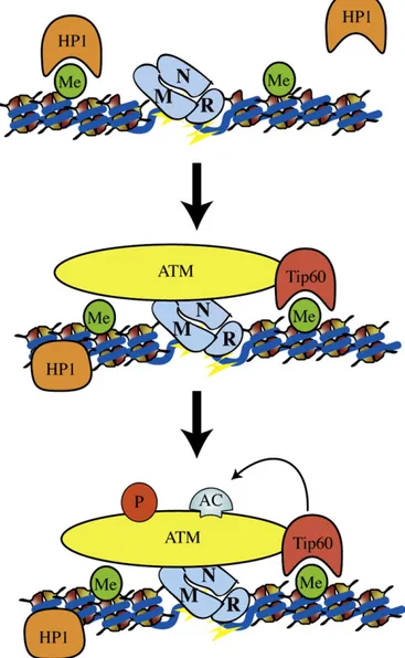
Fig. 2. Early steps of DNA damage response: a potential model for ATM activation by Tip60. Following DNA damage, MRN is recruited to DSB. In parallel, HP1 proteins are released from H3K9me3 (heterochromatin histone mark). MRN promotes targeted recruitment of the inactive ATM-Tip60 complex at DSBs. This event facilitates the interaction between the chromodomain of Tip60 and H3K9me3, enhancing Tip60's HAT activity through an allosteric mechanism. Interaction between MRN and Abl, together with acetylation of ATM mediated by Tip60, activates the kinase activity of ATM. Me = methylation, Ac = acetylation, p = phosphorylation.
Baldeyron, C., Soria, G., Roche, D., Cook, A. J., & Almouzni, G. (2011). HP1alpha recruit-ment to DNA damage by p150CAF-1 promotes homologous recombination repair. J Cell Biol 193, 81–95.
Bannister, A. J., Zegerman, P., Partridge, J. F., Miska, E. A., Thomas, J. O., Allshire, R. C., et al. (2001). Selective recognition of methylated lysine 9 on histone H3 by the HP1 chromo domain. Nature 410, 120–124.
Bao, Y. (2011). Chromatin response to DNA double-strand break damage. Epigenomics 3, 307–321.
Barski, A., Cuddapah, S., Cui, K., Roh, T. Y., Schones, D. E., Wang, Z., et al. (2007). High-resolution profiling of histone methylations in the human genome. Cell 129, 823–837.
Bekker-Jensen, S., Rendtlew Danielsen, J., Fugger, K., Gromova, I., Nerstedt, A., Lukas, C., et al. (2010). HERC2 coordinates ubiquitin-dependent assembly of DNA repair fac-tors on damaged chromosomes. Nat Cell Biol 12, 80–86 (sup pp 81–12). Bensimon, A., Aebersold, R., & Shiloh, Y. (2011). Beyond ATM: the protein kinase
land-scape of the DNA damage response. FEBS Lett 585, 1625–1639.
Boucas, J., Riabinska, A., Jokic, M., Herter-Sprie, G. S., Chen, S., Hopker, K., et al. (2012). Posttranscriptional regulation of gene expression-adding another layer of complexity to the DNA damage response. Front Genet 3, 159.
Boyarchuk, E., Montes de Oca, R., & Almouzni, G. (2011). Cell cycle dynamics of histone variants at the centromere, a model for chromosomal landmarks. Curr Opin Cell Biol 23, 266–276.
Cairns, B. R. (2005). Chromatin remodeling complexes: strength in diversity, precision through specialization. Curr Opin Genet Dev 15, 185–190.
Campos, E. I., & Reinberg, D. (2009). Histones: annotating chromatin. Annu Rev Genet 43, 559–599.
Cerosaletti, K., Wright, J., & Concannon, P. (2006). Active role for nibrin in the kinetics of ATM activation. Mol Cell Biol 26, 1691–1699.
Chapman, J. R., Sossick, A. J., Boulton, S. J., & Jackson, S. P. (2012). BRCA1-associated exclu-sion of 53BP1 from DNA damage sites underlies temporal control of DNA repair. J Cell Sci 125, 3529–3534.
Chiolo, I., Minoda, A., Colmenares, S. U., Polyzos, A., Costes, S. V., & Karpen, G. H. (2011). Double-strand breaks in heterochromatin move outside of a dynamic HP1a domain to complete recombinational repair. Cell 144, 732–744.
Chowdhury, D., Keogh, M. C., Ishii, H., Peterson, C. L., Buratowski, S., & Lieberman, J. (2005). gamma-H2AX dephosphorylation by protein phosphatase 2A facilitates DNA double-strand break repair. Mol Cell 20, 801–809.
Chowdhury, D., Xu, X., Zhong, X., Ahmed, F., Zhong, J., Liao, J., et al. (2008). A PP4-phosphatase complex dephosphorylates gamma-H2AX generated during DNA replication. Mol Cell 31, 33–46.
Ciccia, A., & Elledge, S. J. (2010). The DNA damage response: Making it safe to play with knives. Mol Cell 40, 179–204.
Clapier, C. R., & Cairns, B. R. (2009). The biology of chromatin remodeling complexes. Annu Rev Biochem 78, 273–304.
Cook, P. J., Ju, B. G., Telese, F., Wang, X., Glass, C. K., & Rosenfeld, M. G. (2009). Tyrosine dephosphorylation of H2AX modulates apoptosis and survival decisions. Nature 458, 591–596.
de Wit, E., Greil, F., & van Steensel, B. (2007). High-resolution mapping reveals links of HP1 with active and inactive chromatin components. PLoS Genet 3, e38. De Zio, D., Bordi, M., & Cecconi, F. (2012). Oxidative DNA damage in neurons:
Implication of ku in neuronal homeostasis and survival. Int J Cell Biol 2012, 752420.
Difilippantonio, S., Celeste, A., Fernandez-Capetillo, O., Chen, H. T., Reina San Martin, B., Van Laethem, F., et al. (2005). Role of Nbs1 in the activation of the ATM kinase re-vealed in humanized mouse models. Nat Cell Biol 7, 675–685.
Doil, C., Mailand, N., Bekker-Jensen, S., Menard, P., Larsen, D. H., Pepperkok, R., et al. (2009). RNF168 binds and amplifies ubiquitin conjugates on damaged chromo-somes to allow accumulation of repair proteins. Cell 136, 435–446.
Downs, J. A., Allard, S., Jobin-Robitaille, O., Javaheri, A., Auger, A., Bouchard, N., et al. (2004). Binding of chromatin-modifying activities to phosphorylated histone H2A at DNA damage sites. Mol Cell 16, 979–990.
Du, Y. C., Gu, S., Zhou, J., Wang, T., Cai, H., Macinnes, M. A., et al. (2006). The dynamic alterations of H2AX complex during DNA repair detected by a proteomic approach reveal the critical roles of Ca(2+)/calmodulin in the ionizing radiation-induced cell cycle arrest. Mol Cell Proteomics 5, 1033–1044.
Dudley, D. D., Chaudhuri, J., Bassing, C. H., & Alt, F. W. (2005). Mechanism and control of V(D)J recombination versus class switch recombination: Similarities and differ-ences. Adv Immunol 86, 43–112.
Dupre, A., Boyer-Chatenet, L., & Gautier, J. (2006). Two-step activation of ATM by DNA and the Mre11–Rad50–Nbs1 complex. Nat Struct Mol Biol 13, 451–457. Elkon, R., Rashi-Elkeles, S., Lerenthal, Y., Linhart, C., Tenne, T., Amariglio, N., et al.
(2005). Dissection of a DNA-damage-induced transcriptional network using a combination of microarrays, RNA interference and computational promoter analy-sis. Genome Biol 6, R43.
Falck, J., Coates, J., & Jackson, S. P. (2005). Conserved modes of recruitment of ATM, ATR and DNA-PKcs to sites of DNA damage. Nature 434, 605–611.
Fan, J., Yang, X., Wang, W., Wood, W. H., 3rd
, Becker, K. G., Gorospe, M., et al. (2002). Global analysis of stress-regulated mRNA turnover by using cDNA arrays. Proc Natl Acad Sci U S A 99, 10611–10616.
Fei, P., & El-Deiry, W. S. (2003). P53 and radiation responses. Oncogene 22, 5774–5783. Fierz, B., Chatterjee, C., McGinty, R. K., Bar-Dagan, M., Raleigh, D. P., Muir, T. W., et al. (2011). Histone H2B ubiquitylation disrupts local and higher-order chromatin compaction. Nat Chem Biol 7, 113–119.
Francia, S., Michelini, F., Saxena, A., Tang, D., de Hoon, M., Anelli, V., et al. (2012). Site-specific DICER and DROSHA RNA products control the DNA-damage response. Nature 488, 231–235.
Gallinari, P., Di Marco, S., Jones, P., Pallaoro, M., Steinkuhler, C., et al. (2007). HDACs, histone deacetylation and gene transcription: From molecular biology to cancer therapeutics. Cell Res 17, 195–211.
Gasch, A. P., Huang, M., Metzner, S., Botstein, D., Elledge, S. J., & Brown, P. O. (2001). Ge-nomic expression responses to DNA-damaging agents and the regulatory role of the yeast ATR homolog Mec1p. Mol Biol Cell 12, 2987–3003.
Gonfloni, S. (2010a). DNA damage stress response in germ cells: role of c-Abl and clinical implications. Oncogene 29, 6193–6202.
Gonfloni, S. (2010b). Modulating c-Abl nuclear activity as a strategy to preserve female fertility. Cell Cycle 9, 217–218.
Gonfloni, S., Di Tella, L., Caldarola, S., Cannata, S. M., Klinger, F. G., Di Bartolomeo, C., et al. (2009). Inhibition of the c-Abl-TAp63 pathway protects mouse oocytes from chemotherapy-induced death. Nat Med 15, 1179–1185.
Grewal, S. I., & Jia, S. (2007). Heterochromatin revisited. Nat Rev Genet 8, 35–46. Guenther, M. G., Levine, S. S., Boyer, L. A., Jaenisch, R., & Young, R. A. (2007). A
chroma-tin landmark and transcription initiation at most promoters in human cells. Cell 130, 77–88.
Heo, K., Kim, H., Choi, S. H., Choi, J., Kim, K., et al. (2008). FACT-mediated exchange of histone variant H2AX regulated by phosphorylation of H2AX and ADP-ribosylation of Spt16. Mol Cell 30, 86–97.
Hofmann, K. (2009). Ubiquitin-binding domains and their role in the DNA damage response. DNA Repair (Amst) 8, 544–556.
Huen, M. S., Grant, R., Manke, I., Minn, K., Yu, X., Yaffe, M. B., et al. (2007). RNF8 trans-duces the DNA-damage signal via histone ubiquitylation and checkpoint protein assembly. Cell 131, 901–914.
Ikura, T., Tashiro, S., Kakino, A., Shima, H., Jacob, N., Amunugama, R., et al. (2007). DNA damage-dependent acetylation and ubiquitination of H2AX enhances chromatin dy-namics. Mol Cell Biol 27, 7028–7040.
Jackson, S. P., & Bartek, J. (2009). The DNA-damage response in human biology and disease. Nature 461, 1071–1078.
Jakob, B., Splinter, J., Conrad, S., Voss, K. O., Zink, D., Durante, M., et al. (2011). DNA double-strand breaks in heterochromatin elicit fast repair protein recruitment, his-tone H2AX phosphorylation and relocation to euchromatin. Nucleic Acids Res 39, 6489–6499.
Jiang, Z., Kamath, R., Jin, S., Balasubramani, M., Pandita, T. K., & Rajasekaran, B. (2011). Tip60-mediated acetylation activates transcription independent apoptotic activity of Abl. Mol Cancer 10, 88.
Jiang, X., Sun, Y., Chen, S., Roy, K., & Price, B. D. (2006). The FATC domains of PIKK pro-teins are functionally equivalent and participate in the Tip60-dependent activation of DNA-PKcs and ATM. J Biol Chem 281, 15741–15746.
Johnson, C. A., White, D. A., Lavender, J. S., O'Neill, L. P., & Turner, B. M. (2002). Human class I histone deacetylase complexes show enhanced catalytic activity in the pres-ence of ATP and co-immunoprecipitate with the ATP-dependent chaperone pro-tein Hsp70. J Biol Chem 277, 9590–9597.
Keeney, S., & Neale, M. J. (2006). Initiation of meiotic recombination by formation of DNA double-strand breaks: Mechanism and regulation. Biochem Soc Trans 34, 523–525.
Keogh, M. C., Kim, J. A., Downey, M., Fillingham, J., Chowdhury, D., Harrison, J. C., et al. (2006). A phosphatase complex that dephosphorylates gammaH2AX regulates DNA damage checkpoint recovery. Nature 439, 497–501.
Kerscher, O., Felberbaum, R., & Hochstrasser, M. (2006). Modification of proteins by ubiquitin and ubiquitin-like proteins. Annu Rev Cell Dev Biol 22, 159–180. Kim, J. A., Kruhlak, M., Dotiwala, F., Nussenzweig, A., & Haber, J. E. (2007).
Heterochro-matin is refractory to gamma-H2AX modification in yeast and mammals. J Cell Biol 178, 209–218.
Kolas, N. K., Chapman, J. R., Nakada, S., Ylanko, J., Chahwan, R., Sweeney, F. D., et al. (2007). Orchestration of the DNA-damage response by the RNF8 ubiquitin ligase. Science 318, 1637–1640.
Kouzarides, T. (2007). Chromatin modifications and their function. Cell 128, 693–705. Krebs, J. E. (2007). Moving marks: Dynamic histone modifications in yeast. Mol Biosyst
3, 590–597.
Kryston, T. B., Georgiev, A. B., Pissis, P., & Georgakilas, A. G. (2011). Role of oxidative stress and DNA damage in human carcinogenesis. Mutat Res 711, 193–201. Lee, J. H., & Paull, T. T. (2005). ATM activation by DNA double-strand breaks through
the Mre11–Rad50–Nbs1 complex. Science 308, 551–554.
Li, X., Corsa, C. A., Pan, P. W., Wu, L., Ferguson, D., Yu, X., et al. (2010). MOF and H4 K16 acetylation play important roles in DNA damage repair by modulating recruitment of DNA damage repair protein Mdc1. Mol Cell Biol 30, 5335–5347.
Lieber, M. R. (2010). The mechanism of double-strand DNA break repair by the nonhomologous DNA end-joining pathway. Annu Rev Biochem 79, 181–211. Luijsterburg, M. S., & van Attikum, H. (2011). Chromatin and the DNA damage
response: The cancer connection. Mol Oncol 5, 349–367.
Lukas, J. (2010). The interface between the ubiquitin family and the DNA damage response. EMBO Rep 11, 907–909.
Lukas, J., Lukas, C., & Bartek, J. (2011). More than just a focus: The chromatin response to DNA damage and its role in genome integrity maintenance. Nat Cell Biol 13, 1161–1169. Maiani, E., Di Bartolomeo, C., Klinger, F. G., Cannata, S. M., Bernardini, S., Chateauvieux, S., et al. (2012). Reply to: Cisplatin-induced primordial follicle oocyte killing and loss of fertility are not prevented by imatinib. Nat Med 18, 1172–1174. Maiani, E., Diederich, M., & Gonfloni, S. (2011). DNA damage response: The emerging
role of c-Abl as a regulatory switch? Biochem Pharmacol 82, 1269–1276. Mailand, N., Bekker-Jensen, S., Faustrup, H., Melander, F., Bartek, J., Lukas, C., et al.
(2007). RNF8 ubiquitylates histones at DNA double-strand breaks and promotes assembly of repair proteins. Cell 131, 887–900.
Margueron, R., & Reinberg, D. (2010). Chromatin structure and the inheritance of epigenetic information. Nat Rev Genet 11, 285–296.
Matsuoka, S., Ballif, B. A., Smogorzewska, A., McDonald, E. R., 3rd
, Hurov, K. E., & Luo, J. (2007). ATM and ATR substrate analysis reveals extensive protein networks respon-sive to DNA damage. Science 316, 1160–1166.
Meltser, V., Ben-Yehoyada, M., & Shaul, Y. (2011). c-Abl tyrosine kinase in the DNA damage response: Cell death and more. Cell Death Differ 18, 2–4.
Miller, K. M., & Jackson, S. P. (2012). Histone marks: repairing DNA breaks within the context of chromatin. Biochem Soc Trans 40, 370–376.
Minucci, S., & Pelicci, P. G. (2006). Histone deacetylase inhibitors and the promise of epigenetic (and more) treatments for cancer. Nat Rev Cancer 6, 38–51. Mohammad, D. H., & Yaffe, M. B. (2009). 14-3-3 proteins, FHA domains and BRCT
domains in the DNA damage response. DNA Repair (Amst) 8, 1009–1017. Morrison, A. J., Highland, J., Krogan, N. J., Arbel-Eden, A., Greenblatt, J. F., Haber, J. E.,
et al. (2004). INO80 and gamma-H2AX interaction links ATP-dependent chromatin remodeling to DNA damage repair. Cell 119, 767–775.
Murr, R., Loizou, J. I., Yang, Y. G., Cuenin, C., Li, H., Wang, Z. Q., et al. (2006). Histone acetylation by Trrap-Tip60 modulates loading of repair proteins and repair of DNA double-strand breaks. Nat Cell Biol 8, 91–99.
Nazarov, I. B., Smirnova, A. N., Krutilina, R. I., Svetlova, M. P., Solovjeva, L. V., Nikiforov, A. A., et al. (2003). Dephosphorylation of histone gamma-H2AX during repair of DNA double-strand breaks in mammalian cells and its inhibition by calyculin A. Radiat Res 160, 309–317.
Negrini, S., Gorgoulis, V. G., & Halazonetis, T. D. (2010). Genomic instability—An evol-ving hallmark of cancer. Nat Rev Mol Cell Biol 11, 220–228.
Panier, S., & Durocher, D. (2009). Regulatory ubiquitylation in response to DNA double-strand breaks. DNA Repair (Amst) 8, 436–443.
Papamichos-Chronakis, M., Krebs, J. E., & Peterson, C. L. (2006). Interplay between Ino80 and Swr1 chromatin remodeling enzymes regulates cell cycle checkpoint adaptation in response to DNA damage. Genes Dev 20, 2437–2449.
Paulsen, R. D., Soni, D. V., Wollman, R., Hahn, A. T., Yee, M. C., Guan, A., et al. (2009). A genowide siRNA screen reveals diverse cellular processes and pathways that me-diate genome stability. Mol Cell 35, 228–239.
Pellegrini, M., Celeste, A., Difilippantonio, S., Guo, R., Wang, W., Feigenbaum, L., et al. (2006). Autophosphorylation at serine 1987 is dispensable for murine ATM activa-tion in vivo. Nature 443, 222–225.
Peng, J. C., & Karpen, G. H. (2008). Epigenetic regulation of heterochromatic DNA stability. Curr Opin Genet Dev 18, 204–211.
Peters, A. H., O'Carroll, D., Scherthan, H., Mechtler, K., Sauer, S., Schofer, C., et al. (2001). Loss of the Suv39h histone methyltransferases impairs mammalian heterochroma-tin and genome stability. Cell 107, 323–337.
Pokholok, D. K., Harbison, C. T., Levine, S., Cole, M., Hannett, N. M., Lee, T. I., et al. (2005). Genome-wide map of nucleosome acetylation and methylation in yeast. Cell 122, 517–527.
Polo, S. E., & Jackson, S. P. (2011). Dynamics of DNA damage response proteins at DNA breaks: A focus on protein modifications. Genes Dev 25, 409–433.
Quivy, J. P., Roche, D., Kirschner, D., Tagami, H., Nakatani, Y., & Almouzni, G. (2004). A CAF-1 dependent pool of HP1 during heterochromatin duplication. EMBO J 23, 3516–3526. Ramaekers, C. H., & Wouters, B. G. (2011). Regulatory functions of ubiquitin in diverse
DNA damage responses. Curr Mol Med 11, 152–169.
Reinhardt, H. C., Cannell, I. G., Morandell, S., & Yaffe, M. B. (2011). Is post-transcriptional stabilization, splicing and translation of selective mRNAs a key to the DNA damage response? Cell Cycle 10, 23–27.
Rieger, K. E., & Chu, G. (2004). Portrait of transcriptional responses to ultraviolet and ionizing radiation in human cells. Nucleic Acids Res 32, 4786–4803.
Roos, W. P., & Kaina, B. (2012). DNA damage-induced apoptosis: From specific DNA le-sions to the DNA damage response and apoptosis. Cancer Lett [Epub ahead of print].
http://www.cancerletters.info/article/S0304-3835(12)00032-8/abstract
Ruthenburg, A. J., Li, H., Patel, D. J., & Allis, C. D. (2007). Multivalent engagement of chro-matin modifications by linked binding modules. Nat Rev Mol Cell Biol 8, 983–994. Shahbazian, M. D., & Grunstein, M. (2007). Functions of site-specific histone acetylation
and deacetylation. Annu Rev Biochem 76, 75–100.
Shanbhag, N. M., Rafalska-Metcalf, I. U., Balane-Bolivar, C., Janicki, S. M., & Greenberg, R. A. (2010). ATM-dependent chromatin changes silence transcription in cis to DNA double-strand breaks. Cell 141, 970–981.
Sharma, G. G., So, S., Gupta, A., Kumar, R., Cayrou, C., Avvakumov, N., et al. (2010). MOF and histone H4 acetylation at lysine 16 are critical for DNA damage response and double-strand break repair. Mol Cell Biol 30, 3582–3595.
Shogren-Knaak, M., & Peterson, C. L. (2006). Switching on chromatin: Mechanistic role of histone H4-K16 acetylation. Cell Cycle 5, 1361–1365.
Shrivastav, M., De Haro, L. P., & Nickoloff, J. A. (2008). Regulation of DNA double-strand break repair pathway choice. Cell Res 18, 134–147.
Soria, G., Polo, S. E., & Almouzni, G. (2012). Prime, repair, restore: The active role of chromatin in the DNA damage response. Mol Cell 46, 722–734.
Stewart, G. S., Panier, S., Townsend, K., Al-Hakim, A. K., Kolas, N. K., & Miller, E. S. (2009). The RIDDLE syndrome protein mediates a ubiquitin-dependent signaling cascade at sites of DNA damage. Cell 136, 420–434.
Stucki, M., Clapperton, J. A., Mohammad, D., Yaffe, M. B., Smerdon, S. J., & Jackson, S. P. (2005). MDC1 directly binds phosphorylated histone H2AX to regulate cellular re-sponses to DNA double-strand breaks. Cell 123, 1213–1226.
Suganuma, T., & Workman, J. L. (2011). Signals and combinatorial functions of histone modifications. Annu Rev Biochem 80, 473–499.
Sun, Y., Jiang, X., Chen, S., Fernandes, N., & Price, B. D. (2005). A role for the Tip60 his-tone acetyltransferase in the acetylation and activation of ATM. Proc Natl Acad Sci U S A 102, 13182–13187.
Sun, Y., Jiang, X., Xu, Y., Ayrapetov, M. K., Moreau, L. A., Whetstine, J. R., et al. (2009). Histone H3 methylation links DNA damage detection to activation of the tumour suppressor Tip60. Nat Cell Biol 11, 1376–1382.
Sun, Y., Jiang, X., & Price, B. D. (2010). Tip60: connecting chromatin to DNA damage sig-naling. Cell Cycle 9, 930–936.
Sun, Y., Xu, Y., Roy, K., Price, B. D., et al. (2007). DNA damage-induced acetylation of ly-sine 3016 of ATM activates ATM kinase activity. Mol Cell Biol 27, 8502–8509. Svejstrup, J. Q. (2002). Mechanisms of transcription-coupled DNA repair. Nat Rev Mol
Cell Biol 3, 21–29.
Szenker, E., Ray-Gallet, D., & Almouzni, G. (2011). The double face of the histone vari-ant H3.3. Cell Res 21, 421–434.
Takata, M., Sasaki, M. S., Sonoda, E., Morrison, C., Hashimoto, M., Utsumi, H., et al. (1998). Homologous recombination and non-homologous end-joining pathways of DNA double-strand break repair have overlapping roles in the maintenance of chromo-somal integrity in vertebrate cells. EMBO J 17, 5497–5508.
Talbert, P. B., & Henikoff, S. (2010). Histone variants–ancient wrap artists of the epigenome. Nat Rev Mol Cell Biol 11, 264–275.
Ulrich, H. D., & Walden, H. (2010). Ubiquitin signalling in DNA replication and repair. Nat Rev Mol Cell Biol 11, 479–489.
Uziel, T., Lerenthal, Y., Moyal, L., Andegeko, Y., Mittelman, L., & Shiloh, Y. (2003). Require-ment of the MRN complex for ATM activation by DNA damage. EMBO J 22, 5612–5621.
Vakoc, C. R., Sachdeva, M. M., Wang, H., & Blobel, G. A. (2006). Profile of histone lysine methylation across transcribed mammalian chromatin. Mol Cell Biol 26, 9185–9195.
van Attikum, H., Fritsch, O., Hohn, B., & Gasser, S. M. (2004). Recruitment of the INO80 complex by H2A phosphorylation links ATP-dependent chromatin remodeling with DNA double-strand break repair. Cell 119, 777–788.
van Attikum, H., & Gasser, S. M. (2005). The histone code at DNA breaks: A guide to repair? Nat Rev Mol Cell Biol 6, 757–765.
van Attikum, H., & Gasser, S. M. (2009). Crosstalk between histone modifications during the DNA damage response. Trends Cell Biol 19, 207–217.
Vichi, P., Coin, F., Renaud, J. P., Vermeulen, W., Hoeijmakers, J. H., Moras, D., et al. (1997). Cisplatin- and UV-damaged DNA lure the basal transcription factor TFIID/TBP. EMBO J 16, 7444–7456.
Wang, B., & Elledge, S. J. (2007). Ubc13/Rnf8 ubiquitin ligases control foci formation of the Rap80/Abraxas/Brca1/Brcc36 complex in response to DNA damage. Proc Natl Acad Sci U S A 104, 20759–20763.
Wang, X., Zeng, L., Wang, J., Chau, J. F., Lai, K. P., Jia, D., et al. (2011). A positive role for c-Abl in Atm and Atr activation in DNA damage response. Cell Death Differ 18, 5–15. Ward, I. M., & Chen, J. (2001). Histone H2AX is phosphorylated in an ATR-dependent
manner in response to replicational stress. J Biol Chem 276, 47759–47762. Whetstine, J. R., Nottke, A., Lan, F., Huarte, M., Smolikov, S., Chen, Z., et al. (2006).
Re-versal of histone lysine trimethylation by the JMJD2 family of histone demethylases. Cell 125, 467–481.
Xiao, A., Li, H., Shechter, D., Ahn, S. H., Fabrizio, L. A., Erdjument-Bromage, H., et al. (2009). WSTF regulates the H2A.X DNA damage response via a novel tyrosine ki-nase activity. Nature 457, 57–62.
Xu, Y., & Price, B. D. (2011). Chromatin dynamics and the repair of DNA double strand breaks. Cell Cycle 10, 261–267.
Yamagata, K., & Kitabayashi, I. (2009). Sirt1 physically interacts with Tip60 and nega-tively regulates Tip60-mediated acetylation of H2AX. Biochem Biophys Res Commun 390, 1355–1360.
Yuan, J., Adamski, R., & Chen, J. (2010). Focus on histone variant H2AX: To be or not to be. FEBS Lett 584, 3717–3724.
Yuan, J., & Chen, J. (2010). MRE11-RAD50-NBS1 complex dictates DNA repair indepen-dent of H2AX. J Biol Chem 285, 1097–1104.
Yun, M., Wu, J., Workman, J. L., & Li, B. (2011). Readers of histone modifications. Cell Res 21, 564–578.
Zou, L., & Elledge, S. J. (2003). Sensing DNA damage through ATRIP recognition of RPA-ssDNA complexes. Science 300, 1542–1548.