Hypophysitis Outcome and Factors Predicting
Responsiveness to Glucocorticoid Therapy:
A Prospective and Double-Arm Study
Sabrina Chiloiro,1* Tommaso Tartaglione,2,3* Ettore Domenico Capoluongo,4,5 Flavia Angelini,6 Vincenzo Arena,7 Antonella Giampietro,1 Antonio Bianchi,1 Angelo Zoli,8 Alfredo Pontecorvi,1 Cesare Colosimo,7 and Laura De Marinis1 1
Pituitary Unit, Department of Endocrinology, Catholic University of the Sacred Heart, School of Medicine, 00135 Rome, Italy;2Department of Radiology, Istituto Dermopatico dell’Immacolata, 00167 Rome, Italy; 3
Catholic University of the Sacred Heart, School of Medicine, 00135 Rome, Italy;4Department of Laboratory Medicine, Genetic and Toxicology, Istituto Dermopatico dell’Immacolata, 00167 Rome, Italy; 5
Institute of Biochemistry and Clinical Biochemistry, Catholic University of the Sacred Heart, School of Medicine, 00135 Rome, Italy;6Laboratory of Vascular Biology and Genetics, Department of Medicine, Catholic University of the Sacred Heart, School of Medicine, 00135 Rome, Italy;7Department of Pathology, Catholic University of the Sacred Heart, School of Medicine, 00135 Rome, Italy; and 8Institute of Rheumatology and Affine Sciences, Division of Rheumatology, Catholic University of the Sacred Heart, School of Medicine, 00135 Rome, Italy
Context: Primary autoimmune hypophysitis (PAH) evolves in most untreated cases in irreversible hypopituitarism. PAH outcome, instead, after immunosuppressive treatment has not been completely clarified.
Objective: To evaluate hypophysitis and pituitary function outcomes. Design: A prospective, double-arm study with a 2-year follow-up. Setting: Referral center for pituitary disease.
Patients: Twenty PAH cases.
Interventions: Oral prednisone 50 mg/d or conservative strategy by observation.
Main Outcome Measures: Primary endpoint was the improvement/stabilization/worsening of PAH from baseline to a 2-year visit. Secondary endpoint was the improvement/stabilization/worsening of pituitary function from baseline to a 2-year visit.
Results: Twelve patients (57.1%) were treated with a glucocorticoid-immunosuppressive therapy, and eight patients (42.9%) were observed. At the 2-year visit, PAH improvement/recovery occurred in eight immunosuppressive-treated (66.7%) patients and in two untreated patients (25%). PAH worsened in three untreated patients (37.5%) and was considered stable in four immunosuppressive-treated (33.3%) and three untreated patients (37.5%). Improvement/recovery of pituitary function occurred more frequently in immunosuppressive-treated patients (58.3%) compared with untreated ones (25%;P = 0.04). Responsiveness to immunosuppressive treatment is correlated with antipituitary antibody presence (P = 0.01), occurrence of diabetes insipidus at PAH diagnosis (P = 0.01), absence of the physiological neuropituitary “bright spot” on T1-weighed images (P = 0.01), and pituitary stalk at optical chiasm larger than 3.9 mm (area under the curve:
ISSN Print 0021-972X ISSN Online 1945-7197 Printed in USA
Copyright © 2018 Endocrine Society Received 9 May 2018. Accepted 27 July 2018. First Published Online 1 August 2018
*These authors contributed equally to this study and share first authorship. Abbreviations: ACTH, adrenocorticotropic hormone; AH, adenohypophysitis; AHA, antihypothalamus antibody; APA, antipituitary antibody; AUC, area under the curve; GHD, growth hormone deficit; GHRH, growth hormone-releasing hormone; INH, infundibulo-neurohypophysitis; IQR, interquartile range; MR, magnetic resonance; PAH, primary autoimmune hypophysitis; PH, pan-hypophysitis; pMR, pituitary magnetic res-onance; T1w, T1 weighed.
doi: 10.1210/jc.2018-01021 J Clin Endocrinol Metab, October 2018, 103(10):3877–3889 https://academic.oup.com/jcem 3877
0.97, sensibility: 100%, specificity: 100%;P = 0.04). On the other hand, we failed to identify factors predicting the outcome, among untreated patients.
Conclusions: Glucocorticoid treatment of hypophysitis improves pituitary secretion and should be encouraged in accordance with the evaluation of endocrine-, immunological-, and morphological-predictive markers. (J Clin Endocrinol Metab 103: 3877–3889, 2018)
P
rimary autoimmune hypophysitis (PAH) is an auto-immune inflammatory disease characterized by an acute and a subsequent chronic phase. During the acute phase, the pituitary gland appears enlarged, as a result of the infiltration of T and B lymphocytes. On the other hand, a progressive glandular fibrosis typically occurred during the chronic phase of the disease (1). PAHs are characterized by secretory pituitary dysfunction, as a result of a direct and immune-mediated damage of neuroendocrine cells secreting hormones, rather than because of an indirect effect as a result of the pituitary enlargement (2) and the consequent compression or ar-chitectural distortion of the pituitary stalk and vessels (3, 4). Consequently, in the early stage of the disease, iso-lated hormone deficit occurred more frequently (3). In the later/chronic stage of the disease, instead, an extensive and irreversible hypopituitarism is described (5, 6), with a possible evolution in a secondary empty sella syndrome (7). Frequency of pituitary axis deficit ranged widely according to different studies, involving;80% to 90% of the cases (8, 14). Despite the increased number of reported cases, PAH pathogenesis is still poorly un-derstood. Likewise, the treatment strategies are different and controversial (15). The PAH treatment should be focalized on symptoms, replacement of pituitary hor-monal deficits, and reduction of inflammatory process (16). PAH treatment should be scheduled according to the acute or chronic phase of the disease (17). Particu-larly, in the acute phase, high doses of methylpredniso-lone, followed by cycles of reduced doses, are suggested for a lower pituitary enlargement and improvement of the pituitary function. Different immunosuppressive agents, as azathioprine, methotrexate, cyclosporine A, and more recently, rituximab and infliximab, have been used and proposed in glucocorticoid-resistant cases or in patients with a major contraindication to glucocorticoid treatment (14, 16). Pituitary neurosurgery, instead, may be considered in patients with severe, persisting, or sudden/rapid progressive neurologic symptoms or mor-phological signs of compression of nearby structures, as optical chiasm and nerves of the cavernous sinus (14, 16). In PAH chronic phase, hormonal replacement therapy is required for the hypopituitarism. However, PAH out-come after immunosuppressive treatment is not com-pletely clarified: pituitary function and morphological improvements were observed between 15% and 90% ofcases (8–14, 18–20). Treatment outcome seems influ-enced by pharmacological doses of glucocorticoids, short standing disease, and diagnosis of diabetes insipidus (20–22). Currently, the debate on the indication, benefits, optimal timing, and dosage of glucocorticoids for PAH treatment remains still open (23), and the therapies of hypophysitis present several disputes, without available guidelines or consensus of treatment protocol, according to the rarity of the disease, the lack of prospective studies, and the undefined natural history of the disease (24).
Consequently, we aimed to analyze and compare PAH outcome, among our monocentric series of affected pa-tients, according to treatment choice (high-dose gluco-corticoids or clinical observation). At least, we tried to identify prognostic markers of treatment responsiveness in glucocorticoid-treated patients and markers of disease outcome in untreated patients.
Patients and Methods
Study designA monocentric, 2-year prospective and cross-sectional study was conducted on patients affected by PAH. The study was approved by the Bioethics Committee of the Catholic University of the Sacred Heart.
Patients
Eligible patients were at least 18 years of age and diagnosed with PAH. Among our series of PAH-affected patients (2, 11, 25), were consecutively enrolled patients who satisfied all of the following inclusion criteria:
(1) clinical diagnosis of PAH conducted from November 2008 to April 2015;
(2) immunofluorescence-positive determination of serum for antipituitary or antihypothalamus autoantibodies [respectively, antipituitary antibody (APA) and anti-hypothalamus antibody (AHA)], at PAH diagnosis; (3) clinical, endocrine, and radiological follow-up (of
at least 2 years) conducted at our Hypothalamic and Pituitary Disease Outpatient and Radiological Department.
Key exclusion criteria included debulking pituitary neurosurgery.
All patients who were enrolled signed a consent form.
Clinical PAH diagnosis and anatomical classification
Clinical diagnosis of hypophysis was made if the following criteria were satisfied:
(1) occurrence of hypopituitarism and/or hyperprolactinemia and/or diabetes insipidus and/or visual field deficit and/or headache;
(2) exclusion of focal hypothalamic-pituitary lesions/masses (not-secreting and prolactin-secreting pituitary adenomas, craniopharyngioma, germinoma, meningioma, glioma, pituitary apoplexy, pituitary and infundibular metastasis, physiological pituitary hypertrophy, or pituitary hyper-plasia as a result of primary hormonal deficits);
(3) identification of the typical PAH findings (4) through a magnetic resonance (MR) as pituitary enlargement in the absence of intraglandular focal lesions/masses and/or pituitary stalk swelling with the absence of the poste-rior pituitary“bright spot” on T1-weighed (T1w) images; (4) positivity detection of APA or AHA.
The primary autoimmune etiology of hypophysitis was confirmed after ruling out secondary causes as granulomatous vasculitis, sarcoidosis, Langerhans cell histiocytosis, and tu-berculosis, according to diagnostic criteria (8).
PAH cases were anatomically classified (4) through pituitary MR (pMR) images as the following:
(1) adenohypophysitis (AH) in cases with involvement of adeno-pituitary (pituitary enlargement) and without signs of involvement of the neuropituitary gland; (2) infundibulo-neurohypophysitis (INH) in cases with
signs of the infundibulum, pituitary stalk, and neuro-pituitary involvement (neuro-pituitary stalk thickness and absence of the posterior pituitary bright spot on T1w images) without the involvement of the adeno-pituitary; (3) pan-hypophysitis (PH) in cases of adeno-pituitary, neuropituitary, pituitary stalk, and infundibulum involvement.
Baseline endocrine, immunological, and radiological features
All patients underwent a basal endocrine test of the pituitary function and when indicated, a dynamic test, as adrenocorti-cotropic hormone (ACTH) stimulation (Synacthen 1mg eV) and growth hormone-releasing hormone (GHRH; 1 mg/kg) plus arginine (0.5 g/kg, until a maximum dosage of 30 g) tests. Secondary hypothyroidism was diagnosed according to Jostel thyroid-stimulating hormone index (26), whereas diagnosis of secondary hypogonadism was based on low follicle-stimulating hormone and testosterone levels in males, absence of menses in females, and low follicle-stimulating hormone in postmenopausal females. In most cases, clinical history, along with the measurement of 24 hours urinary volume and osmolality during ad libitum fluid intake, was an indicator of diabetes insipidus. All patients underwent an APA and AHA determination, according to a previous described protocol (11, 25, 27) (Biosystem, S.A., Barcelona 2010). More-over, all patients underwent pMR exami-nations with a 1.5 T MR scanner before and
after the administration of intravenous gadolinium. Pituitary volume, pituitary stalk thickness (both at pituitary insertion and at optical chiasma levels), and the physiological posterior pituitary bright spot on T1w MR images were evaluated in all cases.
Patients’ management and data collection
After PAH diagnosis, patients were proposed with an im-munosuppressive glucocorticoid treatment (prednisone 50 mg/d) or conservative management (observation), according to the pi-tuitary secretory status, age of patients, and clinical conditions, as summarized in Fig. 1. Glucocorticoid-immunosuppressive treat-ment was proposed and recommended to (1) young patients (aged,60 years), (2) patients suffering from partial or complete hypopituitarism and diabetes insipidus, and (3) patients who presented with signs and symptoms of ophthalmological and neurologic involvement. A conservative approach was specifically reserved to patients (1) without signs and symptoms of oph-thalmological and neurologic involvement, (2) with major con-traindication to glucocorticoid-immunosuppressive treatment, and (3) with whom the multidisciplinary clinical evaluation of specific risk/benefit analysis indicated a high risk of adverse events. In the risk/benefit analysis, the potential adverse effects and benefits of the high dose glucocorticoid treatment were evaluated, according to the clinical condition of patients. Hormonal re-placement treatment with hydrocortisone, levothyroxine, and desmopressin acetate was prescribed in the case of pituitary deficit. All patients underwent a rheumatology evaluation, both at hypophysitis diagnosis and during follow-up, to evaluate the concomitant autoimmune disorders and manage the immuno-suppressive treatments and their potential adverse effects. All patients were clinically followed up and underwent basal endo-crine evaluation of pituitary function at 1 and 3 months and then every 3 months from PAH diagnosis, prescription of hormonal
Figure 1. Flow chart summarizing the indication for glucocorticoid-immunosuppressive treatment or conservative management follow-up. *Two patients aged.60 y were affected by congestive heart failure as a result of arterial systemic hypertension, without signs and symptoms of PAH-related ophthalmological/neurologic involvement.§A single patient was affected by isolated and slight hyperprolactinemia with a previous history of peptic ulcer disease and was conservatively managed after a cost-benefit analysis. pts, patients.
replacement, and/or immunosuppressive treatments. A pMR was planned at 3 months from PAH diagnosis and then every 6 months in all patients. An additional pMR was scheduled in cases of worsening of symptoms and/or pituitary function laboratory tests. Prednisone dosage was reduced by 50%, 3 months after the beginning of the treatment, according to the improvement of neuroradiological signs, and then prednisone dosage was reduced by 50% every 2 months (treatment duration: 13 months). On the other hand, in the case of disease progression, prednisone/ azathioprine-associative therapy was prescribed (azathioprine dosage: 5 mg/kg/die).
After the completion of the immunosuppressive treatment, endocrine tests were performed at 1 and 3 months and then every 6 months. A pMR was scheduled after 3 and 6 months and then annually. According to the prospective design of the study, clinical and radiological data were collected at the time of PAH diagnosis and at the 2-year follow-up examination.
Endocrine outcome
With the comparison of baseline endocrine features with those collected at the 2-year follow-up examination, endocrine outcome included the following:
(1) resolved in cases of endocrine-deficit resolution, according to the laboratory features or to the withdrawal of the hormonal replacement therapies;
(2) improved in cases of endocrine-deficit improvement, according to laboratory features or to the reduction of the required dosages of hormonal replacement therapies;
(3) stable in cases of endocrine feature stabilization, according to laboratory features or to the maintenance of required dosages of hormonal replacement therapies; (4) worsened in cases of endocrine-deficit increase, according to laboratory features or to the increase of required dosages of hormonal replacement therapies.
Those patients with a diagnosis of growth hormone defi-cit (GHD) or secondary hypoadrenalism were retested, re-spectively, with GHRH plus arginine and ACTH stimulation tests.
Radiological outcome
Baseline and follow-up neuroradiological features
With the comparison of baseline radiological findings with those collected at 2-year follow-up examination, the following radiological outcome was considered:
(1) recovery in the case of reappearance of the physiological posterior pituitary bright spot on T1w MR images and in the case of normalization of pituitary volume and pituitary stalk thickness, according to the value of our reference ranges;
(2) improvement if at least one of the radiological features had improved, and none had worsened;
(3) worsening if at least one of the radiological features had worsened.
Our previous published reference range was established from 74 consecutive age-matched, healthy subjects (2) and was compared with those of other authors (28).
Hypophysitis outcome
The following patients were considered:
(1) cured if all of the endocrine and radiological features had recovered;
(2) improved if at least one of the endocrine or radiological outcome had improved, and none had worsened; (3) worsened if at least one of the endocrine or radiological
outcome had worsened.
Statistical analysis
A descriptive analysis was carried out by median and interquartile ranges (IQRs) for continuous variables and absolute and relative frequencies for qualitative variables. Fisher exact test was applied to compare qualitative vari-ables. The Mann-Whitney and Kruskal-Wallis tests were performed to compare continuous variables. Nonparametric tests were applied because of the non-normal distribution of data. Logistic regression analysis was performed to identify the factors influencing endocrine and PAH outcomes. To obtain the optimal threshold of pituitary volume and pitui-tary stalk diameter values able to predict the PAH radio-logical improvement, the receiver operating characteristic analysis was performed. Statistical significance was assumed when P # 0.05. Data were analyzed using SPSS software, version 22.
Results
Among 29 patients diagnosed for PAH in our Pituitary Unit, 20 patients met the inclusion criteria: two patients were excluded, as they underwent pituitary neurosur-gery, two patients had a follow-up shorter than 1 year, and five patients were lost at follow-up. All patients had at least a 2-year follow-up, a single patient reached a 3-year up, and two patients had a 6-year follow-up. Thirteen patients are still in follow-up (median du-ration: 123 IQR: 99 months).
As shown in Fig. 1, eight patients (42.9%) were managed in a conservatory manner through observation: two patients refused the glucocorticoid treatment, three patients carried a major contraindication to high-dose glucocorticoid treatments (oral candidiasis and un-stable diabetes mellitus), and three patients were not treated with an immunosuppressive because of a multidisciplinary medical decision after a risk-benefit analysis (two patients over 60 years were affected by congestive heart failure as a result of arterial systemic hypertension, without signs and symptoms of PAH-related ophthalmological/neurologic involvement, and a sin-gle patient was affected by isolated and slight hyper-prolactinemia with a previous history of peptic ulcer disease). Twelve patients (57.1% of cases) were treated with prednisone. As a result of radiological worsening, three of the 12 patients (14% of all cases) started prednisone/azathioprine-associative therapy. At last
examination, these patients were still considered unre-sponsive to immunosuppressive treatments. Regarding patients who had hormonal replacement therapy, eight were treated with hydrocortisone (in six cases in associ-ation with prednisone; median dosage 18.7 mg daily; IQR: 16.5), two with Levothyroxine (median dosage: 50 mg daily; IQR: 0), and six with Desmopressin Acetate (60 mg table as occurred, median dosage: 60 mg daily).
Clinical features of the study population, according to the treatment choice, were summarized in Table 1. Particularly, both APAs and AHAs were detected in 11 patients, only APAs in three patients, and only AHAs in six patients.
Endocrine outcome
Endocrine outcome of the entire study population and of the two different treatment groups was summarized in Tables 2 and 3. At the 2-year follow-up examination, observing the group of patients affected by hypopitu-itarism who underwent the immunosuppressive treat-ment, four cases showed complete recovery of pituitary function, and improvement of pituitary function was noted in three patients. Particularly, secondary hypo-gonadism recovered in all patients, GHD in two patients, secondary hypoadrenalism and hyperprolactinemia in one patient, and diabetes insipidus improved in three cases. Worsening of the pituitary function did not occur in any of these patients. After the completion of the immunosuppressive treatment, in no case had the pitu-itary function worsened.
Among the hypopituitarism-affected patients not treated with immunosuppressive drugs, hyperprolactinemia re-covered in a single patient, and secondary hypogonadism improved in another one. On the other hand, worsening of pituitary function resulted in three patients, with the occurrence of secondary hypoadrenalism in all cases and of secondary hypothyroidism in one case. Endocrine out-come is correlated to the treatment choice: improvement/ recovery of pituitary function occurred more frequently in immunosuppressive-treated patients compared with untreated ones (respectively, 58.3% vs 25%; P = 0.04), and worsening of pituitary dysfunction occurred only in patients untreated with immunosuppressive therapy (Fig. 2).
Radiological outcome
At the 2-year follow-up examination, among the 18 patients with pituitary enlargement, a volume normali-zation occurred in six patients, improvement in eight cases, and worsening in four cases.
Pituitary stalk thickness normalized in 11 cases, im-proved in five cases, and worsened in two cases, among the 18 patients who at diagnosis had a pituitary stalk
thickening. The physiological posterior pituitary bright spot on T1w MRIs did not reappear in any cases. Moreover, a complete recovery of pituitary morphology occurred in two patients, improvement of radiological features was demonstrated in 13 cases, and worsening was in five cases. Among the 12 patients who underwent immunosuppressive treatment, radiological features im-proved in eight cases and worsened in four cases. Among the eight patients untreated with immunosuppressive drugs, radiological features improved in six cases and worsened in two cases.
Hypophysitis outcome
At the 2-year follow-up examination, among the 12 patients treated with immunosuppressive drugs, eight im-proved and were consider to respond to the glucocorticoid treatment, and four patients did not improve and were considered resistant to immunosuppressive treatment. Among the eight patients who did not receive immuno-suppressive therapy, two cases experienced a spontaneous hypophysitis improvement, three patients were considered stable, and three patients progressively worsened.
Adverse events
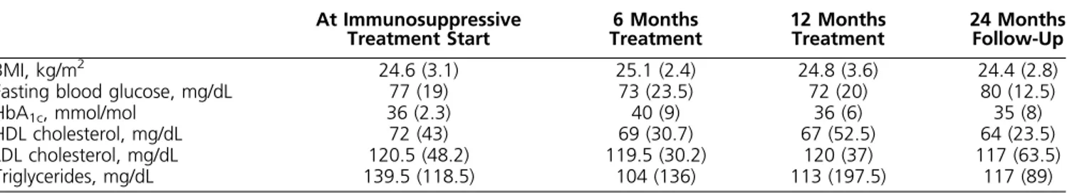
The adverse events that occurred in the glucocorticoid-immunosuppressive patients are summarized in Table 4. No serious adverse event occurred. The most frequent adverse event was the increase in weight that was transient and occurred during the first 6 months of treatment. Likewise, metabolic parameters did not sig-nificantly change during the treatment period (Table 5). Other adverse events reported were mild hypokalemia, transient psychiatric symptoms, and occurrence of in-fection (flu syndrome in two patients and cystitis in a single case).
Prognostic factors of hypophysitis outcome
Hypophysitis outcome, according to the treatment choice, is summarized in Table 6. Sex, age at diagnosis, antinuclear antibody, anti-extractable nuclear antigen, and anti-double-stranded DNA autoantibodies did not correlate to endocrine and hypophysitis outcome, both in patients treated and untreated with immunosuppressive therapy. Moreover, among the eight patients who did not undergo the immunosuppressive therapy, we did not identify factors predicting endocrine and hypophysitis outcome. On the other hand, among the 12 patients who underwent immunosuppressive therapy, it was noted that the responsiveness to immunosuppressive treatment, both at univariate and logistic regression analysis (Tables 7 and 8), is correlated with the presence of APA, oc-currence of diabetes insipidus at PAH diagnosis, absence of the physiological neuropituitary bright spot on T1w
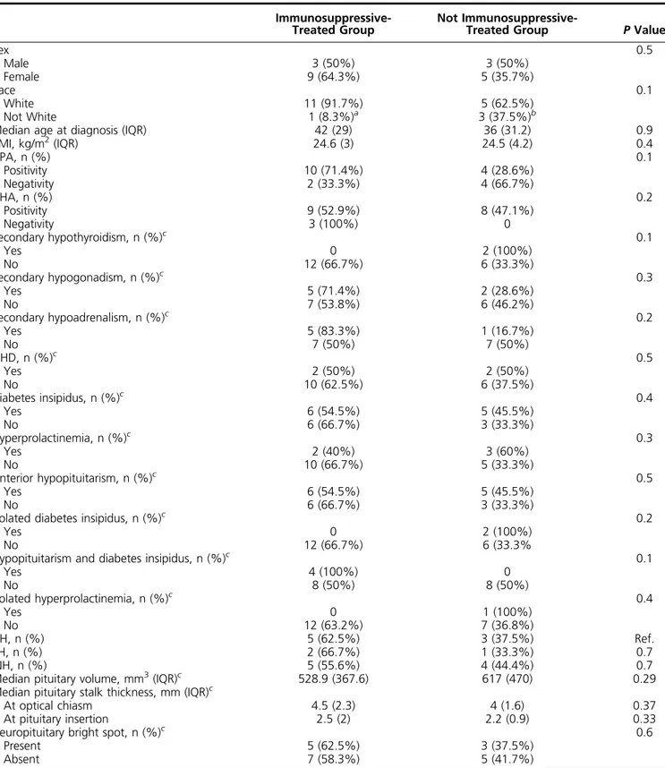
Table 1. Baseline Clinical Features of Study Population According to Treatment Choice
Immunosuppressive-Treated Group
Not
Immunosuppressive-Treated Group P Value
Sex 0.5 Male 3 (50%) 3 (50%) Female 9 (64.3%) 5 (35.7%) Race 0.1 White 11 (91.7%) 5 (62.5%) Not White 1 (8.3%)a 3 (37.5%)b
Median age at diagnosis (IQR) 42 (29) 36 (31.2) 0.9
BMI, kg/m2(IQR) 24.6 (3) 24.5 (4.2) 0.4 APA, n (%) 0.1 Positivity 10 (71.4%) 4 (28.6%) Negativity 2 (33.3%) 4 (66.7%) AHA, n (%) 0.2 Positivity 9 (52.9%) 8 (47.1%) Negativity 3 (100%) 0 Secondary hypothyroidism, n (%)c 0.1 Yes 0 2 (100%) No 12 (66.7%) 6 (33.3%) Secondary hypogonadism, n (%)c 0.3 Yes 5 (71.4%) 2 (28.6%) No 7 (53.8%) 6 (46.2%) Secondary hypoadrenalism, n (%)c 0.2 Yes 5 (83.3%) 1 (16.7%) No 7 (50%) 7 (50%) GHD, n (%)c 0.5 Yes 2 (50%) 2 (50%) No 10 (62.5%) 6 (37.5%) Diabetes insipidus, n (%)c 0.4 Yes 6 (54.5%) 5 (45.5%) No 6 (66.7%) 3 (33.3%) Hyperprolactinemia, n (%)c 0.3 Yes 2 (40%) 3 (60%) No 10 (66.7%) 5 (33.3%) Anterior hypopituitarism, n (%)c 0.5 Yes 6 (54.5%) 5 (45.5%) No 6 (66.7%) 3 (33.3%)
Isolated diabetes insipidus, n (%)c 0.2
Yes 0 2 (100%)
No 12 (66.7%) 6 (33.3%
Hypopituitarism and diabetes insipidus, n (%)c 0.1
Yes 4 (100%) 0 No 8 (50%) 8 (50%) Isolated hyperprolactinemia, n (%)c 0.4 Yes 0 1 (100%) No 12 (63.2%) 7 (36.8%) AH, n (%) 5 (62.5%) 3 (37.5%) Ref. PH, n (%) 2 (66.7%) 1 (33.3%) 0.7 INH, n (%) 5 (55.6%) 4 (44.4%) 0.7
Median pituitary volume, mm3(IQR)c 528.9 (367.6) 617 (470) 0.29
Median pituitary stalk thickness, mm (IQR)c
At optical chiasm 4.5 (2.3) 4 (1.6) 0.37
At pituitary insertion 2.5 (2) 2.2 (0.9) 0.33
Neuropituitary bright spot, n (%)c 0.6
Present 5 (62.5%) 3 (37.5%)
Absent 7 (58.3%) 5 (41.7%)
Univariate analysis. qualitative variables are reported as absolute value and percent (%). Continuous variables are reported as median6 IQR. Abbreviations: BMI, body mass index; Ref, reference.
aEritrean ethnicity.
bEgyptian, Bengali, and Philippine ethnicity. cEvaluation conducted at PAH diagnosis.
images, and pituitary stalk at optical chiasm larger than 3.9 mm [area under the curve (AUC): 0.97, sensibility: 87.5%, specificity: 100%; P = 0.01]. Likewise, as shown in we Tables 7 and 8, we found that the improvement of pituitary function during follow-up is predicted by the presence of APA, occurrence at PAH diagnosis of sec-ondary hypogonadism, diabetes insipidus, PH and INH anatomical hypophysitis classification, pituitary volume lower than 493 mm3 (AUC: 0.86, sensibility: 100%, specificity: 71.4%; P = 0.04), pituitary stalk at optical chiasm larger than 3.9 mm (AUC: 0.97, sensibility:
100%, specificity: 100%; P = 0.008), and absence of the physiological posterior pituitary bright spot on T1w MRIs.
Discussion
In this study, we analyzed PAH outcome in our monocentric series of affected patients to identify immunological, clinical, and morphological markers of immunosuppressive treatment responsiveness. We found that the occurrence of diabetes insipidus, the absence of Table 2. Study Population Endocrine Assessment at Baseline and 2-Year Follow-Up
Baseline 2-Year Follow-Up
Secondary hypothyroidism 2/20 3/20
Jostel indexa 1.8 (0.8) 1.8 (0.9)
Jostel indexb 0.94 (2) 1.2 (2)
Secondary hypogonadism 7/20 2/20
Secondary hypoadrenalism 6/20 4/20
Cortisol (mg/dL) after ACTH stimulationb – Cortisol (microgr/dL) after ACTH stimulation b is 12 (5) Cortisol (mg/dL) after ACTH stimulationc – Cortisol (microgr/dL) after ACTH stimulation c is 24 (2) Cortisol (mg/dL) after ACTH stimulationd Cortisol (microgr/dL) after ACTH stimulation d is 11 (7)
GHD 4/20 2/20
GH peakb 4.5 (2) 15 (2)e
Diabetes insipidus 11/20 8/20
Plasmatic osmolarity, mOsm/kgb 304 (9) 293 (7)
Urinary osmolarity, mOsm/kgb 40 (23) 254 (345)
Hyperprolactinemia 5/20 3/20
PRL, ng/mLa 16 (13.5) 9 (10.25)
PRL, ng/mLb 24 (2) 11.4 (2)
Qualitative variables are reported as absolute value and percent (%). Continuous variables are reported as median6 IQR. Abbreviations: GH, growth hormone; PRL, prolactin.
aValue calculated in entire study population.
bValue calculated in corresponding pituitary dysfunction-affected patients.
cValue referred to the single patient who recovered from secondary hypoadrenalism.
dValue referred to the test performed in the three patients with worsening of ACTH–cortisol axis during the follow-up.
eValue referred to the median value of GH peak at follow-up GHRH plus arginine test of the two patients who recovered from GHD.
Table 3. Endocrine Assessment at Baseline and 2-Year Follow-Up According Treatment Choice
Immunosuppressive-Treated Patients Conservatively Managed Patients
Baseline
2-Year Follow-Up
Baseline
2-Year Follow-Up Recovered/
Improved Stable Worse/NewDiagnosis Recovered/Improved Stable Worse/NewDiagnosis Secondary hypothyroidism 0 Na Na 0 2 0 2 (66.7%) 1 (33.3%) Secondary hypogonadism 5 5 (100%) 0 0 2 1 (50%) 1 (50%) 0 Secondary hypoadrenalism 5 1 (20%) 4 (80%) 0 1 0 1 (25%) 3 (75%) GHD 2 2 (100%) 0 0 2 0 2 (100%) 0 Diabetes insipidus 6 3 (50%) 3 (50%) 0 5 0 5 (100%) 0 Hyperprolactinemia 2 1 (50%) 1 (50%) 0 3 1 (33.3%) 2 (66.7%) 0
Qualitative variables are reported as absolute value and percent (%). Abbreviation: Na, not acceptable.
the neuropituitary bright spot on T1w MR images, and the pituitary stalk thickness larger than 3.9 mm played an important role in building the positive prognostic markers of hypophysitis and pituitary function im-provement at follow-up. Moreover, the occurrence of secondary hypogonadism, as well as pituitary volume lower than 493 mm3, can predict improvement of pi-tuitary function at follow-up. As previously demon-strated (11), an actual study confirms that secondary hypogonadism and diabetes insipidus, absence of the neuropituitary bright spot, and pituitary stalk thickness larger than 3.9 mm are frequently associated with the diagnosis of PH and INH (Table 9). Likewise, a pituitary volume lower than 493 mm3is correlated to the diagnosis of INH (P = 0.002). Consequently, our data suggest that both the diagnosis of INH or PH and the indirect clinical and radiological signs of inflammatory involvement of the pituitary stalk and of neuropituitary (such us the pituitary stalk thickness and the absence of the neuro-pituitary bright spot) can positively predict the hypo-physitis outcome. In fact, according to our results, in patients treated with immunosuppressive therapy, INH
and PH are associated with a better prognosis compared with AH cases. However, although autoimmune eti-ology of primary hypophysitis has been widely described, until now, its pathogenesis is not completely defined, and it is not predictable if INH, PH, and AH differ in disease natural his-tory and treatment responsiveness. In fact, it is possible to speculate that INH, AH, and PH should be supported by different autoantigens, able to trigger the autoimmune inflammation (29). However, several proteins have been suggested as possible PAH anti-gens, as prohormones and hormones, nuclear and cytoplasmic enzymes, and transcriptional factors, all character-ized by a different pituitary cellular expression, involving both the neuroendocrine cells and the T-lymphocytes (30).
Moreover, the different responsiveness to glucocorti-coid treatment in cases with inflammatory involvement of pituitary stalk (as INHs and PHs) should be justified by the different vascular system of the pituitary stalk compared with the pituitary glands, which should modify the bio-distribution of the drugs (31). In fact, it was previously reported that glucocorticoids almost always improve the swelling of the pituitary and of the pituitary stalk, then inducing a recovery of the anterior hypopituitarism (32). Moreover, for the first time, our data suggest a prognostic and predictive role of APA on the immu-nosuppressive treatment responsiveness, reinforcing the hypothesis of the autoimmune inflammation etiology and pathogenesis of PAH (33). APA positivity can consequently suggest an activation of the immune system and an increased sensibility to glucocorticoid treatment. However, until now, APAs have been considered a disease marker rather than causative agent (17). If confirmed on other similar cohorts of patients, use of APA could therefore guide the treatment decisionmak-ing. Up until now and based on the experience reported in the present manuscript, we can consider APA as a surrogate biomarker of response to treatment.
Prognostic markers of treatment responsiveness were evaluated in our series among patients treated according to the same therapeutic protocol. In fact, the three patients treated with the prednisone/azathioprine-associative ther-apy were still considered nonresponders to immunosup-pressive treatments. Moreover, according to our data, glucocorticoid treatments resulted in usefulness for the improvement of pituitary function and hypophysitis out-come. We found a substantial improvement of gonadal
Figure 2. Histogram representing patients’ endocrine outcome according to treatment choice. Bars indicated patients’ outcome. Fisher test proved a statistically significant high frequency of worsening of pituitary function among conservatively managed patients compared with immunosuppressive-treated ones (*P = 0.04).
Table 4. Adverse Events Occurred During Glucocorticoid Treatment
Patients, n (%) Transient body weight increase 8 (66.7%)
Transient psychiatric symptoms (anxiety, nervousness)
3 (25%)
Diabetes mellitus 0
Infections 3 (25%; 2 flu syndromes
and 1 cystitis)
Mild hypokalemia 3 (25%)
function, GHD, and diabetes insipidus, without occurrence of adverse events. Our evidences underline the impor-tance of an etiological-immunosuppressive treatment,
particularly in young, fertile-PAH affected patients with a long life expectancy. Hypopituitarism, in fact, is a very serious clinical condition characterized by a high mortality Table 5. Metabolic Assessment in the 12 Patients Treated With Immunosuppressive Glucocorticoid at Baseline and During Follow-Up
At Immunosuppressive
Treatment Start Treatment6 Months 12 MonthsTreatment 24 MonthsFollow-Up
BMI, kg/m2 24.6 (3.1) 25.1 (2.4) 24.8 (3.6) 24.4 (2.8)
Fasting blood glucose, mg/dL 77 (19) 73 (23.5) 72 (20) 80 (12.5)
HbA1c, mmol/mol 36 (2.3) 40 (9) 36 (6) 35 (8)
HDL cholesterol, mg/dL 72 (43) 69 (30.7) 67 (52.5) 64 (23.5)
LDL cholesterol, mg/dL 120.5 (48.2) 119.5 (30.2) 120 (37) 117 (63.5)
Triglycerides, mg/dL 139.5 (118.5) 104 (136) 113 (197.5) 117 (89)
Continuous variables are reported as median6 IQR.
Abbreviations: BMI, body mass index; HbA1c, hemoglobin A1c; HDL, high-density lipoprotein; LDL, low-density lipoprotein.
Table 6. Factors Predicting Hypophysitis Outcome
Hypophysitis Outcome Patients on Immunosuppressive
Treatment (12)
Patients Not on Immunosuppressive Treatment (8)
Responder Nonresponders P Value Improved/Stable Worse P Value
APA, n (%) 0.03 0.5 Positivity 8 (80%) 2 (20%) 3 (75%) 1 (25%) Negativity 0 2 (100%) 2 (50%) 2 (50%) AHA, n (%) 0.3 Na Positivity 5 (55.6%) 4 (44.4%) 5 (62.5%) 3 (37.5%) Negativity 3 (100%) 0 0 0 Secondary hypothyroidism, n (%)a Na 0.4 Yes 0 0 0 2 (100%) No 8 (66.7%) 4 (33.3%) 4 (66.7%) 2 (33.3%) Secondary hypogonadism, n (%)a 0.04 0.7 Yes 5 (100%) 0 1 (50%) 1 (50%) No 3 (42.9%) 4 (57.1%) 3 (50%) 3 (50%) Secondary hypoadrenalism, n (%)a 0.6 0.5 Yes 3 (60%) 2 (40%) 1 (100%) 0 No 5 (71.4%) 2 (28.6%) 3 (42.9%) 4 (57.1%) GHD, n (%)a 0.4 0.7 Yes 2 (100%) 0 1 (50%) 1 (50%) No 6 (60%) 4 (40%) 3 (50%) 3 (50%) Diabetes insipidus, n (%)a 0.03 0.5 Yes 6 (100%) 0 2 (40%) 3 (60%) No 2 (33.3%) 4 (66.7%) 2 (66.7%) 1 (33.3%) Hyperprolactinemia, n (%)a 0.4 0.5 Yes 2 (100%) 0 1 (33.3%) 2 (66.7%) No 6 (60%) 4 (40%) 3 (60%) 2 (40%) AH, n (%) 1 (25%) 4 (75%) Ref. 3 (100%) 0 0.2 PH, n (%) 2 (100%) 0 0.1 0 1 (100%) 0.6 INH, n (%) 5 (100%) 0 0.02 2 (50%) 2 (50%) Ref.
Median pituitary volume, mm3(IQR)a 422 (487) 631 (309) 0.2 537 (508) 644 (1375) 0.5 Median pituitary stalk thickness,
mm (IQR)a
At optical chiasm 4.9 (2) 3.3 (0.9) 0.02 3.9 (1.8) 4.1 (1.9) 0.5
At pituitary insertion 2.6 (3.3) 2.1 (2.2) 0.4 2.1 (0.7) 2.8 (1) 0.1
Neuropituitary bright spot, n (%)a 0.01 0.2
Present 1 (20%) 4 (80%) 3 (100%) 0
Absent 7 (100%) 0 2 (40%) 3 (60%)
Univariate analysis. Qualitative variables are reported as absolute value and percent (%). Continuous variables are reported as median6 IQR.
aEvaluation conducted at PAH diagnosis.
risk and consequently, required an expensive hormonal replacement therapy (34).
In our group of untreated patients, a spontaneous recovery of hypophysitis and pituitary function occurred rarely: in most of cases, hypopituitarism did not change or worsened during the follow-up stage. However, our data did not allow us to identify a prognostic marker of hypophysitis outcome in patients who were not treated with glucocorticoids.
In previous studies, among patients who underwent an immunosuppressive treatment, pituitary secretion im-provement was proven between 15% and 41% and ra-diological improvement between 36% and 89% of cases
(14, 19, 20, 35). Among patients who did not undergo an immunosuppressive treatment, instead, the improvement of pituitary function was reported in 33% (8) and the spontaneous radiological improvement in 72.7% of cases (19). This variability is justified by sample size, study population selection bias, different disease stage, treatment choice and drug dosage, and definition of the treatment responsiveness and of the outcome. Moreover, most of these studies were retrospectively designed. The positive outcome proven in our series can be explained by an early hypophysitis diagnosis and initiation of immunosuppres-sive treatment and the availability of a tertiary referral center with a medical team devoted to pituitary disease. Table 7. Factors Predicting Endocrine Outcome
Endocrine Outcome Patients on Immunosuppressive
Treatment Patients Not on Immunosuppressive Treatment Resolved/
Improved Stable P Value Resolved/Improved Stable Worse P Value (a) P Value (b) P Value (c)
APA, n (%) 0.06 0.7 0.8 0.7 Positivity 7 (70%) 3 (30%) 1 (20%) 2 (40%) 2 (40%) Negativity 0 2 (100%) 1 (33.3%) 1 (33.3%) 1 (33.3%) AHA positivity, n (%) 5 (41.7%) 7 (58.3%) Na 2 (25%) 3 (37.5%) 3 (37.5%) Na Na Na Secondary hypothyroidism, n (%)a Na 0.6 0.8 0.6 Yes 0 0 0 1 (50%) 1 (50%) No 7 (58.3%) 5 (41.7%) 2 (33.3%) 2 (33.3%) 2 (33.3%) Secondary hypogonadism, n (%)a 0.03 0.4 0.5 0.7 Yes 2 (28.6%) 5 (71.4%) 1 (16.7%) 3 (50%) 2 (33.3%) No 5 (100%) 0 1 (50%) 0 1 (50%) Secondary hypoadrenalism, n (%)a 0.3 0.6 0.5 Na Yes 2 (40%) 3 (60%) 0 1 (100%) 0 No 5 (71.4%) 2 (28.6%) 2 (28.6%) 2 (28.6%) 3 (42.9%) GHD, n (%)a 0.3 0.4 0.5 0.7 Yes 2 (100%) 0 1 (50%) 0 1 (50%) No 5 (50%) 5 (50%) 1 (16.7%) 3 (50%) 2 (33.3%) Diabetes insipidus, n (%)a 0.008 0.7 0.2 0.4 Yes 6 (100%) 0 1 (20%) 1 (20%) 3 (60%) No 1 (16.7%) 5 (83.3%) 1 (33.3%) 2 (66.7%) 0 Hyperprolactinemia, n (%)a 0.3 0.4 0.2 0.7 Yes 2 (100%) 0 1 (33.3%) 0 2 (66.7%) No 5 (50%) 5 (50%) 1 (20%) 3 (60%) 1 (20%)
AH, n (%) 0 5 (100%) Ref. 1 (33.3%) 2 (66.7%) 0 Ref. Ref. Ref.
PH, n (%) 2 (100%) 0 0.05 0 0 1 (100%) Na Na Na
INH, n (%) 5 (100%) 0 0.004 1 (25%) 1 (25%) 2 (50%) 0.5 0.1 0.5
Median pituitary volume, mm3(IQR)a
358.9 (467) 705 (300) 0.04 607 (2) 678 (2) 557 (2) 0.6 0.4 0.7 Median pituitary stalk
thickness, mm (IQR)a
5 (1.4) 3.5 (0.75) 0.005 3.9 (2) 3.8 (2) 4.4 (2) 0.9 0.5 0.7 At optical chiasm 2.3 (3.9) 2.6 (1.95) 0.5 2.2 (2) 2 (2) 3.1 (2) 0.6 0.1 0.3 At pituitary insertion
Neuropituitary bright spot,
n (%)a 0.001 0.5 0.1 0.4
Present 0 5 (100%) 1 (33.3%) 2 (66.7%) 0
Absent 7 (100%) 0 1 (20%) 1 (20%) 3 (60%)
Univariate analysis. Qualitative variables are reported as absolute value and percent (%). Continuous variables are reported as median6 IQR. P value (a), compared the group of patients with resolved pituitary dysfunction with patients with stable endocrine assessment; P value (b), compared the group of patients with stable endocrine assessment with those with worsened pituitary endocrine function; P value (c), compared the group of patients with improved/resolved endocrine assessment with those with worsened pituitary endocrine function.
aEvaluation conducted at PAH diagnosis.
According to previous literature, the role of surgery in treatment of hypophysitis is limited to selected cases, such as those with diagnosis in doubt or those with a rapid/ sudden progression of neurologic symptoms (20). In fact, although some authors suggested that pituitary neuro-surgery may improve PAH morphologically (19), the same does not allow the improvement of pituitary function. Immunosuppressive treatment with pharma-cological doses of glucocorticoid is considered ideal compared with pituitary surgery and conservative man-agement (clinical observation) for restoring pituitary secretory function (8, 20). Likewise, in our two patients who underwent debulking pituitary neurosurgery for the
worsening of the neurologic symptom (11), we did not obtain a pituitary secretory improvement. However, according to the different treatment and to the well-defined postsurgery pituitary region anatomical modi-fications, we decided to exclude these two patients in the current study.
In our series, PAH recurrence did not take place. However, data of hypophysitis recurrence after immu-nosuppressive treatment are variable in previous reports, ranging from 18% to 40% (14, 19, 20). These data can be influenced by the glucocorticoids dosage and treat-ment period. In our series, duration of glucocorticoid treatment was at least 13 months, according to a slow Table 8. Factors Predicting Hypophysitis and Endocrine Outcome
Hypophysitis Worsening at Follow-Up
Worsening of Pituitary Function at Follow-Up
APA positivitya P value: 0.01 P value: 0.05
OR: 5 OR: 3.3
95% CI: 1.4–17.3 95% CI: 1.3–8.6
Secondary hypogonadisma P value: 0.09 P value: 0.03
OR: 0.4 OR: 0.3
95% CI: 0.2–1 95% CI: 0.09–0.9
Diabetes insipidusa P value: 0.01 P value: 0.002
OR: 0.3 OR: 0.2
95% CI: 0.1–1 95% CI: 0.02–0.9
Absence of neuropituitary bright spot P value: 0.01 P value: 0.002
OR: 4 OR: 0.3
95% CI: 0.7–21.8 95% CI: 0.1–0.9
Pituitary volume,493 mm3a P value: 0.09 P value: 0.03
OR: 2.3 OR: 3.5
95% CI: 1–5.5 95% CI: 1.1–11.3
Pituitary stalk at optical chiasm.3.9 mma P value: 0.04 P value: 0.01
OR: 0.3 OR: 0.2
95% CI: 0.1–1 95% CI: 0.02–0.9
PH diagnosis P value: 0.8 P value: 0.01
OR: 0.2 OR: Na
95% CI: 0.03–1.2 95% CI: Na
INH diagnosis P value: 0.09 P value: 0.03
OR: 0.4 OR: 0.3
95% CI: 0.2–1 95% CI: 0.1–0.9
Logistic regression.
aEvaluation conducted at PAH diagnosis.
Table 9. Prognostic Factors Predicting Hypophysitis and Endocrine Outcome, According to Anatomical Hypophysitis Classification
AH PH INH AH vs PHP value AH vs INHP value PH vs INHP value
Secondary hypogonadisma 0 1 (16.7%) 5 (83.3%) 0.3 0.01 0.3
Diabetes insipidusa 0 3 (27.3%) 8 (72.7%) 0.08 ,0.001 Na
Absence of neuropituitary bright spota 0 3 (25%) 9 (75%) 0.08 ,0.001 Na
Pituitary stalk at optical chiasma.3.9 mma 0 2 (22.2%) 7 (77.8%) 0.02 ,0.001 0.3
Pituitary volume,493 mm3a 0 0 6 (100%) Na 0.002 0.002
Univariate analysis. Qualitative variables are reported as absolute value and percent (%).
aEvaluation conducted at PAH diagnosis.
reduction of glucocorticoid pharmacological dosage. In fact, treatment with glucocorticoid can reduce the in-flammatory edema in an early phase of the disease and prevent the development of a chronic inflammation and related fibrotic process in a later disease stage (36).
The main limitations of our paper are the following: (1) the small size of the study population, (2) the low frequency of the histopathological diagnosis of PAH, and (3) the nonrandomized study design. Our study cohort was chosen with the application of a very strict inclusion criteria, to select a homogeneous study group of patients diagnosed, treated, and followed up according to a univocal protocol, by a unique medical team devoted to pituitary disease (namely, the Pituitary Board). However, our study cohort results are very similar to those in-vestigated by other research groups, also reflecting the reported PAH prevalence.
Moreover, despite the nonrandomized study design, our paper represents a real-life scenario in which PAHs were treated with immunosuppressive therapy according to pituitary secretory status and the age of the patients, clinical condition, and preference. Nevertheless, our study was conducted in a cross-sectional and prospective view in the two groups of patients classified as immu-nosuppressive treated and untreated without differences of baseline disease aspects.
Although our data should be confirmed on larger patient series, our actual evidences suggest that hypo-pituitarism and hypophysitis can improve through glucocorticoid immunosuppressive administration, par-ticularly in patients affected by INH and PH. Conse-quently, according to the evaluation of the risk/benefit ratio of glucocorticoid-immunosuppressive treatment, candidate patients should be consequently treated, also according to the endocrine, immunological, and morpho-logical potential biomarker of treatment responsiveness.
Acknowledgments
Correspondence and Reprint Requests: Laura De Marinis, MD, Pituitary Unit, Department of Endocrinology, Catholic University of the Sacred Heart, School of Medicine, 00135 Rome, Italy. E-mail:[email protected].
Disclosure Summary: The authors have nothing to disclose.
References
1. Lin HH, Gutenberg A, Chen TY, Tsai NM, Lee CJ, Cheng YC, Cheng WH, Tzou YM, Caturegli P, Tzou SC. In situ activation of pituitary-infiltrating T lymphocytes in autoimmune hypophysitis. Sci Rep. 2017;7:43492.
2. Tartaglione T, Chiloiro S, Laino ME, Giampietro A, Gaudino S, Zoli A, Bianchi A, Pontecorvi A, Colosimo C, De Marinis L.
Neuro-radiological features can predict hypopituitarism in primary autoimmune hypophysitis. Pituitary. 2018;21(4):414–424. 3. Falorni A, Minarelli V, Bartoloni E, Alunno A, Gerli R. Diagnosis
and classification of autoimmune hypophysitis. Autoimmun Rev. 2014;13(4-5):412–416.
4. Caturegli P, Newschaffer C, Olivi A, Pomper MG, Burger PC, Rose NR. Autoimmune hypophysitis. Endocr Rev. 2005;26(5): 599–614.
5. Molitch ME, Gillam MP. Lymphocytic hypophysitis. Horm Res. 2007;68(5, Suppl 5):145–150.
6. Honegger J, Fahlbusch R, Bornemann A, Hensen J, Buchfelder M, M ¨uller M, Nomikos P. Lymphocytic and granulomatous hypo-physitis: experience with nine cases. Neurosurgery. 1997;40(4): 713–722, discussion 722–723.
7. Chiloiro S, Giampietro A, Bianchi A, Tartaglione T, Capobianco A, Anile C, De Marinis L. Diagnosis of endocrine disease: primary empty sella: a comprehensive review. Eur J Endocrinol. 2017; 177(6):R275–R285.
8. Khare S, Jagtap VS, Budyal SR, Kasaliwal R, Kakade HR, Bukan A, Sankhe S, Lila AR, Bandgar T, Menon PS, Shah NS. Primary (autoimmune) hypophysitis: a single centre experience. Pituitary. 2015;18(1):16–22.
9. Gutenberg A, Hans V, Puchner MJ, Kreutzer J, Br ¨uck W, Caturegli P, Buchfelder M. Primary hypophysitis: clinical-pathological cor-relations. Eur J Endocrinol. 2006;155(1):101–107.
10. Honegger J, Schlaffer S, Menzel C, Droste M, Werner S, Elbelt U, Strasburger C, St ¨ormann S, K ¨uppers A, Streetz-van der Werf C, Deutschbein T, Stieg M, Rotermund R, Milian M, Petersenn S; Pituitary Working Group of the German Society of Endocrinology. Diagnosis of primary hypophysitis in Germany. J Clin Endocrinol Metab. 2015;100(10):3841–3849.
11. Chiloiro S, Tartaglione T, Angelini F, Bianchi A, Arena V, Giampietro A, Mormando M, Sciandra M, Laino ME, De Marinis L. An overview of diagnosis of primary autoimmune hypophysitis in a prospective single-center experience. Neuroendocrinology. 2017;104(3):280–290.
12. Kyriacou A, Gnanalingham K, Kearney T. Lymphocytic hypo-physitis: modern day management with limited role for surgery. Pituitary. 2017;20(2):241–250.
13. Rao S, Mahadevan A, Maiti T, Ranjan M, Shwetha SD, Arivazhagan A, Saini J. Granulomatous and lymphocytic hypophysitis—are they immunologically distinct? APMIS. 2016; 124(12):1072–1077.
14. Angelousi A, Cohen C, Sosa S, Danilowicz K, Papanastasiou L, Tsoli M, Pal A, Piaditis G, Grossman A, Kaltsas G. Clinical, en-docrine and imaging characteristics of patients with primary hypophysitis. Horm Metab Res. 2018;50(4):296–302.
15. Faje A. Hypophysitis: evaluation and management. Clin Diabetes Endocrinol. 2016;2(1):15.
16. Fukuoka H. Hypophysitis. Endocrinol Metab Clin North Am. 2015;44(1):143–149.
17. Bellastella G, Maiorino MI, Bizzarro A, Giugliano D, Esposito K, Bellastella A, De Bellis A. Revisitation of autoimmune hypo-physitis: knowledge and uncertainties on pathophysiological and clinical aspects. Pituitary. 2016;19(6):625–642.
18. Lupi I, Manetti L, Raffaelli V, Lombardi M, Cosottini M, Iannelli A, Basolo F, Proietti A, Bogazzi F, Caturegli P, Martino E. Di-agnosis and treatment of autoimmune hypophysitis: a short review. J Endocrinol Invest. 2011;34(8):e245–e252.
19. Honegger J, Buchfelder M, Schlaffer S, Droste M, Werner S, Strasburger C, St ¨ormann S, Schopohl J, Kacheva S, Deutschbein T, Stalla G, Flitsch J, Milian M, Petersenn S, Elbelt U; Pituitary Working Group of the German Society of Endocrinology. Treat-ment of primary hypophysitis in Germany. J Clin Endocrinol Metab. 2015;100(9):3460–3469.
20. Wang S, Wang L, Yao Y, Feng F, Yang H, Liang Z, Deng K, You H, Sun J, Xing B, Jin Z, Wang R, Pan H, Zhu H. Primary lymphocytic hypophysitis: clinical characteristics and treatment of 50 cases in a
single centre in China over 18 years. Clin Endocrinol (Oxf). 2017; 87(2):177–184.
21. Abe T. Lymphocytic neurohypophysitis and infundibulo-panhypophysitis regarded as lymphocytic hypophysitis variant. Brain Tumor Pathol. 2008;25(2):59–66.
22. Lupi I, Cosottini M, Caturegli P, Manetti L, Urbani C, Cappellani D, Scattina I, Martino E, Marcocci C, Bogazzi F. Diabetes insipidus is an unfavorable prognostic factor for response to glucocorticoids in patients with autoimmune hypophysitis. Eur J Endocrinol. 2017; 177(2):127–135.
23. Johnston PC, Chew LS, Hamrahian AH, Kennedy L. Lymphocytic infundibulo-neurohypophysitis: a clinical overview. Endocrine. 2015;50(3):531–536.
24. Allix I, Rohmer V. Hypophysitis in 2014. Ann Endocrinol (Paris). 2015;76(5):585–594.
25. Chiloiro S, Capoluongo ED, Tartaglione T, Bianchi A, Giampietro A, Angelini F, Arena V, Pontecorvi A, De Marinis L. Human leucocyte antigens coeliac haplotypes and primary autoimmune hypophysitis in caucasian patients. Clin Endocrinol (Oxf). 2018; 88(5):692–699.
26. Jostel A, Ryder WD, Shalet SM. The use of thyroid function tests in the diagnosis of hypopituitarism: definition and evaluation of the TSH Index. Clin Endocrinol (Oxf). 2009;71(4):529–534. 27. Bellastella G, Rotondi M, Pane E, Dello Iacovo A, Pirali B, Dalla
Mora L, Falorni A, Sinisi AA, Bizzarro A, Colao A, Chiovato L, De Bellis A; Italian Autoimmune Hypophysitis Network Study. Pre-dictive role of the immunostaining pattern of immunofluorescence and the titers of antipituitary antibodies at presentation for the occurrence of autoimmune hypopituitarism in patients with au-toimmune polyendocrine syndromes over a five-year follow-up. J Clin Endocrinol Metab. 2010;95(8):3750–3757.
28. Pecina HI, Pecina TC, Vyroubal V, Kruljac I, Slaus M. Age and sex related differences in normal pituitary gland and fossa volumes. Front Biosci (Elite Ed). 2017;9:204–213.
29. Iwama S, Sugimura Y, Kiyota A, Kato T, Enomoto A, Suzuki H, Iwata N, Takeuchi S, Nakashima K, Takagi H, Izumida H, Ochiai H, Fujisawa H, Suga H, Arima H, Shimoyama Y, Takahashi M, Nishioka H, Ishikawa SE, Shimatsu A, Caturegli P, Oiso Y. Rabphilin-3A as a targeted autoantigen in lymphocytic infundi-bulo-neurohypophysitis. J Clin Endocrinol Metab. 2015;100(7): E946–E954.
30. Guaraldi F, Giordano R, Grottoli S, Ghizzoni L, Arvat E, Ghigo E. Pituitary autoimmunity. Front Horm Res. 2017;48:48–68. 31. Stanfield JP. The blood supply of the human pituitary gland. J Anat.
1960;94:257–273.
32. Sugihara H. Review on recent topics in hypophysitis. J Nippon Med Sch. 2017;84(5):201–208.
33. Akahori H, Sugimoto T. Lymphocytic hypophysitis with a long latent period from onset of central diabetes insipidus to devel-opment of pituitary enlargement. Intern Med. 2010;49(15): 1565–1571.
34. Jasim S, Alahdab F, Ahmed AT, Tamhane S, Prokop LJ, Nippoldt TB, Murad MH. Mortality in adults with hypopituitarism: a sys-tematic review and meta-analysis. Endocrine. 2017;56(1):33–42. 35. Kristof RA, Van Roost D, Klingm ¨uller D, Springer W, Schramm J.
Lymphocytic hypophysitis: non-invasive diagnosis and treat-ment by high dose methylprednisolone pulse therapy? J Neurol Neurosurg Psychiatry. 1999;67(3):398–402.
36. Tzou SC, Lupi I, Landek M, Gutenberg A, Tzou YM, Kimura H, Pinna G, Rose NR, Caturegli P. Autoimmune hypophysitis of SJL mice: clinical insights from a new animal model. Endocrinology. 2008;149(7):3461–3469.