www.elsevier.nl / locate / jinorgbio
Cu(II) and Zn(II) complexes with hyaluronic acid and its sulphated
derivative
Effect on the motility of vascular endothelial cells
a ,
*
a a b bRolando Barbucci
, Agnese Magnani , Stefania Lamponi , Stefania Mitola , Marina Ziche ,
c b
Lucia Morbidelli , Federico Bussolino
a
Department of Chemical and Biosystem Sciences and Technologies, University of Siena, Pian dei Mantellini 44, 53100 Siena, Italy
b
Institute for Cancer Research and Treatment(I.R.C.C.), Department of Genetics, Biology and Biochemistry, University of Torino,
Strada Provinciale142, Km 3.95, 10060 Candiolo, Torino, Italy
c
Interuniversity Center for Molecule Medicine and Applied Biophysics(C.I.M.M.B.A.), University of Firenze, Viale Pieraccini 6, 50139 Firenze, Italy Received 28 March 2000; received in revised form 12 June 2000; accepted 28 June 2000
Abstract
With the aim of improving the compatibility of biomaterials to be used for the construction of cardiovascular prosthesis, we have designed bioactive macromolecules resulting from chemical modifications of hyaluronic acid (Hyal). The stability constants of Cu(II) and Zn(II) complexes with the sulphated derivative of hyaluronic acid (HyalS3.5) were evaluated. Two different complexes have been found for each metal ion, CuL, Cu(OH) L and ZnL, Zn(OH) L (L means the disaccharide unit of the ligands) in aqueous solution at 378C. The2 2
dihydroxo Cu(II) complex was present in high percentage at pH57.4. On the contrary, the Zn(II) ion was present with a relatively low percentage of both complexes. The ability to stimulate endothelial cell adhesion and migration was evaluated for Hyal, HyalS3.5and their
( 4.5 )2
complexes with Cu(II) and Zn(II) ions. The results revealed that Hyal and [Cu(OH) HyalS2 3.5] induced cell adhesion, while
( 2.5 )2 ( 4.5 )2
[ZnHyalS3.5] and [Zn(OH) HyalS2 3.5] inhibited the process. The chemotactic activity of increasing concentrations of the above
( 4.5 )2
complexes was also evaluated, demonstrating that [Cu(OH) HyalS2 3.5] complex at 1 mM concentration was the most active in inducing cell migration. These results have been also strengthened by analysing adherent cell migration in agarose. In conclusion, sulphated hyaluronic acid coordinated to Cu(II) seems to be a promising matrix molecule for the construction of cardiovascular prosthesis.
2000 Elsevier Science S.A. All rights reserved.
Keywords: Metal ion complexes; Sulphated hyaluronic acid; Cu(II) and Zn(II) complexes; Endothelial cells; Adhesion; Migration; Chemotaxis
1. Introduction confers a major stability to hyaluronic acid, leading to an
increased half-life [3]. The design of new anticoagulant In recent years a big effort has been made to improve molecules which can replace heparin is of extreme impor-the cellular compatibility of biomaterials to be used for impor-the tance to avoid the complications due to continuous ad-construction of cardiovascular, orthopedic, plastic and juvant therapy and to produce novel blood compatible reconstructive surgery prosthesis. Recently our group has materials to be linked to the surface of medical devices. actively been involved in the demonstration that some One of the events relevant for the reduction of xenograft chemical modifications of polysaccharides are able to rejection in humans is the cellular biocompatibility of the change their biological activity. Sulphation of hyaluronic matrix. Specifically, in the case of cardiovascular pros-acid (Hyal) [1] allows for the acquisition of a biological thesis the adhesion, attachment and spreading of vascular activity (inhibition of platelet aggregation, anticoagulant endothelial cells on the biomaterial are necessary, along activity and increased endothelial cell proliferation rate) in with the absence of procoagulant activity. Many are the comparison with the native molecule, leading to a potential growth and adhesion factors able to stimulate endothelial endothelium-dependent action [2–4]. Moreover sulphation cell adhesion, movement and proliferation. A great number of vascular endothelial growth factors are polypeptides (i.e. vascular endothelial growth factors, fibroblast growth
*Corresponding author. Tel. / fax: 139-577-232-033.
E-mail address: [email protected] (R. Barbucci). factors, angiopoietins, interleukin-8, transforming growth
0162-0134 / 00 / $ – see front matter 2000 Elsevier Science S.A. All rights reserved. P I I : S 0 1 6 2 - 0 1 3 4 ( 0 0 ) 0 0 1 2 7 - 6
factor, tumor necrosis factor), or to a less extent lipid molecules (prostaglandin E1, platelet-activating factor) [5– 7]. A pivotal role is played by extracellular matrix components such as collagen, fibronectin and heparan sulphates. Heparin, and generally proteoglycans, play an important role in angiogenesis and vascularization, being low affinity receptors for angiogenic inducers such as basic
fibroblast growth factor, vascular endothelial growth fac- Scheme 1. Disaccharide unit of hyaluronic acid. tor–A and transforming growth factor-beta [8–11] and
modulating endothelial cell adhesion and migration
[12,13]. We have recently shown that hyaluronic acid can easily
Other components important for endothelial cell func- be sulphated and molecules with a certain number of tions are oligoelements and metals such as copper and sulphates per repeating unit (disaccharide unit) can be
2
zinc. They are essential constituents of macromolecules as obtained ranging from 1 to 4 OSO3 groups. The most enzymes, soluble proteins and matrix components. They sulphated terms of the series, i.e. HyalS3.5 and HyalS4.0, play an important role both for the conformation and the show quite good anticoagulant activities [2]. The precise catalytic activity of the macromolecules. Micromolar con- structure of hyaluronic acid allowed us to study the centrations of copper induce endothelial cell migration [14] protonation behaviour in aqueous solution by a thermo-and proliferation [15] by unknown mechanisms that could dynamic point of view, as well as the ability to form require the ion binding to fibroblast growth factor receptors complexes with Cu(II) and Zn(II) ions (Scheme 1). [16] or to angiogenin [17]. It has been also observed that The aim of the present study was to evaluate the the accumulation of copper preceded the rabbit cornea stability and the biological activity of hyaluronic acid vascularization induced by implanted tumor cells [18] and complexes with Cu(II) and Zn(II) ions and its sulphated that chelation of copper ions inhibits the angiogenesis derivative and to assess the effect of Cu(II)-HyalS3.5 and associated with the growth of a experimental gliosarcoma Zn(II)-HyalS3.5 complexes present at physiological pH on in rats [19]. Furthermore, copper as salt [20] or coordi- cultured endothelial cells. The studies were integrated with nated with some biological molecules, such as cerulo- analogous tests performed on Cu(II), Zn(II) complexes plasmin, is an angiogenic inducer [14,21,22]. Actually, with non sulphated Hyal. The protonation and Cu(II), heparin-copper (II) complex is an efficient angiogenic Zn(II) stability constants were determined at 378C to factor in rabbit cornea and induces the in vitro migration of identify the stoichiometry of the complexes and their capillary endothelial cells [23,14,18]. Zinc is another percentage at body temperature. Endothelial cell fuctions element which modulates some endothelial cell functions relevant for the biocompatibility of the materials under related to angiogenesis. It is an essential component of study (i.e. adhesion and migration) were studied.
metalloproteinases, key enzymes involved in endothelial cell invasiveness during the early stages of angiogenesis.
When used as soluble form, zinc stimulates the prolifer- 2. Materials and methods
ation of endothelial cells [24] and promotes the repair of
wounded monolayers of this cell type [25]. 2.1. Reagents
On this basis, the development of macromolecules
coordinated to metal ions seems promising for the develop- Sulphated hyaluronic acid samples, with 3.5 sulphate ment of matrix for improving endothelial cell biocom- groups per disaccharide unit (HyalS3.5), were obtained as patibility and reducing blood clotting in vivo. However, previously reported [1,29] and determined by C, H, N, S, heparin is not entirely understood from the chemical point elemental analysis. Hyaluronic acid (M.W.5180 000) was of view, and due to its variety, it is still difficult to define kindly provided by FAB (Fidia Advanced Biopolymers-the stoichiometry of Biopolymers-the metal ion complex. Some authors Padova, Italy).
have found the N-sulphate, O-sulphate and the carboxyl
groups are involved in the Cu(II) binding process [26,27], 2.2. Potentiometric measurements whereas our group ascertained the presence of only
carboxyl and acetyl groups in Cu(II) binding [1]. By using 2.2.1. Protonation studies
low molecular weight saccharide molecules, it has been Potentiometric titrations were performed according to a reported that the heparin cell-modulating activities depend previously described procedure [30]. A Crison MicropH-primarily on a minimum intramolecular density of neigh- 2002 potentiometer, equipped with a combined electrode bouring anionic groups (sulphate) [28]. All these activities (Crison mod. 6.0204.000) was used together with an are associated with the multi-ionic complex formation automatic Crison microburette (mod. 2031) connected to a between the clusters of anionic and cationic sites on the PC 386 DX 40 MHz. For each titration experiment, the
in which a known amount of solid polymer was dissolved isolated from umbilical veins by collagenase digestion by magnetic stirring under nitrogen stream. The titration following a standard protocol [33]. Cells were maintained data were automatically stored on a floppy disk for further in a 5% CO atmosphere at 378C in medium 199 (Sigma,2
processing. The basicity constants were computed by the Italy) with 20% FCS, L-glutamine and gentamicin. Cells
HYPERQUAD package program [31] on the 386 DX 40 were identified as endothelial by their polygonal morpholo-MHZ computer, once it was ascertained that the basicity gy. For migration experiments cells were used when
2
constant of the COO of the repeating unit does not cultures had reached confluence. depend on the protonation degree of the whole
macro-molecules [32]. 2.4. Adhesion assay
2.2.2. Zn(II) and Cu(II) complex-formation To assess the ability of test substances to interfere with The potentiometric titrations were carried out at 378C by cell adhesion, endothelial cells were let to adhere to the same potentiometric apparatus described above. The polystyrene plastic wells in the presence of test substances. stability constants of the Cu(II)-polymer and Zn(II)-poly- Elisa 96-multiwell plates were coated with 10 mg / ml mer complexes were determined by adding standard fibronectin. Cells were detached from confluent cultures
4
sodium hydroxide solution to a solution containing the and suspended at the density of 5310 / ml. 100 ml of cell polysaccharide and the metal ion. Computation of the suspension in the presence of test substances were put in
21
stability constants was performed by the HYPERQUAD each well and incubated at 378C for 30 min h. Cells
program [31]. were washed with PBS and then fixed in methanol and
The experimental details for the potentiometric titrations stained in Diff-Quick (Harleco, Gibbstown, NJ, USA).
are summarised in Table 1. Adherent cells were counted at 1003 magnification with
2
the aid of an ocular grid (21 mm ) [38]. Data are
2.3. Cell culture expressed as total cell number counted / well.
H.end.FB cell line was obtained from heart cells taken 2.5. Motility assays
th
from a mouse fetus DBA / 2Xc57b1 / 6 at the 13 day of
gestation and immortalised by infection with N-TKmT 2.5.1. Chemotaxis assay
retroviral vector containing mT antigen of polyoma virus Chemotaxis experiments were performed with the [33–35]. This cell line was characterised as endothelial on Boyden chamber technique (48-well microchemotaxis the basis of CD31, VE-cadherin and factor VIII expression, chamber) using polycarbonate filters (5 mm pore size, uptake of low density acetylated lipoproteins, synthesis of polyvinylpyrrolidone-free, Nucleopore Costar Italia, platelet activating factor and monocyte chemotactic Milano,Italy) [16–18]. Three different concentrations of peptide-1 after stimulation with interleukin-1 and tumor Hyal, HyalS3.5, Cu(II)-Hyal, Zn(II)-Hyal, Cu(II)-HyalS3.5, necrosis factor [36,37]. H.end.FB cells were kept in Zn(II)-HyalS3.5, CuCl and ZnCl (0.1, 1 and 10 mM) in2 2
DMEM (Dulbecco’s Modified Eagle Medium, Gibco- 2% FCS-DMEM were placed in the lower compartment of
5
Europe Paisley, Scotland) supplemented wih 10% fetal the chamber, and 1310 cells suspended in DMEM
bovine serum (FCS) (Sigma Chemical Co., St. Louis, containing 2% FCS were put in the upper compartments.
MO). After 6 h of incubation at 378C, the upper surface of the
Human umbilical vein endothelial cells (HUVEC) were filter was scratched in order to remove non-migrated cells.
Table 1
Experimental details for the protonation and complex-formation potentiometric measurements at 378C in NaCl 0.1 M
a a a
Compound System T , mmolL T , mmolM added HCl C , mMT Range of pH
mmol ( 4.5 )2 1 HyalS3.5 H 1L 0.03552 – 0.09110 0.0993 2.5–10.8 0.03547 – 0.08733 0.0993 2.5–10.5 21 Cu 1L 0.03134 0.02846 0.09534 0.1000 2.5–10.8 0.03066 0.00605 0.08364 0.1000 2.7–10.9 0.03152 0.00608 0.08419 0.1000 2.6–11.3 0.03115 0.00616 0.08096 0.1000 2.6–11.3 21 4.52 Zn 1L 0.02660 0.01330 0.12769 0.0998 2.4210.6 21 Zn 1L 0.02491 0.01245 0.11306 0.0998 2.4210.8 0.02660 0.00532 0.13103 0.0998 2.4211.0 0.02696 0.00539 0.13089 0.0998 2.4210.9 0.02600 0.02340 0.12388 0.0998 2.4210.8 0.02708 0.02708 0.12273 0.0998 2.4210.8 a
Filters were fixed and stained with Diff-Quick. The directed toward one side; 3, 95–100% of the cells directed number of cells present in five oil immersion fields / well toward one side.
was counted at 1003 magnification. DMEM containing 2% FCS was used as negative control and complete M199
2.6. Statistical analysis containing 10% FCS as positive control.
Analysis of variance (ANOVA) was applied to the data 2.5.2. Migration assay
for cell adhesion and motility; P values ,0.05 were The migration tests were performed on agarose gels as
considered statistically different. Post hoc comparisons previously described [23]. Briefly, tissue culture dishes
between groups (95% confidence for mean) were carried were coated with gelatin and agarose solution was poured
out by a least squares difference test (LSD test) when on the gelatin and allowed to harden at room temperature.
appropriate. Then, three wells were cut with a suitable template.
Endothelial cells were seeded into the central well and dishes were incubated for 5 h (378C, 5% CO ) to allow2
3. Results
attachment. The sample test solution was put into one well while the sample control solution in the controlateral well.
3.1. Protonation constant and Cu(II) and Zn(II) stability The samples containing Hyal / Cu(II) and Cu(II)-HyalS3.5
constants
were dissolved in phosphate buffered saline (PBS) solu-tion. The sample containing CuCl 2H O salt was dissolved2 2
2
The protonation constant relative to the COO group of in Tris solution. The molar ratio of Cu(II)-Hyal and
the disaccharide unit of hyaluronic acid derivative HyalS
HyalS3.5/ Cu(II) was nearly 1. All the solutions were 3.5
at 378C (log K 3.99) was very close to the analogous sterilised by filtering with 0.2 mm filters. Incubation (378C,
constant determined at 258C (log R53.94) [39]. Thus, the 5% CO ) followed for 12 h. Cells were then fixed and2
2
protonation reaction of the COO group in this molecule stained with Diff-Quick. Cell migration toward the stimuli
was almost athermic, not being influenced by the tempera-was quantified microscopically at a magnification of 403
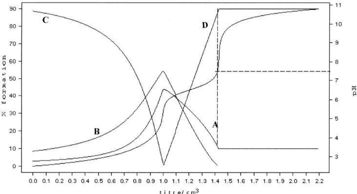
ture. The Cu(II) complex species found at 378C were the in a blind manner by two independent operators. Cell
52
same ones previously seen at 258C for HyalS [27], i.e.
migration was quantified by using the following score: 0, 4
( 2.5 )2
the simple [CuHyalS ] complex and the dihydroxo
no directional movement; 1, at least 10% of the cells 3.5
( 4.5
)-directed toward one side; 2, at least 50% of the cells [Cu(OH) HyalS2 3.5] complex (Fig. 1). Zn(II) ion with
Fig. 1. Cu(II)-HyalS3.5complex: Potentiometric titration curve and distribution curves of the species in solution at different pH’s at 378C in 0.1 M NaCl;
4.52 2.52 3.52 4.52 4.52
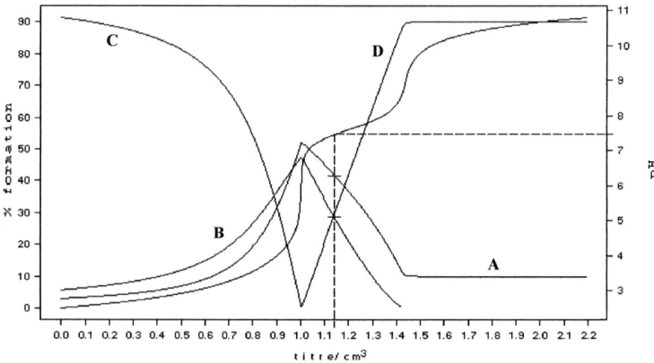
Fig. 2. Zn(II)-HyalS3.5complex: Potentiometric titration curve and distribution curves of the species in solution at different pH’s at 378C in 0.1 M NaCl;
4.52 2.52 3.52 4.52 4.52
A5L ; B5ZnL ; C5LH ; D5Zn(OH) L2 ; [L5HyalS3.5 ].
( 2.5 )2
HyalS3.5 formed two complexes [ZnHyalS3.5] and 3.2. Adhesion of endothelial cells in the presence of
( 4.5 )2
[Zn(OH) HyalS2 3.5] at 378C (Fig. 2), the same species Hyal and HyalS3.5, and their complexes with Cu(II) and found with Cu(II), but not exactly the same found with Zn(II) ions
52
Zn(II) ion and HyalS4 at 258C, where only the dihydroxo
complex was present [39]. Cellular adhesion is one of the early events during the
Knowing the stability constants at 378C, the percentage attachment of endothelial cells to the extracellular matrix. of the complex species at physiological pH (7.4) could be The number of cells adhered to polystyrene plates coated calculated (Table 2). The dihydroxo Cu(II) complex was with fibronectin was evaluated after 30 min of stimulation present in a very high percentage (90%); thus this com- of endothelial cells in suspension with the different
pound could be considered responsible for any of the compounds at 1 mM concentration. Two-way ANOVA
biological effects. On the contrary, Zn(II) ion at pH57.4 showed significant differences among the basal and Hyal, was present with a relatively low percentage of both Cu(II)-HyalS3.5 and Zn(II)-HyalS3.5 (Fig. 3). Specifically,
( 2.5 )2
complexes, 33% [ZnHyalS3.5] and 26% as the compounds most effective in inducing endothelial cell
( 4.5 )2
[Zn(OH) HyalS2 3.5] . adhesion were Hyal (P,0.05) and
Table 2
( 4.5 )2 21 21 a
Stability constants of the sulphated hyaluronic acid (HyalS3.5 ) with Cu and Zn at 378C in 0.1 M NaCl
b
Reaction Log b * % species at 378C and pH57.4
( 2.5 )2 ( 4.5 )2 ( 4.5 )2
ML M(OH) L2 L
21 ( 4 – 5 )2 ( 2.5 )2
Cu 1HyalS3.5 á[Cu HyalS3.5] 3.58(1) 0 90 10
21 ( 4 – 5 )2 2 ( 4.5 )2
Cu 1HyalS3.5 12OH á[Cu(OH) HyalS2 3.5] 17.52(2)
21 ( 4 – 5 )2 ( 2.5 )2
Zn 1HyalS3.5 á[Zn HyalS3.5] 3.37(2) 31 26 41
21 ( 4 – 5 )2 2 ( 4.5 )2
Zn 1HyalS3.5 12OH á[Zn(OH) HyalS2 3.5] 15.17(2)
a 21 21
The potentiometric titration were carried out at 378C. The stability constants of the Cu polymer and Zn polymer complexes were determined by adding standard sodium hydroxide solution to solution containing the polysaccharide and the metal ion at pH57.4.The values of the stability constants are expressed as mean1S.D. *Values in parentheses are standard deviation.
b ( 4.5 )2 21 21 ( 4.5 )2 1 ( 3.5 )2
Fig. 3. Adhesion of endothelial cells in the presence of 1 mM CuCl , ZnCl , Hyal, HyalS2 2 3.5, Cu(II)-Hyal, Zn(II)-Hyal, Cu(II)-HyalS3.5 and Zn(II)-HyalS3.5. H.end.FB cells were let to adhere to polystyrene plates coated with 10 mg / ml fibronectin in the presence of test substances for 30 min. Data are expressed as adherent cells counted in six random fields / well. Numbers represent mean6S.E. (n53); *P,0.05 vs. basal condition.
( 4.5 )2
[Cu(OH) HyalS2 3.5] (P,0.05). On the contrary, the Zn(II)-HyalS3.5 complexes were the most effective in inhibiting cell adhesion (P,0.05). All the other com-pounds stimulated cell adhesion without any significant differences when compared to basal condition.
Furthermore, after 1 h a complete adhesion of endotheli-al cells was observed in endotheli-all the wells with no difference among the tested compounds (data not shown).
3.3. Chemotactic activity by Hyal and HyalS3.5
complexes with Cu(II) and Zn(II) ions
Cell migration is another early function of endothelial cells during vascularization. Chemotaxis of endothelial cells in suspension toward a gradient of stimuli can be studied in vitro by the Boyden Chamber procedure. Increasing concentrations of test substances (0.1, 1 and 10 mM) were assayed. In Fig. 4a endothelial cell migration in response to Hyal and HyalS3.5 is reported. While Hyal did not affect endothelial cell migration at any tested con-centration, HyalS3.5 significantly (P,0.05) increased cell chemotaxis in comparison to Hyal both at 1 and 10 mM concentration. The effect was concentration-dependent with maximal activity at 1 mM. A plateau phase was observed with higher concentration (10 mM).
Endothelial cell migration was then evaluated in re-sponse to different concentrations of Cu(II)-HyalS3.5 and
( 4.5 )2
CuCl . As evident from Fig. 4b, [Cu(OH) HyalS2 2 3.5] complex, even at the lowest tested concentration (0.1 mM) (P,0.05), strongly induced cell migration. This effect was stronger with a higher concentration (up to 1 mM, P, 0.05), which represented the maximal effective concen-tration. Also at 10 mM cell migration induced by
( 4.5 )2
[Cu(OH) HyalS2 3.5] complex was significantly
differ-ent with respect to control (P,0.05). Fig. 4. Endothelial cell migration in the presence of (a) Hyal and HyalS , (b) HyalS ,Cu(II)-HyalS and CuCl and (c) HyalS
,Zn(II)-We then assessed the chemotactic activity of increasing 3.5 3.5 3.5 2 3.5
HyalS3.5 and ZnCl . Test substances were assayed at increasing con-2
concentrations of Zn(II)-HyalS3.5 and ZnCl in compari-2 centrations (0.1, 1 and 10 mM). Migration of H.end.FB cells was
son with HyalS3.5. While ZnCl2 at all the three tested evaluated by the modified Boyden chamber technique after 6 h of concentrations was devoid of any chemotactic effect, incubation. The number of migrated cells is expressed as mean6S.E.
Table 4
0.05). Maximal effect was observed at 1 and 10 mM
a
Endothelial cell migration in agarose gel
concentration (P,0.05). As shown in the distribution
System Score System
curves at 378C, two complexes were contemporaneously
present in solution at pH57.4 (Table 2), thus we were PBS ←0→ Cu(II)
unable to determine which species induced cell migration. Cu(II)-HyalS3.5 ←0.5 Cu(II)-Hyal
Cu(II)-HyalS ←2.5 HyalS
Besides, as shown in Fig. 4c, the extent of cell migration in 3.5 3.5
a
the presence of Zn(II)-HyalS3.5 might be considered HUVEC were seeded in the central well while test substances (at 1 mM concentration) were seeded in the lateral wells. Following 12 h of
approximately the sum of that induced by the HyalS3.5 and
incubation, cells were fixed and stained. Cell migration was quantified as
by Zn(II) ion (Fig. 4c).
reported in the Material and method section. The arrow indicates the
Data related to the chemotactic ability of Cu(II)-Hyal
direction of the cells toward the indicated stimulus. Data are
representa-and Zn(II)-Hyal demonstrated that neither of the two tive of three plates with similar results. species seemed to be able to stimulate cell migration at the
concentrations tested (Table 3).
All these data taken together demonstrate the marked 4. Discussion ( 4.5 )2
chemotactic activity of [Cu(OH) HyalS2 3.5] complex
which proves to be the most effective in inducing endo- In this study we demonstrate, for the first time, that thelial cell chemotaxis. This effect was specific for the sulphated hyaluronic acid derivatives coordinated with complex present in solution, since CuCl was not able to2 Cu(II), beside their anticoagulant and antithrombotic
ac-induce cell migration. tivity, facilitate endothelial cell functions relevant for
vascularization as adhesion, migration and spreading. 3.4. Migration of endothelial cells toward Hyal and These characteristics are fundamental for the potential use
HyalS3.5 complexes with Cu(II) and Zn(II) metal ions of these compounds in the construction of medical devices to be used in cardiovascular surgery. Until now the major Cell migration and spreading on extracellular matrix can problem of cardiovascular prosthesis, once implanted, has be studied on endothelial cells by using the agarose assay. been blood clotting also due to the absence of a whole and This test differs from the chemotaxis assay for three functioning endothelial cell monolayer in the inner surface reasons: (i) adherent cells are used; (ii) the effect of two of the artificial graft. The criteria for the design of an different compounds is compared in the same plate; (iii) artificial basement membrane for vascular graft must the result of the test is the general tendency of cells to include: (1) Structural matrix, (2) enhanced adhesion and migrate towards a gradient of the compound which dif- growth of endothelial cells and (3) ensured antithrom-fuses through the gel. Based on chemotaxis results, in this bogenicity. Our results indicate that these criteria have all
( 4.5 )2
been satisfied by the Cu(II)-coordinated to suphated hy-assay the motogenic effect of [Cu(OH) HyalS2 3.5] was
aluronic acid. compared to the activity elicited by HyalS3.5 and
Cu(II)-The use of hyaluronic acid in reconstructive surgery has
Hyal complex. The results obtained showed that
( 4.5 )2
already been reported. Non-modified hyaluronic acid has [Cu(OH) HyalS2 3.5] was more powerful than HyalS3.5
been used for the development of artificial skin material and Cu(II)-Hyal complex in inducing cell migration,
and has been demonstrated to accelerate granulation tissue whereas Cu(II) was devoid of any migratory activity
ingrowth and to increase the number of capillaries present (Table 4). These observations confirm the results obtained
in the matrix [40]. in the chemotaxis assay, underlining the high ability of
( 4.5 )2
Copper and zinc are two metal ions which activate in [Cu(OH) HyalS2 3.5] in stimulating cell migration.
vitro a pro-angiogenic program in vascular endothelial cells (i.e. migration and proliferation) [7–15,24,25].
Cop-Table 3
per as salt or coordinated to proteins or proteoglycans is
Endothelial cell migration in the presence of Zn(II)-Hyal and
Cu(II)-a
Hyal also angiogenic in vivo [7,20–22]. The results presented
here identify another class of compounds, i.e. the sulphated
Samples Number of
hyalurane derivatives which, once coordinated to Cu(II)
migrated cells
ion, are able to induce cell migration. The first chemically
Control 30.363.4
defined complex species between HyalS and Cu(II), i.e.
Zn(II)-Hyal 0.1 mM 31.062.8 3.5
( 4.5 )2
Zn(II)-Hyal 1 mM 33.763.1 [Cu(OH) HyalS2 3.5] , is able to activate adhesion of
Zn(II)-Hyal 10 mM 31.764.2 endothelial cells to fibronectin as well as their migration.
Cu(II)-Hyal 0.1 mM 29.761.9
Furthermore we have found evidence that the system
Cu(II)-Hyal 1 mM 32.763.3
Zn(II) and HyalS3.5 slacken the adhesion and do not
Cu(II)-Hyal 10 mM 28.362.1
promote the migration of endothelial cells. In solution at
a
Test substances were assayed at increasing concentrations (0.1, 1 and
pH57.4 Zn(II) is present with two complexes:
10 mM). Migration of H.end.FB cells was evaluated by the modified ( 2.5 )2 ( 4.5 )2
[ZnHyalS ] and [Zn(OH) HyalS ] . Thus we
Boyden chamber technique after 6 h of incubation. The number of 3.5 2 3.5
biological effect found. Furthermore, while the stability ever, a complete assessment of biocompatibility of these constant of the dihydroxo complex species is high enough biomaterials on vascular endothelial cells both in vitro and to presume that it is present in solution even with the in vivo is still absent.
contemporaneous presence of other competitive ligands such as aminoacids, proteins etc. of the medium, the
simple complex shows a very low stability which does not Acknowledgements
assure its presence in the medium. In conclusion, with
Zn(II) we also attribute the biological effects to the The authors would like to thank Progetto Finalizzato dihydroxo complex species even if present in low per- MSTA II of the National Research Council and Italian
( 4.5 )2
centage in solution. [Cu(OH) HyalS2 3.5] , which pre- Association for Cancer Research (AIRC) for financial sents a precise structure in solution [29], is the first defined support.
coordination complex between copper and a polysac-charide eliciting a biological response on endothelial cells.
It is possible to speculate on the mechanisms responsible References
for the facilitation of endothelial cell adhesion, migration
and proliferation. The presence of numerous sulphate [1] R. Barbucci, A. Magnani, M. Casolaro, N. Marchettini, C. Rossi, M. Bosco, Gazz. Chim. Ital. 125 (1995) 169–180.
groups may provide an anion group rich surface which
[2] A. Magnani, A. Albanese, S. Lamponi, R. Barbucci, Thromb.Res.
allows for the electrostatic interaction and attachment of
81 (1996) 383–395.
the endothelial cell membrane on the matrix. It is widely [3] G. Abatangelo, R. Barbucci, P. Brun, S. Lamponi, Biomaterials 18 known that surface charge on endothelial cells is critical (1997) 1411–1415.
for their interaction with soluble factors, the extracellular [4] R. Barbucci, S. Lamponi, A. Magnani, L.F. Paoletti, N.P. Rhodes, M. Sobel, D.F. Williams, J. Thrombosis Thrombolysis 6 (1998)
matrix and circulating blood cells. Concerning the degree
109–115.
of sulphatation of oligosaccharides, it has been
demon-[5] F. Bussolino, A. Mantovani, G. Persico, Trends Biochem. Sci. 22
strated that non- or low-sulphated saccharides have anti- (1997) 251–256.
angiogenic activity compared to highly sulphated mole- [6] N. Ferrara, T. Davis-Smyth, Endocrinol. Rev. 18 (1997) 4–25.
cules [41]. We can speculate that the same surface charge [7] J. Folkman, Nature Med. 1 (1995) 27–31.
[8] J. Folkman, Y. Shing, Angiogenesis. J. Biol. Chem 267 (1992)
on the macromolecule could be responsible for the correct
10931–10934.
refolding and ‘‘presentation’’ of the complex to the cells.
[9] S. Gengrinovitch, B. Berman, G. David, L. Witte, G. Neufeld, D.
Another hypothesized mechanism might be the following: Ron, Glypican-1 is a VEGF binding proteoglycan that acts as an
165
endothelial cells incorporate the sulphated complex on extracellular chaperone for VEGF165, J. Biol. Chem. 274 (1999)
their surface (pericellular coating) and then interact with 10816–10822.
[10] F.J.L.W. Lopez-Casillas, J. Massague, Cell 73 (1993) 1435–1444.
the extracellular matrix components. All the experiments
[11] J. Schlessinger, I. Lax, M. Lemmon, Cell 83 (1995) 357–360.
were performed by using sulphated Hyal complexes in
[12] C.A. Henke, U. Roongta, D.J. Mickelson, J.R. Knutson, J.B.
solution, and thus this latter mechanism cannot be ex- McCarthy, J. Clin. Invest 97 (1996) 2541–2552.
cluded. [13] U. Lindahl, K. Lindhalt, D. Spillmann, L. Kjellen, Tromb. Res 75
There have been many different attempts in the literature (1994) 1–32.
[14] G. Alessandri, K. Raju, P.M. Gullino, Cancer Res. 43 (1983)
to develop functioning artificial vascular grafts which
1790–1797.
could facilitate endothelization: impregnation of basic
[15] G.G. Hu, J. Cell Biochem. 69 (1998) 326–335.
fibroblast growth factor and heparin [42], coating with [16] G.M.P. Patstone, J. Biol. Chem. 271 (1996) 3343–3346.
single or mixed extracellular matrix components [43]. [17] F. Soncin, J.D. Guitton, T. Cartwright, J. Badet, Biochem. Biophys.
Even if these grafts were demostrated to be endothelized Res. Commun. 236 (1997) 604–610.
[18] P.M. Gullino, M. Ziche, G. Alessandri, Cancer Metastasis Rev. 9
both in vitro and in vivo and to be non-thrombogenic
(1990) 239–251.
surfaces, no information is provided on the stability of
[19] D. Yoshida, Y. Ikeda, S. Nakazawa, Neurosurgery 37 (1995) 287–
coating substances. Our previous results indicate the 292.
stability of sulphated hyaluronic acid derivatives to the [20] A. Parke, P. Bhattacherjee, R.M. Palmer, N.R. Lazarus, Am. J.
enzymatic digestion by hyaluronidase and chondroitinase Pathol. 130 (1988) 173–178.
[21] T.F. Lane, M.L. Iruela-Arispe, R.S. Johnson, E.H. Sage, J. Cell.
[3].
Biol. 125 (1994) 929–943.
Since HyalS compounds are not subject to enzymaticx [22] K.S. Raju, G. Alessandri, M. Ziche, P.M. Gullino, J. Natl. Cancer
degradation, they may be used in the preparation of blood- Inst. 69 (1982) 1183–1188.
compatible materials. Different chemical routes have been [23] G. Alessandri, K. Raju, P.M. Gullino, Endothelium Lymphatics 1
developed to immobilize HyalS and Cu(II)-HyalS com-x x (1984) 329–346.
[24] T. Kaji, Y. Fujiwara, C. Yamamoto, M. Sakamoto, H. Kozuka, Life
plexes [unpublished results] on various materials,
includ-Sci. 55 (1994) 1781–1787.
ing adsorption on positively charged polymeric films [44],
[25] Y. Fujiwara, S. Watanabe, M. Sakamoto, T. Kaji, Toxicol. Lett. 94
grafting by photoimmobilization [45] and glow discharge (1998) 181–188.
treatment [46] and the preparation of networks with [26] S.S. Stivala, Fed. Proc 36 (1977) 83–88.
[28] B. Casu, Adv. Carbohydrate Chem. Biochem z43 (1985) 51. Mantovani, G. Ciliberto, F. Bussolino, J. Immunl. 157 (1996) [29] R. Barbucci, M. Benvenuti, M. Casolaro, S. Lamponi, A. Magnani, 2618–2623.
J. Mat. Sci. 5 (1994) 830–833. [38] M. Ziche, L. Morbidelli, R. Choudhuri, H.-T. Zhang, S. Donnini, [30] R. Barbucci, M. Casolaro, A. Magnani, C. Roncolini, Polymer 323 H.J. Granger, R. Bicknell, J. Clin. Invest. 99 (1997) 2625–2634.
(1991) 897–903. [39] A. Magnani, V. Silvestri, R. Barbucci, Macromol. Chem. Phys. 200 [31] P. Gans, A. Sabatini, A. Vacca, Coord. Chem. Rev. 120 (1992) (1999) 2003–2014.
389–405. [40] T. Murashita, T. Nakayama, T. Hirano, S. Ohashi, J. Plast. Surg. 49 [32] R. Barbucci, M. Casolaro, A. Magnani, Coord. Chem. Rev 120 (1996) 58–63.
(1992) 29–50. [41] R. Hahnenberger, A.M. Jakobson, A. Ansari, T. Wehler, C.M. [33] F. Bussolino, M.F. Renzo, M. Ziche, E. Bocchietto, M. Olivero, L. Svahn, U. Lindahl, Glycobiology 3 (1993) 567–573.
Naldini, G. Gaudino, L. Tamagnone, A. Coffer, P.M. Comoglio, J. [42] K. Doi, T. Matsuda, J. Biomed. Mater. Res. 34 (1997) 361–370. Cell. Biol. 119 (1992) 629–641. [43] A. Schneider, R.N. Melmed, H. Schwalb, M. Karck, I. Vlodavsky, [34] R.L. Williams, W. Risau, H.G. Zerwes, H. Drexler, A. Aguzzi, E.F. G. Uretzky, J. Vasc. Surg. 15 (1992) 649–656.
Wagner, Cell 1053 (1989) 57. [44] A. Magnani, R. Barbucci, L. Montanaro, C.R. Arciola, S. Lamponi, [35] D. Ghigo, M. Arese, R. Todde, A. Vecchi, F. Silvagno, C. Cos- J. Biomed. Sci. Polym. Ed., in press
tamagna, O. Dong, M. Alessio, S. Heller, R. Soldi, F. Trucco, G. [45] G. Chen, Y. Ito, Y. Imanishi, A. Magnani, S. Lamponi, R. Barbucci, Garbarino, G. Pescarmona, A. Mantovani, F. Bussolino, A. Bosia, J. Bioconjugate Chem. 8 (1997) 730–734.
Exp. Med. 9 (1995) 181. [46] P. Favia, R. Palumbo, R. d’Agostino, S. Lamponi, A. Magnani, R. [36] E. Bocchietto, A. Guglielmetti, F. Silvagno, G. Taraboletti, G.P. Barbucci, Plasmas Polymers 3 (1998) 77–96.
Pescarmona, A. Mantovani, F. Bussolino, J. Cell. Physiol. 155 [47] R. Barbucci, A. Magnani, S. Lamponi, M. Consumi, J. Mater.
(1993) 89–95. Chem. 9 (1999) 2393–2398.