Ph.D. Course in Biochemistry
Department of Biochemical Sciences “A. Rossi Fanelli” XXXII cycle
Characterisation of the key enzymes involved
in vitamin B
6Salvage Pathway
in Escherichia coli and humans
Tutor
Prof. Roberto Contestabile
Ph.D. Student Anna Barile
Coordinator Prof. Stefano Gianni
Abstract
The catalytically active form of vitamin B6, pyridoxal 5'-phosphate (PLP),
acts as a coenzyme in a variety of different enzymatic reactions. Organisms which are not able to synthesize PLP de novo acquire B6 vitamers from
nutrients and interconvert them through a salvage pathway, which involves pyridoxine 5'-phosphate oxidase (PNPOx) and pyridoxal kinase (PDXK). PNPOx converts pyridoxine phosphate (PNP) and pyridoxamine 5'-phosphate (PMP) to PLP, using flavinmononucleotide (FMN) as coenzyme. Both Escherichia coli and human PNPOx are homodimers and, although these enzymes share only 39% of sequence identity, have very similar structural and functional properties. PNPOx plays a crucial role in the regulation of PLP metabolism. It has been proposed that PLP inhibits the catalytic activity of both E. coli and human PNPOx by binding at the active site and acting as a competitive inhibitor. However, PLP can also bind tightly at a secondary site. Our kinetics characterisation suggests that PLP inhibition results from binding of this vitamer at an allosteric site, in both E. coli and human enzymes. This interpretation was confirmed by the analysis of mutated forms of E. coli PNPOx, in which PLP binding at the active site is impaired. Crystallographic studies carried out by other authors on the E. coli PNPOx indicated a possible location of the secondary PLP binding site in two surface pockets of the protein, but site-directed mutants of amino acid residues putatively critical for this interaction showed that this hypothesis is wrong. Molecular docking analyses identified a possible alternative PLP binding site, which is a cleft on the protein surface mainly delimited by arginine residues and located near the subunit interface. Characterisation of mutant forms of this site and crystallographic studies suggested that this might be the allosteric PLP binding site.
Concerning human PNPOx, it is known that missense mutations in the gene encoding this enzyme lead to the onset of a rare neurological disease, the neonatal epileptic encephalopathy (NEE); however, the molecular reason of most PNPOx mutations remains to be established. We expressed PNPOx mutants as recombinant proteins in E. coli, purified and characterised them with respect to structural and functional properties, in order to better understand the molecular basis of the disease.
The other key enzyme involved in the salvage pathway is PDXK, which converts pyridoxal (PL), pyridoxamine (PM) and pyridoxine (PN) into PLP, PMP and PNP, respectively. In Drosophila, mutations in the dPdxk gene encoding PDXK cause chromosome aberrations (CABs) and increase glucose content in larval haemolymph. This observation suggests that PDXK mutations in humans may be involved in diseases such as cancer and diabetes. We analysed the effect of the expression of four PDXK human variants in Drosophila dPdxk mutants: three of them (D87H, V128I and H246Q) are listed in databases, and one (A243G) was found in a genetic screening of patients with gestational diabetes. None of the variants was able to completely rescue CABs and glucose content. Our biochemical analysis revealed reduced catalytic activity and different affinity of these variants for PLP precursors. Overall, our findings suggest that, when PLP levels are reduced by the presence of these PDXK variants, cancer and diabetes risk may be increased.
I
Index
1. Introduction
1.1 Pyridoxal 5′-phosphate: the catalytically active form of vitamin B6 3
1.2 Vitamin B6 metabolism 5
1.2.1 Vitamin B6 de novo biosynthesis 6
1.2.2 Vitamin B6 metabolism in humans 8
1.2.3 Key enzymes in pyridoxal 5′-phosphate synthesis 10
Pyridoxine 5′-phosphate oxidase 10
Pyridoxal kinase 12
1.3 Pyridoxal 5′-phosphate homeostasis 13
1.3.1 Feedback inhibition of pyridoxal 5′-phosphate synthesis 16 1.4 Pyridoxine 5′-phosphate oxidase from Escherichia coli 17 1.4.1 Non-catalytic secondary PLP binding site 20 1.5 Pyridoxal 5'-phosphate deficiency and related diseases in humans 22
1.6 Human pyridoxine 5'-phosphate oxidase 26
1.6.1 PNPOx mutations and neonatal epileptic encephalopathy 30
II
1.7.1 Human pyridoxal kinase in cancer and diabetes 35
1.8 Aims of the work 37
Part I 37
Part II 38
Part III 39
2. Materials and Methods
2.1 Materials, bacteria strains, plasmids and growth conditions 42
2.1.1 Growth media and supplements 42
2.1.2 Bacteria strains and plasmids 42
2.1.3 Site-directed mutagenesis of PNPOx from Escherichia coli 45 2.1.4 Site-directed mutagenesis of human PNPOx 47 2.1.5 Site-directed mutagenesis of human PDXK 48 2.2 Methods used in the studies on Escherichia coli and human PNPOx 49 2.2.1 Expression and purification of E. coli PNPOx 49 2.2.2 Expression and purification of human PNPOx 50
Preparation of apo-PNPOx 52
III
2.2.4 Differential Scanning Fluorimetry (DSF) assays 53
2.2.5 Kinetic studies 54
2.2.6 Stopped-flow experiments on Escherichia coli PNPOx 55 2.2.7 Measurement of PLP content of the PNPOx-PLP complex 55 2.2.8 Determination of the dissociation constants of FMN and 56 PLP binding equilibria
2.2.9 Data analysis 57
2.3 Methods for the studies on human PDXK variants 60
2.3.1 Expression and purification 60
2.3.2 Differential Scanning Fluorimetry (DSF) assays 61
2.3.3 Kinetic studies 62
3. Results
Part I Studies on E. coli PNPOx
3.1 Regulation and PLP binding properties of pyridoxine 67 5'-phosphate oxidase from Escherichia coli
3.1.1 Effect of pyridoxal supplementation on E. coli PNPOx 67 knock-out strain
IV
Kinetics of pyridoxal 5'-phosphate formation 69
from pyridoxine 5'-phosphate
Mechanism of PLP product inhibition 75
Stopped-flow kinetics 79
3.1.3 Analysis of PLP binding equilibrium 82
3.1.4 Retention of PLP by PNPOx and activity of the PLP- 83 PNPOx complex
3.2 Location of the PLP binding site 85
3.2.1 Characterisation of the potential PLP binding site identified 85 through crystallographic studies
Differential scanning fluorimetry analysis 86
Kinetic studies on crystallographic site mutants 87
of PNPOx
Analysis of FMN and PLP binding equilibria 88
Retention of PLP by crystallographic site mutants 90
of E. coli PNPOx
3.2.2 Characterisation of E. coli PNPOx mutants identified 91 through molecular docking
V
Thermal denaturation and kinetic studies on 92
molecular docking site mutants of PNPOx
Analysis of PLP binding equilibrium and retention 93
3.2.3 Analysis of PLP binding in active site mutants of E. coli 95 PNPOx
Studies of stability and catalytic activity on active 96
site mutants of E. coli PNPOx
Binding of PLP at the active site 97
Part II Studies on human PNPOx
3.3 Allosteric regulation of human PNPOx 103
3.3.1 Kinetic studies of the wild type human PNPOx 103
Allosteric feedback inhibition 103
Analysis of the PLP binding equilibrium using 109
different fluorimetric methods
3.4 Different mutations in human PNPOx are related to the neonatal 112 epileptic encephalopathy (NEE)
3.4.1 Identified mutations in human PNPOx show different 112 outcomes
VI
3.4.2 Characterisation of human PNPOx mutants 114
Differential scanning fluorimetry analysis 114
Kinetic studies on human PNPOx mutants 116
Allosteric binding site in the R225H PNPOx 118
mutant
3.4.3 Analysis of FMN and PLP binding equilibria in human 120 PNPOx mutants
Active site mutants impair the FMN 120
binding equilibrium
PLP binding analysis of R225H mutant 121
through differential scanning fluorimetry
Part III Studies of pyridoxal kinase human variants
3.5 Pyridoxal kinase human variants are related to DNA damage and 125 cancer
3.5.1 Chromosome aberrations and hyperglycaemia caused by the 126 expression of PDXK human variants in dPdxk1 flies
VII
Differential scanning fluorimetry 129
Kinetic studies of the PDXK variants 130
4. Discussion
Part I E. coli PNPOx: a key enzyme in the regulation of vitamin B6
biosynthesis
4.1 E. coli PNPOx binds PLP at an allosteric binding site 139 4.2 The crystallographic PLP binding site does not coincide with 145 the PLP allosteric binding site
4.3 Molecular docking experiments identified an excellent candidate 146 for the PLP allosteric binding site
Part II human PNPOx: a relevant enzyme in the onset of neonatal epileptic encephalopathy
4.4 The impairment of the human PNPOx active site is related to the 151 NEE onset
4.5 Both wild type and R225H PNPOx bind PLP at the allosteric site 155
Part III human PDXK: connection between vitamin B6 metabolism and
diabetes
VIII
4.7 PDXK variants respond differently to PLP precursors 162 4.8 PLP is the vitamer that best reduces glucose levels and CABs 164 formation
5. Conclusions and future perspectives
1696. References
173Collaborations
1891
3
1.1 Pyridoxal 5′-phosphate: the catalytically active form of vitamin B6
Vitamin B6 is water-soluble vitamin essential for normal growth and
development. This term denotes all six B6 vitamers, which share the
2-methyl-3-hydroxypyridine structure; the structural difference between them is due to the substituents on C4 and C5 carbons.1 The six vitamers are pyridoxine (PN); pyridoxamine (PM), pyridoxal (PL) and their 5′-phosphorylated forms (PNP, PMP and PLP, respectively), which differ in the identity of the chemical group present at the C4 position (Fig. 1.1), and are interconvertible thanks to the action of different enzymes, such as pyridoxal kinase and pyridoxine 5′-phosphate oxidase.2
Figure 1.1 Structure of the B6 vitamers. The carbon atoms numbering is shown
on the PLP structure.3
The catalytically active form of the vitamin, pyridoxal 5′-phosphate (PLP) acts as a cofactor in numerous enzymatic reactions, such as amino acid metabolism and biosynthesis of many neurotransmitters, including dopamine, norepinephrine, histamine, serotonin, and γ-aminobutyric acid.4 The reactions carried out by PLP-dependent enzymes include the transfer of the amino
4
group, decarboxylation, interconversion of L- and D-amino acids, and removal or replacement of chemical groups bound at the β- or γ-carbon.5
In fact, PLP is a very versatile molecule that covalently binds the substrate and then acts as an electrophilic catalyst, thereby stabilizing different types of reaction intermediates.6 More than 140 enzymatic reactions are PLP-dependent, corresponding to approximately 4% of all classified enzyme activities; about 70 of these occur in humans.1 Each holo-enzyme contains PLP attached by a Schiff base link to the ε-amino group of a lysine residue at the active site.7 A classification of PLP-dependent enzymes based on the chemical characteristics of the catalysed reaction was suggested and PLP-dependent enzymes were divided into α, β and γ classes, depending on the carbon atom involved in the transformation.8 Grishin and collaborators9 classified PLP-dependent enzymes into five different fold types on the basis of amino acid sequence comparisons, predicted secondary structure elements and available three-dimensional structural information. As mentioned above, PLP participates as a cofactor in numerous enzymatic functions. For example, PLP is very important for the synthesis of neurotransmitters, such as dopamine, γ-aminobutyric acid and adrenaline, acting as a cofactor of enzymes such as decarboxylases.4 Then, PLP also acts as a cofactor in transamination reactions, fundamental in the amino acids metabolism and, moreover, the keto acids corresponding to some amino acids (such as pyruvate and oxaloacetate, derived from alanine and aspartate amino acid residues, respectively) are important metabolic intermediates.10 And again, this cofactor participates in the transfer of one-carbon units in the biosynthesis of purines and to glycogenolysis, in which PLP is required for the activity of glycogen phosphorylase.10 Also in the biosynthesis of heme, PLP is a cofactor of aminolevulinate synthase which catalyses the production of 5-aminolevulinic acid (ALA), the universal precursor of the tetrapyrrole
5
biosynthesis pathway.11 In mammals, the condensation of succinyl-CoA with glycine produces this precursor.10 On the other hand, plants, algae and most bacteria synthesize ALA by two-step transformation: the NADPH-dependent glutamyl-tRNA reductase (HemA) produces the glutamate-1-semialdhyde (GSA), and then, GSA is isomerized to ALA by the PLP-dependent glutamate-1-semialdehyde-2,1-aminomutase (HemL).11
Besides the role of PLP as a cofactor, this vitamer can act as reactive oxygen species scavenger in plants, quenching singlet oxygen at rates comparable to vitamins C and E,12,13 and in Plasmodium falciparum.14 PLP can also modify the expression and action of steroid hormone receptors15 and may have an effect on the immune function.16 Moreover, PLP is a transcriptional regulator in Eubacteria17 and a virulence factor in Helicobacter pylori18 and
Mycobacterium tuberculosis.19 Finally, of interest because of its antiepileptic activity,20 PLP is an antagonist of ATP at P2 purinoceptor7 (P2X7).21 It has been suggested that when neuroinflammation triggers cellular ATP release, this can lead to epilepsy by activation of P2X7 receptors;22 and PLP has the potential to block this activation. This could explain the action of PLP on drug-resistant epilepsies, besides the role of this vitamer in the genetic disorders discussed in detail below.23,24
1.2 Vitamin B6 metabolism
The vitamin B6 is of fundamental importance for all living beings, however,
only microorganisms and plants are able to synthesize it de novo, using two different biosynthetic routes, the deoxyxylulose 5-phosphate (DXP)-dependent pathway and the DXP-in(DXP)-dependent pathway. All other organisms
6
have to acquire vitamers from nutrients and interconvert them in order to match their needs, using a salvage pathway.25
1.2.1 Vitamin B6 de novo biosynthesis
As mentioned before, only microorganisms and plants are able to synthesize vitamin B6 de novo. Two independent de novo biosynthetic routes are known
(Fig. 1.2).26 The first to be discovered was extensively studied in Escherichia
coli and for a long time assumed to be ubiquitous, however it has been shown
to be restricted to some Eubacteria. This pathway, also called deoxyxylulose 5-phosphate (DXP)-dependent pathway, is articulated in two branches; the first one starts from D-erythrose 4-phosphate, while the second one from pyruvate and glyceraldehyde 3-phosphate. The two branches converge in the last reaction catalysed by PNP synthase (coded by the pdxJ gene), forming PNP.27 In addition to the latter enzyme, other five enzymes are required in this pathway and are encoded by gapB, pdxB, pdxF, pdxA and dxpS genes. PNP is then oxidized to PLP by PNP oxidase (PNPOx), encoded by the pdxH gene.26 In the second route, the so-called DXP-independent pathway, PLP is directly formed from glutamine, either ribose or ribulose 5-phosphate and either glyceraldehyde 3-phosphate or dihydroxyacetone phosphate by the action of the PLP synthase complex (encoded by the pdx1 and pdx2 genes).28,29 After the second route was serendipitously discovered in fungi, it became clear that it is much more widely distributed than the first one, being found in Archaea, most Eubacteria and plants.13,30
7
Figure 1.2 Vitamin B6 biosynthetic routes. DXP-dependent pathway: gapB, D-erythrose
4-phosphate dehydrogenase; pdxB, erythronate-4- 4-phosphate dehydrogenase; pdxF/serC, phosphoserine aminotransferase; pdxA, 4-hydroxythreonine-4-phosphate dehydrogenase;
dpxS, 1-deoxy-D-xylulose-5-phosphate synthase; PNP synthase, from pdxJ gene.
DXP-independent pathway: PLP synthase complex: synthase domain from pdx1 gene; glutaminase domain from pdx2 gene. Salvage pathway: PLK, pyridoxal kinase from the pdxK gene and pyridoxal kinase 2 from the pdxY gene; PNPOx, pyridoxine 5′-phosphate oxidase from the
pdxH gene.3
A further metabolic pathway, that is not only present in microorganisms and plants but also in mammals, is the salvage pathway, in which PLP is recycled from protein turnover or from B6 vitamers present in foods.26
8 1.2.2 Vitamin B6 metabolism in humans
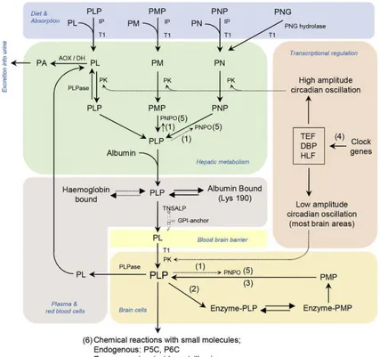
The human intestine only absorbs non phosphorylated B6 vitamers (Fig. 1.3).31 Therefore, phosphorylated vitamers are first hydrolysed by intestinal phosphatases.32 Once within the intestine cells, PN and PM can either be converted prior to transport to the liver33 or once within the liver to pyridoxal phosphate. In the liver (or intestine) PM, PN and PL, are first phosphorylated by the ATP-dependent pyridoxal kinase. The flavinmononucleotide (FMN)-dependent enzyme, pyridoxine 5′-phosphate oxidase (PNPOx) converts the phosphorylated derivative of PM and PN to PLP. Through the “salvage pathway” PLP is formed from the other vitamers taken from the diet or by recycling the cofactor from degraded enzymes34 (Fig. 1.2; Fig. 1.3). Although PNPOx is mainly expressed in the liver, this enzyme is expressed in all cell types. PLP is exported from the liver bound to albumin.35 When B6 vitamers intake exceeds requirements, PLP is dephosphorylated (mainly in the liver) and the resulting PL is oxidized by an aldehyde dehydrogenase or aldehyde oxidase (AOX)36 to pyridoxic acid prior to excretion in urine. To enter the brain, PLP must dissociate from albumin and be dephosphorylated to PL at the blood-brain barrier (BBB). Dephosphorylation is carried out by tissue non-specific alkaline phosphatase, an enzyme tethered to cell membranes at the BBB by a glycophosphatidylinositol (GPI) anchor. PL crosses the BBB, probably by facilitated diffusion, and is “trapped” as PLP in the brain cells and the choroid plexus by the action of pyridoxal kinase37 (Fig. 1.3). A similar mechanism occurs in other target cells. Within tissues, PNP or PMP can be converted back to PLP by PNPOx (salvage pathway)3 (Fig. 1.2).
9
Figure 1.3 Human PLP synthesis and homeostasis. AOX/DH, aldehyde oxidase (Mo
cofactor)/ β-NAD dehydrogenase; IP, intestinal phosphatases; ; E-PLP, enzyme bound PLP; E-PMP, enzyme-bound PMP; GPI, glycosylphosphatidylinositol anchor; PA, 4-pyridoxic acid; PK, pyridoxal kinase; PL, pyridoxal; PLP, pyridoxal 5′-phosphate; PLPase, pyridoxal-phosphatase; PLPBP/PROSC, pyridoxal 5′-phosphate binding protein; PM, pyridoxamine; PMP, pyridoxamine 5′-phosphate; PN, pyridoxine; PNG, pyridoxine-5′-β-D-glucoside; PNP, pyridoxine 5′-phosphate; PNPO, pyridoxamine 5′-phosphate oxidase; T1, transporter (identity unknown); TNSALP, tissue non-specific alkaline phosphatase. (1) feedback inhibition of PNPOx by PLP; (2) PLP functions as a cofactor; (3) salvage pathway; (4) PLP levels maintained by circadian-clock-controlled transcription factors (DBP, HLF, and TEF) targeting PK; (5) PNPOx mutations cause a B6-dependent epilepsy; (6) decreasing PLP
bioavailability resulting from accumulation of L-Δ1-pyrroline-5-carboxylic acid (P5C) and Δ1
10
1.2.3 Key enzymes in pyridoxal 5′-phosphate synthesis
The biosynthetic pathway leading to the formation of PLP is present in bacteria, plants and fungi. Particularly, in E. coli metabolism a key enzyme is pyridoxine 5'-phosphate oxidase (PNPOx), which catalyses the last step of vitamin B6 biosynthesis. However, the oxidation of PNP to PLP is an
important step in the recycling of this vitamer in all living beings.38 PLP can be synthesized by three different enzymes, which are not present in all organisms: pyridoxine 5′-phosphate oxidase (PNPOx); pyridoxal kinase (PDXK), which converts all vitamers into their related phosphorylated derivatives; and pyridoxal synthase, that is not present in γ-proteobacteria and in mammals.25 In mammals, PLP is recycled from degraded B6-enzymes and
from vitamers acquired from nutrients in the salvage pathway, which essentially involves two ubiquitous enzymes: the ATP-dependent pyridoxal kinase and the FMN-dependent PNPOx.3 Once it is made, PLP is targeted to apo-B6 enzymes that are being synthesized in the cell.39 The mechanism and
regulation of the salvage pathway and the mechanism of PLP transfer to PLP-dependent enzymes are poorly understood. Moreover, PDXK and PNPOx play key roles in regulating the level of PLP formation.3,40
Pyridoxine 5′-phosphate oxidase
PNPOx is a flavinmononucleotide (FMN)-dependent enzyme that uses molecular oxygen as electron acceptor. This protein is crucial in the PLP salvage pathway; it is responsible for the transfer of a pair of electrons from the C4′ carbon of PNP or PMP to a tightly bound molecule of FMN forming FMNH2. These two electrons are subsequently transferred in a second
half-11
reaction to molecular oxygen, regenerating FMN and forming H2O2.41
Kinetic studies carried out on rabbit PNPOx have led to the proposal of two possible catalytic mechanisms for oxidizing PNP and PMP to PLP (Fig. 1.4). The first one is a direct hydride transfer from C4′ of the substrate to N5 of FMN, to generate PLP and FMNH2. The second possible mechanism
involves the presence of a base at the active site that removes a proton from C4′ of the substrate, which then attacks FMN forming a covalent complex; collapse of this complex would generate PLP and FMNH2 (Fig. 1.4).
Figure 1.4 Description of pyridoxine 5′-phosphate oxidase (PNPOx) mechanism. A)
Reaction catalysed by PNPOx; B1) Direct hydride transfer of C4′ proR hydrogen of PNP (or PMP) substrate to FMN; B2) Removal of the proR proton at C4′ of PNP (or PMP) to generate a carbanion.3
In either cases, FMN is regenerated by the transfer of the two electrons to oxygen, forming hydrogen peroxide. Experiments performed with
site-12
directed mutants of the active site and isotope-labelled PMP have shown that the oxidation process involves direct hydride transfer from PNP or PMP to FMN.3
Pyridoxal kinase
The other essential enzyme involved in PLP synthesis is pyridoxal kinase, which, in the presence of MgATP, catalyses the addition of a phosphate group to the 5′-alcohol of pyridoxine, pyridoxamine and pyridoxal to form PNP, PMP and PLP, respectively (Fig. 1.5).3 In this reaction, both substrates are bound to the enzyme forming a ternary complex. The mechanism of phosphorylation has been previously elucidated for the sheep and E. coli enzymes and is characterised by a random sequential substrates addition.42 On the other hand, it has not been reported whether, in human pyridoxal kinase, the ternary complex is formed in an ordered or random sequence.42
13
In pyridoxal kinases, a conserved Asp235 residue is observed that makes a hydrogen-bond interaction with the C5′-OH group of substrates and deprotonates this group, thus resulting in a negatively charged O5′ atom making a direct nucleophilic attack on the ATP γ–phosphate.43 For the function of many kinases, both monovalent and divalent cations are known to be essential, providing forces for ATP-binding and substrate catalysis.42 Particularly, in human pyridoxal kinase, the affinity for the ATP and PL substrates increases in presence of K+ when compared to Na+, and then, the monovalent metals seem to lower the energetic barriers in the basal and transition state. Moreover, Mg2+ and Na+ cations act in tandem to anchor ATP at the active site, thus suggesting that both monovalent and divalent cations are necessary for the enzymatic function of pyridoxal kinase.3,42
1.3 Pyridoxal 5′-phosphate homeostasis
Pyridoxal 5′-phosphate (PLP) is a highly reactive aldehyde, that can react with several nucleophilic compounds in the cell.31 Intracellular free PLP concentrations are maintained at approximately 1 μM to prevent inappropriate reactions (“aldehyde stress” or “carbonyl stress”); in fact, the very reactive aldehyde group at the C4′ position can easily form aldimines with primary and secondary amines3 and, moreover, it can react with thiol groups. In humans, toxicity is usually observed when vitamin B6 uptake
exceeds 200 mg/day.44 Very high doses of supplementation in humans lead to toxicity that presents as sensory neuropathy, whereas low status of this vitamin has been associated with severe malnutrition, venous thromboembolism, many neurological diseases, such as epilepsy, Alzheimer's, Parkinson's and even diabetes and cancer.45,46 Levels of vitamin
14
B6 could also be raised as a result of an environmental insult or genetic
defects. Furthermore, the toxicity of PLP has been previously described not only in humans but also in other organisms, such as Candida utilis, where PLP can inhibit the enzymatic activity of 6-phosphogluconic dehydrogenase forming a Schiff base with a lysine residue located in the active site of the enzyme.2,47 Given the toxicity of PLP, different mechanisms are needed in the cells to strictly control the levels of this vitamer.
Several mechanisms are involved in maintaining low concentrations of PLP; at the same time, large amounts of this cofactor are required to saturate the many PLP-dependent enzymes. One regulation mechanism could be the conversion of PL into pyridoxic acid, catalysed by the aldehyde oxidase and the NAD-dependent dehydrogenase enzymes.36 PL results from the action of phosphatases, which catalyse the removal of phosphate groups. A PLP-specific phosphatase is involved in PLP homeostasis in mammals,48 and in E.
coli a specific phosphatase, encoded by the ybhA gene, has been identified
that maintains PLP homeostasis, reducing PLP toxicity.2 On the other hand, a candidate for an important missing player in PLP homeostasis is the PLP-binding protein (PLPBP), also called YggS in E. coli or PROSC in humans.31,49 YggS was first identified in E. coli as a member of the COG0325 family.50,51 The aforementioned family shares with alanine racemases and certain decarboxylases a similar folding,1 and has been shown to bind PLP.50 In crystal structures of yeast and E. coli COG0325 proteins it was observed that, like the N-terminal domain of alanine racemase and ornithine decarboxylase, these proteins fold as TIM barrel, displaying eight β–strands alternate to eight α-helices, and bind PLP in a similar mode.50
Prunetti and collaborators49 observed that an E. coli K12 ΔyggS strain accumulated PNP and that high levels of exogenous PN are toxic to this mutant strain; PLP, PM and PL are not toxic and, the latter vitamer
15
suppresses the PN toxicity, as also observed expressing the E. coli yggS gene in trans. The mechanism of this toxicity is unknown, although it was suggested that the high concentration of PNP, perhaps due to the retroactive inhibition of PNPOx by PLP, could inhibit PLP-dependent enzymes, including a particular transaminase involved in the synthesis of branched-chain amino acids.49 Moreover, in the ΔglyA ΔyggS strain was observed a defect in the cell division, probably due to lower levels of D-alanine. Thus, it has been suggested that the phenotypes observed in E. coli in the absence of yggS are caused by a lower activity of PLP-dependent enzymes.49 Furthermore, in humans other proteins such as glycogen phosphorylase in muscle,52 haemoglobin in erythrocytes,53 and albumin in plasma,35 can bind PLP and help maintain PLP homeostasis. Interestingly, also the enzymes that produce PLP (PDXK and PNPOx) can bind it tightly.34,54 Concerning pyridoxal kinase, in E. coli it was observed that PLP forms a complex with the enzyme and inactivates it. Interestingly, this complex can be partially reactivated by transferring the tightly bound PLP to apo-B6 enzymes, which
has been suggested to be an important pathway for B6 enzyme activation.40 In vitro studies suggest that PLP is protected intracellularly by being transferred
from these enzymes directly to PLP-dependent enzymes, such as serine hydroxymethyltransferase (SHMT).39,40,54,55 Safo and collaborators56 observed that PLP tightly binds at a non-catalytic binding site of PNPOx and remains bound during size exclusion chromatography.54 Furthermore, the crystal structure of E. coli PNPOx, obtained from crystals soaked in a concentrated solution of PLP showed a PLP molecule bound at the protein surface, about 11 Å from the active site.56 The actual involvement of this surface pocket, which is present as two symmetric sites in the dimeric PNPOx, has never been confirmed experimentally.
16
1.3.1 Feedback inhibition of pyridoxal 5′-phosphate synthesis
It was previously reported that PLP inhibits both pyridoxal kinase and PNPOx.57,58 In Escherichia coli PNPOx, a competitive PLP product inhibition was observed by Zhao and Winkler, and also substrate inhibition by PNP, but not PMP, was described, that can be reversed by increasing oxygen concentration.58 PLP product inhibition of E. coli PNPOx has been reported to take place with a KI of 8 µM and attributed to PLP binding at the
active site.58 Also, in previous studies on sheep brain PNPOx, a competitive nature of PLP inhibition with respect to PNP substrate was suggested; the inactivation of the enzyme was apparently due to a modification of a lysine residue, that is located very close to the substrate binding site.59 Furthermore, steady-state data, obtained from kinetic measurements on rabbit liver PNPOx, indicated that the catalytic mechanism to form the PLP product follows two different kinetic mechanisms depending on the substrate used, with oxygen in both cases functioning as the electron acceptor. Thus, PNP oxidation proceeds via a ping-pong mechanism, while PMP oxidation follows a ternary sequential mechanism.59 In humans, although the enzyme shares only 39% of sequence identity with E. coli PNPOx, a remarkable similarity has been found between the two enzymes in the structure and in the catalytic properties. Particularly, these two enzymes differ in substrate specificity, with the E. coli PNPOx displaying a greater preference for PNP; however, in both enzymes PLP acts as product inhibitor.34 The KI of PLP for the human
enzyme is lower than that of the E. coli PNPOx58 and was estimated to be about 3.2 µM.34 Since this KI value is not close enough to that of a putative
high affinity binding site, it has been proposed by Musayev and collaborators34 that PLP inhibits the enzyme by binding at the active site. This product inhibition is probably an important regulatory mechanism of
17
PLP biosynthesis, however further investigations are required to gain better insight into this mechanism.
Concerning E. coli pyridoxal kinase, it has been observed that PLP is a slow tight binding inhibitor of this enzyme.40 It is known that in E. coli two kinases are present, one of which is able to catalyse the phosphorylation of all B6 vitamers, while the other is specific for PL.60 In the first pyridoxal kinase,
encoded by the pdxK gene, the mechanism of PLP inhibition was described in detail, and it is characterised by the formation of a Schiff base between PLP and an active site lysine residue (Lys229). The inactivation is faster when both PLP and MgADP are present compared to when PLP is present alone.40 The human enzyme is slowly inhibited by PLP34 and this inhibition is probably due to the formation of a non-covalent enzyme-PLP complex.43
1.4 Pyridoxine 5′-phosphate oxidase from Escherichia coli
Pyridoxine 5′-phosphate oxidase (PNPOx) is encoded by the pdxH gene and has been purified from sheep, rat and pig brain, rabbit liver, and E.
coli 58,59,61,62 with the most extensive studies being performed on the rabbit liver, sheep brain, and E. coli enzymes. In Escherichia coli, PNPOx is a relatively abundant enzyme,58 and there is enough enzyme to serve as a significant reservoir of PLP. It was previously reported that the pdxH gene in
E. coli forms a complex operon with a downstream gene, tyrS, which encodes
the essential enzyme tyrosyl-tRNA synthetase.63 The characterisation of
pdxH mutants revealed several unusual phenotypes, like a block in cell
division, excretion of L-glutamate and inhibition of L-isoleucine biosynthesis.63 Moreover, the absence of growth of pdxH mutants aerobically
18
and anaerobically, also when supplemented with PL, suggested that the PNPOx is not only essentially required for PLP biosynthesis in cells, however, the enzyme could play a role in an alternative pathway.63 In Eubacteria, PNPOx catalyses the final step of vitamin B6 metabolism and is a
member of the oxidoreductase enzymes, that catalyse a simultaneous oxidation-reduction reaction, where the molecular oxygen is thought to be the sole electron acceptor in the reaction.25,64 The E. coli enzyme is a homodimer, and each monomer counts 218 amino acid residues.38
Figure 1.6 Monomeric structure of E. coli PNPOx. The eight β-strands are shown in pink,
the five α-helices in cyan and loops in magentas; FMN and PLP molecules at the active site are shown as sticks with atom-based colours (PDB code: 1G7956).
The folding of each monomer (Fig. 1.6) can be described in terms of two domains: the larger domain, which consists of β–strands and two α–helices; and the smaller one, that is formed by the remaining three α–helices and is near the β–barrel core.38
The binding site of FMN is located in a deep cleft formed by the two subunits and is conserved in the PNPOx structures. In the dimeric structure, two molecules of FMN are bound at the interface and are
19
also involved in several hydrogen-bond interactions with the protein; particularly, the phosphate group of this cofactor interacts extensively with highly conserved arginine and lysine residues from both subunits of the protein.38,65 PLP and PNP bind at the re face of FMN, with the phosphate group pointing out of the catalytic cavity, in contrast to the FMN phosphate moiety that points downward into the cavity bottom.56 In previous studies, different crystal structures describing the E. coli PNPOx complexed with FMN and PLP were reported.38,56 The crystal structure of the enzyme without ligands shows an open active site;65 however, binding of either PNP or PLP elicits a protein conformational change that partially closes the active site.56 In this close conformation, three conserved residues, Tyr129, Arg133 and Ser137, come closer to PLP phosphate moiety, and together with the Lys72 help the interaction between PLP and FMN, consisting in extensive hydrogen-bonds and van der Waals contacts.38 All these amino acid residues are highly conserved in most members of the PNPOx family.65 The invariant Arg197 and His199 residues from the second monomer are situated in front of the pyridine ring of PLP, and make direct hydrogen bond interactions with the carbonyl oxygen on C4′ and the O3′-hydroxyl group of PLP, respectively. Arg197 makes further interactions with the phosphate moiety of PLP.56 These two residues act as a clamp on PLP sandwiching the pyridine moiety onto the isoalloxazine ring while catalysis takes place.38 Interestingly, the monoclinic crystal form of the enzyme in complex with PLP showed previously unobserved N-terminal residues that fold over the active site to completely close it and sequester the ligand from the solvent; in fact it has been suggested that the monoclinic crystal structure may represent the catalytic state conformation.38
20
1.4.1 Non-catalytic secondary PLP binding site
It has been previously reported in crystallographic studies carried out in the presence of PLP that there is one molecule of PLP per monomer of PNPOx, and that this PLP is located at the active site, nearby the FMN cofactor. However, soaking crystals with a higher concentration of PLP, the protein structure displayed two molecules of PLP per monomer; one PLP molecule was at the active site, while the second one was located at a non-catalytic PLP binding site56 (Fig. 1.7). This secondary binding site, was firstly described on the basis of experiments carried out in solution by Yang and Schirch.54 In this case there were experimentally evidences: for instance, when the E. coli PNPOx was incubated with PLP and then passed through a size exclusion chromatography, it retains PLP and shows a normal catalytic activity. Moreover, the spectral property of PLP bound at the apo-PNPOx suggested a non-Schiff base binding (a non-covalent binding of PLP); thus, in both apo- and holo-PNPOx, PLP binds at a non-catalytic site.54 An unanswered question is the location of the non-catalytic secondary PLP binding site. In the trigonal crystal structure of the E. coli enzyme, a second PLP molecule binds to PNPOx at about 11 Å from the active site, prompting speculation that this crystallographic secondary binding site corresponds to the functionally observed secondary PLP binding site (PDB code: 1G79).56 The plane of the pyridine ring is sandwiched between the side-chains of Phe177 and Lys145 (which also makes a hydrogen bond with the PLP phosphate moiety) and is located close to what seems to be a tunnel that leads to the active site.38 Adjacent to Phe177, and also guarding the tunnel, is Asn84, which makes a close hydrogen bond to the pyridine nitrogen of PLP (Fig. 1.7). The putative tunnel between the two PLP binding sites is formed almost entirely by one subunit and it is mainly composed by glycine residues
21
and small side-chains.56 There are structural water molecules located inside the tunnel. The presence of the side-chains of Asn84, Arg133, Phe177 and Trp178 in the cavity narrows the tunnel. Although the structures show the tunnel to be small for the passage of PLP, the channel could easily open up, since most of the surrounding protein structures are flexible.56 The possible role of the non-catalytic site in channelling PLP to enzymes that requires it as a cofactor is still an important field of investigations. Binding of PLP at the non-catalytic site of PNPOx might be one method for cells to regulate free PLP concentration in vivo.
Figure 1.7 Enlargement showing the non-catalytic secondary PLP binding site. The protein
chain is shown in cyan (PDB code: 1G7956). The residues of the putative secondary PLP binding site are shown as sticks, in blue. FMN and both PLP molecules are shown as sticks and coloured by atoms.
22
1.5 Pyridoxal 5'-phosphate deficiency and related diseases in humans
Because of the essential role of PLP in neurotransmitters metabolism, including dopamine, serotonin, histamine, D-serine, epinephrine and GABA, it is not surprising that inborn errors leading to PLP deficiency result in a pathological phenotype.66 PLP deficiency occurs through different mechanisms. An example is PNPOx deficiency, caused by inborn defects of the PNPOx enzyme, the neonatal epileptic encephalopathy, a disorder affecting PLP synthesis and recycling. The patients affected by this kind of disease display a particular electroencephalogram pattern and some of them promptly respond to PLP, but not to pyridoxine treatment.67 Another disorder affecting PLP import into the brain is alkaline phosphatase deficiency, or hypophosphatasia, which is associated with a molecular defect in the gene encoding the tissue non-specific alkaline phosphatase (TNSALP). TNSALP hydrolyses several substances, including inorganic pyrophosphate and PLP. The latter must be dephosphorylated by TNSALP before it can cross the cell membrane.68,69 Vitamin B6 deficiency in the brain impairs the
synthesis of neurotransmitters, which can cause seizures.70 Moreover, PLP binding protein (PLPBP) deficiency is a disorder likely due to abnormal intracellular PLP transport, given the fundamental role of this protein in the homeostatic regulation of free PLP levels. The clinical phenotype is similar to that observed in patients with PNPOx deficiency.31 In other cases, the accumulation of metabolites that inactivate PLP causes the pyridoxine-dependent epilepsy (PDE), that is characterised by ALDH7A1 deficiency (also known as α-aminoadipic semialdehyde dehydrogenase or antiquitin deficiency) or by ALDH4A1 (also known as P5C dehydrogenase) deficiency.71 ALDH7A1 deficiency results in the build-up of an intermediate with a nucleophilic carbon atom, Δ1-piperideine 6-carboxylic acid (P6C),
23
which reacts with PLP thereby inactivating it and resulting in PLP deficiency.72 Accumulation of Δ1-pyrroline 5-carboxylic acid (P5C) in ALDH4A1 deficiency has a similar effect in hyperprolinaemia type II.73 All the deficiencies described above (PNPOx, ALDH7A1, ALDH4A1 and PLPBP), in their most severe forms (neonatal presentations) can have other expected consequences including anaemia, lactic acidosis, and hypoglycaemia.71 Furthermore, PLP not only acts as a cofactor of numerous enzymatic reactions, but also works as an antioxidant molecule by quenching oxygen reactive species.13 Several studies suggest that DNA damage is a possible link between metabolism and cancer. In particular, it has been proposed that under metabolic stress conditions or in the case of reduced availability of necessary nutrients some cellular processes such as DNA acetylation/methylation, synthesis of DNA precursors and ROS production can be altered causing DNA damage, which can drive cells toward cancer.74 Although many studies converge towards a protective role of B6 in cancer,
the molecular mechanisms are not completely understood. PLP, as antioxidant molecule, could play an important role in mediating the cross talk between metabolism and DNA damage, in fact PLP acts against the formation of advanced glycation end products (AGEs), that are genotoxic compounds associated with senescence and diabetes.75 This relationship between PLP deficiency and diabetes has been previously described,76–78 but the molecular and cellular mechanisms underlying this relationship have not yet been completely understood. PLP deficiency may impact on diabetes in different ways; for example, Kotake and collaborators79 previously reported that metabolites produced in the altered tryptophan degradation pathway can interfere with insulin activity, causing insulin resistance. Moreover, given the important role of PLP in controlling the expression of genes involved in adipogenesis and in the metabolic pathway of homocysteine, a deficiency of
24
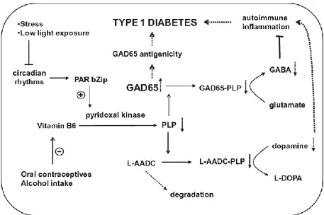
this vitamin could cause insulin resistance.80,81 Rubì and collaborators78 reported that PLP deficiency may play a role in type I diabetes onset and this is probably due to the function of this vitamer as a cofactor for two enzymes both present in the pancreatic islet: glutamic acid decarboxylase (GAD65), which synthesizes γ–aminobutyric acid (GABA), that, as part of the GABA cycle, may represent means by which the cell can utilize glutamate as a reserve energy source; and L-aminoacid decarboxylase (L-AADC), a key enzyme in dopamine synthesis (Fig. 1.8).78 Dopamine and serotonin produced by L-AADC could be involved in the regulation of insulin synthesis and secretion, thus, lower levels of dopamine can favour the onset of the type I diabetes.82 Furthermore, GAD65 can act as autoantigen in pancreatic cells, in fact, it has previously been observed that amino acids 250-273 of this enzyme show sequence similarity with amino acids 28-50 of the coxsackievirus B protein P2-C.83 It was proposed that, as a result of this molecular mimicry, cross-reactive T-cell proliferation will occur, leading to autoimmune destruction of the B cells.82 Also, the GAD65 apo- to holo-enzyme change is key for the regulation of this protein. PLP binding domain surrounds the auto-antigenic region described above, thus, a PLP-deficiency results in higher levels of apo-GAD65, which is antigenic and not able to synthesize GABA (Fig. 1.8).78
25
Figure 1.8 Hypotetical model for PLP deficiency and the onset of type I diabetes. B6
vitamers are converted into PLP by pyridoxal kinase, whose expression is promoted by the transcription factor PAR bZip. Alteration in circadian cycle, alcohol consumption and use of contraceptives decrease PLP synthesis. GAD65 devoid of PLP does not synthesize GABA. Alteration of L-AADC function lowers dopamine levels. GAD65, glutamic acid decarboxylase; PLP, pyridoxal 5'-phosphate; PAR bZip, transcription factor belonging to proline and acidic amino acid-rich basic leucine zipper transcription factor family; L-AADC, L-aminoacid decarboxylase; L-DOPA, levodopa.78
Furthermore, it has been reported that high expression levels of pyridoxal kinase positively correlate with survival of non-small cell lung cancer (NSCLC) patients.84 In previous studies, mutations in Drosophila dPdxk gene caused chromosome aberrations (CABs), that can be rescued by PLP supplementation.85 The same effect is produced by treating wild type flies with PLP analogues such as 4-deoxypyridoxine (4-DP) or inhibitors of PLP-dependent enzymes like cycloserine and penicyllamine. The alteration of pyridoxal kinase functionality increases the glucose content in larval
26
haemolymph, and hyperglycaemia and CABs are interconnected by a cause-effect relationship, in which high glucose is largely responsible for CABs.85
1.6 Human pyridoxine 5'-phosphate oxidase
As the E. coli enzyme, human PNPOx has been crystallized and its structure determined.34 This protein, in humans, is coded by the pdxH gene located on chromosome 17q21.2. As mentioned above, the enzyme has a primary role in the vitamin B6 salvage pathway and a non-correct functioning of it brings to
an abnormal activity of several B6–dependent enzymes. The
three-dimensional fold of the human enzyme is very similar to that of the E. coli enzyme. The human and E. coli enzymes share 39% sequence identity, but the binding sites for FMN and substrate are highly conserved.34 As observed with the E. coli enzyme, also in this case, thanks to chromatography studies, it has been demonstrated that the human enzyme binds tightly one molecule of pyridoxal 5′-phosphate on each subunit and this tight binding occurs at a non-catalytic binding site.34 However, unlike the E. coli PNPOx,38,56 no crystallographic data of the secondary PLP binding site in the human enzyme have been reported. In the light of all these considerations, a parallel study of the human and E. coli PNPOx would have an extraordinary importance in order to understand more in depth the mechanism of regulation of these enzymes. Human PNPOx is a homodimer and each monomer has a length of 261 amino acids. In crystallographic studies, the first 48 N-terminal residues are not visible in the electron density map because of disorder. The 1.95 Å three-dimensional structure of human PNPOx in the presence of PLP reveals a very similar protein fold to that of E. coli 65 (PDB code: 1NRG34). The monomer structure of human PNPOx shows the typical two-domain architecture (domain 1 and domain 2), previously observed for E. coli. The
27
larger domain 1 of the E. coli enzyme is formed by eight β-strands (β1–β8) and two α-helices (α1 and α2), with the human PNPOx having one additional β-strand, β7′, while the smaller domain 2 of both PNPOx structures is made up of 3 helices (α3, α4 and α5). The β7′ strand and part of the turn and loop regions associated with it are absent in the E. coli PNPOx structure because of a 15-residue insertion at residue 238 (Fig. 1.9).34 The role of these additional structures in human PNPOx might be solely structural, because they have no direct catalytic role and are also not involved in dimer formation. These unique regions are open to the solvent, with the four-amino-acid residue loop that connects the strands β7 to β7′ highly disordered.34
Figure 1.9 Structure of the human PNPOx monomer. FMN and PLP molecules are shown as
sticks with atom-based colours. The loops are shown in violet; -helices are shown in blue;
-strands are in light blue. The 7' strand is present only in human PNPOx (PDB code: 1NRG34).
One of the most relevant differences in the amino acid sequence between E.
coli and human PNPOx is that the human enzyme contains six Cys residues,
whereas the E. coli enzyme contains only a single Cys residue. Unfortunately, only four of the six residues are modelled in the crystal
28
structure. The other two are located in the missing N-terminal region. Two of the cysteine residues (Cys82 and Cys86, that in the E. coli PNPOx correspond to Val54 and Val58, respectively) are located at the dimer interface, each facing its symmetry-related counterpart. The distance from sulfhydryl groups of Cys82 and Cys86 and their related counterpart are 3.6 Å and 4.2 Å, respectively. These distances are significantly longer than an expected disulfide bond, but there is evidence that some disulfide bonds are formed between the monomers. The two remaining cysteine residues, Cys72 and Cys156 (Ala45 and Lys128 in E. coli PNPOx, respectively), are located on the surface of the protein and possibly both of them make a disulfide interaction with 2-mercaptoethanol of the buffer used to purify and crystallize the protein.34
Other differences between the two structures mainly occur in turns, loops, the N-terminal segment, and the α5 helix. There are two pairs of inter-subunit salt-bridge interactions, including Arg116–Glu143 and Arg181–Asp228 (Arg88-Ile115 and Arg153-Asp200, respectively in E. coli PNPOx). The latter is conserved in both PNPOx structures, whereas the former does not occur in the E. coli enzyme. In E. coli, the Glu143 residue, present in the human enzyme, is replaced by isoleucine, necessitating the rearrangement of Arg116 to make an inter-subunit salt bridge with Glu217 (Glu189 in E. coli PNPOx). Interestingly, the latter amino acid residue is strictly conserved in both PNPOx structures, but the salt-bridge interaction between Arg116 and Glu217 is only observed in the E. coli structure. Contrarily to the β7 and β8 strands, that are involved in extensive inter-subunit interactions, the new β7′ strand, makes only one weak inter-subunit hydrogen-bond interaction from Met246 to Pro179 (Ser151 in E. coli PNPOx).34 The binding site of FMN is identical to that of the E. coli PNPOx structure, with the FMN located in a deep cleft formed by the two subunits with extensive hydrogen-bond
29
interactions to the protein.65 These interactions involve both subunits. Most of the salt-bridge and hydrogen-bond interactions between the protein and the FMN are strictly conserved in both PNPOx structures. In particular, these structures have very similar FMN geometry, active-site environment, and bond distances. The bound PLP molecule occupies the same position in the active site and has essentially unaltered conformation in both the human and the E. coli PNPOx structures. In addition, the two crystals show similar interactions between the protein and the bound PLP, with almost all of the residues and interactions strictly conserved. The interactions involve both protein subunits; the phosphate moiety of PLP makes salt-bridge and hydrogen-bond interactions with Lys100, Arg161, Tyr157 and Ser165, belonging to a monomer, and Arg225, belonging to the other (Lys72, Arg133, Tyr129, Ser137 and Arg197, respectively in E. coli PNPOx).34 The PLP pyridine ring is stacked parallel against the FMN isoalloxazine ring, with extensive van der Waals contacts between the two. As observed in the E. coli structure, C4' of PLP and N5 of FMN are separated by approximately 3.4 Å. The O3′- hydroxyl group of PLP makes a hydrogen-bond interaction with His227 (His199 in E. coli PNPOx), whereas the carbonyl oxygen on C4′ makes a water-mediated interaction with Glu77 (Asp49 in E. coli PNPOx), and these residues belong to different monomers of the protein. Lastly, the pyridine nitrogen makes a water-mediated hydrogen-bond interaction with Trp206 (Trp178 in E. coli PNPOx). The phosphate moiety of PLP is oriented toward the N-terminus of the α4 helix, which further compensates for the PLP negative charge in addition to the salt-bridge interactions. Among the active site residues that make direct or water-mediated contact with the PLP, Glu77 is the only non-conserved residue among the two structures, and it is replaced by Asp49 in E.
30
1.6.1 PNPOx mutations and neonatal epileptic encephalopathy
Maintenance of a correct balance among B6 vitamers inside the cell is of
fundamental physiological importance in all organisms, including humans where PLP imbalance causes severe neurological dysfunctions as described above. Among those, of particular significance is the neonatal epileptic encephalopathy (NEE), a severe neurological disorder which usually manifests a few hours after birth, with seizures that can be fatal and do not respond to conventional anticonvulsant treatments. This particular form of epilepsy (an autosomal recessive disorder) is caused by mutations in human PNPOx, that lead to insufficient levels of PLP and a subsequent decreased activity of PLP-dependent enzymes. In one of the first studies about NEE, the authors86 described a neonate patient whose seizures were not controlled by pyridoxine but were well controlled by PLP. Clayton and collaborators87 described the same phenomenon in an infant whose levels of B6 vitamers
suggested deficient PLP-dependent enzyme activities: the aromatic amino acid decarboxylase deficiency causes low concentrations of homovanillic acid (HVA) and 5-hydroxyindoleacetic acid (5HIAA) with raised 3-methoxytyrosine and urinary vanillactate. Then, defects in the PLP-dependent pathways for catabolism of threonine and glycine determine an increase in plasma levels of these amino acids.87 Other patients with similar findings were identified and were shown to have PNPOx mutations leading to reduced enzyme activity.88 Treatment of PNPOx deficiency was found to be difficult. Reports have emphasized the possibility of normal developmental outcome with early treatment.89,90 By 2014, it was clear that some patients with PNPOx deficiency respond to treatment with pyridoxine, and treatment with PLP may even aggravate seizures.90,91 Certain genotypes (R225H/C and D33V) appear more likely to result in seizures responsive to
31
pyridoxine. Other mutations seem to be associated with infertility, miscarriage, and prematurity. To date, 62 genetically confirmed PNPOx-deficient patients have been reported with 27 different mutations in the gene encoding PNPOx 92–97Most of them are homozygous missense mutations, but also stop codon suppression, deletions and splice site mutations have been reported in the literature.40,87,88,90,98–102 So far, the only NEE-related human PNPOx mutations characterised from a functional and structural point of view are R229W,100 R95C39 and R116Q.92 Both R229W and R95C variants resulted to be 350-fold less catalytically efficient than the wild type enzyme and presented a 50-fold and 15-fold reduction in affinity for the FMN cofactor, respectively. The crystal structure of the R229W mutant showed that the mutation prevents the proper binding of the PNP substrate due to the absence of two essential hydrogen bond interactions of His227 and Arg225 with PNP. Moreover, the binding of the FMN cofactor is weakened because of the loss of a hydrogen bond and salt-bridge interactions between FMN and Arg229 and Ser175 residues.100 Also Arg95 makes salt-bridge and hydrogen interactions with the FMN phosphate and its substitution to Cys95 likely destabilize FMN binding.39 Recently, the functional effects of the controversial c.347G>A (p.R116Q) mutation of human PNPOx gene have been studied and its pathogenic role in epileptic encephalopathy has been discussed.92 Patients carrying this mutation present a peculiar clinical feature, namely a later epilepsy onset. In vitro biochemical characterisation of the R116Q mutant showed that although the catalytic properties are not drastically changed, the mutation affects the thermal stability of the enzyme. This altered stability of the R116Q enzyme with temperature could be linked to the increased susceptibility to febrile seizures frequently observed in children affected by NEE.92
32 1.7 Human pyridoxal kinase
Human pyridoxal kinase (PDXK) is a member of the ribokinase family and catalyses the ATP-dependent phosphorylation reaction of vitamin B6. This
enzyme is encoded by the highly conserved pdxK gene, that is present also in
E. coli, and in humans is located on the 21q22.3 chromosome.103 The structures of pyridoxal kinase from sheep brain and E. coli were solved in complex with substrates and products.104,105 Cao and collaborators103 reported for the first time the crystal structure of human PDXK at 2.8 Å resolution (PDB code: 2F7K), and then, a structure at a better resolution (2.0 Å and 2.2 Å, in absence and in complex with MgATP, respectively; PDB codes: 2YXT and 2YXU) was described by Musayev and collaborators.42 Human pyridoxal kinase is a homodimer, containing 312 amino acid residues per monomer, related by a non-crystallographic twofold axis in an asymmetric unit and the two monomers interact through hydrogen binding, salt bridges and hydrophobic interactions.103 Each monomer contains nine α-helices, named α1-9, and 11 β-strands, named β1-11. This structure shows the typical ribokinase superfamily overall folding pattern, with the αβα three-layer sandwich,106 characterised by a central core of β-strands, surrounded by α-helices (Fig. 1.10).42 The overall structure is particularly conserved in E. coli, sheep brain and human PDXK.42,103,105 The dimer interface is formed between α1, α9, β1 and β3 from each monomer, and the residues involved in this interaction are substituted by more hydrophobic residues in sheep brain PDXK, thus indicating a stronger hydrophobic interaction between monomers.103 On the enzyme surface, there is a cavity with negative charge that is favourable for binding the positive charged substrates, such as the pyridine ring of the vitamers and the adenine ring of ATP. ATP interacts with the hydrophobic side chains of Val201, Ile223 and Leu263, and,
33
interestingly, these data are consistent with the biochemical results obtained for this enzyme. In fact, human PDXK displayed higher affinity for ATP than the sheep brain enzyme, where the aforementioned residues are replaced with less hydrophobic residues (Ala201, Met223 and Met263).103 Comparison of the structures in absence or in complex with MgATP shows that a loop region between strand β11 and helix α7 (residues 224-228) have rotated away from the active site to allow binding of the ATP. Moreover, the so-called “flap” (residues Gly117-Val128), which is formed by strand β6, Loop I and strand β7, moved closer to the bound ATP, thus suggesting that this flap plays an important role in preventing the unproductive hydrolysis of ATP in the absence of vitamin B6; in fact, it provides hydrogen-bond interactions to
the ATP β- and γ-phosphates.42,104,105 Metals, both monovalent and divalent cations, are known to be absolute requirements for the function of many kinases, providing driving forces for ATP binding and substrate catalysis.107 Safo and collaborators105 observed in previous studies on E. coli PDXK that K+ is the required monovalent cation for the enzyme activity, and in humans, both ATP and PL bind with higher affinity in the presence of K+.42 However, Na+ elicits the maximum enzyme activity, which results to be twofold higher than in the presence of K+. It is possible that the replacement of K+ with Na+ changes the geometry of the active site to a more optimal orientation of catalytic residues, leading to greater enzyme activity in the presence of the latter cation. The Mg2+ ion in human PDXK is located between the ATP α- and β-phosphate groups (Fig. 1.10), and this observation is in contrast to the sheep brain PDXK structure, where the ATP phosphate groups interact with a divalent Zn2+ cation.42 In previous studies, it has been reported that even for the mammalian enzymes, ZnATP appears to be a better substrate only when the metal nucleotide concentrations are low, and thus, under physiological conditions, MgATP is the favoured substrate.108
34
Figure 1.10 Monomeric structure of human pyridoxal kinase complexed with MgATP. ATP
is shown as sticks with atom-based colours; α-helices are shown in green, β-strands in light blue and loops in blue. Also the Mg2+ cations are shown at the active site as spheres (PDB code: 3KEU42).
In human pyridoxal kinase, as in the ribokinase superfamily, a conserved amino acid residue (Asp235) of the active site is observed to make a hydrogen-bond interaction with the C5'-OH group of the PL substrate.43 These data were also confirmed by kinetic studies carried out on two variants of this aspartate residue, D235A and D235N, showing a decrease in catalytic activity and PL affinity, while ATP binding has remained unchanged in both cases.43 As mentioned above, PDXK is an ATP-dependent kinase and catalyses the conversion of PN, PM and PL to their related phosphorylated derivatives, PNP, PMP and PLP.109,110 The kinetic constants in the human enzyme are similar for the different vitamers and the estimated parameters are the following: KM values for PN, PM and PL are 20, 35 and 30 µM,
respectively; kcat values are 20, 20 and 45 min-1, respectively; and the KM
35
are 210, 330 and 420 µM, respectively.108 These data confirm the higher affinity of the enzyme for the vitamer than the ATP as observed previously on human PDXK variants of the active site Asp235 residue.43 PL binding at the active site involves Ser12, Thr47, Asp235, Val19 and Tyr84, that are totally conserved residues, with the exception of Thr47, which is replaced with a proline residue in the E. coli structure.42 The importance of these residues was also highlighted by a work on some pyridoxal kinase inhibitors, such as the ginkgotoxin (4'-O-methylpyridoxine), an analog of vitamin B6. In
fact, Thr47, Tyr84 and Val19 together with Phe43, Val231 and His46 residues are found to make hydrophobic interactions with this inhibitor, that like the PL, entirely places in the PL binding site.110
1.7.1 Human pyridoxal kinase in cancer and diabetes
In previous studies, pyridoxal 5'-phosphate has been proposed to influence carcinogenesis through different pathways, including those involved in DNA metabolism, suggesting a protective role of vitamin B6 against DNA
damage.111 PLP supplementation was inversely correlated with the colorectal cancer risk112 and the low PLP levels are also correlated to the onset of diabetes.78 Moreover, an incorrect intake of vitamin B6 could represent a risk
factor for diabetic patients, as it enhances DNA damage. PLP deficiency can result in chromosome aberrations and, thus, it can be related to an increased formation of genotoxic compounds, such as the advanced glycation end products (AGEs).85 Chromosome aberrations (CABs), such as deletions, duplications, translocations, DNA amplification and formation of aberrant gene fusions can contribute to cancer development.113 These CABs are mainly generated by unrepaired double strand breaks (DSBs), which can be
36
induced by external agents (such as chemical mutagens) or by endogenous factors (such as oxidative metabolism).85 In literature, several works suggested that vitamin B6 has a protective role against DNA damage and
cancer.114–116 Previously, it has been reported that the mutation dPdxk1 in the
Drosophila gene, encoding pyridoxal kinase (dpdxK), causes chromosome
aberrations. Moreover, the haemolymph of dPdxk1 larvae contains nearly twice as much glucose as that of the wild type larvae.85 The hyperglycaemia is interconnected with CABs by a cause-effect relationship, in which high glucose is largely responsible for CABs. High glucose triggers AGEs formation, which through ROS production leads to the formation of chromosome aberrations.85 Interestingly, this relationship between glucose and CABs, in PLP depleted cells, is evolutionarily conserved as glucose supplementation enhances chromosome damage also in human cells.46 Furthermore, hyperglycaemia can be considered a cancer risk in diabetic patients by different ways: the excess of glucose can promote cell growth or it can also cause oxidative stress, thus inducing DNA damage.46 The role of PDXK in chromosome integrity maintenance has also been demonstrated in yeast showing that mutations in the BUD16 gene, the pdxK orthologue, cause gross chromosome rearrangements largely mediated by altered DNA synthesis.117 A dysfunction of PDXK results in PLP deficiency. It has been reported by Merigliano and collaborators111 that a decrease in PLP levels strongly impacts on chromosome integrity in both diabetes models, thus suggesting a combined genotoxic effect of low PLP and high glucose levels. The impact of low PLP levels on genome integrity has also been tested on human cells. HeLa cells deprived of PLP by RNA interference directed against the pdxK gene showed chromosome aberrations. Moreover, the treatment of HeLa cells with 4-deoxypyridoxine causes chromosome aberrations, confirming the relationship between PLP deficiency and CABs