European Journal
of Case Reports in
Internal Medicine
DOI: 10.12890/2020_001525 European Journal of Case Reports in Internal Medicine © EFIM 2020
Doi: 10.12890/2020_001525 - European Journal of Case Reports in Internal Medicine - © EFIM 2020
Necrotizing Fasciitis Causing Acute Compartment Syndrome
after Radial Artery Catheterization
Filipa Cabral Amado1, Carlos Noversa2, Andreia Moura2, Luís Carvalho1, Leila Cardoso3
1 Serviço de Medicina 1, Centro Hospitalar de Leiria, Leiria Portugal
2 Serviço de Anestesiologia, Centro Hospitalar e Universitário de Coimbra, Coimbra, Portugal 3 Serviço de Medicina Interna, Centro Hospitalar e Universitário de Coimbra, Coimbra, Portugal
Received: 27/01/2020 Accepted: 03/02/2020 Published: 27/02/2020
How to cite this article: Cabral Amado F, Noversa C; Moura A, Carvalho L, Cardoso L. Necrotizing fasciitis causing acute compartment syndrom after radial artery catheterization. EJCRIM 2020;7: doi:10.12890/2020_001525.
Conflicts of Interests: The Authors declare that there are no competing interests. This article is licensed under a Commons Attribution Non-Commercial 4.0 License
ABSTRACT
Necrotizing fasciitis is a rare but potentially fatal infection involving the subcutaneous tissue and fascia with the development of necrosis of these structures. Acute compartment syndrome occurs when increased pressure within a closed muscle compartment compromises the circulation and function of the tissues within that space. We report the case of a male patient who was admitted to the intensive care unit for the management of urosepsis due to an acute obstructive pyelonephritis complicated by cardiopulmonary arrest. A radial arterial catheter in the left arm was urgently inserted, under suboptimal aseptic technique. His clinical condition progressively deteriorated, and swelling of the left arm with extension to the forearm with incipient signs of compromised perfusion were observed. The diagnosis of necrotizing fasciitis with acute compartment syndrome was made and an emergency fasciectomy performed. Following this, the patient gradually improved, organ dysfunction resolved, and he was discharged without sequelae.
LEARNING POINTS
• Necrotizing fasciitis is a rare but potentially fatal infection that can cause substantial tissue destruction and lead to sepsis.
• In rare cases, it can cause acute compartment syndrome, which occurs when increased pressure within a closed muscle compartment compromises the circulation and function of the tissues within that space.
• Both necrotizing fasciitis and acute compartment syndrome can be a complication of an arterial catheterization performed under suboptimal aseptic technique; hence, rigorous daily physical examination of the site is crucial for timely diagnosis.
KEYWORDS
Necrotizing fasciitis, acute compartment syndrome, arterial catheterization
INTRODUCTION
Necrotizing fasciitis (NF) is a rare but potentially lethal infection of the fascia, rapidly progressive, with the development of necrosis of the subcutaneous tissues[1]. It may occur as a complication of a variety of surgical procedures or medical conditions but can also be idiopathic.
It is associated with a high mortality rate and requires aggressive treatment. Acute compartment syndrome (ACS) occurs when increased pressure within a closed muscle compartment compromises the circulation and function of the tissues within that space. It may affect any compartment and almost any injury can cause this syndrome[2]. It is a surgical emergency, where an emergency fasciectomy is the definitive
surgical therapy. We report the case of a patient who developed NF of the left arm after arterial line placement under suboptimal aseptic technique, which led to ACS.
European Journal
of Case Reports in
Internal Medicine
DOI: 10.12890/2020_001525 European Journal of Case Reports in Internal Medicine © EFIM 2020
CASE DESCRIPTION
A 54-year-old male patient with a clinical history of hypertension and nephrolithiasis was admitted to the emergency department with an acute pyelonephritis associated with an obstructing calculus and abscess causing septic shock. He underwent emergency cystourethroscopy with removal of the calculus, abscess drainage and ureteral catheterization in the operating room. Immediately after the procedure, the patient went into cardiopulmonary arrest and was successfully resuscitated. A radial arterial catheter in the left arm and a central venous catheter in the right subclavian vein were urgently inserted and, given the cardiovascular instability, the aseptic technique was suboptimal. The patient was subsequently admitted to the intensive care unit. He first developed multiple organ dysfunction syndrome, requiring sedation, mechanical ventilation and vasopressor support. He received antibiotic therapy according to the antibiogram, when positive blood and urine cultures were identified. After 5 days, during which the patient progressively recovered from the septic shock, his clinical condition began to deteriorate without explanation, with high fever, refractory shock and multiple organ dysfunction.
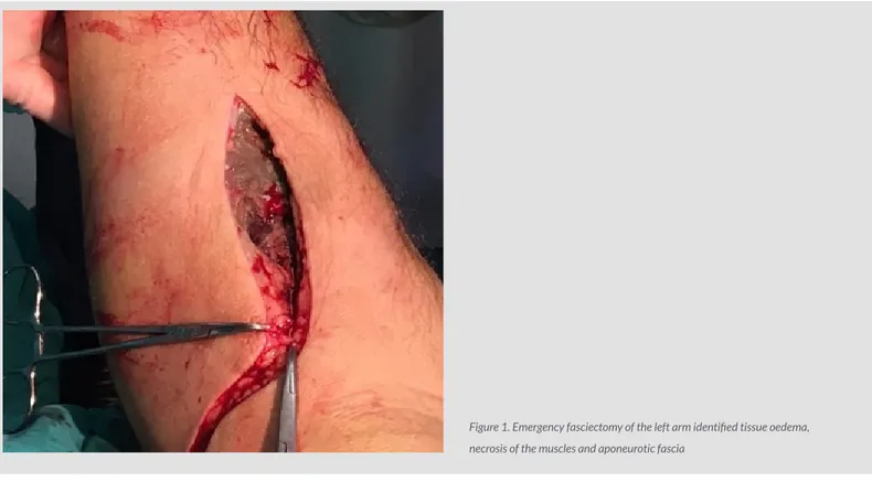
Laboratory analysis revealed hyperleukocytosis (19,000/µL), thrombocytopenia (platelets 86,000/µL), cytolysis (ASAT 343 U/L, ALT 486 U/L), cholestasis (GGT 251 U/L, ALP 244 U/L), and CPK 800 U/L. Gas exchange worsened, lactates increased to 4.6 mmol/L, and ventilation was necessary with 100% oxygen, high PEEP and frequent recruitment manoeuvres. Clinical examination showed swelling of the left arm with extension to the forearm, under tension, with incipient signs of compromised perfusion where the arterial line had been inserted. Ultrasound revealed extensive thickening of fat and subcutaneous tissue of the entire left arm and forearm and an ill-defined hypoechogenic heterogenic area, involving the biceps brachii and other muscles of the anterior compartments of the forearm. These findings, given the clinical scenario, were consistent with the diagnosis of extensive NF of the superior limb. An emergency fasciectomy was performed, where surgical exploration identified tissue oedema and necrosis of the muscles and aponeurotic fascia, which confirmed the suspected diagnosis (Fig. 1). Following fasciectomy and debridement, the signs of compromised perfusion resolved, septic shock gradually improved, organ dysfunction resolved, and the patient was discharged without sequelae.
Figure 1. Emergency fasciectomy of the left arm identified tissue oedema, necrosis of the muscles and aponeurotic fascia
DISCUSSION
Critically ill patients require arterial lines to monitor blood pressure, obtain blood samples for arterial blood gases and laboratory studies, and titrate drug therapies. Invasive catheterization of arteries typically includes radial, brachial and femoral arteries. This invasive procedure may have serious complications and should be performed under strict aseptic conditions and the insertion site must be monitored daily[3].
European Journal
of Case Reports in
Internal Medicine
DOI: 10.12890/2020_001525 European Journal of Case Reports in Internal Medicine © EFIM 2020
as the clinical situation allows; if the indication for invasive monitoring of blood pressure is still present, replace the catheter with a new one, under aseptic technique. NF is an infection of a fascia with secondary necrosis of the surrounding tissues[4-6]. It is a rare condition that
usually occurs after a skin injury, whether traumatic or iatrogenic. It can cause substantial tissue destruction and lead to sepsis. In rare cases it can cause ACS. This occurs when the pressure within a muscle compartment increases with sufficient duration and intensity to cause ischaemic and neurological compromise that can threaten the viability of the affected limb and the patient’s life. It is more frequent in the anterior and posterior deep compartments of the leg and anterior compartment of the forearm. The most common cause of this syndrome is trauma, but it can be caused by any event that increases compartment pressure. Following that, venous drainage from the compartment may decrease, leading to a cycle of increased pressure. Eventually, arterial perfusion becomes impaired. It is a surgical emergency that can result in irreversible tissue damage and treatment consists of an emergency fasciotomy releasing pressure from the compartment[7]. Despite the
low incidence of complications, arterial lines are not risk-free[8, 9]. The more frequent arterial line complications are usually less severe and
without serious risk for the patient. Local or systemic infections are extremely rare, making this a more difficult diagnosis, especially in critically ill patients with potentially multiple sources of infection, and with clinical signs and symptoms often limited by patient sedation. When a patient presents with an obvious source of infection, such as in our case, but fails to improve when appropriate therapy is instituted, it is important to never assume that the identified site is the only source of systemic infection. Thus, rigorous daily physical examination, together with complementary diagnostic tests, is crucial for timely diagnosis[8-10].
REFERENCES
1. Puvanendran R, Chan Meng Huey S, Pasupathy S. Necrotizing fasciitis. Can Fam Physician 2009;55(10):981–987.
2. Davis ML, Della Rocca GJ, Brenner M, Cribari C, Gill Cryer H, Ficke JR, et al. Best Practices in the Management of Orthopaedic Trauma. Chicago, IL: American College of Surgeons (ACS), 2015, pp.15–18.
3. Zerrouki N, Es-Saad O, Housni B, Dikhaye S, Zizi N. Necrotizing fasciitis after central venous catheter placement. Our Dermatol Online [Internet] 2019;10(3):289–291. 4. Peetermans M, de Prost N, Eckmann C, Norrby-Teglund A, Skrede S, De Waele JJ. Necrotizing skin and soft-tissue infections in the intensive care unit. Clin Microbiol Infect
2020;26(1):8–17.
5. Taylor LA. Necrotizing fasciitis. In: Rosenbach M, Wanat KA, Micheletti RG, Taylor LA (eds) Inpatient Dermatology. 1st ed. Cham, Switzerland: Springer Nature, 2018, pp. 111–114.
6. Shaikh N. Necrotizing Fasciitis: Facts and Figures. 1st ed. Doha, Qatar. Scientific Research Publishing, 2016, pp. 3–32
7. Stevens DL, Bisno AL, Chambers HF, Dellinger EP, Goldstein EJ, Gorbach SL, et al. Practice guidelines for the diagnosis and management of skin and soft tissue infections: 2014 update by the Infectious Diseases Society of America. Clin Infect Dis 2014;59(2):147–159.
8. Burnham JP, Kirby JP, Kollef MH. Diagnosis and management of skin and soft tissue infections in the intensive care unit: a review. Intensive Care Med 2016;42(12):1899–1911. 9. Misiakos EP, Bagias G, Papadopoulos I, Danias N, Patapis P, Machairas N, et al. Early diagnosis and surgical treatment for necrotizing fasciitis: a multicenter study. Front Surg
2017;4:5.