1
R
ESEARCH
P
ROJECT
2007-2009
Student: Alessia Ciucci
Doctorate Course: Molecular Biotechnology
Cycle: XXII
TITLE OF THE PROJECT
BCL6 and LRF crosstalk in Dohh2 cells
Laboratory: laboratory of Gene and Molecular Therapy
Supervisor: Giuseppe Rainaldi
Tutor: Giuseppe Rainaldi
BIOS
2
3
4
Indice ... 3
Abstract ... 7
Introduction ... 10
1. Short-interfering RNAs (siRNAs) ... 12
2. Piwi-interacting RNAs (piRNAs) ... 14
3. 21U-RNAs ... 14
4. microRNAs ... 15
4.1 miRNAs classification ... 15
4.2 miRNAs localization ... 16
4.3 Biogenesis: miRNAs Transcription ... 18
4.4 From pri-miRNA to pre-miRNA ... 20
4.5 Nuclear export by exportin-5... 22
4.6 From pre-miRNA to miRNA...23
4.7 miRNAs's action...25
4.8 miRNAs and cancer...28
5. non-Hodgkin's lymphomas ... 32
6. POK proteins ... 32
6.1 B cell Lymphoma 6...34
6.2 Leukaemia/lymphoma related factor...35
7. The circuit ... …37 7.1 p53...38 7.2 miR-34a...39 7.3 miR-145...40 7.4 c-Myc...40 7.5 E2F1...42 7.6 miR-20a...43 7.7 p14...44
8. Aim of the project...44
Material and Methods ... 45
1. Cell colture ... 46
5
3. RT-PCR...47
4. Etoposide treatment...47
5. Cell cycle profile analysis ...48
6. Proliferation assay...48
7. MTT assay...48
8. Methyl cellulose assay...48
9. Transfection with miR-145...49
10. Stably silenced Dohh2...49
11. Predictive algorthm...50
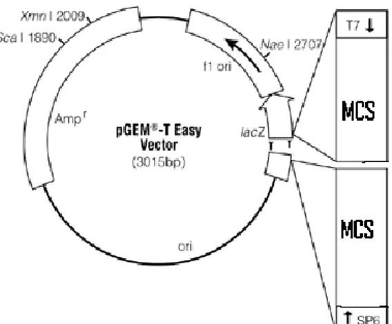
12. Expression vectors...51
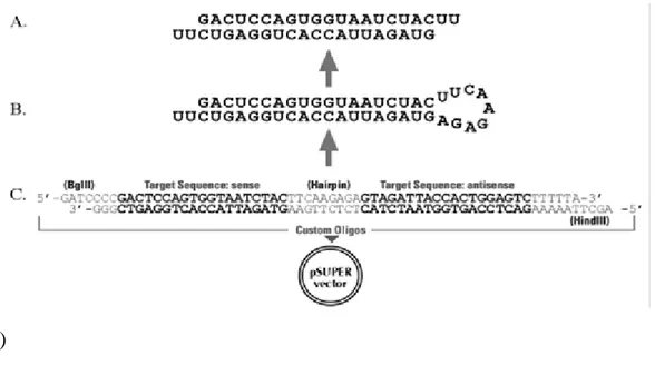
13. E-GFP reporter assay...53
14. Statistical analysis...53
Results ... 54
1. BCL6 and LRF expression in non-Hodgkin’s lymphoma cell lines...55
1.1 BCL6...55
1.2 LRF...56
1.3 The pathway BCL6-LRF...57
1.4 Etoposide treatment as an approach to test the BCL6-LRF pathway via TFs and miRNAs...58
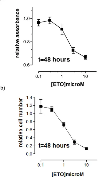
2. Dohh2 cells responsiveness to etoposide treatment...60
2.1 Time and dose dependent experiments...60
3. BCL6 and LRF responses to etoposide treatment...63
3.1 The segment BCL6-P53...64
3.2 The segment p53-miR-145/miR-34a...66
3.3 The segment miR-145-c-Myc...66
3.4 The segment c-Myc-miR-20a-E2F1...67
3.5 The segment miR-20a-LRF-p14-miR-28...69
4. Could miR-145 transfection mimic the effect obtained with etoposide treent?...70
4.1 The segment c-Myc- miR-20a-E2F1...70
4.2 The segment LRF-p14-miR-28...72
6
5. The pathway BCL6-LRF in a clone with stably silenced BCL6...76
5.1 Analysis of stably silenced clone...77
5.2 The pathway in BCL6 stably down-regulated Dohh2 cells...78
Supplementary...83
1 BCL6 reduction induced p21 after etoposide treatment...83
2 miR-30 family members after etoposide exposure in Dohh2...84
3 miR-30 family members after miR-145 transient transfection……...84
4 miR-30 family members in stably silenced cells………...85
5 miRNAs predicted by TargetScan on BCL6 and LRF……….85
6 miR-30 and miR302 target BCL6 3’UTR………86
Discussion...87
7
8 B cell Lymphoma 6 (BCL6) and Leukaemia/lymphoma related factor (LRF) are Pok-proteins over-expressed in some types of Non-Hodgkin lymphoma, such as Diffuse large B cell lymphoma (DLBCL) and Follicular lymphoma (FL).
BCL6 is located on the chromosome 3 in the breakpoint affecting 3q27 band which is the most frequent translocation in Non-Hodgkin’s lymphomas. This gene is a transcription repressor whose principal effect, in human B cell, is to thwart the response to DNA damage by inhibiting both a p53-dependent and a MIZ-1 dependent pathway. In the germinal centre BCL6 is constitutively expressed to suppress the p53 action. In B-cell lines BCL6 prevents apoptosis induced by DNA damage.
LRF, encoded by the Zbtb7a gene, is also known as Pokemon and it is a transcription repressor involved in many cellular processes as differentiation, inflammation and oncogenesis. LRF indirectly controls p53, acting through p14 and MDM2 repression, and it also plays an important role as protooncogene in Non-Hodgkin's lymphomas. Both BCL6 and LRF repression act at transcriptional level on the target genes to regulate protein expression.
It has been demonstrated microRNAs, a class of endogenous 22-25 nt single stranded RNA molecules, work to regulate target gene expression at post-transcriptional level. In fact, these molecules share a partial complementarity with 3’UTR region of expressed genes and act repressing translation.
In this project we focused our attention on the possible correlation between BCL6 and LRF over-expression in cell lines derived from Non- Hodgkin's lymphomas.
Since miRNAs play an important role in tumours and in regulatory circuits in which are involved, we focused our attention on the behaviour of the miRNAs belonging to the hypothetical circuit that correlates BCL6 and LRF. Using our data and the data taken from the literature demonstrated in different model systems, we reconstructed a hypothetical circuit connecting BCL6 and LRF to each other. A negative feed back loop should link BCL6 and LRF (following the interaction showed in the circuit), while in Non-Hodgkin's lymphomas these two genes are found both over-expressed. We hypothesized that this double increase could be involved in the high proliferation rate typical of tumor cells. The BCL6-LRF network is very intriguing because there are transcription factors and miRNAs: both are described as fundamental actors in the regulation of the cell cycle, often deregulated in tumours and all are fundamental in the
9 cellular response to stimuli. We treated the Dohh2, a cell line derived from a Non-Hodgkin's lymphomas, with etoposide, a chemotherapeutic agent, that is able to reduce BCL6. We observed that in this condition the Dohh2 cells are not able to proliferate. We focused our attention on the circuit and in particular on the behaviour of the molecules belonging to it. We noticed that there is an over-expression of miR-145. It is interesting because it has been described as a tumour suppressor miRNA and it is under p53 control. This transcriptional factor is under BCL6 direct control and LRF indirect control. We decided to over-express miR-145 through transient transfection to try to reproduce the effect of etoposide. We analyzed the network 24 hours after transfection: the transcription factors and miRNAs on the pathway, that hypothetically connects BCL6 and LRF, were influenced by the transfection but we did not find any change in the proliferation rate of Dohh2 cells. We hypothesized that it was due to the fact the over-expression was not sufficiently long. So we tried to prolong the increased expression level of miR-145 in Dohh2 cells. We performed a preliminary analysis of the circuit in a population stably silenced for BCL6 in which we had a prolonged down-regulation of BCL6: in this case the molecules belonging to BCL6-LRF circuit were modified too but the proliferation did no change. We focused on the possible differences between the etoposide treatment and the others. We noticed that in the former there was not only BCL6 and LRF decrease but also miR-145 up-regulation accompanied by p53 and E2F1 increase, not present in the other treatments. We can hypothesize that the BCL6 and LRF down-regulation are necessary, together with the increase of miR-145, p53 and E2F1, to affect proliferation in Dohh2 cells.
We hypothesized that the alteration of the correlation between BCL6 and LRF could have been developed in lymphoma cells in order to increase their proliferation and escape from apoptosis or cell death.
10
11 The main dogma of genetics was that information flows from DNA to RNA and after this from RNA to proteins. The researchers focused their attention on DNA, as a molecule that hosts the information of life, and afterwards on proteins considered as effectors of such information. The RNA molecules were considered as simple tools of connection between them. In the past few years RNA molecules were greatly reassessed: they are not just a linker between the information (DNA) and its application (proteins), but they represent a complex regulatory system that we started to understand only twenty years ago. In fact in the past 20 years researchers found out that the larger part of DNA is noncoding RNAs: in highly developed organisms, about 97% of the transcription output is represented by noncoding RNAs. This large group of noncoding RNAs can be divided into two sub-groups: one of housekeeping RNAs and the other regulatory RNAs. In the first group there are for example molecules such as transfer RNA (tRNAs), small nuclear RNA (snRNA), ecc.. In the second one there are short-interfering RNAs (siRNAs), piwi-interacting RNAs (piRNAs), 21U-RNAs and microRNAs (miRNAs) (Table n.1).
Table n.1 Comparison between different small RNA species.
(Modified from International Journal of Biological Sciences. Qazi Mohd. Rizwanul Haq, 2009.)
12 siRNAs and miRNAs are present both in plants and animals while piRNAs and 21-U RNAs have been described so far as limited to animals. Initially the short RNAs were considered a product derived from the degradation of larger molecules. Starting from 1993 with the lin-4 discovery of Ambros and co-workers in Caenorhabditis elegans as an endogenous regulator of genes that control developmental timing a new field of science started1. Today it is known that these molecules are involved in the control of genes and genomes: in fact, they were described as able to regulate chromatin structure, chromosome segregation, transcription, RNA processing, RNA stability, and translation. This discovery explained one of the strangest issues of biology: the redundancy of DNA and the presence of the junk DNA in the genome. In fact this explains the usage of genome and the number of genes present, but it has also complicated the situation in terms of biochemical attributes and functional genesis of the molecules. These small molecules seem to be present in all kingdoms: from unicellular alga Chlamydomonas reinhardtii to humans2,3. These regulatory molecules seem also to be present in the prokaryotic world: bacterial short regulatory RNAs have also been referred to as small RNAs, but they are not related to eukaryotic small RNAs. In fact bacteria have few proteins the domains of which are similar to proteins belonging to the RNA interference pathway. Their genome shows sequences of a parasite genome called clustered regularly interspaced short palindromic repeats (CRISPR). These are multiple noncontiguous direct repeats with spacer sequences and make bacteria resistant to phages through the RNA interference pathway: this is a sort of primordial immuno system against them4,5. Unfortunately, it is not known how these sequences work.
1. Short-interfering RNAs (siRNAs)
Short-interfering RNAs (siRNAs) are ~20-24 nt long RNA molecules acting as a primitive immune system protecting cells from intrusion of any exogenous nucleic acid, as for example viruses, and that are involved in maintaining genome integrity by stopping the transcription of retrotransposon and repeat sequences. They move from a cell to the other through the phloem, the vascular system of plants6. They are characterized by the perfect match between the siRNA and its target RNA. This perfect
13 complementarily is necessary for the recognition of the target. siRNAs are transcribed through the RNA dependent RNA polymerase (RdRP) activity on aberrant transcripts or transcript with full or partial complementarity 7. RdRP is present in plants, fungi and
Caenorhabditis elegans, but not in humans7. The main function of this enzyme is the
generation of secondary siRNAs, a step termed signal amplification in siRNA pathway7. RdRP can recognize aberrant RNA molecules to produce dsRNAs either in a primer dependent or in an independent manner. In the nucleus siRNAs are processed by Dicer, a RNaseIII type endonuclease, that cut the molecules to their characteristic length. Animals own one type of this enzyme. The exceptions are represented by
Drosophila and C. elegans, each encoding two Dicers, while the plants have many of
them8. Some researches have demonstrated that the presence of many Dicers is redundant: each enzyme is dedicated to the processing of a specific type of siRNA, but when one Dicer is mutated, its activity is partially taken over by the others9,10. The siRNA-Dicer complex is then exported to cytoplasm where it binds to the Argonaute proteins. The functions of this family of proteins are mostly unknown: Ago-2 is one of the most important members in the siRNAs pathway in plants. Argonaute proteins unwind the double strand of RNA: the process starts from the end with lower thermal energy. One of the two strands, the passenger strand, is degraded by exonucleases and the other, the guide strand, remains in the RISC complex. This complex binds with a perfect match to the RNA target and it cuts at the position between the 10 and 11 nt from the 5′ end of the siRNA. siRNAs are divided into different classes: trans-acting short interfering RNAs (tasiRNA), repeat-associated short interfering RNAs (rasi-RNAs), scan RNA (scn RNA), long siRNAs (lsiRNAs).
Trans-acting short interfering RNAs (tasiRNA) are like miRNAs: they are about 21 nt long small RNAs that require endogenous transcript as a template and they do not have a perfect match with their targets11,12. They cut an endogenous mRNA, but the target gene is different from the one originating the siRNA7. They are not present in humans because they do not have the RdRP enzyme that transcribes the double strand of RNA13. They work against possible erroneous transcription to preserve cells7.
Repeat-associated short interfering RNAs (rasi-RNAs) are longer than the previous class of si-RNA: 24-26 nt. They need the amplification step of the
14 RdRP enzyme. They are important during gametogenesis in flies, worms and mammals to regulate the chromatin status, silencing viral transcripts by recruiting histone modifying proteins14-17.
Scan RNA (scn RNA) are relatively long, about 29 nt and they are described in the Tetrahymena thermophila. These protozoans have two nuclei and during the conjugation the sncRNA of the micronucleus eliminates the corresponding loci from its own genome: this produces the macro-nucleus18,19.
Long siRNAs (lsiRNAs) were discovered in Arabidopsis and they are longer RNA molecules : 30-40 nt. They are produced in response to bacterial infection and growth conditions20.
2. Piwi-interacting RNAs (piRNAs)
Piwi-interacting RNAs (piRNAs) were divided into two categories depending on their length: 24-28 nt and 29-31 nt. These kinds of molecules are specific to germline cells
21-24
. In fact, the Argonauta protein family was divided into two sub-groups through studies on amino acid sequences: the AGOs, that are present in all cells and that bind to all kinds of small RNAs, and the PIWI (P element-induced wimpy testes) that are germline-specific25-27. The main part of piRNAs were mapped in the genomic regions previously thought to be non-transcribed, while others are found in intergenic, exonic, intronic and repeat regions16,23. Generally the transcription of these molecules follows an unknown mechanism started by a mono- or bidirectional promoter. It seems that the biogenesis of piRNAs does not depend on Dicer action. Unlike the others small RNA molecules, it is thought that piRNAs can promote the stability of their mRNA target and enhance translation18.
3. 21U-RNAs
21U-RNAs were identified so far only in C. elegans, C. briggsae28. They are
characterized by their sequence: 21 nt long with uridine at its 5' end. The majority were mapped within intergenic or intronic regions and probably they play a role in chromatin reorganization and genome stability since their sequences have no homology with any transcript.
15
4. microRNAs
microRNAs (miRNAs) are a very large family of molecules that were described in literature for the first time in 1981 by Ambros and Ruvkun and their collaborators in a study about loss of function in Caenorhabditis elegans29. They found that the gene lin-4 originates two different RNA molecules: a long molecule of around 61 nt and a smaller one of around 22 nt1, where by the first is the precursor of the second; the latter is required to inhibit the expression of lin-14 messenger and this small RNA is able to down-regulate it during the transition from the first to the second larval stage of development in the worm C.elegans30,31. In fact, it recognizes complementary sites in the 3' untranslated region (3'UTR) of this mRNA target. Moreover it was demonstrated that the same lin-4 is able to regulate lin-28 at a later stage of development32. Nowadays it is known that miRNAs are a class of endogenous 22-25nt single-strand RNA molecules that regulate gene expression at the post-transcriptional level33,34. miRNAs are involved in the most varied processes: from the control of leaf and flower development in plants35 to pluripotency of embryonic stem cells36,37, from cells proliferation to cells death38, from neuronal patterning in nematodes to cancer34. This strategy of translational control represents a very fine tuning process which is very rapid and not very expensive in the economy of cell energy. This hypothesis is supported by the fact that this kind of regulatory system has evolved in different kingdoms in an apparently independent way33,39. In fact, miRNAs genes represent the largest class of regulatory molecules since they represent 1% of total coding genes40,41.
4.1 miRNAs classification
In 2003 Sam Griffiths-Jones established at Welcome Trust’s Sanger Institute a miRNA register in which all miRNAs discovered are annotated42. The miRNA Registry provides a service for the assignment of miRNA gene names prior to publication. This is a database of published miRNA sequences that are accessible via a web interface and all sequences and annotation data are freely available for download. The classification is standard: mammalian miRNA genes are distinguished by the prefix miR- followed by a number; while in the miRNA genes of C.elegans the prefix is lin- or let- 43. Moreover miRNAs that are transcribed from different loci but showing the same sequence are noted under the same name but with a different suffix ( e.g. miR-16-1,
16 miR-16-2), whereas two miRNAs that have one or two different bases are recorded with the same number but with a different letter ( e.g. miR-16a e miR-16b). The less represented form of miRNAs in the cells is marked with * (e.g. miR.9*). miRNAs having the same seed are clustered in a family because the perfect pairing between 7 bases of miRNAs's seed and the locus on the 3'UTR of the mRNA target (seed match) is the main rule to match the mRNA target44,45. In the remaining pairing between miRNA and mRNA mismatch, bubbles, non conventional pairing (as G-U) are allowed, even if other pairings (out of the seed match) in an other part of mRNA make the match stronger and the repression more efficient46,47. A mRNA can have more seed matches for the same or different miRNA, which can produce a synergic action of repression on mRNA of miRNAs32,48-51. A candidate gene to be annotated as novel miRNA must have the following characteristics: the first criterion (expression) plus the second criterion (structure), or the first criterion (expression) plus the third criterion (conservation).
4.2 miRNAs localization
miRNAs can be localized in different parts of genome: they can be categorized according to their genomic locations relative to exon and intron positions (Fig. 1)52-55.
Figure 1. Structures of primary microRNA transcripts (pri-miRNAs). miRNA
host transcripts may be unspliced, or miRNAs may be located within introns, exons, or untranslated regions (UTRs) of spliced transcripts (Modified from Mendell ,Cell Cycle 2005).
17 In fact the miR-985 hairpin is found in the last exon of CACNG8 mRNA a protein-coding transcript56, but it is well known that more than 80% of miRNAs are localized in introns of genes that codify or do not codify for proteins (Fig. 2)54.
Figure 2. Schematic diagram to depict differences between mirtron and canonical miRNA generation. Introns that assume foldback structures are
recognized and cleaved by DROSHA. These stem-loop lariats are then acted upon by Lariat debranching enzyme that cleaves the phospho-diester bond formed during the splicing event. The primiR thus formed joins the mainstream miRNA flux, before making exit to cytoplasm (Modified from Rizwanul Haq. International Journal of Biological Sciences 2009).
These miRNAs are called mirtrons, for example miR-106b, miR-93 and miR-25 that are localized in introns of the gene that codifies for protein mcm-757. When miRNAs are found in introns their expression profile follows that of the gene in which they are localized: after transcription mRNA undergoes splicing and miRNAs are processed53,58,59. Introns are characterized by flanking regions in which conserved nucleotide sequences, for example GU-AG, are present. They are recognized by the proteins removing the intron. This sequence folds in a lariat with a typical 2'-5' phosphodiester bond that is performed by Lariat debranching enzyme that cleaves it60. Mirtrons do not need DROSHA/DGCR8 maturation: they can leave the nucleus through the Exportin-5 after the action of Lariat debranching enzyme. The mirtrons have not been identified in plants and other organisms yet. Since the introns are not subjected to selective pressure it is logical to assume that they are unlikely to preserve
18 their sequences and this could explain their specificity. Other miRNAs are localized in introns or exons of genes that do not codify for proteins. For example the miR-15a and miR-16-1 cluster is found in the intron of a well-defined non-coding RNA gene, DLEU261, or the miR-155 is located in BIC: a previously defined non-coding RNA gene. All these miRNAs can be orientated in a sense or non-sense with respect to the gene in which they are found58. 30% of miRNAs have an independent origin of transcription44,62,63. In the literature there are examples of the same miRNA that is transcribed on a different chromosome: the precursor miR-1 is miR-1-1 that is expressed in the atrium of heart during the embryonic development of mouse while miR-1-2 is expressed in the ventricle64. Similar examples are 1 and miR-30c-265. The action of miRNAs plays in trans: they target a mRNA transcribed from a gene located in another part of DNA. They can be grouped in a cluster and in this case scientists have supposed that they are expressed together and if they belong to the same family they have probably originated from a tandem duplication66-68. Approximately 50% of mammalian miRNA loci are found near to other miRNAs. These clustered miRNAs are transcribed from a single polycistronic transcription unit. miRNAs belonging to different families could also be present in a same cluster: they may be involved in the regulation of genes related each others. A transcript may encode clusters of distinct miRNAs, or it may encode a miRNA and messenger .
4.3 Biogenesis: miRNAs Transcription
miRNAs's biogenesis is effected by diverse enzymes in different subcellular compartments. In fact maturation occurs through sequential processing events: miRNAs are first transcribed as a large RNA molecule, termed a primary miRNA (pri-miRNA), which is sequentially processed in the nucleus, to give the approximately 65-nt pre-miRNA hairpin i65-ntermediate, and then in the cytoplasm, to give the mature miRNA (Fig. 3). A pri-miRNA hallmark is the stem-loop hairpin where imperfect pairings in the stem take place63. In the lower part of harping there are flanking sequences that are important for the following step of miRNA's maturation.
19 Most miRNAs are located in intergenic regions (>1 kb away from annotated/predicted genes), although a sizeable minority was found in the intronic regions of known genes in the sense or antisense orientation. miRNA genes can be transcribed from their own promoters, and the clustered miRNAs are generated as polycistronic primary transcripts (pri-miRNAs). Transcription of miRNA genes is mediated by RNA polymerase II (pol II)54,66,69,70 a minor group of miRNAs that are associated with Alu repeats can be transcribed by Pol III 71. Furthermore, chromatin immunoprecipitation analysis shows that pol II is physically associated with a miRNA promoter66. miRNA gene
Figure 3. MicroRNA biogenesis. pri-miRNA, transcribed by polII, are processed
by Drosha in the nucleus. Pre-miRNAs undergo cytoplasmic translocation, which is mediated by exportin 5 in conjunction with Ran-GTP, and are subsequently processed into RNA duplexes of about 22 nucleotides by Dicer. The RISC-miRNA complex is guided to the mRNA target for translational repression or degradation (Modified from Kim, Nature 2005).
20 transcription can be controlled by various pol-II-associated regulatory factors, so that a specific set of miRNAs can be expressed during development as well as under specific conditions and in certain cell types34. In addition, the expression of miRNA and protein-coding genes might be coordinated, especially when a miRNA and a protein- coding region both reside in a single transcript. miRNAs can have their own promoter as for example let-7 in C.elegans that depends on an element enhancer called TRE (Temporal Regulatory Element) that is located at about 1200 bp upstream of the mature miRNA72. From the literature it is well known that there are transcription factors able to transcribe some miRNAs independently as for example miR-1 that is transcribed by Serum Response Factor (SRF)64 or cluster miR-17 that is transcribed by c-Myc73. The primary miRNA transcript (pri-miRNAs) contains a cap structure as well as a poly(A) tail, which are the unique properties of class II gene transcripts66,69,74,75.
4.4 From pri-miRNA to pre-miRNA
Pri-miRNA undergoes a first processing in the nucleus by means of an enzyme called Drosha76: a RNase III protein of about 160 kDa that is well conserved in the animal kingdom77-79. The stem-loop structure is cleaved by the nuclear RNase III Drosha to release the precursor of miRNA (pre-miRNA). The remnants, the flanking fragments, are thought to be degraded in the nucleus. Drosha contains two tandem RNase III domains (RIIIDs) and a double-stranded RNA-binding domain (dsRBD) that are crucial for catalysis (Fig. 4)80,81.
Figure 4. Domain Organization of Human Drosha. RIII, RNase III catalytic
domain; D, dsRNA binding domain; PRORICH, proline-rich domain; RS-RICH, arginine/serine rich domain (Modified from Bryan R. Cullen. Molecular Cell,2004).
The central region of the protein, adjacent to the RIIIDs, is also essential for pri-miRNA processing. RNase III are divided into three different classes: first class
21 enzymes are found in bacteria and in yeast and they have one side of binding at double RNA strand and one domain RNase III82. Enzymes of class II and III instead have a helicasic domain and a Paz (Piwi/ Argonaute/ Zwille) domain that is typical for Argonaute proteins. Drosha is RNasi III enzyme of II class. Drosha forms a large complex of ~500 kDa in D. melanogaster83 or ~650 kDa in humans82,84 called Microprocessor complex. In this complex Drosha interacts with a cofactor: the DiGeorge syndrome critical region gene 8 (DGCR8) protein in humans (also known as Pasha in D. melanogaster and C. elegans). When DGCR8 protein is mutated in humans it causes the DiGeorge syndrome, provoking specific heart malformations, facial malformations, anomaly of the endocrine and immune systems84. DGCR8/Pasha is a ~120 kDa protein that contains two dsRBDs83. It also contains a putative WW domain, which is known to be an interaction module for specific proline-rich sequences. It remains to be determined if this domain interacts with the proline rich region of Drosha. Although the biochemical role of DGCR8/Pasha is currently unclear, it is believed to assist Drosha in substrate recognition. Through interaction with DGCR8, Drosha cuts dsRNA to create a 5' phosphate and 2 nt 3' loops currently used to define miRNA 85(Fig. 5). This product is called pre-miRNA and its protruding end is important for the following step of processing. Drosha depends upon its protein cofactor for the efficiency and accuracy of this processing (see below). This enzyme can cleave not only pri-miRNAs but also mRNAs that contain long hairpins. Drosha negatively regulates its own cofactor, DGCR8, by cleaving the hairpins in the second exon of the DGCR8 mRNA86. The introns containing miRNA are spliced more slowly than the adjacent introns for unknown reasons. The splicing commitment complex is thought to tether the introns ( exon-tethering model) while Drosha cleaves the miRNA hairpin87. The pre-miRNA enters the miRNA pathway, while the rest of the transcript undergoes pre-mRNA splicing and produces mature mRNA for protein synthesis. Another way the miRNAs located in the introns are processed is bypassing the Drosha step. The mRNA is spliced and the intron is debranched. The mirtrons show similar protruding ends. Sometimes some pri-miRNAs are edited by Adenosine Deaminase Acting on RNA (ADARs) at specific positions (generally +4 and +44) changing adenine to inosine88,89. This process increases the variability of miRNAs in the cells. After this passage these molecules are also processed by Drosha.
22
4.5 Nuclear export by exportin-5
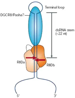
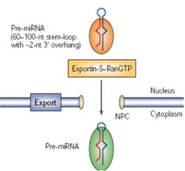
Nuclear export of pre-miRNAs is a crucial step in miRNA biogenesis: pre-miRNA is actively transported from the nucleus to the cytoplasm by Ran-GTP and the export receptor Exportin-5. Members of the nuclear export receptor family bind cooperatively to the cargo as well as to the GTP bound form of the cofactor Ran (RAS-related nuclear protein) in the nucleus (Fig. 6)90-93.
Following export, hydrolysis of GTP to GDP results in the release of the cargo from the export complex. A structural motif can be found in pre-miRNA stem-loops, which typically comprise a stem of ~22 bp, a terminal loop and a 3′ overhang of ~2 nucleotides. By introducing mutations in the pre-miR-30a, Cullen and colleagues confirmed that a RNA stem of >16 bp and a short 3′ overhang are significant structural requirements for pre-miRNA export34,94.
Figure 5. Drosha/pri-miRNA complex. Drosha, interacting with DGCR8
cuts the ends at the lower part of pri-miRNA ( Modified from Kim, Nature 2005).
23
Figure 6. Pre-miRNA transit from the nucleus to cytoplasm. Pre-miRNA is a
transport complex together with exportin-5 and its cofactor Ran (the GTP-bound form) ( Modified from Kim, 2005 Nature).
4.6 From pre-miRNA to miRNA
When pre-miRNA arrives in the citosol it undergoes another transformation performed by another endonuclease: Dicer95-98. This is an enzyme of 160 kDa, a highly conserved RNase III which is well conserved in eukaryotes (Fig. 7).
Figure 7. Domain Organization of Human Dicer. Helicase and Paz domain; RIII,
RNase III catalytic domain; D, dsRNA binding domain. (Modified from Bryan R. Cullen. Molecular Cell, 2004)
Dicer joins with other proteins of the Argonaute family to stabilize pre-miRNAs99,100.
D. melanogaster Dicer 1 requires Loquacious, which contains three dsRNA-binding
domains (dsRBDs) for pre-miRNA processing101.Three splice variants of Loquacious are known, of which only two isoforms interact with Dicer-1. Interestingly, the third
24 isoform lacks the non-canonical dsRBD, suggesting that this domain may be essential for association with Dicer-1102. Loquacious plays a critical role in the maintenance of germ-line stem cells: mutant female flies are sterile and lack germ-line stem cells103. Human Dicer interacts with two closely related proteins, TRBP (TAR RNA-binding protein; also known as TARBP2)104,105 and PACT (also known as PRKRA)106. So far the biochemical role of these proteins is unclear: neither is required for processing activity itself. They seem to contribute to formation of the RNA-induced silencing complex. In addition to the binding site for double stranded RNA on Dicer there are catalytic sites that perform the cut to generate the new miRNA end. Dicer, through the Paz domain, recognizes the end cuts by Drosha and binds pre-miRNA, but the reaction centre lies in the RNase III domains107. Each of the two RNase III active sites cleaves one of the two strands, leading to a staggered duplex scission to generate new ends with 2 nt 3′ overhangs. The reaction leaves a 5′ monophosphate on the product end and 2 nt overhang at 3' (Fig. 8)108. This 5’ monophospate is later required for loop removal.
Figure. 8. Dicer/pre-miRNA complex. Dicer interacting with pre-miRNA takes
off removes the loop (Modified from Kim, Nature 2005).
Dicer releases a double strand of RNA with imperfect pairing. Some organisms, including mammals and nematodes, have only a single Dicer that does double duty in the biogenesis of both miRNAs and siRNAs, whereas other organisms divide this task among multiple Dicer proteins101. After the Dicer action has taken place the double RNA molecules are loaded in a multiprotein complex called RNA induced silencing
25 complex (RISC)109-114. Argonaute proteins are the core of this complex. They are characterized by four domains: the N-terminal (N), the PAZ domain, that is common in Dicer, the PIWI domain that is unique to the Argonaute family115, and middle (Mid) domains. The Argonaute PAZ domain has RNA 3′ terminus binding activity, and the 5′-phosphate engages pocket in the Mid domain.
When this miRNA-RISC complex is formed, the RNA duplex is unwound, based on thermodynamic stability of the duplex's ends116. In mammals, miRNAs having nuclear signal sequences can traffic back to the nucleus where they can target many under-processed transcripts or bring about silencing of genomic regions.
4.7 Action of miRNA
miRNA is the guide for the identification of mRNA by the RISC complex. The main rule is the almost perfect match between the seed on seven nt of miRNA, included between its second and eighth base (miRNA seed), and the seed match on mRNA target (Fig. 9)31,32,46,117-120.
Figure 9. miRNA recognition of target mRNAs. A lin-4 binding site in the 3’
UTR of lin-14 is depicted to illustrate typical features of miRNA:target interactions. In the figure the perfect match that occurs in the seed region is shown. (Modified from Mendell ,Cell Cycle 2005).
Out of the seed some mismatches are allowed in animals, while in plants the binding is near-perfect. The complex miRNA-RISC-mRNA moves to particular foci in the citosol called Processing bodies (P-bodies) or GW-bodies (Fig. 10)121,122.
26 a) b)
Figure 10. mRNA processing in P-bodies. mRNA linked with one or more
miRNAs in the P-bodies can undergo repression of translation or degradation (Modified from Zamore, Science 2005 ).
In fact, the GW-182 protein is one of most abundant protein in P-bodies and when it is silenced through si-RNA the researchers observed these foci failed to assemble121,122. Similar structures were discovered in plants where they are called Cajal bodies123. These are a kind of storage sites for translationally suppressed mRNAs that are released when required and can actively translate into the nucleus. If the match between the miRNA and its target is perfect scission of the mRNA occurs. An example of this action is represented by the miR-196a that degrades HOXB8 transcript 124,125. Instead, if the binding presents mismatches there is repression of translation by unexplained mechanisms.
According to a proposed model there is competition between miRISC and eIF4E for binding to the mRNA 5′ cap structure (Fig. 11)74,75,126,127
27
Figure 11. Possible mechanisms of miRNA’s repression. Non-repressed mRNAs
recruit initiation factors and ribosomal subunits and form circularized structures that enhance translation (a). When miRISCs bind to mRNAs, they can repress initiation at the cap recognition stage (b) or the 60S recruitment stage (c). Alternatively, they can induce deadenylation of the mRNA and thereby inhibit circularization of the mRNA (d). They can also repress a post-initiation stage of translation by inducing ribosomes to drop off prematurely (e). Finally, they can promote mRNA degradation by inducing deadenylation followed by decapping (f) (Modified from Erik J. Sontheimer. Cell. 2009).
eIF4E is a sub-unit of eIF4F complex that starts the translation. eIF4E binds the methylated base of the cap between two tryptophan residues. The Mid domain of human Ago2, with two phenylalanine residues, is hypothesized to compete with eIF4E. A second model suggests miRISC should promote deadenylation of the mRNA tail. The cap and PABP1-free tail of the deadenylated mRNA are unable to circularize so translation is repressed128-131.
A third model proposes that miRISC blocks association of the 60S ribosomal subunit with the 40S pre-initiation complex. Human AGO2 binds eIF6 and 60S ribosomal subunits in vitro and eIF6 is involved in the biogenesis and the maturation of 60S ribosomal subunits, thus preventing their binding with 40S subunits132.
a b c d e f
28 But all these models present some contradictions and none has the capability of explaining the action of miRNAs completely.
4.8 miRNAs and cancer
Cancer is one of the most common diseases in the world: cancer-related global mortality follows only cardiovascular and infectious illnesses133. Tumors are a class of disease affecting people at all ages and also animals. The sickness is caused by hyper proliferation and an inappropriate survival of damaged cells that are not controlled by normal cell signals. These cells are able to grow beyond normal limits: they are capable of metastasizing, invading and colonizing other tissues in the body. There are many causes for the onset of cancer: exposure to carcinogens, genetic errors in the DNA replication, inheritance or chance. Tumor suppressor genes are inactivated in cancer cells, resulting in the loss of normal functions, such as accurate DNA replication, control over the cell cycle, orientation and adhesion within tissues, and interaction with protective cells of the immune system. Oncogenes are typically activated in cancer cells, giving them some advantages, such as faster growth and division, protection against programmed cell death, the ability to form metastases. A new large class of molecules that is important in these kinds of disease are microRNAs (Table n.2)134. The first evidence of aberrant miRNA expression in human cancers was described in B-cell chronic lymphocytic leukaemia. In this illness the loss or reduction of miR-15 and miR-16 expression was found, which causes the B-cell chronic lymphocytic leukaemia61. There is other evidence in this direction, as for example the over-expression of miR-17-92 cluster, located at chromosome 13q31, a region that is amplified in B-cell lymphomas135.
29
Table 2. Oncogenic or tumor-suppressive miRNAs and their direct target genes (Modified from Anindya Dutta The Annual Review of Pathology 2009)
30 Post translational modifications of miRNAs, as for example methylation, could cause the tumour formation. In fact, a wrong miRNA gene methylation could bring about the deregulation of their expression in cells136. Moreover in human genome many miRNAs
Figure 12. miRNA oncogenes and tumour suppressors. (a) Reduced miRNA
levels, reflecting defects at any stage of miRNA biogenesis (indicated by question marks), produce inappropriate expression of target oncoproteins (purple squares). The resulting defects in homeostasis increase proliferation, invasiveness or angiogenesis, or decrease the levels of apoptosis or differentiation, potentiating tumour formation. (b) Conversely, over-expression of an oncogenic miRNA eliminates the expression of tumour-suppressor genes (pink), leading to cancer progression (Modified from Slack FJ Nature review 2006).
31 are located at fragile sites, which in particular types of tumour, are associated with DNA rearrangements suggesting a correlation between miRNAs and cancer137. Another case in which a defect in the miRNA biogenesis machinery can produce the deregulation of miRNAs and as consequently the tumour formation if this miRNA has a tumour gene or a tumour suppressor gene as a target.138,139. It can be affirmed that miRNAs can act as tumour suppressors and oncogenes (Fig.12)134,137,140-146. Tumour suppressor miRNAs can target an oncogene stimulating apoptosis of cells or inhibiting cell proliferation. On the contrary, miRNAs that negatively regulate tumour-suppressor genes for example by stimulating cell proliferation and inhibiting apoptosis act as oncogenes. miRNAs could promote the tumour formation also by affecting the proteins involved in the cell cycle. For example miR-221 and miR-222, that are over-expressed in many tumours, are able to target p27 and p57 mRNAs147,148. These two proteins are two negative cell-cycle regulators: they bind to Cdk/cyclin complexes and inhibit the G1/S phase switch. miRNAs are involved not only in tumour formation but also in its diffusion. In fact, miR-10 was demonstrated to promote metastases in breast cancer cells. This miRNA has HOXD10 as a target whose reduction produces higher levels of RHOC, which stimulates cancer cell motility149. But miRNAs could be a good ally to distinguish different tumours and different subtypes of tumours as well as to predict their clinical behaviour. Some studies on the expression profile of miRNAs in bone marrow samples from patients revealed that it is possible to distinguish the different origins of tumours150. Moreover miRNAs could play an important role as specific miRNAs for identifying disease progression. In fact, researchers demonstrated that high miR-21 expression was associated with poor survival of patients with pancreas endocrine tumours or colon adenocarcinomas151,152. miRNAs could also be a therapeutic tool in tumours: a lot of attempts reconstituting in reduced gene expression, for example blocking a transcriptional repressor increasing a miRNA expression that target it, or reducing over-expressed genes through miRNAs are being made. miRNAs are one of the fundamental elements in the integrated regulation of gene expression since they are involved in the balance of gene regulating networks that determine the cells fate. Disruption of these pathways may contribute to cancer progression. Therapeutics as well as diagnostics and prognostics can take advantage of this new knowledge about miRNAs and their actions: clinical applications exploiting our
32 understandings of miRNA's functions will be the next great challenge in cancer research.
5. Non-Hodgkin's lymphomas
The lymphoproliferative diseases are a group of illnesses due to neoplastic transformation of a lymphocytic clone. By examining the characteristics of these clones the researchers can determine the kind of disease: if the cells are circulating a lymphatic leukaemia affects the patients, whereas a lymphoma is present if the cells are found in lymphatic organs. The transformation causing this illness could be due to several different factors: exposure to toxic agents, genetic mutation, inheritance or chance. The lymphomas are a large group of diseases. The first discrimination is to distinguish between Hodgkin and Non-Hodgkin lymphomas. This division is based on the presence of a characteristic kind of cells: Reed-Sternbeng cells. If these cells are present, it is a Hodgkin lymphoma, if they are not it is a Non-Hodgkin lymphoma. The first group has a better characterization as a disease thanks to this classification. Hodgkin lymphomas are twice as common as Hodgkin lymphomas. Moreover, Non-Hodgkin lymphomas contain a heterogeneous group of diseases, the majority of which, around 80%, originate from B lymphocytes153. The most common types are the follicular lymphomas and the diffuse large B-cell lymphomas, respectively at 22% and 35%154. These diseases are often discovered at an advanced stage in different loci because no symptoms are recognisable during the initial period. For this reason it could be interesting to gain a better knowledge of these diseases so as to determine possible treatments.
6. POK proteins
POK proteins are a large family of proteins involved in the development, differentiation, and oncogenesis of living beings. A subgroup of these proteins is characterized by an N-terminal Poz domain and C-terminal Krüppel-type zinc finger domain. The POZ domain, also known as the BTB domain, mediates homodimerization and heterodimerization plus recruitment of corepressor/HDAC complexes155. This is a protein-protein interaction domain, originally identified in some zinc finger proteins of D.melanogaster and Pox virus. The POZ domain is a versatile
33 protein-protein interaction motif that participates in a wide range of cellular functions, including transcriptional regulation, cytoskeleton dynamics, ion channel assembly and gating, and targeting proteins for ubiquitination (Fig. 13).
Figure 13. Backbone superposition of the POZ domains of LRF, BCL6, and PLZF (Modified from David S. Waugh Elsevier Inc. 2006).
The C-terminal Krüppel-type zinc finger domain mediates specific DNA recognition and binding. Many members of this large family, like B cell Lymphoma 6 (BCL6) and leukaemia/lymphoma related factor (LRF) have been characterized as important transcription factors, implicated in the development and oncogenesis.
LRF is often aberrantly over-expressed in association with BCL6 in several kinds of tumours such as diffuse large B cell lymphoma (DLBCL) and follicular lymphoma (FL), the most common types of Non-Hodgkin lymphomas156. Moreover they have a common target: p53. In fact, as it will be explained in the following paragraphs, BCL6 blocks this transcriptional factor in a direct way157 while LRF acts indirectly156. These evidences suggest a possible crosstalk between these factors in these kinds of tumours: they could be connected through a circuit to each other.
34
6.1 B cell Lymphoma 6
B cell Lymphoma 6 (BCL6) is a protooncogene located on the chromosome 3 in the breakpoint affecting band 3q27158,159, the most frequent translocation in Non-Hodgkin’s lymphomas, characterized by a heterogeneous group of malignant diseases, the majority of which originates from B lymphocytes160,161. BCL6 is a 95 KDa protein and it is a transcriptional repressor162-164. It is able to directly bind the DNA through six C-terminal zinc finger domains, each separated by a conserved stretch of seven amino acids (Fig. 14).
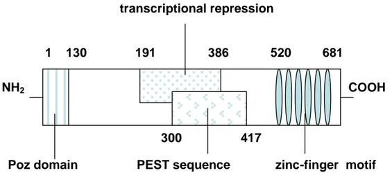
Figure 14. Functional domain of the BCL6 protein. Starting from N terminus
there are: the Poz domain(1-130), the domain involved in the transcritional
repression(191-386), the PEST domain (300-417), and at the C terminus (520-681) the zing finger domain.
Moreover BCL6 is able to cooperate with other proteins through the N-terminal POZ domain: this is an evolutionary conserved interaction domain required for homo- and hetero-dimerization of this family of proteins. Some of these molecules are the silencing mediator of retinoic acid and thyroid hormone receptor (SMRT), which in turn associates with both SIN3A and histone deacetylases (HDACs) to constitute a large repressing complex164,165. Other domains are present in the central part of BCL6 as for example the PEST motif that is critical for BCL6 protein stability.
In fact, BCL6 protein is also phosphorylated by exogenous kinases in the PEST domain: this produces the fast degradation of the BCL6 protein through its ubiquitinization and proteasome's action166,167. Another mechanism that negatively controls BCL6 is acetylation by p300: it blocks the capability of this transcriptional
transcriptional repression
1 130 191 386 520 681
300 417
Poz domain PEST sequence zinc-finger motif
35 repressor to interact with for example HDAC2168. Another important effect of BCL6, in human B cell, is to thwart the response to DNA damage by inhibiting both a p53-dependent and a MIZ-1 p53-dependent pathway169. In fact by physically interacting with the transcriptional activator Miz-1, BCL6 suppresses the transcription of the cell-cycle arrest gene p21169. The BCL6 expression is present in different tissues, for example, in sub-groups of epidermal and neuronal cells170, but its main role is in the germinal centre: here BCL6 is constitutively expressed and its action is necessary for the development of germinal centre and the suppression of p53 action157,171,172. In fact, BCL6 binds to the p53 promoter by silencing the transcription and by cooperating with MIZ-1 it blocks p21 cyclin dependent kinase, that produces cell cycle arrest. Germinal centres are dynamic structures within secondary lymphoid tissues that are responsible for the generation of B cell memory and high-affinity antibodies. These processes, in which BCL6 level increases, are necessary for B cells after the activation of T-cell dependent antigens and they allow the class switch recombination and the somatic hypermutation which physiologically occur in germinal centre cells of lymphatic organs173,174. In this way B cells can produce antibodies with high affinity for the antigen. BCL6 is also an essential requirement for GC formation, because mice lacking BCL6 cannot form these structures. Moreover in B-cell lines BCL6 prevents apoptosis induced from DNA damage.
When cells differentiate the down-regulation of BCL6 occurs. This gene is involved in chromosomal translocations in 40% of diffuse large B cell lymphoma and approximately 5–10% of follicular lymphoma cases175-178. Under these conditions the mutation is not present in the coding region but in the promoter or in the 5' part. So BCL6 can not be regulated at the end of the cell differentiation.
6.2 Leukaemia/lymphoma related factor
Leukemia/lymphoma related factor (LRF)179, that is encoded by the Zbtb7a gene, is also known as pok erythroid myeloid ontogenic factor (Pokemon)156, factor binding to IST-1 (FBI-1)180-183 or osteoclast derived zinc finger (OCZF)184. LRF is a transcriptional repressor of 67 KDa involved in many cellular processes such as viral infection180,181,185, differentiation179, inflammation186 and oncogenesis156. LRF is a
36 member of the POK family of transcriptional repressors and it is over-expressed in some kinds of tumours (Fig. 15).
Figure 15. Functional domain of the LRF protein. At the N terminus there is the
Poz domain and at the C terminus the zinc-finger domain.
In mouse embryonic fibroblast it is necessary for the transformation induced by two oncogenes. When LRF is deleted, these cells are resistant to oncogene-mediated cellular transformation. Instead the cells transformation is produced when LRF is co-expressed with another oncogene187. LRF is a transcriptional repressor of ARF that in turn is able to activate p53 in an indirect manner. In this way cells can be induced toward senescence or apoptosis, if at the same time there is an oncogene over-expression. Moreover LRF inactivation in mice produces the death of embryos due to a severe anaemia and an impaired cellular differentiation in many tissues. Microarray analysis shows that LRF is over-expressed in many different kinds of tumours: breast, lung, colon, prostate and bladder carcinomas. LRF is also one of the main regulatory genes in determining the development of common lymphoid progenitor cells in B or T cells: in the bone marrow, where LRF is not expressed, the cells are committed towards B development while in the thymus LRF is expressed and is able to repress the Notch pathway188. The Notch is a family of transmembrane proteins with an extracellular domain that induce proteolytic cleavage and release an intracellular signal. These proteins are involved in many different processes including T cell lineage commitment from common lymphoid precursor188. LRF plays an important role as a protooncogene also in Non- Hodgkin lymphomas: it is over-expressed in 80% of FL and in 60% of DLBCL156. LRF is involved in many other different processes. In fact it is necessary for the differentiation from preosteoclasts to osteoclasts184 and from preadypocytes to
NH2 COOH
37 adypocytes189. It seems to produce the exit from the cell cycle through the repression of genes such as Cyclin A189. Moreover it represses the cartilaginous markers in non-condrocytic cells190. During the mouse embryogenesis until the 11.5th day LRF is expressed just in haematopoietic tissue and afterwards it extends its expression to limb buds, pharyngeal arches, tail bud and neuronal tube179. In inflammation processes LRF is able to interact with Nuclear Factor-kappaB (NF-kappaB) transcriptional factor. In this way NF-kappaB moves from the cytoplasm to the nucleus where it acts as a transcriptional factor in response to inflammatory stimuli186.
7. The circuit
We followed the hypothesis that the over-expression of the two protooncogenes BCL6 and LRF are causally correlated in Non- Hodgkin's lymphomas as suggested in Pandolfi's paper156. To verify this idea we tested these two genes in the Dohh2 cell line: a cell line derived from a human B cell lymphoma which over-expresses both LRF and BCL6. Using our data and data taken from the literature demonstrated in different model systems, we reconstructed a hypothetical network that includes both these genes ( Fig. 16). We briefly described the members of this circuit and the data that are present in literature:
1. p53, which is under BCL6 direct negative control157 and LRF indirect negative control156 viap14-ARF down-regulation;
2. miR-34a, which is under p53 positive control191;
3. miR-145, which is an important link between p53 and c-Myc192, is under p53 positivecontrol;
4. c-Myc, which negatively controls miR-30 family transcription73;
5. miR-20a, which is involved in a complex feed back loop with c-Myc and E2F173;
6. E2F1, which induces p14-ARF transcription252 thus promoting p53 up-regulation;
7. LRF, which is negatively controlled by miR-20a193;
38
Figure 16. Hypothetical circuit connecting BCL6 and LRF.
7.1 p53
p53 is negatively regulated by BCL6 in a transcriptional manner as demonstrated by Dalla Favera157. The p53 gene is one of the most important tumour suppressor genes in human cancer transcribed from gene TP53: its molecular mass is 53 KDa. It encodes a gene regulatory protein that is activated by damage to DNA: in this manner it is involved in the cell-cycle control, in apoptosis, and in the maintenance of genetic stability194,195. These functions are fundamental in protecting the organism against cellular damage and disorder: p53 is mutated in about half of all human cancers196. The p53 protein has a dual role, regulating both progression through the cell cycle and, if the damage is extensive, it promotes senescence, or the initiation of apoptosis. Normal cells stop dividing after a finite number of cell divisions because they lacks of endogenous telomerase: the enzyme that elongates telomere sequences in DNA. This provokes telomere shortening and beyond a certain limit it triggers a the activation of the ATM/ATR–p53 pathway inducing the replicative senescence197,198. Under normal conditions very little p53 protein is found in most of the body's cells: it is not required for development. p53 interacts with another protein, Mdm2, that acts as a ubiquitin ligase: it targets p53 for destruction by proteasomes. On the contrary, after exposure to radiation or certain chemicals, when chromosomes are damaged, phosphorylation of
BCL6 p53 miR-34
miR-145 c-Myc miR-30 miR-20 E2F1 LRF p14 miR-28 MDM2
39 p53 occurs: this decreases its degradation because it blocks its binding to Mdm2199,200. As a consequence p53 increases and promotes the transcription of its target genes. For example p53, binding specific sequences on the DNA, induces the transcription of p21: a regulatory gene whose protein product binds to Cdk complexes required for entry into and progress through S-phase201. By blocking the kinase activity of these Cdk complexes, the p21 protein prevents the cell from entering the S phase, replicating its DNA and arresting them in G1 phase202. p53 is also involved in the activation of apoptosis because it promotes the transcription of genes of BCL2 family: these proteins promote the release of cytochrome c from mitochondria203. Cells defective in p53 fail to show these responses. In fact, the loss of p53 activity can be dangerous: it may allow faulty mutant cells to continue through the cell cycle, it may allow them to escape apoptosis and it may lead to the genetic instability characteristic of cancer cells, allowing further cancer-promoting mutations to accumulate as the cells divide. For example the rare genetic disease known as ataxia telangiectasia is caused by a defect in one of the protein kinases that phosphorylates and activates p53 in response to X-ray-induced DNA damage. Patients affected by this disease are very sensitive to X-rays due to the loss of the DNA damage checkpoints, and they consequently suffer from X-rays-induced cancer in an higher percentage. Another way through which p53 operates is the miRNAs transcription. In fact, p53 could modulate the cell fate not only as previously described but also through miRNAs, involving them in a complex network. Some miRNAs under p53 control are miR-15 and miR-1661, targeting the anti-apoptotic BCL2 protein, let-7, that down-regulates Ras and miR-221147, which, in turn, down-regulate the CDK inhibitor p27. Researchers have demonstrated that miR-34a191 and miR-145192 transcription is also regulated by p53 action.
7.2 miR-34a
The miR-34 family is composed by three members: miR-34a, miR-34b and miR-34c that are directly regulated by p53191. miR-34a resides on chromosome 1p36 and both miR-34b and miR-34c are generated by processing of a bicistronic transcript derived from chromosome 11q23 (termed miR-34b/c). These miRNAs are described as tumour suppressors: they are down-regulated in some tumour specimens as miR-34b/c was reported in a subset of lung cancer patients191, while miR-34a expression frequently
40 occurs in pancreatic cancer cells, and was detected in 36% of clinical colon cancer specimens204. c-Myc is one of the targets of miR-34a73. It has been demonstrated that enforced expression of miR-34a promotes apoptosis and up-regulation of miR-34b/c reduces proliferation and adhesion-independent growth of lung cancer cells191. Besides, in animal studies, the introduction of miR-34a also suppresses the cell proliferation of human colon cancer cells204.
7.3 miR-145
miR-145 is located on chromosome 5 (5q32-33) within a 4.09 Kb region. This is a well-known fragile site in the human genome: it is deleted in 11% of sporadic breast cancers205. Human miR-145 is enriched in germ line and mesoderm-derived tissues, such as uterus, ovary, testis, prostate, spleen, and heart206. Although the primicroRNA structure has not been identified, it is suggested to be co-transcribed with miR-143 because both are localized close to each other in the chromosome207. The transcriptional induction of miR-145 by p53 takes place in response to stress such as serum starvation192. miR-145 is described as a tumour suppressor. In fact, it is under-expressed in both breast and colon cancer specimens, compared to matched normal tissue samples192. It is worth mentioning that, this down-regulation of miR-145 seems to depend on the type of tissue: its down-regulation is described as more prominent in colon cancer than in breast cancer. Researchers demonstrated that miR-145 has a profound inhibitory effect on two colon cancer cell lines in vitro model by suppressing insulin receptor substrate-1 through binding to its 3’-UTR208. Consistent with this finding, miR-145 can also significantly reduce cell growth in B-cell lymphoma cell lines. This miR-145-mediated inhibition of cell proliferation is likely due to direct targeting c-Myc oncogene by directly binding to its 3’-UTR and down-regulating it. It has been described that miR-145 is inversely correlated to levels of c-Myc in breast cancer and in B lymphoma cell lines 192.
7.4 c-Myc
c-Myc is a basic helix-loop-helix transcription factor which directly regulates the transcription of a diverse array of target genes. Through this target gene network, c-Myc globally reprograms cells to drive proliferation and, in some settings, induce cell
41 death209. c-Myc can be activated by the small GTPase Ras, which leads to the activation of a MAP kinase cascade that, by uncertain mechanisms, leads to increased levels of the gene regulatory protein Myc210. Myc promotes cell-cycle entry: it increases the transcription of genes that encode D cyclins, thereby increasing G1-Cdk (cyclin DCdk4) activity211. In addition, Myc increases the transcription of a gene that encodes a component of the SCF ubiquitin ligase. This mechanism promotes the degradation of the CKI protein p27, leading to increased cyclin E-Cdk2 activity212. c-Myc can also play a role in tumour formation: c-Myc is frequently amplified in cancers, it can also be made active by a chromosomal translocation, amplification or virus infection. For example, as a consequence of a rearrangement, in Burkitt's lymphoma a translocation brings the c-Myc gene under the control of sequences that normally drive the expression of antibodies in B cells. As a result, the mutant B cells proliferate to excess and form tumours. Similar specific chromosome translocations are common in lymphomas and leukemias213.
High levels of c-Myc cause the activation of p14ARF, which binds and inhibits Mdm2 and thereby causes increased p53 levels: this could cause cell-cycle arrest or apoptosis. c-Myc may also promote E2F activity directly by stimulating the transcription of the E2F gene214-216. It has been demonstrated that c-Myc is able to regulate both positively and negatively the miRNAs transcription. c-Myc represses miR-30 family and it induces the expression of microRNAs within the miR-17–92 cluster: through chromatin immunoprecipitation researchers confirmed that c-Myc binds to its recognition sites, E-boxes, upstream of this cluster217. This regulation is a part of a complex feed back loop network that involves c-Myc and E2F1, that have been shown to activate each other’s transcription, and the miRNAs belonging to the cluster of miR-1773. Some of these miRNAs, such as miR-20a, are also able to target E2F1 mRNA. Moreover, the E2F transcription factors can also induce the microRNAs in the miR-17– 92 cluster: the over-expression of E2F1 or E2F3 results in increased miR-17–92 promoter activity. This complex network can be explained by the c-Myc and E2F1 roles: miR-17–92 may act as a brake on this possible positive feedback loop, thus helping to ensure a tightly controlled expression of c-Myc and E2F1 proteins.
42
7.5 E2F1
E2F are a family of proteins binding to specific DNA sequences in the promoters of many genes that encode proteins required for the S-phase entry, including G1/S-cyclins and S-cyclins. Members of the E2F family are downstream effectors of the retinoblastoma protein (Rb) tumour suppressor: an inhibitor of cell-cycle progression218. When cells are stimulated to divide by extracellular signals, active G1-Cdk accumulates and phosphorylates Rb, reducing its affinity for E2F. The Rb then dissociates, allowing E2F to activate S-phase gene expression. Rb plays a fundamental role in controlling cell cycle progression. Transcriptional activation of S phase-associated genes is only one facet of E2F activity: it is known that E2Fs both transactivate and repress gene expression to regulate a wide range of biological processes, including DNA replication, mitosis, the function of DNA damage checkpoints, DNA repair, differentiation, autophagy and apoptosis219-221. E2F transcription factors can increase the transcription of its own gene: E2F-dependent transcription of G1/cyclin and cyclin genes leads to increased G1/Cdk and S-Cdk activities, which, in turn, increase Rb phosphorylation and promote further E2F release. Moreover, the increase in G1/S-Cdk and S-Cdk activities enhances the phosphorylation of Hct1 and p27, leading to their inactivation or destruction. In mammals, the E2F family comprises eight genes (E2F1–8), which give rise to nine distinct proteins221. E2F family members have been categorized into subfamilies on the basis of their transcriptional activity, structure and interaction with Rb family members. E2F1, E2F2 and E2F3A, which interact only with Rb, constitute one subfamily and are often referred to as the ‘activator E2Fs’, as they are believed to work mainly in activating gene expression. E2F4–8 largely operate in the repression of gene expression and are generally referred to as the ‘repressor E2Fs’. E2F1 is particularly interesting: it could induce apoptosis by both p53-dependent and p53-independent pathways220. It is generally believed that up to a certain threshold, E2F1 expression drives cellular proliferation, but an excessive expression of this protein appears sufficient to induce cell death. E2F1 activates p53222, p53 and E2F1 co-regulate pro-apoptotic genes (for example the BH3-only protein-encoding genes PUMA and NOXA)223,224 and p53 and E2F1 negatively regulate anti-apoptotic genes such as BCL2 and MCL1225,226.