R E V I E W
Electromagnetic Field Therapy: A Rehabilitative
Perspective in the Management of Musculoskeletal
Pain
– A Systematic Review
This article was published in the following Dove Press journal: Journal of Pain Research
Teresa Paolucci1 Letizia Pezzi 1
Antonello Marco Centra 1 Niki Giannandrea 1
Rosa Grazia Bellomo2 Raoul Saggini1
1Department of Medical Oral Sciences
and Biotechnology (DiSmob), Physical Medicine and Rehabilitation Unit,
G. D’Annunzio University of
Chieti-Pescara, Chieti, Italy;2Department of
Biomolecular Sciences, University of Study of Urbino Carlo Bo, Urbino, Italy
Abstract: Electromagneticfields (EMFs) provide a non-invasive, safe, and easy method to
treat pain with respect to musculoskeletal diseases. The purpose of this systematic review
was to describe the use of electromagnetic therapy in the rehabilitationfield by investigating
the efficacy in acute and chronic pain in the musculoskeletal disorders. A database search
was conducted using the following resources: PubMed, Cochrane, PEDro, SCOPUS, and
WoS. The following MESH terms were used: [Electromagnetic field AND/OR
Rehabilitation], [Electromagnetic field AND/OR Pain], [Pulsed Magnetic field AND/OR
Rehabilitation] and [Pulsed Magnetic field AND/OR Pain], [Pulsed Electromagnetic field
AND/OR Rehabilitation] and [Pulsed Electromagneticfield AND/OR Pain], per the
guide-lines of the PRISMA statement. Articles published between January 1, 2009 and December 31, 2018 were included as assessment of musculoskeletal pain conditions, rando-mized clinical trial including crossover and prospective design studies, full English text available, population age > 18 years; instead were excluded neurological randomized clinical trials, transcranial magnetic stimulation application, neuropathic pain, animal/in vitro studies, and articles without English abstract or English full text. Three independent investigators (AMC, NG, and LP) retrieved all the information. Twenty-one RTC (N=21) were considered
for the inclusion and exclusion criteria. The results showed as pulsed magneticfields at low
intensity and frequency (from 1 Hz up to 100 Hz) are commonly used with efficacy in
resolving musculoskeletal pain. EMFs therapy is a well tolerated, effective with no negative side effects, which can be integrated with rehabilitation for the treatment of chronic and acute pain in musculoskeletal diseases, but further studies are needed to examine the use of more standardized protocols.
Keywords: pulsed electromagnetic fields, rehabilitation, physical medicine, magnetic
therapy, pain
Introduction
Pain is an unpleasant sensory and emotional experience associated with actual or potential tissue damage or described in terms of such damage.1
Musculoskeletal diseases comprise several conditions that are characterized by pain and limitations in mobility, dexterity and functional ability, reducing people’s ability to work and participate in social roles with associated impacts on mental wellbeing. The most common and disabling musculoskeletal diseases are osteoar-thritis, back and neck pain, tendinopathy,fibromyalgia and myofascial pain. Among the clinically relevant pain conditions treated in rehabilitation, pain with respect to
Correspondence: Teresa Paolucci
G. D’Annunzio University of
Chieti-Pescara, Chieti, Italy
Email [email protected]
open access to scientific and medical research
Open Access Full Text Article
Journal of Pain Research downloaded from https://www.dovepress.com/ by 192.167.13.146 on 03-Jun-2021
the musculoskeletal system is most frequent and has a major impact on people’s quality of life.2Chronic low back pain (CLBP) has a significant impact on musculos-keletal pain with a prevalence increases linearly from the third decade of life on, until the 60 years of age, being more prevalent in women.3–5
The use of electromagneticfields (EMFs) and in particu-lar of the magneto-therapy has had a notable increase in the last decade in rehabilitation treatment and provides a non-invasive, safe, and easy method to directly treat the site of injury, the source of pain and inflammation, and other types of disease.6–8Magneticfield therapy was applied to promote bone healing, treat osteoarthritis and inflammatory diseases of the musculoskeletal system, alleviate pain, enhance heal-ing of ulcers and reduce spasticity9and, also, extremely low frequency (ELF) magneticfields in the pico tesla and milli tesla ranges are aimed at improving neurotransmission and correcting local immune pathology, respectively.10 An analgesic and anti-nociceptive efficacy, similar to the opioid analgesic effect respect of pulsed electromagnetic field (PEMF) is reported by scientist literature but the clear biolo-gical and biochemical mechanism of the effect of magnetic therapy on pain remains unknown.11Also, some studies have shown that short-term exposure to electromagnetic fields influences several inflammatory cellular and neurological processes, such as patterns of cortical activation and inhibi-tion and the activity of various neurotransmitters.12
Above all, the magneto-therapy recognizes an important use in the field of both chronic and acute pain in muscu-loskeletal disorders using protocols with specific intensities and frequencies: magneticfields applied in magneto-therapy for pain, in line with the criteria generally accepted in physical medicine, have the frequency below 100 Hz and magnetic flux density in the range between 0.1 mT and 30 mT.13 When used alone, the PEMF seems to have a good effect in reducing the pain intensity in low back patients, independently of the low-back pain condition. However, when added to other standard therapies (such as standard physiotherapy or analgesic therapy) seems to do not add any additional effect.14–16
Furthermore, the efficacy of magnetotherapy compared to some forms of chronic pain such asfibromyalgia (FM) still remains debated.17,18 Moreover, also in neurological pathology, ELF magnetic field was revealed to induce a significant improvement in functional and mental status in brain stroke patients and clinical parameters had posi-tive correlation with the level of enzymatic antioxidant protection.7
Thus, surely, the efficacy of magneto-therapy is related to the type of electromagnetic fields used and in the rehabilitationfield there are today very different treatment protocols: certainly, in the last decade, the use of ELF magnetic fields has been on its increase. Innovative and still experimental approaches concern, for example, the use of cyclotronic resonance (CR), a kind of specific ultra-weak pulsed electromagnetic fields, in low back pain:19 the theory of the CR considered that the endogenous electromagnetic forces generated by the activity of the cells of the human body are of infinitely low intensity. Then, the effect of ELFfields, however, does not depend directly on their very low frequency and intensity, but more on the fact that if they are structured with a correct form, intensity, frequency and sequence, they synchronize with the frequencies of the biological system that disturb, triggering an effect. CR involves electrically charged ions and molecules that oscillate at specific harmonic frequen-cies, within a continuous feedback system with the cells themselves. This mechanism of interaction between ELF magnetic fields, the earth’s magnetic field, and living organisms has been called Cyclotron Resonance (CR) by Liboff.20
Despite the widespread use of magneto-therapy in rehabilitationfield it is difficult to find standardized treat-ment protocols especially when aimed at treating muscu-loskeletal pain, be it chronic or acute.
Musculoskeletal pain often develops over time result-ing in more hyperalgesia and larger pain areas. Peripheral and spreading sensitizations are probably important mechanisms for the translation of acute local pain to chronic musculoskeletal pain conditions. The transition from acute to chronic musculoskeletal pain is not well understood.21 Acute pain is a direct outcome of the nox-ious event and is reasonably classified as a symptom of underlying tissue damage or disease. Chronic pain may not be directly related to their initial injury or disease condi-tion, but rather to secondary changes including some that occur in the pain detection system itself. Thus, the mechanisms underlying chronic or persistent pain may be quite different from acute pain.22The distinction between acute and chronic pain is sometimes determined by an arbitrary interval of time since onset; the two most com-monly used markers being 3 months and 6 months since onset, though some theorists and researchers have placed the transition from acute to chronic pain at 12 months.
Thus, the aim of this systematic review was to inves-tigate the scientific evidence over the last decade with
Journal of Pain Research downloaded from https://www.dovepress.com/ by 192.167.13.146 on 03-Jun-2021
respect to the use of electromagnetic therapy in the reha-bilitation field by investigating the efficacy in acute and chronic pain in the musculoskeletal disorders.
Materials and Methods
Search Strategy
A systematic review of the literature was performed using the following search engines: PubMed, Cochrane, PEDro, SCOPUS and Web of Science (WoS), per the guidelines of the PRISMA statement.23 In order to perform the search, the following algorithm was developed, based on the PICO acronym,24to evaluate the effects of electromagneticfields respect the reduction of pain (acute and chronic) as the primary outcome in musculoskeletal diseases.
These keywords were used (MESH terms): [Electromagnetic field AND/OR Rehabilitation], [Electromagnetic field AND/OR Pain], [Pulsed Magnetic field AND/OR Rehabilitation] and [Pulsed Magnetic field AND/OR Pain], [Pulsed Electromagnetic field AND/OR Rehabilitation] and [Pulsed Electromagnetic field AND/ OR Pain].
Reference lists of most relevant studies were scanned for additional citations; country, author, affiliated institu-tions, and enrollment periods were extracted and reviewed to identify and exclude duplicate publications from the same cohort.
Study Criteria and Selection
Inclusion criteria were: (1) articles published between January 1, 2009, and December 31, 2018, (2) assessment of musculoskeletal pain conditions, (3) randomized clin-ical trial including crossover and prospective design stu-dies, (4) full English text available, (5) population age > 18 years.
Exclusion criteria were (i) neurological randomized clinical trials, (ii) transcranial magnetic stimulation appli-cation, (iii) neuropathic pain (iv) animal/in vitro studies, and (v) articles without English abstract or English full text.
Data Extraction
Three independent investigators (AMC, NG, and LP) retrieved all the information. The main outcome of interest was the quantification of intensity of pain in musculoske-letal diseases. The secondary outcomes were the recovery of function and quality of life with respect to the disability in musculoskeletal diseases. Thus, after the application of
the eligibility criteria and the included studies were deter-mined, the studies were analyzed based on sample demo-graphics, study’s aim, statement of conflict of interest, study duration and follow-up (period of time and percen-tage), EMF devices used, evaluation time, intervention protocol, and outcome parameters assessed (clinical and functional).
Methodology Quality and Risk of Bias
Assessment
Establish a quality assessment of each study using the PEDro scale (Physiotherapy Evidence Database, 1999): this scale has shown good reliability for scoring RCTs.25 The PEDro scale consists of 11 items related to scientific rigor. The scale’s items 2 to 11 contribute to internal validity, and the study is given 1 point for each of these items that are met. Thefirst item relates to external valid-ity and is not included in the final score. The quality assessment was performed independently by the three reviewers, and any disagreement was discussed until con-sensus was reached. We considered trials with scores of≥6 as having high quality and trials with scores of ≤5 as having low quality.
The assessment of the risk of bias was done indepen-dently by the same three authors (AMC, NG, and LP), and was assessed according to the Cochrane Collaboration’s domain-based evaluation framework.26 Main domains were assessed in the following sequence: 1) selection bias (randomized sequence generation and allocation con-cealment); 2) performance bias (blinding of participants and personnel); 3) detection bias (blinding of outcome assessment); 4) attrition bias (incomplete outcome data, eg, due to dropouts); 5) reporting bias (selective report-ing); 6) other sources of bias. The scores for each bias domain and thefinal score of risk of systematic bias were graded as low, high, or unclear risk.
Results
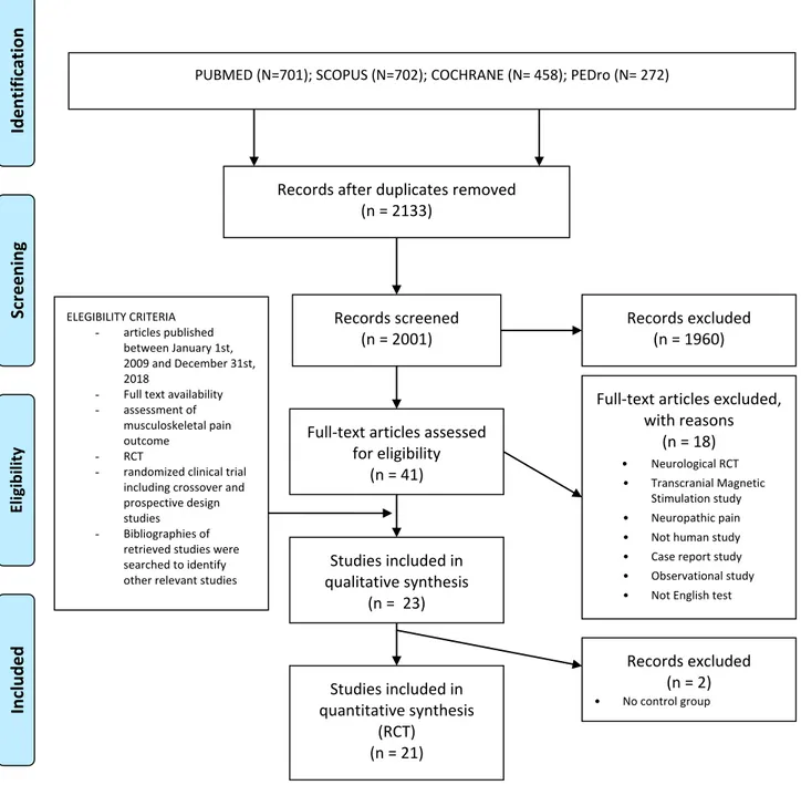
The PRISMAflow-diagram showing the selection of stu-dies is given inFigure 1.
Twenty-one articles (N=21) satisfied the inclusion criteria and were considered in the review: 8 articles treated pain of the knee for osteoarthritis (OA),27–34 2 articles treated Shoulder Impingement Syndrome (SIS),35,365 articles trea-ted spine pain, of which 1 study about chronic mechanical neck pain (CNP),37and 4 studies were about low back pain (LBP),14,16,38,39 3 articles treated Fibromyalgia Syndrome
Journal of Pain Research downloaded from https://www.dovepress.com/ by 192.167.13.146 on 03-Jun-2021
(FM),17,18,40then 1 article showed the effect of EMF respect of patellofemoral pain (PFP),411 article treated Plantar fas-ciitis (PF),42and 1 article treated Hand osteoarthritis (HO).43 The PEDro score values and other characteristics of the included studies were shown inTable 1: the methodological quality of the twenty-one included articles, according to the PEDro scale, with averaged between 4/10 and 10/10, aver-aged 7.57/10 and only 4 studies have a PEDro score ≤5 indicating low quality. Furthermore, in Table 2 were specified type of magnetic field and parameters used in the individual studies included in our review. In detail, 15
studies have used Pulsed Electromagnetic Field (PEMF),14,16,17,27-29,31,33-35,37,38,40,41,43 one study has used Extremely low-frequency magneticfield (ELF-MF),182 stu-dies have used Pulsed Radiofrequency Electromagnetic Field (PRFE),30,42one study has used Electromagnetic transduc-tion therapy (EMTT),36 and another one has used Pulsed electromagnetic energy (PEME),39and Gökşen et al32have used Magnetic resonance treatment. Lastly, the risk of bias was considered High for 4 studies,31,33,34,38 Unclear for 2 studies14,43 and Low forfifteen studies (see Table 3). The most frequent source of potential bias was the performance
PUBMED (N=701); SCOPUS (N=702); COCHRANE (N= 458); PEDro (N= 272)
Screening
Included
Eligibilityn
oi
t
ac
ifi
t
n
e
dI
Records after duplicates removed (n = 2133)
Records screened (n = 2001)
Records excluded (n = 1960)
Full-text articles assessed for eligibility
(n = 41)
Full-text articles excluded, with reasons (n = 18) Neurological RCT • Transcranial Magnetic Stimulation study • Neuropathic pain
• Not human study
• Case report study
• Observational study
• Not English test
Studies included in qualitative synthesis (n = 23) Studies included in quantitative synthesis (RCT) (n = 21) ELEGIBILITY CRITERIA - articles published between January 1st, 2009 and December 31st, 2018
- Full text availability
- assessment of
musculoskeletal pain outcome
- RCT
- randomized clinical trial
including crossover and prospective design studies
- Bibliographies of
retrieved studies were searched to identify other relevant studies
Records excluded (n = 2)
• No control group
•
Figure 1 Flowchart of the included studies in the review according to the PRISMA 2009 guidelines.
Journal of Pain Research downloaded from https://www.dovepress.com/ by 192.167.13.146 on 03-Jun-2021
T able 1 PEDr o Scor e V alues and Other Characteristics of the Included Studies
Author (Year Published) [Ref.]
Dia gnosis N (M/F) Mean± SD Ag e (Y ears) Study N TG (Mean± SD Ag e) CG (Mean± SD Ag e) Inter v ention Outcome P arameters Evaluation Time Conclusions PEDr o Scor e A y et al (2009) 31 O A N=55 (15/40) RCT TG=30 (58,9±8,8) CG=25 (57,7±6,5) TG: PEMF , hot pack, TENS and ex er cise pr ogram; CG: sham-PEMF , hot pack, TENS and ex er cise pr ogram V AS: pain; Lik ert pain scale: pain; LI: function T0: baseline T1: 3 w eeks V AS: TG=CG LI: TG=CG 6/10 Bagnato et al (2016) 30 O A N=60 (43/17) y=67,7 ±10,9 RCT TG=30 (68,6±11,9) CG=30 (66,9±10) TG: PRFE CG: placebo V AS: pain; W OMA C: function, pain, disability SF-36: QoL T0: baseline T1: 1 month V AS, W OMA C: TG>CG SF-36:TG=CG 9/10 Br ook et al (2012) 42 PF N=70 (18/52) RCT TG=42 (53,2±14,7) CG=28 (49,7±15,2) TG: PRFE CG: sham-PRFE V AS: pain T0: baseline T1: 1 da y T2: 2 da ys T3: 3 da ys T4: after 4 da ys T5: 5 da ys T6: 6 da ys T7: 7 da ys V AS: TG>CG 8/10 Dündar et al (2016) 34 O A N=40 (11/29) RCT TG= 20 (56.8 ±14.5) CG= 20 (57.6 ±13.8) TG: hot pack, ultrasound, TENS, isometric knee ex er cise and PEMF CG: hot pack, ultrasound, TENS, isometric knee ex er cise and sham-PEMF V AS: pain W OMA C: function, pain Ultrasonographic effusion of the knee Serum YKL-40 a nove l biomark er of osteoarthritis T0: befor e tre atment T1: 1 month W OMA C: TG=CG VAS: TG=CG 5/10 Galace de Fr eitas et al (2014) 35 SIS N=56 (20/36) y=50,5 ±8,9 RCT TG=26 (50,1±8,2) CG=30 (50,8±9,6) TG: PEMF and ex er cises; CG: placebo and ex er cises V AS: pain; CMS: function; UCLA: function; Handheld dynamometr y: str ength T0: baseline T1: 3 we eks T2: 9 w eeks T3: 3 months V AS, UCLA, CMS, Dynamometr y: TG>CG 9/10 (Continued )
Journal of Pain Research downloaded from https://www.dovepress.com/ by 192.167.13.146 on 03-Jun-2021
T
able
1
(Continued).
Author (Year Published) [Ref.]
Dia gnosis N (M/F) Mean± SD Ag e (Y ears) Study N TG (Mean± SD Ag e) CG (Mean± SD Ag e) Inter v ention Outcome P arameters Evaluation Time Conclusions PEDr o Scor e Giombini et al (2013) 37 CNP N=45 (14/31) Pr ospectiv e RCT TG=15 (44.0±9.6) CG1=15 (40.5±7.4) CG2=15 (43.0±9.4) TG: PEMF CG1: neck balance system CG2: neck balance system with negligible balancing V AS: pain NDI: disability NPDS: QoL T0: baseline; T1: end of 8 w eek T2: after follow-up 12 w eeks V AS: CG1>TG>CG2 NDI: CG1>TG>CG2 NPDS: CG1> CG2 > TG 8/10 Gök şen et al (2016) 32 O A N=97 RCT TG=49 (54.02 ±6.79) CG= 48 (54.92 ±7.5) TG: magnetic treatme nt CG: sham-magnetic treatme nt V AS: pain W OMA C: function, pain, disability SF-36: QoL T0: baseline; T1: end of 2 w eek T2: follow-up after 12 w eeks V AS, W OMA C, SF-36: TG=CG 9/10 Kanat et al (2013) 43 HO N= 50 RCT TG= 25 (64±2.6) CG= 25 (62±2.4) TG: PEMF + ex er cise CG: sham PEMF + ex er cise Lik ert scale: pain at res t, pain at motion, joint stiffness SF-36: QoL Duruöz: function AU SC AN: pain, stiffness and disability HG and PG: str ength T0: baseline T1: after tr eatment T3: follow-up one month after tr eatment SF-36: TG>CG Duruöz: TG>CG AU SC AN: TG>CG HG: TG=CG, PG: TG=CG 7/10 Klüter et al (2018) 36 SIS N=86 (41/45) RCT TG=44 (50,21±8,5) CG=42 (49,21±7,3) TG: EMTT/ESWT ; CG: sham-EMTT /ESWT V AS: pain; CMS: function T0: baseline T1: 6 we eks T2: 12 we eks T3: 24 w eeks V AS: TG>CG CMS: TG>CG 9/10 Krammer et al (2015) 39 LBP N=40 (20/20) RCT TG=20 (35,7) CG=20 (30,2) TG: PEMF and ph ysiotherap y treatme nt; CG: sham -PEMF and ph ysiotherap y treatme nt NRS: pain ODI: disability PSFS: function T0: baseline T1: 1 w eek T2: 4 w eeks NRS, ODI, PSFS: TG=CG 8/10 Külcü et al (2009) 33 O A N=45 RCT TG =15 (65.8 ±10.3) TG1 = 15 (63.1±13.6) CG =15 (62.0±6.0) TG: PEMF TG1: US CG: contr ol V AS: pain W OMA C: function, pain, stiffness T0: baseline T1: end of thr ee we eks V AS, W OMA C: TG, TG1>CG 5/10
Journal of Pain Research downloaded from https://www.dovepress.com/ by 192.167.13.146 on 03-Jun-2021
Multanen et al (2018) 17 FM N=108 y=47±10 RCT (cr oss-ov er) TG=57 CG=51 TC: PEMF CG: sham-PEMF V AS: pain, stiffness FIQ: QoL and disability T0: baseline T1: end of 12 we ek T2: after washout 16 w eek T3: end of 28 we ek V AS: TG= CG FIQ: TG= CG 10/10 Nelson et al (2013) 27 O A N=34 RCT TG=15 (55.5±2.5) CG= 19 (58.4±2.5) TG: PEMF CG: Sham-PEMF V AS: pain T0: baseline T1: end of da y 3 T2: end of da y 14 T3: end of da y 29 T4: end of da y 42 V AS: TG>CG 10/10 Ok e et al (2013) 38 BP N=16 (9/ 7) y= 26.00 ± 8.62 RCT TG=8 CG=8 TG: PEMF + FANS CG: FANS NRS: pain Functional Activity Scale T0: baseline T1: end of tre atment (after 5– 9 da ys) NRS, Functional Activity Scale: TG>CG 4/10 Omar et al (2012) 14
LBP (Unilateral Radicular Pain)
N=40 RCT TG=20 (37.5±8.5) CG= 20 (40.0±8.3) TG: PEMF CG: sham-PEMF V AS: pain OSW: disability SSEPs: nerve conduction T0: baseline T1: end of 8 w eek T2: after follow-up 12 w eeks V AS, OSW , SSEPs: TG>CG 5/10 Özgüçlü et al (2010) 28 O A N=40 RCT TG=20 (60.55±7.7) CG= 20 (62.15 ±8.1) TG: isometric knee+ hot pack+ therapeutic ultrasound+ PEMF ex er cise CG: isometric knee+ hot pack+ therapeutic ultrasound+ sham PEMF V AS: pain W OMA C: function, pain, disability T0: baseline T1: end of 2 w eek V AS, W OMA C: TG=CG 6/10 Paolucci et al (2016) 18 FM N=37 (F) y=50.33 ±10.94 RCT (cr oss-ov er) TG1=16 (49.5 ±9.38) TG2 =17 (51.12±12.47) TG1: ELF/sham-ELF TG2: sham-ELF/ELF V AS: pain FAS: pain, fatigue and quality of sleep FIQ: QoL and disability HA Q: QoL and daily activities T0: baseline; T1: end of 1 tr eatment cycle T2: beginning of 2 tr eatment cycle (after a 1-month washout) T3: end of 2 tr eatment cycle T4: follow-up after 1 month V AS, FAS: T1:TG1<TG2 T3:TG2<TG1 FIQ, HA Q: T1: TG1<TG2 T3: TG2<TG1 8/10 (Continued )
Journal of Pain Research downloaded from https://www.dovepress.com/ by 192.167.13.146 on 03-Jun-2021
T
able
1
(Continued).
Author (Year Published) [Ref.]
Dia gnosis N (M/F) Mean± SD Ag e (Y ears) Study N TG (Mean± SD Ag e) CG (Mean± SD Ag e) Inter v ention Outcome P arameters Evaluation Time Conclusions PEDr o Scor e Park et al (2014) 16 LM (Lumbar Myalgia) N= 38 (11/27) y=31.95 ±12.30 RCT TG=19 (33.00 ±11.06) CG=19 (30.89 ±13.66) TG: PEMF CG: placebo V AS: pain ODI: function SF-36, EQ-5D: QoL BDI: depre ssion RMDQ: disability T0: baseline T1: after the 6 th tre atment T2: follow-up after 1 w eek V AS, RMDQ: TG>CG ODI, SF-36, EQ-5D , BDI: TG=CG 9/10 Ser vodio Iammarr one et al (2016) 41 PFP N= 31 y=22.5 RCT TG: 13 (21±7) CG: 17 (24±8) TG: PEMF , Home ex er cise pr ogram CG: Home ex er cise pr ogram V AS: pain VISA: pain, functional mobility , QoL T0: baseline T1: 2 months T2: 6 months T3: 12 months VISA T1: TG=CG VISA T2-T3: TG>CG VAS: TG<CG 6/10 Sutbe yaz et al (2009) 40 FM N=56 RCT TG=28 (42.96 ±9.57) CG=28 (40.89 ±6.88) TG: PEMF CG: sham-PEMF V AS: pain FIQ: QoL and disability BDI: depre ssion SF-36: QoL PGAR T : patient ’s global assessment T0: baseline; T1: end of 3we ek T2: follow-up after 12 w eeks V AS: TG>CG FIQ, SF-36: TG >CG BDI: CG>TG PGAR T : debated 8/10 W uschech et al (2015) 29 O A N=57 RCT TG=44 (63.4±12.1) CG=13 (55.5±10.8) TG: PEMF CG: placebo V AS: pain W OMA C: function, pain, disability T0: baseline; T1: end of 18 da ys V AS, W OMA C: TG>CG 9/10 Abbre viations: O A, knee osteoarthritis; PF , plantar fasciitis; SIS, shoulder impingement syndr ome; CNP , chr onic mechanical neck pain; HO , hand osteoarthritis; L BP , low back pain; FM, fibr om yalgia; BP , back pain; LM, lumbar m yalgia; PFP , patellofemoral pain; PPS, parallel pr ospective study; RCT , randomized contr olled trial; TG, tr eatment gr oup; CG, contr ol gr oup; PEMF , pulsed electr omagnetic fields; PRFE, pulsed radiofr equency electr omagnetic field therap y; US, therapeutic ultrasound; EMTT/ESWT , electr omagnetic transduction therap y/extracorporeal shockwa ve therap y; V AS, visual analog scale; W OMA C , W estern Ontario and McMaster Univ ersities Osteoarthritis Index; RMS, Roles – Maudsle y scor e; CMS, Constant –Murle y scale; UCLA, Univ ersity of California/Los Angeles; SF-36, Short-Form 36 version-2; NDI, Neck Disability Index; NRS, numerical rating scale ; LI, Lequesne algofunctional index; FIQ, Fibr om yalgia Impact Questionnair e; FAS, Fibr om yalgia Assessment Scale; HA Q, Health Assessment Questionnaire; BDI, Beck Depr ession In ventory ; PGAR T , patient ’s global assessment rating scale; NPDS, neck pain disability scale; OSW , Modi fied Oswe str y Low Back Pain Disability Questionnair e; SSEPs, somatosensor y e vok ed potentials; FSS, Fatigue Sev erity Scale; MFIS, Modi fied Fatigue Impact Scale; R OM, range of motion; VISA, Victorian Institute of Sport Assessment score; A USC AN, Australian Canadian Osteoarth ritis Hand Index; Duruöz, Duruöz Hand Index; HG, hand grip; PG, pinch grip; ODI, Osw estr y Disability Index; EQ -5D , Eur oQol-5 Dimension; RMDQ, Roland-Morris Disability Questionnaire; PSFS, Patient Speci fic Functional Scale.
Journal of Pain Research downloaded from https://www.dovepress.com/ by 192.167.13.146 on 03-Jun-2021
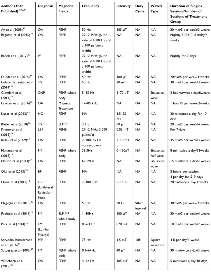
Table 2 Type of Magnetic Field and Parameters Used in the Included Studies
Author (Year
Published)[Ref.]
Diagnosis Magnetic
Fields
Frequency Intensity Duty
Cycle Wave’s Type Duration of Singles Session/Number of Sessions of Treatment Group
Ay et al (2009)31 OA PEMF 50 Hz 105 µT NA NA 30 min/5 per week/3 weeks
Bagnato et al (2016)30 OA PRFE 27,12 MHz (pulse
rate of 1000 Hz and a 100 µs burst width)
NA NA NA Nightly/11,3± 0, 8 h/day/4
weeks
Brook et al (2012)42 PF PRFE 27,12 MHz (pulse
rate of 1000 Hz and a 100 µs burst width)
NA NA NA Nightly for 7 days
Dündar et al (2016)34 OA PEMF 50 Hz 100 µT NA NA 20min/5 per week/4 weeks
Galace de Freitas et al
(2014)35
SIS PEMF 50 Hz 20 mT NA NA 30 min/3 per week/3 weeks
Giombini et al (2013)37 CNP PEMF whole body 5–25 Hz 5–70 µT NA Sinusoidal wave 2 hours/twice a day/8weeks Gökşen et al (2016)32 OA Magnetic Treatment
17–85 kHz NA NA NA 1 hour/5 per week/2weeks
Kanat et al (2013)43 HO PEMF NA 3.5–25
mT
NA NA 20 min/once a day for 10
days
Klüter et al (2018)36 SIS EMTT 3 Hz 80 µT NA NA 20 min/2 per week/4 weeks
Krammer et al (2015)39 LBP PEME 27.12 MHz (1000 pulses/s) 0.03 mT NA NA For 7 days Külcü et al (2009)33 OA PEMF 2–100–25 Hz consecutively
2–10 mT NA NA 35 min/5 per week/3 weeks
Multanen et al (2018)17 FM PEMF whole body 33,3Hz 0–150µT NA Sinusoidal half-wave
8 min twice a day/12weeks
Nelson et al (2013)27 OA PEMF 6.8 MHz NA NA Sinusoidal
wave
15 min/twice a day/2 weeks
Oke et al (2013)38 BP PEMF NA NA NA NA 2 hours per session;
4 per day for 5–9 days
Omar et al (2012)14 LBP
(Unilateral Radicular Pain)
PEMF 7–4000 Hz 5–15 G NA NA 20min/once a day/3 weeks
Özgüçlü et al (2010)28 OA PEMF 50 Hz 30 G 90-s
interval
NA 30min/5 per week/2 weeks
Paolucci et al (2016)18 FM ELF-MF
whole body
1–80Hz 100 µT NA NA 30 min/3 per week/4 weeks
Park et al (2014)16 LM
(Lumbar Myalgia)
PEMF 8.56 kHz 820 mT NA NA 10 min/3 per week/2 weeks
Servodio Iammarrone
et al (2016)41
PFP PEMF 75 Hz 1.5 mT 10% Square
waveform
4 h per day/6 weeks
Sutbeyaz et al (2009)40 FM PEMF whole
body
0.1–64Hz 40 µT NA NA 30 min/twice a day/3 weeks
Wuschech et al
(2015)29
OA PEMF 4–12 Hz 105 mT NA NA 5 min/twice a day/18 days
Abbreviations: OA, knee osteoarthritis; PF, plantar fasciitis; SIS, shoulder impingement syndrome; CNP, chronic mechanical neck pain; HO, hand osteoarthritis; LBP, low
back pain; FM,fibromyalgia; BP, back pain; LM, lumbar myalgia; PFP, patellofemoral pain; PEMF, pulsed electromagnetic field; PRFE, pulsed radiofrequency electromagnetic
field; ELF-MF, extremely low-frequency magnetic field; EMTT, electromagnetic transduction therapy; PEME, pulsed electromagnetic energy.
Journal of Pain Research downloaded from https://www.dovepress.com/ by 192.167.13.146 on 03-Jun-2021
bias related to incomplete outcome data, due to not mention-ing adverse events, and the inadequate blindmention-ing participant and personnel. Furthermore, the articles analyzed share the same contraindications to the use of magnetotherapy as patients with pacemakers (or other electrical devices) and/ or with cancer or in pregnant women.
Outcomes of Interest
Regarding the primary outcome, acute and chronic pain, in the included studies, the Visual Analogue Scale (VAS)44 was the main evaluation tool, except for Kanat et al,43 which presented 10-point Likert Scale to quantify pain at rest and at motion of the hand. Also, Oke et al38used the numeric rating scale for pain (NRS).45
Regarding the evaluation of the recovery of function, the authors use specific scales depending on the muscu-loskeletal diseases. In fact, to evaluate the patients with OA was administrated the Western Ontario and McMaster Universities Osteoarthritis Index (WOMAC),46 or Lequesne Algofunctional index of knee (LI).47
Instead, to measure pain in SIS35,36 was used the Constant–Murley Scale (CMS)48 and University of California/Los Angeles scale (UCLA);49in addition, to eval-uate the hand’s function was used Duruöz and Auscan Hand Osteoarthritis Indexes (AUSCAN).50,51 The evaluation of function in neck and low-back pain was assessed with the Neck Disability Index (NDI),52the Modified Oswestry Low Back Pain Disability Questionnaire (OSW),53 the Korean version of Oswestry Disability Index (ODI)54and Modified Version Functional Activity Scale.38 The Fibromyalgia Assessment Status scale (FAS)55 and the Fibromyalgia Impact Questionnaire (FIQ)56have been administered speci-fically in FM to quantify pain and the secondary outcomes like recovery of function, disability and quality of life. Last, the Victorian Institute of Sport Assessment score (VISA)57 was used to quantify mobility and function in patellofemoral pain.
Finally, in order to evaluate the increase in quality of life (QoL) perceived by patients, five studies16,30,32,40,43 used the 36-item Medical Outcomes Study Short-Form 36
Table 3 Risk of Bias Summary of 21 Included Studies
Random Sequence Generation Allocation Concealment Blinding of Participants and Personnel Blinding of Outcome Assessment Incomplete Outcome Data Selective Reporting Other Bias Ay et al (2009)31 High − ? − + + − − Bagnato et al (2016)30 Low + + + + + − ? Brook et al (2012)42 Low + + + + + + ? Dündar et al (2016)34 High ? − − + − + ? Galace de Freitas et al (2014)35 Low + + + + + + ? Giombini et al (2013)37 Low + + − − + + ? Gökşen et al (2016)32 Low + + + + + + ? Kanat et al (2013)43 Unclear ? ? + − + − ? Klüter et al (2018)36 Low + + – + + + ? Krammer et al (2015)39 Low + + + + − − ? Külcü et al (2009)33 High + − − − − + ? Multanen et al (2018)17 Low + + + + + + − Nelson et al (2013)27 Low + ? + + + − ? Oke et al (2013)38 High ? − − − − + ? Omar et al (2012)14 Unclear ? − ? ? − + ? Özgüçlü et al (2010)28 Low + − + ? − + ? Paolucci et al (2016)18 Low + + + + − + − Park et al (2014)16 Low + + + + + + ? Servodio Iammarrone et al (2016)41 Low + + + − − ? ? Sutbeyaz et al (2009)40 Low + + + + + + ? Wuschech et al (2015)29 Low ? + + + − ? ?
Notes: The“+” means low risk of bias; the “−” means high risk of bias; the “?” means unknown risk of bias. Trials involving three or more high risks of bias were considered
as poor methodological quality.
Journal of Pain Research downloaded from https://www.dovepress.com/ by 192.167.13.146 on 03-Jun-2021
(SF-36);58 then the Health Assessment Questionnaire (HAQ)59or the EuroQol-5 Dimension (EQ-5D).60
Discussion
The purpose of this systematic review was to investigate the scientific evidence of the last decade regarding the use of EMF in rehabilitation about its efficacy of acute and chronic pain in musculoskeletal disorders. The results suggest as EMF therapy is an optional treatment in the management of musculoskeletal pain disease that can reduce pain intensity and improves function.
Our review shows that PEMF are the most widely used magneticfields, particularly in knee OA.27–34Among non-pharmacological treatments, PEMF have beneficial thera-peutic effects on knee joint tissue.61 PEMF signals modulate calmodulin (CaM)-dependent nitric oxide (NO) signaling cascades in articular chondrocytes and other cells, as demonstrated by a previous study that used CaM antagonists and NO downstream inhibitors.62,63 This mechanism could promote the resolution of pain by accel-erating the removal of inflammatory substances. PEMF stimulates chondrocyte proliferation, differentiation, and extracellular matrix synthesis through the release of ana-bolic morphogens, such as bone morphogenetic proteins and anti-inflammatory cytokines, by adenosine receptors A2A and A3: in clinical translational study, a beneficial effect was observed in improving function in knee OA.64 In 4 studies dealing with knee OA,27,29,30,33 there were good results in the reduction of pain and improvement of function compared to the control group. In detail, Nelson et al27 proposed a short protocol of 2 weeks (15 mins per session, twice daily) with 6.8 MHz and an intensity of 30 Gauss in OA: patients in the PEMF group had a mean self-reported maximum daily VAS pain score at baseline of 6.85 ±0.33 and 4.19±0.71 at the end of treat-ment, compared with 7.18±0.31 and 6.11±0.54 for the sham group. Thus, PEMF effects significant and rapid reductions in pain in early knee OA (p=0.036). Also, in Wuschech’s et al29 study, a PEMF portable device was used and a total of 57 patients were enrolled, 44 patients randomly assigned to the treatment group but only 13 patients to placebo group. At the end, the PEMF group showed a great improvement in pain, disability and func-tion. Another study33 compared PEMF respect of thera-peutic ultrasound (US) and authors suggested as PEMF and US were more effective than no treatment and PEMF may be a good alternative to other physical therapies in the management of knee pain in the osteoarthritis. A further
study to the efficacy of the PEMF in knee OA was a double-blind, placebo-controlled, randomized clinical trial by Bagnato et al30 randomly allocated 60 patients with OA of the knee into 2 groups (treatment PEMF and control placebo-PEMF), reporting a decrease in pain but not in the quality of life (SF-36 in the PEMF group) after 12 hrs/daily for 1 month of treatment. The results showed a decrease in pain in the PEMF group but quality of life (SF-36) did not change in a statistically significant way in the two groups.
In the RCT study of Ay et al31 55 patients have been recruited and randomly assigned to PEMF group and pla-cebo-PEMF group. Both groups have carried out hot pack, transcutaneous electric nerve stimulation (TENS) for 20 mins and isometric quadriceps exercise program. PEMF was applied for 30 mins, treatment consists of 15 sessions, 5 times/week for 3 weeks. After the treatment period both groups improve their symptoms (pain and functional capa-city) but without statistically significant difference in two groups. Only morning stiffness and daily living activity have shown a statistical difference in the two groups in favor of the treatment group. Also, in Özgüçlü’s study28 forty patients with OA were randomized into two groups: both groups received 20-min hot pack, 5-min therapeutic ultrasound instead treatment group underwent also PEMF therapy for 30 mins. A bias was that patients could take the acetaminophen as needed. Their results conclude that PEMF did not show additional effect on reducing knee pain. This study showed that were no statistically signi fi-cant differences between groups in WOMAC pain, stiff-ness, and physical function scores after treatment (p = 0.906, p = 0.855, p = 0.809, respectively).
Only one study of Dundar et al34provide a measure of a YKL-40, a serum marker made in OA by chondrocytes and neutrophils in inflamed joints. Forty patients were included and randomized into two groups: group 1 (PEMF) and group 2 (sham-PEMF). Both groups receive a physical therapy consist of therapeutic US, TENS, hot pack and isometric exercisefive-session/week for 4 weeks. Each patient underwent to knee ultrasound to assess the knee effusion and has compiled a VAS score and WOMAC questionnaire at baseline and at the end of the treatment (1 month). A venous blood sample was collected for each patient before and after the protocol period to measure YKL-40 serum marker. Results showed no effect or additional result from PEMF therapy with conventional therapy. Also, there was a significant improvement in both groups for VAS and WOMAC. The only significant
Journal of Pain Research downloaded from https://www.dovepress.com/ by 192.167.13.146 on 03-Jun-2021
correlation among these parameters was between delta-VAS and delta-WOMAC (r = 0.512, p = 0.001). Moreover, YKL-40 level was not correlated with the change in VAS, WOMAC questionnaire scores, as well as knee effusion. In conclusion, PEMF therapy had no additional effect on knee OA when associated with other physical therapy, but this study still has a low risk of bias but a Pedro score of 5. In fact, Vavken et al65 in their review suggested that PEMF has clinical relevance as a successful adjuvant option in the management of OA rather than a stand-alone therapy.
While being a magneticfield therapy method, Gökşen et al32 used the effects of magnetic resonance therapy (MRT) in OA versus placebo-MRT with a high frequency of 17–85 kHz. They enrolled 100 patients and randomized them equally into 2 groups (MRT and placebo-MRT). The treatment consisted of magneticfields, 1 hr per day 2 two weeks. Their results did not show positive effects in favour of the treatment group, because both groups improved significantly regarding pain after 2 and 12 weeks: thus, MRT was safe but not superior to placebo.
Conversely, Brook et al42 analysed a pulsed radiofre-quency electromagnetic field device in treating plantar fasciitis, noting positive results with respect to morning pain; however, this study had several limitations, such as the lack of long-term follow-up and the lack of interceptor analysis.
Kanat et al43evaluated the efficacy of magnetotherapy in hand osteoarthritis. In this study, the treatment group received magnetotherapy with flux intensity from 3.5 to 25 mT, 450 pulse/s, and 5–80 G, for 10 days, 20 min/day, combined with exercises for the hand. The control group received sham-magnetotherapy for 20 min/day for the same duration, combined with the same exercises. Pain and quality of life for SF-36 scale improved in favour of the treatment group.
Servodio-Iamarrone et al41 assessed whether the com-bination of a home exercise program with PEMFs was more effective than the program alone in patellofemoral pain syndrome, concluding that PEMFs improve the reduction of pain favouring the earlier start of the exercise the therapeutic exercises, accelerating the recovery and reducing pain in this condition.
Other groups have reported the use of PEMFs at fre-quencies of 50 Hz,35reporting good results in SIS, as with standard physical therapy, with no negative effects. Patients in the treatment PEMF group showed a higher level of function and less pain at all follow-up time frames
compared with baseline. On the contrary, the placebo-PEMF group had increased function and reduced pain only at the 9-week and 3-month follow-ups that is, after performing the associated exercises. Instead, in Klüter et al36 86 patients with SIS were randomized to undergo 3 sessions of extracorporeal shock wave therapy in combination with 8 sessions of electromagnetic trans-duction therapy or sham-electromagnetic transtrans-duction therapy. Therefore, the two treatment modalities seem to have a synergetic effect and electromagnetic transduction therapy can be useful to improve the results after extra-corporeal shock wave therapy. For example, a study com-pared PEMF and therapeutic ultrasound (US),33suggesting these modalities are more effective than no treatment and that PEMF is a good alternative to other.
Other groups have proposed very-high-frequency treat-ment protocols of 27.12 MHz,30,39,42because in 1947, the Federal Communications Commission assigned 3 frequen-cies at the short end of the radiofrequency band for med-ical use. McGaughey et al66 underline that pulsed electromagnetic energy encloses the terms pulsed short-wave diathermy, pulsed electromagneticfield (PEMF) and diapulse, but if we move on frequencies of 27.12 MHz, then we considered diathermy effect related to therapy. Energy is emitted in a sequence of impulses with the “off” period much longer than the “on” period which entails a lower dose that is given to the patient and any heat produced is dissipated by the circulation.67 For this reason, Goats et al suggest that “pulsed electromagnetic energy therapy cannot correctly be called diathermy because little or no heating of the tissue occurs”.67 An electric field is influenced by a magnetic field and vice versa, for this reason, an exogenous electromagneticfield influences many biologic processes which are important for therapeutic interventions.68 Markov et al68 in their work they showed that the magnetic and electromagnetic fields that are used today can be classified as follows: permanent magnetic fields (created by several permanent magnets or by passing a direct current through a coil); electromagnetic fields from low-frequency sine waves; pulsed electromagnetic fields usually low-frequency and forms of looking specific signal; pulsed radiofrequency fields with selected frequencies in the radiofrequency range and short but intensive magnetic pulses for transcra-nial magnetic stimulation.68 In literature, other studies report the efficacy of other forms of energy that are based on magnetic fields in rehabilitation for shoulder pain that exploit other clinical effects of magnetic fields
Journal of Pain Research downloaded from https://www.dovepress.com/ by 192.167.13.146 on 03-Jun-2021
but equally effective. For example, short-wave diathermy utilizes electromagnetic waves to convert energy to deep heat, and diathermy is thought to exert its therapeutic effects by both thermal and nonthermal mechanisms. The primary nonthermal mechanism associated with the use of therapeutic short-wave diathermy occurs via vibration induction of tissue molecules when exposed to radio waves. By changing the characteristics of the shortwave applicator, the therapist can target the specific type of tissue he or she wants to heat.69,70
Concerning LBP, four studies analysed the efficacy of electromagnetic fields.14,16,38,39 Krammer et al39 investi-gated PEMFs with the frequency 27.12 MHz for the treat-ment of LBP, generating uncertain results on the effectiveness of magneticfields in combination with typi-cal physiotherapy. To avoid the thermal effect, the ana-lysed studies proposed a pulse rate of 1000 Hz and a 100-microsecond burst width. Also, Oke et al38 assessed the efficacy of PEMF in the treatment of LBP without speci-fying the frequency, and their results were positive, as in Krammer et al.39 Moreover, Park et al16 performed a randomized-controlled trial to determine the efficacy of PEMFs in alleviating lumbar myalgia in acute LBP in 38 patients. All patients were treated on the lumbar muscle and acupuncture points, 3 times per week for 2 weeks with a frequency of 8.56 kHz; versus placebo, the PEMF group showed better results for pain but not quality of life. Also, Omar et al14 have evaluated the effect of PEMF in the management of patients with LBP in 40 patients randomly assigned: 20 in a study group who received PEMF therapy, and 20 patients in a control group who received placebo treatment. The authors concluded that PEMF should be effective for conservative treatment of lumbar radiculopa-thy caused by lumbar disc prolapse and seems effective in reducing nerve root compression as evidenced by the improvement of somatosensory evoked potentials (SSEPs) parameters after treatment. The results are in line with the review by Andrade et al15 in which the authors underline how PEMF can reduce pain and increase functionality in patients with different LBP conditions, when added to standard therapy, it seems not to add any beneficial effect.
Using a different approach, Giombini et al37 rando-mized 45 patients to 3 groups with chronic neck pain. Groups A and B (control groups) used an NBS-DM2/RW (neck balance system-Del Monte 2/regular weight) and NBS-DM2/NW (neck balance system-Del Monte 2/negli-gible weight) helmet systems with balancing weight,
respectively, whereas Group C (treatment group) under-went electromagnetic therapy with whole-body PEMF in supine position with a low frequency of 5–25 Hz and very low intensity of 5–70 µT. The authors concluded that PEMF therapy has no significant effect on reducing pain and disability in chronic mechanical neck pain. According to these results, Wu et al71showed that PEMFs are useless for reducing pain and improving function in cervical OA, as there is no evidence that PEMFs act on reducing the formation of osteophytes, which may induce nerve root compression that can lead to deterioration of pain and function.
Based on our review, PEMFs in whole-body mode have found innovative and unique use, especially in com-plex and widespread musculoskeletal chronic pain condi-tions, such as FM.
Multanen et al17 analysed the effects on FM of low-energy PEMF therapy, with a signal that consisted of 5 sessions of pulses of half-wave-shaped sinusoidal variations, with a range of intensities of 0–150 µT and a frequency of 33.3Hz, in a sample of 108 women (47±10) who were randomized to 2 groups (TG, CG) in a crossover study. They found that treatment with an active device elicited no significant improvement in pain, stiffness, or FIQ index over sham treatment. Also, there was no correlation between the frequency of using the device and the decrease in pain with active (r =−0.11, 95% CI [−0.31, 0.10]) or sham treatment (r =−0.10, 95% CI [−0.31, 0.12]).
In contrast, Paolucci et al18 described the efficacy of administering extremely-low-frequency magnetic fields (not pulsed ELF) to the entire body in FM patients in decreasing chronic pain. Thirty-seven (N=37) women (50.33±10.94) were randomized to 2 groups (TG1, TG2) in a crossover study. One group was first exposed to systemic ELF-MF therapy (100 µT, 1 to 80 Hz) and then sham therapy, and the other group received the opposite sequence of interventions. Regarding the primary out-come, ELF-MF treatment significantly lowered pain (p=0.001), which rose after the end of treatment but remained significantly lower than baseline levels (p=0.001) in both groups. Short-term benefits were also observed in terms of the secondary outcome measures, but the medium-term effects were less significant; FAS and FIQ scores generally declined by 40% for scores between pre-and post-ELF-MF versus sham treatment.
Also, Sutbeyaz et al40analysed the effects of PEMF on pain in 56 patients with FM, administered to the entire body using a mat with a mean intensity of 40 µT and
Journal of Pain Research downloaded from https://www.dovepress.com/ by 192.167.13.146 on 03-Jun-2021
frequency that ranged from 0.1 to 64 Hz. For pain, VAS scores in the TG improved significantly from baseline (73.3±14.0) to after treatment (38.07±16.9) and at the follow-up (59.4±9.8). In contrast, in the CG, VAS scores improved significantly only from baseline (68.4±12.1) to after the treatment (63.4±13.8), not at the follow-up (67.4 ±11.8). Also, FIQ scores in the TG were enhanced at the end of therapy compared with baseline and were signi fi-cant than in the CG at the end of therapy.
The three groups used very different protocols in FM: Paolucci et al18used a non-pulsedfield, administered once per day, whereas Multanen et al17 and Sutbeyaz et al40 administered a pulsed-field twice daily. In addition, the results of Paolucci et al18 and Sutbeyaz et al40should be interpreted with caution due to the small sample sizes in each treatment arm. However, there is evidence that expo-sure to electromagnetic fields affects pain, nociception, and opiate-mediated analgesia.12 However, all three groups used frequencies lower than 100 Hz and very low intensities between 0 and 150 µT.
In the literature, the duration of disease in FM directly influences the efficacy of the treatment, because drugs and rehabilitation treatment are less sensitive to the placebo effect. This finding implies that the longer a person has FM, the more entrenched the condition becomes, the lower the patient expectancy is, and the harder it is to improve outcomes by active treatment or placebo or other factors that govern contextual responses.72
Conclusion
In conclusion, this systematic review suggests that electro-magneticfield therapy relieves pain and improves function in patients with various pain musculoskeletal diseases. Electromagnetic field therapy is well tolerated with no reported negative side effects in the analyzed studies. Then, it could be a helpful component during drug therapy for chronic and acute pain in musculoskeletal disease. A limitation of our study is that the included studies analysed have high-level criticism about the standardized protocols especially with respect to the length and exposi-tion time and Hz frequency of the magneticfield applied. PEMFs at the low weak intensity and low frequency (from 1 Hz up to 100 Hz) are the most commonly used and most effective in resolving pain, but when other physical thera-pies, such as TENS, US, and hot pack are added, no additive beneficial effect is observed.
Finally, further studies are needed to examine the use of more standardized protocols respect to the length and
exposition time and frequency characteristics of the magnetic field, applied to specific pathologies to resolve with relatively safe and conservative treatment the musculoskeletal pain.
Disclosure
The authors report no conflicts of interest in this work.
References
1. Merskey H. Pain terms: a list with definitions and notes on usage. Recommended by the IASP subcommittee on taxonomy. Pain.
1979;6(3):249.
2. Hawker GA, Mian S, Kendzerska T, French M. Measures of adult pain: visual analog scale for pain (VAS pain), numeric rating scale for pain (NRS pain), McGill pain questionnaire (MPQ), short-form McGill pain questionnaire (SF-MPQ), chronic pain grade scale (CPGS), short form-36 bodily pain scale (SF-form-36 BPS), and measure of intermittent and constant osteoarthritis pain (ICOAP). Arthritis Care Res (Hoboken).
2011;63(Suppl 11):S240–S252. doi:10.1002/acr.20543.
3. Meucci RD, Fassa AG, Faria NM. Prevalence of chronic low back
pain: systematic review. Rev Saude Publica. 2015;49:S0034–
89102015000100408. doi:10.1590/S0034-8910.2015049005874. 4. Ciafaloni A. Cyclotronic ion resonance therapy and arthralgia.
Electromagn Biol Med. 2007;26(4):299–303. doi:10.1080/153683
70701764418
5. Bachl N, Ruoff G, Wessner B, Tschan H. Electromagnetic
interven-tions in musculoskeletal disorders. Clin Sports Med.2008;27(1):87–
105, viii. doi:10.1016/j.csm.2007.10.006.
6. Markov MS. Magnetic field therapy: a review. Electromagn Biol
Med.2007;26(1):1–23. doi:10.1080/15368370600925342
7. Cichoń N, Bijak M, Miller E, Saluk J. Extremely low frequency
electromagnetic field (ELF-EMF) reduces oxidative stress and
improves functional and psychological status in ischemic stroke
patients. Bioelectromagnetics. 2017;38(5):386–396. doi:10.1002/
bem.22055.
8. Zwolińska J, Gąsior M, Śnieżek E, Kwolek A. The use of magnetic
fields in treatment of patients with rheumatoid arthritis. Review of the
literature. Reumatologia. 2016;54(4):201–206. doi:10.5114/reum.
2016.62475
9. Quittan M, Schuhfried O, Wiesinger GF, Fialka-Moser V. Clinical
effectiveness of magneticfield therapy – a review of the literature.
Acta Med Austriaca.2000;27(3):61–68. doi:10.1046/j.1563-2571.20
00.270210.x
10. Bistolfi F. Extremely low-frequency pulsed magnetic fields and
multi-ple sclerosis: effects on neurotransmission alone or also on
immuno-modulation? Building a working hypothesis. Neuroradiol J.2007;20
(6):676–693. doi:10.1177/197140090702000612
11. Prato FS, Carson JJ, Ossenkopp KP, Kavaliers M. Possible mechanisms by
which extremely low frequency magneticfields affect opioid function.
FASEB J.1995;9(9):807–814. doi:10.1096/fasebj.9.9.7601344.
12. Del Seppia C, Ghione S, Luschi P, Ossenkopp KP, Choleris E,
Kavaliers M. Pain perception and electromagnetic fields. Neurosci
Biobehav Rev.2007;31(4):619–642. doi:10.1016/j.neubiorev.2007.01.003
13. Pasek J, Pasek T, Sieroń-Stołtny K, Cieślar G, Sieroń A.
Electromagneticfields in medicine - the state of art. Electromagn Biol
Med.2016;35(2):170–175. doi:10.3109/15368378.2015.1048549.
14. Omar AS, Awadalla MA, El-Latif MA. Evaluation of pulsed
electro-magneticfield therapy in the management of patients with discogenic
lumbar radiculopathy. Int J Rheum Dis. 2012;15(5):e101–e108.
doi:10.1111/j.1756-185X.2012.01745.x.
15. Andrade R, Duarte H, Pereira R, et al. Pulsed electromagneticfield
therapy effectiveness in low back pain: a systematic review of
ran-domized controlled trials. Porto Biomed J. 2016;1(5):156–163.
doi:10.1016/j.pbj.2016.09.001.
Journal of Pain Research downloaded from https://www.dovepress.com/ by 192.167.13.146 on 03-Jun-2021
16. Park WH, Sun SH, Lee SG, et al. Effect of pulsed electromagnetic field treatment on alleviation of lumbar myalgia: a single center, randomized, double-blind, sham-controlled pilot trial study. J Magn.
2014;19(2):161–169. doi:10.4283/JMAG.2014.19.2.161
17. Multanen J, Häkkinen A, Heikkinen P, Kautiainen H, Mustalampi S,
Ylinen J. Pulsed electromagneticfield therapy in the treatment of pain
and other symptoms infibromyalgia: a randomized controlled study.
Bioelectromagnetic.2018;39(5):405–413. doi:10.1002/bem.22127
18. Paolucci T, Piccinini G, Iosa M, et al. Efficacy of extremely
low-frequency magnetic field in fibromyalgia pain: a pilot study.
J Rehabil Res Dev. 2016;53(6):1023–1034. doi:10.1682/JRRD.20
15.04.0061.
19. Saggini R, Bellomo RG, Saggini A, Iodice P, Toniato E. Rehabilitative treatment for low back pain with external pulsed
electromagnetic fields. Int J Immunopathol Pharmacol. 2009;22
(3_suppl):25–28. doi:10.1177/03946320090220S305.
20. Liboff AR. ION cyclotron resonance: geomagnetic strategy for living
systems? Electromagn Biol Med.2019;38(2):143–148. doi:10.1080/
15368378.2019.1608234
21. Arendt-Nielsen L, Fernández-de-las-Peñas C, Graven-Nielsen T. Basic aspects of musculoskeletal pain: from acute to chronic pain. J Man Manip
Ther.2011;19(4):186–193. doi:10.1179/106698111X13129729551903
22. Henry JL. The need for knowledge translation in chronic pain. Pain
Res Manag.2008;13(6):465–476. doi:10.1155/2008/321510
23. Moher D, Liberati A, Tetzlaff J, Altman DG; The PRISMA Group. Preferred reporting items for systematic reviews and meta-analyses:
the PRISMA statement. PLoS Med.2009;6(7):e1000097. doi:10.13
71/journal.pmed.1000097
24. van Loveren C, Aartman IH. The PICO
(Patient-Intervention-Comparison-Outcome) question. Ned Tijdschr Tandheelkd.2007;114
(4):172–178.
25. Maher CG, Sherrington C, Herbert RD, Moseley AM, Elkins M. Reliability of the PEDro scale for rating quality of randomized
controlled trials. Phys Ther. 2003;83(8):713–721. doi:10.1093/ptj/
83.8.713
26. Higgins JP, Altman DG, Gøtzsche PC, et al. The cochrane
collabora-tion’s tool for assessing risk of bias in randomised trials. BMJ.
2011;343:d5928. doi:10.1136/bmj.d5928
27. Nelson FR, Zvirbulis R, Pilla AA. Non-invasive electromagneticfield
therapy produces rapid and substantial pain reduction in early knee osteoarthritis: a randomized double-blind pilot study. Rheumatol Int.
2013;33(8):2169–2173. doi:10.1007/s00296-012-2366-8
28. Ozgüçlü E, Cetin A, Cetin M, Calp E. Additional effect of pulsed
electromagnetic field therapy on knee osteoarthritis treatment:
a randomized, placebo-controlled study. Clin Rheumatol. 2010;29
(8):927–931. doi:10.1007/s10067-010-1453-z
29. Wuschech H, von Hehn U, Mikus E, Funk RH. Effects of PEMF on patients with osteoarthritis: results of a prospective,
placebo-con-trolled, double-blind study. Bioelectromagnetics. 2015;36(8):57
6–585. doi:10.1002/bem.21942
30. Bagnato GL, Miceli G, Marino N, Sciortino D, Bagnato GF. Pulsed
electromagnetic fields in knee osteoarthritis: a double blind,
placebo-controlled, randomized clinical trial. Rheumatology
(Oxford).2016;55(4):755–762. doi:10.1093/rheumatology/kev426
31. Ay S, Evcik D. The effects of pulsed electromagnetic fields in the
treatment of knee osteoarthritis: a randomized, placebo-controlled
trial. Rheumatol Int.2009;29(6):663–666.
doi:10.1007/s00296-008-0754-x
32. Gökşen N, Çaliş M, Doğan S, Çaliş HT, Özgöçmen S. Magnetic
resonance therapy for knee osteoarthritis: a randomized, double
blind placebo controlled trial. Eur J Phys Rehabil Med.2016;52
(4):431–439.
33. Külcü DG, Gülşen G, Altunok EC. Short-term efficacy of pulsed
electromagneticfield therapy on pain and functional level in knee
osteoarthritis: a randomized controlled study. Turk J Rheumatol.
2009;24:144–148.
34. Dündar Ü, Aşık G, Ulaşlı AM, et al. Assessment of pulsed
electro-magneticfield therapy with serum YKL-40 and ultrasonography in
patients with knee osteoarthritis. Int J Rheum Dis. 2016;19
(3):287–293. doi:10.1111/1756-185X.12565
35. Galace de Freitas D, Marcondes FB, Monteiro RL, et al. Pulsed
electromagneticfield and exercises in patients with shoulder
impin-gement syndrome: a randomized, double-blind, placebo-controlled
clinical trial. Arch Phys Med Rehabil.2014;95(2):345–352. doi:10.
1016/j.apmr.2013.09.022
36. Klüter T, Krath A, Stukenberg M, et al. Electromagnetic transduction therapy and shockwave therapy in 86 patients with rotator cuff tendino-pathy: a prospective randomized controlled trial. Electromagn Biol Med.
2018;37(4):175–183. doi:10.1080/15368378.2018.1499030
37. Giombini A, Di Cesare A, Quaranta F, et al. Neck balance system in the treatment of chronic mechanical neck pain: a prospective randomized
controlled study. Eur J Phys Rehabil Med.2013;49(3):283–290.
38. Oke KI, Umebese PF. Evaluation of the efficacy of pulsed
electro-magnetic therapy in the treatment of back pain: a randomized con-trolled trial in a tertiary hospital in Nigeria. West Indian Med J.
2013;62(3):205–209.
39. Krammer A, Horton S, Tumilty S. Pulsed electromagnetic energy as an adjunct to physiotherapy for the treatment of acute low back pain:
a randomised controlled trial. N Z J Physiother.2015;43(1):16.
40. Sutbeyaz ST, Sezer N, Koseoglu F, Kibar S. Low-frequency pulsed
electromagnetic field therapy in fibromyalgia: a randomized,
double-blind, sham-controlled clinical study. Clin J Pain.2009;25
(8):722–728. doi:10.1097/AJP.0b013e3181a68a6c
41. Servodio Iammarrone C, Cadossi M, Sambri A, Grosso E, Corrado B, Servodio Iammarrone F. Is there a role of pulsed electromagnetic fields in management of patellofemoral pain syndrome? Randomized
controlled study at one year follow-up. Bioelectromagnetics.2016;37
(2):81–88. doi:10.1002/bem.21953
42. Brook J, Dauphinee DM, Korpinen J, Rawe IM. Pulsed
radiofre-quency electromagneticfield therapy: a potential novel treatment of
plantar fasciitis. J Foot Ankle Surg.2012;51(3):312–316. doi:10.105
3/j.jfas.2012.01.005
43. Kanat E, Alp A, Yurtkuran M. Magnetotherapy in hand osteoarthritis:
a pilot trial. Complement Ther Med.2013;21(6):603–608. doi:10.10
16/j.ctim.2013.08.004
44. Bijur PE, Silver W, Gallagher EJ. Reliability of the visual analog
scale for measurement of acute pain. Acad Emerg Med. 2001;8
(12):1153–1157. doi:10.1111/j.1553-2712.2001.tb01132.x
45. Hawker GA. The assessment of musculoskeletal pain. Clin Exp
Rheumatol.2017;35 Suppl 107(5):8–12.
46. Ornetti P, Dougados M, Paternotte S, Logeart I, Gossec L. Validation of a numerical rating scale to assess functional impairment in hip and knee osteoarthritis: comparison with the WOMAC function scale.
Ann Rheum Dis.2011;70(5):740–746. doi:10.1136/ard.2010.135483
47. Lequesne MG, Samson M. Indices of severity in osteoarthritis for
weight bearing joints. J Rheumatol Suppl.1991;27:16–18.
48. Constant CR, Murley AH. A clinical method of functional
assess-ment of the shoulder. Clin Orthop Relat Res.1987;214:160–164.
49. Ellman H, Hanker G, Bayer M. Repair of the rotator cuff. End-result
study of factors influencing reconstruction. J Bone Joint Surg Am.
1986;68:1136–1144. doi:10.2106/00004623-198668080-00002
50. Erçalik T, Şahin F, Erçalik C, Doğu B, Dalgiç S, Kuran B.
Psychometric characteristics of duruoz hand index in patients with
traumatic handflexor tendon injuries. Disabil Rehabil.2011;33(17
–-18):1521–1527. doi:10.3109/09638288.2010.533244
51. Poole JL. Measures of hand function: Arthritis Hand Function Test (AHFT), Australian Canadian Osteoarthritis Hand Index (AUSCAN), cochin hand Function Scale, Functional Index for Hand Osteoarthritis (FIHOA), Grip Ability Test (GAT), Jebsen Hand Function Test (JHFT), and Michigan Hand Outcomes Questionnaire (MHQ).
Arthritis Care Res (Hoboken).2011;63(Suppl 11):S189–S199. doi:10.
1002/acr.20631
Journal of Pain Research downloaded from https://www.dovepress.com/ by 192.167.13.146 on 03-Jun-2021
52. Saltychev M, Mattie R, McCormick Z, Laimi K. Psychometric prop-erties of the neck disability index amongst patients with chronic neck
pain using item response theory. Disabil Rehabil. 2018;40(18):21
16–2121. doi:10.1080/09638288.2017.1325945
53. Baker DJ, Pynsent PB, Fairbank JCT. The oswestry disability index revisited: its reliability, repeatability, and validity, and a comparison with the St Thomas disability index. In: Roland M, Jenner JR, edi-tors. Back Pain: New Approaches to Rehabilitation and Education.
Manchester, UK: Manchester University Press;1989:174–186.
54. Jeon CH, Kim DJ, Kim SK, Kim DJ, Lee HM, Park HJ. Validation in the cross-cultural adaptation of the Korean version of the oswestry
disability index. J Korean Med Sci.2006;21(6):1092–1097. doi:10.
3346/jkms.2006.21.6.1092
55. Salaffi F, Sarzi-Puttini P, Girolimetti R, Gasparini S, Atzeni F,
Grassi W. Development and validation of the self-administered
fibro-myalgia assessment status: a disease-specific composite measure for
evaluating treatment effect. Arthritis Res Ther. 2009;11(4):R125.
doi:10.1186/ar2792
56. Bennett R. The Fibromyalgia Impact Questionnaire (FIQ): a review of its development, current version, operating characteristics and
uses. Clin Exp Rheumatol.2005;23(5 Suppl 39):S154–S162.
57. Visentini PJ, Khan KM, Cook JL, Kiss ZS, Harcourt PR, Wark JD. The VISA score: an index of severity of symptoms in patients with
jumper’s knee (patellar tendinosis). Victorian institute of sport tendon
study group. J Sci Med Sport.1998;Jan(1):22–28. doi:10.1016/S144
0-2440(98)80005-4
58. Ware JE Jr, Sherbourne CD. The MOS 36-item short-form health survey (SF-36): I. Conceptual framework and item selection. Med
Care.1992;30(6):473. doi:10.1097/00005650-199206000-00002
59. Fries JF, Spitz P, Kraines RG, Holman HR. Measurement of patient
outcome in arthritis. Arthritis Rheum.1980;23(2):137–145. doi:10.10
02/art.1780230202
60. Rabin R, de Charro F. EQ-SD: a measure of health status from the
EuroQol group. Ann Med. 2001;33(5):337–343. doi:10.3109/0785
3890109002087
61. McCarthy CJ, Callaghan MJ, Oldham JA. Pulsed electro- magnetic
energy treatment offers no clinical benefit in reducing the pain of
knee osteoarthritis: a systematic review. BMC Musculoskelet Disord.
2006;7:51. doi:10.1186/1471-2474-7-51
62. Pilla A, Fitzsimmons R, Muehsam D, Wu J, Rohde C, Casper D.
Electromagnetic fields as first messenger in biological signaling:
application to calmodulin-dependent signaling in tissue repair.
Biochim Biophys Acta. 2011;1810(12):1236–1245. doi:10.1016/j.
bbagen.2011.10.001
63. Bredt DS. Nitric oxide signaling specificity the heart of the problem.
J Cell Sci.2003;116(Pt 1):9–15. doi:10.1242/jcs.00183
64. Iwasa K, Reddi AH. Pulsed electromagneticfields and tissue
engi-neering of the joints. Tissue Eng Part B Rev.2018;24(2):144–154.
doi:10.1089/ten.TEB.2017.0294
65. Vavken P, Arrich F, Schuhfried O, Dorotka R. Effectiveness of pulsed
electromagneticfield therapy in the management of osteoarthritis of
the knee: a meta-analysis of randomized controlled trials. J Rehabil
Med.2009;41(6):406–411. doi:10.2340/16501977-0374
66. McGaughey H, Dhamija S, Oliver L, Porter-Armstrong A,
McDonough S. Pulsed electromagnetic energy in management of
chronic wounds: a systematic review. Phys Ther Rev. 2009;2:
132–146. doi:10.1179/174328809X435231
67. Goats GC. Pulsed electromagnetic (short-wave) energy therapy. Br
J Sports Med.1989;23(4):213–216. doi:10.1136/bjsm.23.4.213
68. Markov MS. Magnetic and electromagneticfield therapy: basic
prin-ciples of application for pain relief. In: Bioelectromagnetic Medicine.
CRC Press; Taylor & Francis Group 2004:258–270.
69. Paolucci T, Pezzi L, Centra MA, et al. Effects of capacitive and resistive electric transfer therapy in patients with painful shoulder impingement syndrome: a comparative study. J Int Med Res.
2019;4:300060519883090. doi:10.1177/0300060519883090
70. Kim GW, Won YH, Park SH, et al. Effects of a newly developed therapeutic deep heating device using high frequency in patients with shoulder pain and disability: a pilot study. Pain Res Manag.
2019;2019:8215371. doi:10.1155/2019/8215371
71. Wu Z, Ding X, Lei G, et al. Efficacy and safety of the pulsed
electromagneticfield in osteoarthritis: a meta-analysis. BMJ Open.
2018;8:e022879. doi:10.1136/bmjopen-2018-022879
72. Chen X, Zou K, Abdullah N, et al. The placebo effect and its
determinants infibromyalgia: meta-analysis of randomised controlled
trials. Clin Rheumatol.2017;36(7):1623–1630.
doi:10.1007/s10067-017-3595-8
Journal of Pain Research
Dove
press
Publish your work in this journal
The Journal of Pain Research is an international, peer reviewed, open
access, online journal that welcomes laboratory and clinicalfindings in
thefields of pain research and the prevention and management of pain.
Original research, reviews, symposium reports, hypothesis formation and commentaries are all considered for publication. The manuscript
management system is completely online and includes a very quick and fair peer-review system, which is all easy to use. Visit http:// www.dovepress.com/testimonials.php to read real quotes from pub-lished authors.
Submit your manuscript here: https://www.dovepress.com/journal-of-pain-research-journal
Journal of Pain Research downloaded from https://www.dovepress.com/ by 192.167.13.146 on 03-Jun-2021