1
Scuola di Dottorato in Scienze Mediche, Cliniche e
Sperimentali
Dottorato di ricerca in
“Immunoreumatologia e Oncologia Clinica e
Sperimentale, Bioetica ed Epidemiologia dei tumori”
XXVII ciclo
PATHOPHYSIOLOGY OF HCV-RELATED
CRYOGLOBULINEMIC VASCULITIS: MOLECULAR,
IMMUNOLOGICAL AND CLINICAL ANALYSIS
Tutor
Dottoranda
Prof. Gianfranco Lauletta Dott.ssa Vincenza Conteduca
_________________________________________________________________
2
INDEX
HEPATITIS C VIRUS 3
CRYOGLOBULINS 4
HCV CHRONIC INFECTION AND MIXED CRYOGLOBULINEMIA 6
HCV, B CELLS, AND LYMPHOPROLIFERATION 10
CRYOGLOBULINEMIC SYNDROME: IMPACT ON CLINICAL OUTCOME OF
CHRONIC LIVER DISEASE AND THERAPEUTIC APPROACH 14
INTERLEUKIN 28B GENE POLYMORPHISMS AND
MIXED CRYOGLOBULINEMIA 18
OBJECTIVE OF THE STUDY 21
PATIENTS AND METHODS 22
RESULTS 25
DISCUSSION 31
3
HEPATITIS C VIRUS
Hepatitis C virus (HCV) is a Flaviviridae family member, genus Hepacivirus, infecting about 200 million people worldwide [1]. About 80% of HCV-infected patients develop chronic hepatitis and 10-20% evolve into cirrhosis; about 1-5% of cirrhotic patients display an hepatocarcinoma [2].
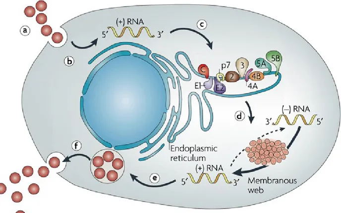
Viral genome is represented by a single strand RNA molecule of about 9,600 Kb length, encoding for a single polyprotein of over 9024 nucleotides. By mean of viral and host proteases, this protein is subsequently cleaved into several structural and non-structural proteins. Starting from the 5’-end, structural proteins are represented by core protein followed by the envelope E1 and E2 proteins. The non structural proteins include the ion channel protein p7, the NS2 protease, the NS3-4A complex harboring protease and NTPase/RNA helicase activities, the NS4B and NS5A proteins, and the NS5B RNA-dependent RNA polymerase. NS4B is a master organizer of replication complex formation; NS5A is a zinc–containing phosphoprotein involved in the regulation of HCV RNA replication/particle production. Proteins from core to NS2 constitute the assembly module, whereas NS3 to NS5B proteins represent the replication module (replicase) [3] Two untranslated regions (UTR) are present at the 5’ and 3’ end of HCV genome, respectively. 5’UTR contains an internal ribosome entry site (IRES), whereas 3’UTR a poly-U/UC stretch. During the replicative stage, after endocytosis, HCV genomic RNA is transcribed into a complementary (negative) RNA strand that represents the template for new genomic synthesis, whose identification provides evidence of active viral replication. Viral proteins are the result of a co- and post-translational cleavage of a single polyprotein. Host peptidases catalyze structural protein cleavage. After genome amplification and viral protein expression, new virions are assembled and released (Figure 1).
4
Figure 1. Schematic representation of HCV replication. Virus binding and internalization (a); cytoplasmic release
and uncoating (b); IRES-mediated translation and polyprotein processing (c); RNA replication (d); packaging and assembly (e); virion maturation and release (f). The topology of HCV structural and non-structural proteins at the endoplasmic reticulum membrane is shown schematically. HCV RNA replication occurs in a specific membrane alteration, the membranous web. Note that IRES-mediated translation and polyprotein processing, as well as membranous web formation and RNA replication, which are illustrated here as separate steps for simplicity, might occur in a tightly coupled fashion. IRES: internal ribosome entry site.
CRYOGLOBULINS
Cryoglobulins were observed for the first time in a patient affected by multiple myeloma [4] and defined as immunoglobulins (Igs) that are insoluble at temperature below 37°C, redissolving after warming [5]. The association of cryoglobulin production to a symptomatologic clinical triad (purpura, weakness and arthralgias), increased serum levels of rheumatoid factor (RF) and/or organ dysfunction, characterize the “cryoglobulinemic syndrome” [6]. Classification of cryoglobulins as single (type I) or mixed (type II or III) is based on their immunochemical composition. Type I is represented by monoclonal Ig, more frequently of IgM (occurring in almost 6% of malignant IgM paraproteinemias) or IgG (characterizing almost 2% of all myelomas) isotype. The presence of monoclonal IgM with rheumatoid factor activity, associated to polyclonal IgG defines type II cryoglobulinemia, whereas in type III there is any monoclonal component [7]. Type II
5
cryoglobulinemia accounts for 50-60% and type III for 30-40% of all cryoglobulins. Mixed cryoglobulins can be detected in the course of connective tissue and autoimmune diseases, and chronic infections. HCV chronic infection is now accepted as the disease most commonly associated to cryoglobulinemic syndrome; in all the cases without an underlying identifiable disease, cryoglobulinemic syndrome is still defined as “essential” [8, 9].
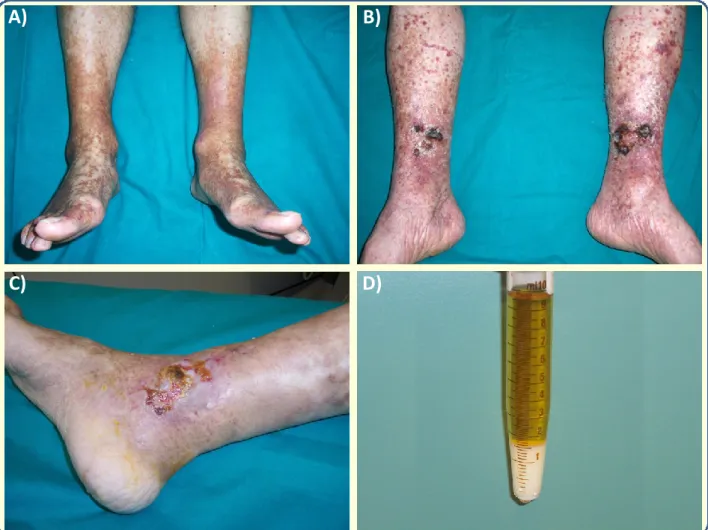
The classical clinical manifestations of cryoglobulinemic syndrome are represented by: 1) skin involvement ranging from palpable purpura of lower limbs to chronic torpid cutaneous ulcers expecially in the supramalleolar regions (Figure 2). Skin reactions may also include Raynaud’s phenomenon, livedo reticularis, urticaria and edema. 2) Arthralgias, more frequently involving hands and knees symmetrically; 3) weakness, that is nearly always present.
Figure 2. Clinical manifestations of cryoglobulinemic vasculitis. A) purpuric manifestations of the legs; B) and C)
skin ulcers of the lower limbs; D) Cryoprecipitate in a Wintrobe’s tube.
A)
B)
6
Renal injury may complicate cryoglobulinemia in almost 30% patients and in 20% of them nephropathy is present at the diagnosis [10]. Cryoglobulinemic nephropathy is considered as a distinct clinical and pathological entity [11] and the etiological role of HCV has been extensively investigated [12] even if the mechanism of HCV-induced renal damage remains unclear. HCV core protein resulted homogeneously distributed along the glomerular capillary wall and tubulo-interstitial blood vessels [13] in association with an anti-core activity, suggesting a major role of these immune complexes in the pathogenesis of renal damage [14].
The involvement of the nervous system in the course of HCV-related MC ranges from 17% to 60% [15, 16]. Peripheral nervous system involvement presenting with sensory-motor neuropathy especially of the lower limbs, is often characterized by paresthesias with loss of strength, pain and burning sensations [17]. Less frequent is central nervous system involvement, characterized by transient dysarthria, hemiplegia and confusional state [18].
Liver is involved in almost 70% of cases, often with a histopathologic picture of chronic active hepatitis with or without cirrhosis [19].
Less common clinical pictures of cryoglobulinemia are represented by gastrointestinal (2-6%) and pulmonary (5%) involvement. Intestinal ischaemia may arise with acute abdominal pain; intestinal perforation is also described as well as symptoms that mimic cholecistitis and/or pancreatitis [20]. Interstitial pneumopathy may characterize patients displaying dyspnea and dry cough, whereas an acute alveolar haemorrhage with haemoptysis, respiratory failure, and a radiologic demonstration of multiple infiltrates is rare [21].
HCV CHRONIC INFECTION AND MIXED CRYOGLOBULINEMIA
HCV infection is characterized not only by chronic liver damage but also by the emergence of several extrahepatic manifestations, the most common of which is represented by mixed
7
cryoglobulinemia (MC). MC is an immune-complex mediated vasculitis involving small vessels, characterized by an underlying B cell proliferation [22].
The relationship between HCV and “essential” MC became evident in the early ‘90s, after the introduction in clinical practice of the first ELISA test for IgG anti-HCV detection [23]. Further studies demonstrated that HCV viral genome was detectable not only in sera of cryoglobulinemic patients, but also in cryoprecipitates with a selective concentration [24].
Incidence of HCV infection in MC ranges from 40 to 90% [25]; HCV-negative MC accounts for about 5-10%. Even if small amounts of cryoglobulins are detectable in approximately 40% of HCV-infected patients, only 12-15% of these patients will develop a full-blown cyoglobulinemic syndrome [26].
The intrinsic mechanism by which HCV promotes cryoglobulin production remains unclear. Probably, virus persistence may represent a constant stimulus for host immune system unable to produce neutralizing antibodies. Cryoglobulins may represent the product of virus-host interactions in HCV-infected patients, in which virus persistence represents a continuous stimulus for host immune system unable to produce neutralizing antibodies [27]. The production of IgM molecules with RF activity is a crucial event in the cryoprecipitating process [25]. The majority of these IgM molecules are almost always associated with light chain cross-idiotype 17.109 and heavy chain cross idiotype G6 [28], that are considered as the product of a restricted expression of germline genes [29].
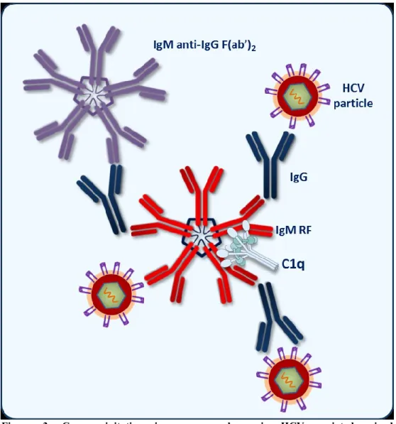
The composition of immune complexes (ICs) in HCV-related cryoglobulinemia may be represented by IgM molecules with RF activity with 17.109 cross-reactive idiotype binding polyclonal IgG with anti-HCV specificity that bind HCV antigen(s) [30]. A schematic representation of cryoprecipitating ICs is reported in Figure 3.
8
Figure 3. Cryoprecipitating immune complexes in HCV-associated mixed cryoglobulinemia. Monoclonal IgM molecules with rheumatoid factor activity bind
specific IgG recognizing HCV antigens (i.e. core protein). A crucial role is also played by complement factors.
HCV core protein seems to play an important role in the constitution of cryoprecipitating immune-complexes, as the more relevant ligand for IgG [31]. In addition, HCV directly interacts with lymphocytes; an in vivo activation and expansion of CD5-positive B cells has been considered the major source of IgM RF molecules in type III MC [32, 33]. It can be inferred that an initial activation of these cells may be followed by the emergence of a dominant clone synthetizing a monoclonal RF and supporting the development of type II MC after a transition phase in which an IgM clonal heterogeneity may define a type II-type III variant [34]. In a subset of HCV-positive patients with MC, a clonal expansion of IgM+CD27+ B cells expressing hyper-mutated RF-like Ig has been demonstrated in peripheral blood in association to VH 1-69/JH4 and VH 3-20 gene segment
9
restriction [35]. These findings have been interpreted as a B-cell proliferation induced by specific antigen stimulation, thus sustaining the notion that persistent B-cell stimulation may represent a first step to malignant evolution that probably requires further cell modifications [36].
Complement system represents a critical factor in the cryoprecipitation phenomenon. Generally, complement binding to setting up ICs decreases their size maintaining them in solution [37]. It has been demonstrated that levels of C3 and C4 fractions in cryoprecipitates are lower than in the soluble phase of MC patients’ sera, thus suggesting the existence of two different compartments characterized by a distinct complement activation [25]. On the contrary, C1q protein and C1q binding activity result significantly enriched in the cryoprecipitates [31]. These data support the hypothesis that an efficient engagement of C1q protein by cryoglobulins may represent a crucial factor in the pathogenetic pathway of MC.
It has been demonstrated that HCV-encoded core protein interacts directly with the receptor for the globular domain of C1q protein (gC1q-R), The wide expression of gC1q-R on the surface of both circulating blood immunocytes and endothelial cells may determine a specific binding to HCV core protein-containing ICs, whereas circulating HCV core protein engagement with gC1q-R expressed on the surface of B-lymphocytes may represent a direct way by which the virus can affect host immunity [38, 39]. Higher levels of soluble gC1q-R have been detected in MC patients reflecting an higher specific mRNA expression in blood mononuclear cells [40]. Soluble gC1q-R can also circulate as a complexed form binding in different sites both C1q and HCV core protein. Interestingly, lower levels of C4d, a low molecular weight fragment derived from the cleavage of C4 complement fraction following classic complement pathway activation, have been found in MC patients’ sera than in chronic HCV carriers or in healthy subjects. Otherwise, C4d fragment deposits characterize almost all skin biopsy samples of cryoglobulinemic vasculitis. These data lead to hypothesize that low circulating C4d levels may be the result of sequestered fragments in the vascular bed [20].
10
Summarizing, it can be hypothesized that, in the presence of high levels of circulating gC1q-R, HCV core protein can exacerbate the inflammatory condition by activation of complement cascade thus determining endothelial cell activation starting an in situ inflammatory response. From a biological point of view, clinical response to therapy should be characterized by a significant reduction of soluble gC1q-R associated to increased levels of C4d and lower viral load.
HCV, B CELLS, AND LYMPHOPROLIFERATION
As previously reported, HCV persistence could represent a continuous stimulus to host immune system, unable to produce neutralizing antibodies, resulting in B cells oligo/monoclonal expansions [36]. Following the initial stimulation, a single dominant clone may arise, probably also in an antigen-independent way. In this context, monoclonal IgM RF production supporting MC development can be considered as the expression of such a dominant clone [33, 34].
An intriguing feature of HCV infection is the peculiar tropism of this virus for the lymphoid tissue, leading to the hypothesis of a direct interaction between HCV and lymphocytes. Among the different binding molecules on cell surface recognized by HCV, one of the most known is CD81, a tetraspanin recognized as the ligand for E2 viral protein, that modulate viral entry into lymphocytes [41]. Interestingly, a significant enrichment of HCV RNA in peripheral blood lymphocytes of MC patients with higher levels of cell-associated viral load has been demonstrated [42], and can be considered as a consequence of higher receptor density and/or polymorphism of receptor genes [43, 44]. In addition, it should be considered that lymphocyte proliferation may be promoted by direct infection and replication of HCV into B cells [45].
On these bases, the detectability of HCV RNA minus strand represents a key factor to demonstrate an active viral replication, whereas the presence of plus strand RNA may only indicate a possible contamination by circulating virions. The availability of an high specific and sensitive method to identify HCV RNA minus strand, leads to the demonstration that HCV actively replicates
11
in lymphoid cells from MC patients [46]. Peripheral blood lymphocytes may act as circulating viral reservoir, representing another compartment for HCV productive infection [47].
The presence of B cell clonal expansions can be evaluated analyzing immunoglobulins’ variable region gene (IgV). In HCV-positive MC patients IgV heavy and light chain genes are always mutated as observed in B cells of germinal or post-germinal center origin [48, 49], sustaining the hypothesis of an antigen-driven B cells expansion, even if no specific viral protein has been identified as BCR ligand [50]. IgV genes hypermutation recognizing a single epitope may randomly arise from from the B cell pool [49].
On the other hand, it has been observed that CDR3 in most cases of B cell expansions displays a significant homology with RF-CDR3 [49, 50], leading to the hypothesis that these clonalities derive from precursors with auto-IgG specificity [51]. In some HCV-positive MC patients BCR may recognize both IgG-Fc and HCV-NS3 domains, suggesting a cross reactivity between viral epitopes and IgG autoantigen [52]. Virus enrichment on lymphoid cells surface observed in MC patients may also be explained by this mechanism, leading to RF-B cells replication and RF molecules secretion [53].
HCV chronic hepatitis is characterized by the development of inflammatory infiltrates involving the portal tracts, often appearing as follicle-like structures resembling germinal centers [54, 55]. Polymerase chain reaction (PCR) against the variable-determining-joining region (VDJ) of immunoglobulins’ heavy chain gene, enable us to identify the unique combination of N-regions along with different DH and JH that represents a clonal marker of cell progeny. It has been demonstrated that in almost 90% of HCV-positive MC patients, B cell clonal expansions are present in the liver compartment if compared with blood and bone marrow [53], showing a VDJ pattern ranging from oligoclonal to monoclonal. This lead to the hypothesis that intrahepatic B-cell expansions raise from very few or single cells and that each focus may derive from different cell of the polyclonal repertoire, thus resulting in the presence of unrelated clones.
12
The clinical spectrum of HCV infection resulted invariably influenced by the presence of intrahepatic B cell clonalities being associated not only with MC but also with high serum levels of rheumatoid factor, monoclonal gammopathy of undetermined significance and frank B-cell non Hodgkin lymphoma. The direct relation with clinical manifestations is confirmed by the restricted V gene usage displayed by B cell clonal expansions [56], whereas the wide range of variations displayed by IgH CDR3 gene segments sequence analyses suggest an antigen-driven response [57]. In conclusion, it has been hypothesized that, based on an up-regulation of IgH-VDJ mutational activity, B cell clones start expanding in the liver and then migrates to other biologic compartments like blood and bone marrow [56].
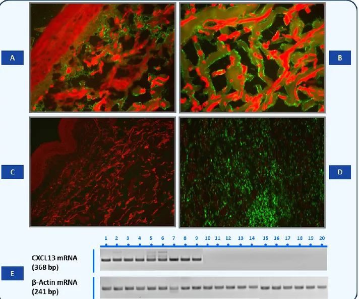
To further explain the emergence of intrahepatic and/or circulating B-cell clones in the course of chronic HCV infection, the possible role of some chemokines has been considered as crucial factor in determining an adequate microenvironment in response to signals provided by antigen-presenting cells [58]. MC patients showed high serum levels of CXC ligand 13 (CXCL13) protein, also known as B-cell attracting chemokine 1 or B-lymphocyte chemoattractant. This protein represents an important factor for the development of secondary lymphoid tissue and lymphocytes distribution [59]. Higher CXCL13 serum levels observed in MC patients correlated with specific mRNA expression in liver and skin tissue, suggesting that this chemokine may contribute to lymphoid homing in the liver by creating a favorable microenvironment and sustaining lymphoid follicle-like B-cell aggregation [60] (Figure 4).
13
Figure 4.Demonstration of CXCL13 protein and CXCL13 mRNA in skin biopsy samples. (A) Hematoxylin and
eosin staining of skin sample from a patient with cryoglobulinemic active vasculitis. Magnification, 10×/0.25. (B) CXCL13 protein immunofluorescence staining in skin biopsy showing feature of active vasculitis. CXCL13- specific signal appears as linear green deposits along collagen bundles. Magnification, 10×/0.25. (C) Complete absence of CXCL13 immunoreactant in skin tissue of an MC patient without active phase of vasculitis. Magnification, 10×/0.25. (D) Positive control staining in human tonsil. Magnification, 20×/0.40. (E) RT-PCR analysis of CXCL13 mRNA extracted from MC patients with and without active cryoglobulinemic vasculitis.
B-cell enrichment may also depend from enhanced cell survival [61]. In this context, serum levels of B-lymphocyte activating factor (BAFF), also known as B-lymphocyte stimulator (BLyS) and secreted by activated monocytes, macrophages and dendritic cells, resulted increased in chronically HCV infected patients, as well as in autoimmune diseases (i.e. systemic lupus erythematosus and rheumatoid arthritis) and correlated to clinical autoimmune vasculitic manifestations. This chemokine may prolong B cell survival by modulating their sensitivity to apoptosis, leading to B cell expansion [62].
14
CRYOGLOBULINEMIC SYNDROME: IMPACT ON CLINICAL OUTCOME OF CHRONIC LIVER DISEASE AND THERAPEUTIC APPROACH
On the basis of all reported above, we can state that patients with chronic HCV infection are at high risk of developing an extrahepatic disorder, most frequently represented by mixed cryoglobulinemia. If we exclude those HCV positive patients in whom some small amounts of cryoglobulins are detectable (approximately 40%), it is important to define if the occurrence of a full-blown cryoglobulinemic vasculitis (12-15% of patients) may represent a prognostic factor influencing the natural history of HCV chronic infection and if an adequate therapeutic approach may influence clinical outcome.
In 2003, Kayali et al, published a meta-analysis of 19 studies including a total number of 2323 patients with chronic HCV infection, 44% of whom with associated cryoglobulinemia, concluding that in these patients the presence of cryoglobulins represented an important prognostic factor for the development of liver cirrhosis [63]. Subsequent studies also by the same group, didn’t clarified the association between fibrosis and cryoglobulins that were not considered as an independent factor for advanced fibrosis [64] that seemed much more influenced by other conditions like age and duration of disease [65]. Furthermore, a ten-years prospective study showed that MC did not influence the clinical course of HCV-related disease in that the rate of progression to cirrhosis was similar in patients with or without cryoglobulins [66]. However, all these studies considered a limited number of patients with cryoglobulinemic vasculitis, whereas in retrospective evaluations multi-organ involvement influencing the severity of MC, resulted in reduced life expectancy [67].
Recently, it has been published a 15-year prospective study evaluating the impact of cryoglobulinemic syndrome on the outcome of chronic HCV infection. Starting from a cohort of 950 chronically HCV-infected patients, MC were diagnosed in 246 (26%) patients, 184 of whom displayed a full-blown cryoglobulinemic vasculitis. After a long-term follow-up of 15 years, 141
15
patients with cryoglobulinemic vasculitis and 601 without MC completed the study. Results showed an estimated liver fibrosis progression rate lower in patients with cryoglobulinemic syndrome than in those without MC (p < 0.05). In addition, the 15-year cumulative probability of developing cirrhosis and/or hepatocellular carcinoma was higher in patients without MC (24.9% vs 14.2%; p < 0.005 and 20.3% vs 75%, p = 0.003, respectively). On the other hand, renal involvement, neurologic impairment and B-cell non Hodgkin lymphoma resulted more common in patients with cryoglobulinemic vasculitis (32.6% vs 3%, p < 0.0001; 31,2% vs 4,8%, p < 0.0001; 15% vs 7.1%, p = 0.003, respectively). Despite different clinical evolution and causes of death, the 15-year survival rate was similar in the two groups (70.2% vs 71.7%), concluding that the presence of a cryoglobulinemic syndrome profoundly influenced the natural history of chronically HCV-infected patients but not the overall survival [68].
An additional interesting consideration emerging from this study is that antiviral therapy had an undisputable impact on patients’ outcome. Considering the long time span covered by the study, antiviral therapy included interferon-α (IFN-α) monotherapy, IFN-α plus ribavirin, pegylated IFN-α (pIFN-α) monotherapy and pIFN-α plus ribavirin. About half of the patients were treated with antiviral therapy showing long term benefits and better outcome, particularly in the case of a sustained virologic response, both in MC negative patients group (p = 0.0002) and in cryoglobulinemic patients group ( p < 0.0001) [68].
These evidences lead to the obvious conclusion that any available therapeutic approach should be considered in MC as well as in chronic liver disease in order to ameliorate long-term clinical outcome and also to prevent malignant evolution like B cell NHL and HCC.
Being MC a complex disease involving a viral infection, autoimmunity, and lymphoproliferation, a complex treatment capable of modifying each clinical and biologic aspect of the disease is required. The main goals of MC therapy are represented by: a) eradication of HCV
16
infection (virological response); b) depletion of cryoproteins (immunological response); c) deletion of the underlying B-cell clonal expansions (molecular response).
In the pre-HCV era, management of MC was conventionally based on the use of corticosteroids and immunosuppressive drugs. In 1987 was demonstrated the effectiveness of recombinant IFN-α as an “immunomodulant” agent in 7 patients with “essential” MC [69]; obviously, after the demonstration of the pathogenetic role of HCV [8], IFN-α became a rational therapeutic strategy. The introduction of pegylated IFN-α, increased virological responses [70, 71] as well as the introduction of ribavirin (RBV), a nucleoside antimetabolite agent [72]. This combination, considered the standard of care (SoC) for HCV management [73], has been shown to be effective in a remarkable proportion of HCV-related MC patients, resulting in a complete clinical response and sustained virological response (SVR) in 78% of the patients [74]. Serum levels of C3 and C4 complement fractions also normalized in 80% and cryoglobulins disappeared in 56% of the patients but no or only partial improvement in neuropathy and glomerulonephritis was observed, suggesting that the clinical outcome may be conditioned by factors other than the virus [25].
As previously reported, the occurrence of B cell clonal expansions is able to influence the clinical expression of HCV infection, in that it is consistently associated with extra-hepatic manifestations, like MC [56, 75-76]. Considering the enrichment of B cell clones in liver, bone marrow and peripheral blood as the biological hallmark of MC, as well as expansion of RF-synthesizing B cells [25], the deletion of B-cell clonalities may provide a rational way to treat MC. CD20 antigen, a transmembrane protein selectively expressed on pre-B and mature lymphocytes, resulted remarkably expressed also on expanded and activated B cells of patients with MC [42, 77].
On these bases, the use of rituximab (RTX), a chimeric monoclonal antibody (moAb) specifically directed to CD20 antigen therapeutically effective in lymphoproliferative and autoimmune disorders [78-80], has been proposed also in HCV-related MC patients refractory to, or relapsing after, conventional antiviral therapy. The first papers about the use of RTX in
HCV-17
related MC [81, 82] showed that it is an effective, safe and well tolerated treatment for type II MC patients. However, a not negligible drawback is represented by the frequently increased viremia almost invariably not associated with increased ALT levels. Several subsequent papers have addressed the issue of the use of RTX, alone or in combination with steroids [83, 84].
In 2010 our group proposed a triple therapeutic combination (pIFN-α plus RBV plus RTX), designated with the acronym PIRR, that was administered to 22 HCV-positive MC patients, whereas 15 additional patients with the same pathology received, by comparison, pIFN-α plus RBV with the exclusion of RTX [85]. After treatment, follow-up was protracted for 36 months. Evaluation criteria were as follows: 1) complete response, defined by clinical response (disappearance of clinical signs of vasculitis) plus virological response (disappearance of viremia) plus immunological response (disappearance of cryoglobulins) plus molecular response (disappearance of expanded B cell clones); 2) partial response, defined arbitrarily as achievement of 3 of the four criteria mentioned for complete response; 3) no response, if any of the criteria was achieved. Results showed a complete response in 54.5% of patients treated with PIRR, and only in 33.3% of those who were given pIFN-α plus RBV without RTX (p<0.05). Despite sustained virological response in 5/22 (22.7%) patients treated with PIRR and in 5/15 (33.3%) patients treated with pIFN-α plus RBV cryoproteins persisted. No response occurred in remaining 5 patients of both groups. Even more interesting were the observations that: a) in the large majority (83·3%) of the responders belonging to the PIRR-treated group, a conversion of B-cell populations from oligoclonal to polyclonal was recorded in the liver, bone marrow and peripheral blood compartments; b) compared with 40% of the control group, in all patients of the PIRR group the CR was maintained throughout the follow-up period [85]. Whether RTX should be administered to patients with cryoglobulinemic vasculitis as first- or second-line therapy, remains to be established [86].
18
capable to obtaining the main goals of HCV-associated cryoglobulinemic vasculitis, which is the lesson from partial/non responders? How should we consider MC patients that do not obtain an SVR and those showing a continuous cryoglobulin production despite virus eradication?. In the first case the use of the first generation direct-acting antivirals (DAAs) like Telaprevir or Boceprevir (approved for the treatment of HCV genotype 1 chronic infection), and more recently second generation protease inhibitors or polymerase inhibitors may represent a further therapeutic option [87]. In the second case, representing an emerging picture following antiviral and B-cell depletive combined therapies [88, 89], a different immunochemical structure of circulating immune-complexes may be postulated and the use of corticosteroids, cyclophosphamide, or Ofatumomab (an IgG1k fully humanized CD20 MoAb) may be considered [90].
Therapeutic apheresis is a palliative procedure that can be extremely useful for the treatment of severe, life-threatening vasculitis [86] as well as for the treatment of chronic leg ulcers in patients resistant to other therapies [91].
Others additional therapeutic approaches for MC have been proposed, like tyrosine kinase inhibitor imatinib, anti-angiogenic drugs like thalidomide, bortezomib (a proteasome inhibitor) and IL-2, but future controlled studies are required to establish if these agents will improve MC therapy [92, 93].
INTERLEUKIN 28B GENE POLYMORPHISMS AND MIXED CRYOGLOBULINEMIA
An important consideration that should be done is about the efficacy of both antiviral and B-cell depletive treatments. According to Simmonds’ classification we can recognize at least 6 major genotypes of HCV and several subtypes [94], but HCV has a very high mutation rate with the development of quasispecies. It is well established that HCV genotypes 1 and 4 are more resistant to SOC than genotypes 2 and 3, with further differences among subtypes. All the previous cited studies evaluating therapeutic approaches in MC showed that HCV genotypes equally distributed among MC and non-MC patients, resembling geographical distribution and response to therapy of
19
each genotype, even if HCV genotype did not represent a discriminant criteria.
In order to better identify patients who are more susceptible to achieve a therapeutic success (i.e. sustained virological response: SVR), in addition to viral factors like “difficult” genotype, other host factors have been considered like sex, steatosis, obesity, iron overload, and at least genetic factors, becoming a high priority in determining response to treatment.
In 2009, genome-wide association studies (GWAS) described single nucleotide polymorphisms (SNPs) on chromosome 19, near the IFN3 encoding region gene (IL28B; rs12980275 and rs8099917), as highly reliable predictors of a spontaneous resolution of acute hepatitis C and of the outcome after antiviral therapy, both in the general population [95-98] and immunocompromised individuals [99]. IL28B encodes for interferon (IFN) 3, a type III IFN involved in host antiviral immunity. IL28B/IFN3 may induce innate antiviral response in vitro through the activation of the IL28 receptor complex expressed on different cytotypes [100, 101].
The SNP on chromosome 19q13 (rs12979860) resulted strongly associated with SVR in genotype 1 HCV infection. As reported by Ge et al [95], three different genotypes can be identified: C/C, C/T, and T/T. C/C genotype resulted associated with 2.5 or greater SVR rate if compared to T/T genotype (also in relation to ethnicity) and the C allele associated with a more favorable spontaneous viral clearance. Therefore, rs12979860 polymorphism may explain differences in SVR among different groups of patients with more favorable response in European than Africans. Furthermore, Rauch et al demonstrated that other SNPs on chromosome 19 in the region of IL28B gene, rs8099917, may predict outcome and treatment response in HCV infection. The rs8099917 minor allele (T/G or G/G) is associated with progression to chronic HCV infection and also with antiviral therapy failure, with the strongest effects in patients with HCV genotype 1, 2, or 4 [98].
In 2013 a large prospective study evaluating IL28B genotype in patients with HCV-related mixed cryoglobulinemia has been published. In this study the rs12979860/rs8099917 IL28B
20
polymorphisms were analyzed in 481 consecutive patients (250 with MC and 231 without), in order to assess for the first time a potential role of these SNPs also in cryoglobulinemic vasculitis. One hundred and fifteen cryoglobulinemic patients underwent standard antiviral therapy and results were evaluated according to IL28B SNP distribution. Similar IL28B SNPs allele frequencies were recorded for patients and controls. In this study the IL28B genotype was confirmed to be a strong independent predictor of response to IFN-based therapeutic regimen also in MC as well as in chronic HCV infection, thus confirming the key role played by HCV in MC. In addition, the IL28B genotype does not seem to influence MC evolution [102].
More recently, IL28B rs12979860 polymorphism has been evaluated in relation to different hepatitis C virus infection statuses. IL28B allelic distribution has been compared in a series of 1050 patients with chronic HCV infection and different outcomes (including spontaneous clearance, chronic hepatitis, hepatocellular carcinoma, mixed cryoglobulinemia and non-Hodgkin’s lymphoma). This study confirmed the association between IL28B C allele and spontaneous viral clearance, but also demonstrated a weak relation between IL28B T allele and progression to hepatocellular carcinoma, in accordance with a carcinogenic model in which IL28B TT genotype could facilitate cancer development by promoting a persistent viral infection. In addition, IL28B CC genotype was more frequent in patients with MC than in those with malignant tumors taken together (HCC and NHL). Results of this study confirmed the hypothesis that persistent HCV infection may represent a risk factor for the development of both liver and lymphoproliferative disorders [103].
21
OBJECTIVE OF THE STUDY
On the basis of previously reported issues, it can be summarized that clinical improvement of MC is almost invariable associated with the inhibition of HCV replication, regression of expanded B cell clones in different compartments, and disappearance of cryoprecipitating proteins. The combination of pIFN- with RBV and rituximab significantly improved clinical response of MC. However, this improvement may be transient and followed by disease relapse. Considering that viral and host factors are predictive of a sustained virological response and that the evaluation of a molecular response (i.e. deletion of activated/expanded B cell clones) as well as the immunological response (disappearance of cryoglobulins) are fundamental in MC patients, it is also important to clarify if IL28B genotype could represent a predictive marker of treatment efficacy in HCV-related MC. It has been reported the observation that in some MC patients cryoglobulins persist despite an SVR, suggesting that antiviral therapy only partially interferes with the B cell clones whose activity directly influence the clinical spectrum of HCV infection.
The aim of this study was to assess the possible association between IL28B single nucleotide polymorphisms (SNPs) rs12979860 and therapeutic response in patients with HCV-related Type II CV. Specifically, the following issues were addressed: a) whether the frequency of IL 28B C/C and non C/C genotypes differed in patients with and without CV; b) whether IL28B C/C homozygosis was predictive of treatment response in HCV-related CV; c) whether IL28B polymorphisms were associated with a higher frequency of B cell clonal expansions, as well as with peculiar clinical manifestations of CV disease; d) whether the second variant of IL28B polymorphisms, rs8099917, played also a predictive role on the clinical outcome in MC and could give additional information in MC patient with no response to treatment, especially in consideration of the availability of new direct antiviral agents.
22
PATIENTS AND METHODS
The study cohort comprised 159 and 172 HCV infected patients with and without MC, referred to both the Liver Unit of Foggia and Bari.
Baseline evaluation included disease history and stage, current signs and symptoms, and previous medications. Physical examination and laboratory values were recorded.
Inclusion criteria were as follows:
positivity for anti-HCV antibodies and polymerase chain reaction (PCR)-based assay to detect HCV RNA in serum of patients with/without palpable purpura;
detection of serum cryoglobulins;
liver biopsy showing chronic hepatitis performed within three months from enrollment; negativity for hepatitis B surface antigen and human immunodeficiency virus antibodies; no previous administration of IFNs or immunosuppressive drugs.
All MC patients were treated with pegylated interferon/ribavirin (pIFN-α/RBV) based antiviral therapy. Subcutaneous injections of 180 mcg pIFN-α2a (Pegasys®, Hoffmann-La Roche Ltd) or 1.5 mcg/Kg pIFN-α2b (Peg-Intron®, Merck) were administered to all MC patients once a week, together with a daily dose of 800-1,200 mg RBV according to body weight. The median length of follow-up was 49 ± 11 months. A complete response (CR) included sustained virological response (SVR), defined as undetectable HCV RNA 24 weeks after the cessation of therapy, in addition to the disappearance of B-cell expanded clones from the blood (molecular response: MR), and the non-detectability of cryoproteins (immunological response: IR). Partial response (PR) was defined as PR-1 including SVR and PR-2 including MR and IR.
Serum cryoglobulins were measured, isolated, and purified. The monoclonal component of mixed cryoglobulins was characterized by immunofixation.
HCV status was evaluated by an HCV RNA qualitative assay with detection limit of 5 IU/mL (Versant, HCV RNA Qualitative assay, Bayer Health Care, Diagnostic Division); HCV
23
RNA levels were measured by a branched-chain DNA assay (Bayer) with detection threshold of 615 IU/mL and HCV genotypes were determined using INNOLIPA HCV assay (Innogenetics).
For the identification of IL28B locus rs12979860 and rs8099917, genomic DNA was extracted from the buffy coat of the patients’ whole blood and IL28B genotyped using the ABI Taq-Man allelic discrimination kit (Applied Biosystems) and the ABI7300 sequence detection system (Applied Biosystems). For rs12979860 was prepared a 25-l Taq-man genotyping master mix including the following primers: forward GCC TGT CGT GTA CTG AAC CA-3’, reverse 5’-GCG CGG AGT GCA ATT CAA C-3’, probe (C allele) 5’-VIC-TGG TTC 5’-GCG CCT TC-3’, probe (T allele) 5’-FAM-CTG GTT CAC GCC TTC-3’. The SNP rs8099917 was genotyped using a predesigned TaqMan SNP Genotyping Assay (Applied Biosystems; assay ID C_11710096_10).
For the evaluation of B cell clonal expansions, DNA was purified from peripheral blood mononuclear cells (PBMC). Twenty ml of reaction mixture were analyzed by electrophoresis on 5% agarose gel in tris-borate-EDTA buffer, stained with ethidium bromide and optically evaluated by UV transillumination. Control mixtures in the reaction were devoid of DNA or included DNA from a clonal cell line. A monoclonal B cell expansion was defined as 1 or 2 (if both alleles were rearranged) discrete narrow bands within the predicted size. Distinction of biclonal monoallelic rearrangements from monoclonal biallelic rearrangements was based on the results of the sequence analyses, in that a nonfunctional rearrangement of 1 of 2 alleles was detected in the case of monoclonal disorders, whereas both dominant bands were representative of a functional IgH rearrangement in biclonal disorders. Because DNA material was sometimes insufficient, IgH VDJ gene segment amplification was carried out in 106 of the 159 patients with MC.
Statistical analyses: data were expressed as counts and percentages for qualitative variables and as a median with interquartile range for quantitative variables. The Mann-Whitney U test was carried out to compare groups. Fisher’s exact test was used to explore associations between different qualitative measures. Pretreatment measures that were identified as significant in the logistic regression model were further assessed in stratified analyses across different categories.
24
Variables were eligible for entry into a multiple logistic regression model if they were significantly associated with the admissions (p < 0.25). The k statistic was used to assess reliability of variables. Given the few comparisons considered sensible, no formal corrections were done. When statistically non-significant (p > 0.05), variables were eliminated from the multivariate model, and calibration was assessed using Hosmer-Lemeshow goodness-of-fit test. All statistical tests were 2-tailed; the significance cutoff was p < 0.05. All the data were analyzed using a statistical package (SAS, version 6.04; SAS Institute).
25
RESULTS
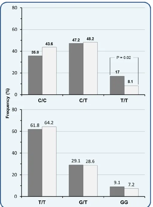
All patients with chronic HCV infection with and without MC were evaluated for the occurrence of SNP rs12979860 and rs8099917 of IL28B genotypes. The frequency rate of IL-28B rs12979860 was compared to the frequency rate of rs8099917. The first observation was that SNPs allele distribution resulted similar in MC patients and in HCV-positive patients without MC (Figure 5).
Figure 5. Frequency of IL28B genotypes at the SNP rs12979860 and rs8099917 in
chronically HCV infected patients with (■) and without (□) cryoglobulinemic vasculitis.
26
However, IL28B rs12979860 TT genotype occurred significantly more often in MC than in non-MC patients (17% vs. 8.1%; p=0.02), while there was no difference in IL28B rs8099917 GG minor allele. We also found that rs12979860 CC homozygosity was less represented in the MC than in the non-MC group (35.8% vs. 43.6%), such as rs8099917 TT major allele (61.8% vs. 64.2%), although these differences were not significant (Figure 5).
In consideration of these results showing a similar behavior between the two polymorphisms of IL28B, and of the emerging feature of TT minor allele increased frequency in MC patients, we focused our analyses only on the more common rs12979860 IL28B polymorphism in both MC and non-MC HCV chronically infected patients.
All patients were of European ancestry and Caucasian race. They were divided into two subgroups depending on their C/C or non-C/C genotype; the latter subgroup comprised C/T as well as T/T variants. Univariate analysis consisted of parameters that were compared with each other in patients with “favorable” C/C allele carriage and in those with “less favorable” T-alleles.
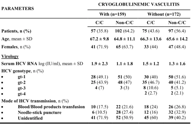
TABLE 1. Epidemiological and virological parameters in chronically HCV-infected patients with and
without cryoglobulinemic vasculitis who were stratified for IL28B rs12979860 C- and non-C alleles.
PARAMETERS CRYOGLOBULINEMIC VASCULITIS
With (n=159) Without (n=172) C/C Non-C/C C/C Non-C/C Patients, n (%) 57 (35.8) 102 (64.2) 75 (43.6) 97 (56.4) Age, mean ± SD 67.2 ± 9.8 64.8 ± 11.1 66.3 ± 13.6 65.6 ± 14.2 Females, n (%) 41 (71.9) 65 (63.7) 33 (44) 47 (48.4) Virology
Serum HCV RNA log (IU/ml), mean ± SD 1.9 ± 2.3 1.1 ± 1.8 1.5 ± 1.2 1.3 ± 1.6 HCV genotype, n (%) gt-1 gt-2 gt-3 gt-4 28 (49.1) 25 (43.9) 4 (7) 51 (50) 48 (47) 3 (3) 30 (40) 35 (46.7) 8 (10.6) 2 (2.7) 50 (51.6) 40 (41.2) 5 (5.1) 2 (2.1) Mode of HCV transmission, n (%)
Blood/Blood products transfusion
Needle-stick puncture Unidentified 10 (17.5) 6 (10.5) 41 (71.9) 22 (21.6) 28 (27.4) 52 (50.9) 18 (24) 12 (16) 45 (60) 26 (26.8) 32 (32.9) 39 (40.2)
27
As shown in Table 1, the median viral load did not significantly differ between IL28B genotype subgroups. HCV genotypes also showed a similar distribution: in subgroups with and without MC, HCV genotype 1 occurred in 28 of 57 (49.1%) and in 30 of 75 (40%) patients with the IL28B C/C variant, and in 51 of 102 (50%) and 50 of 97 (51.6%) patients with the non-C/C variant, respectively. Mean age did not differ among subgroups and the source of HCV infection remained unidentified in the majority of patients.
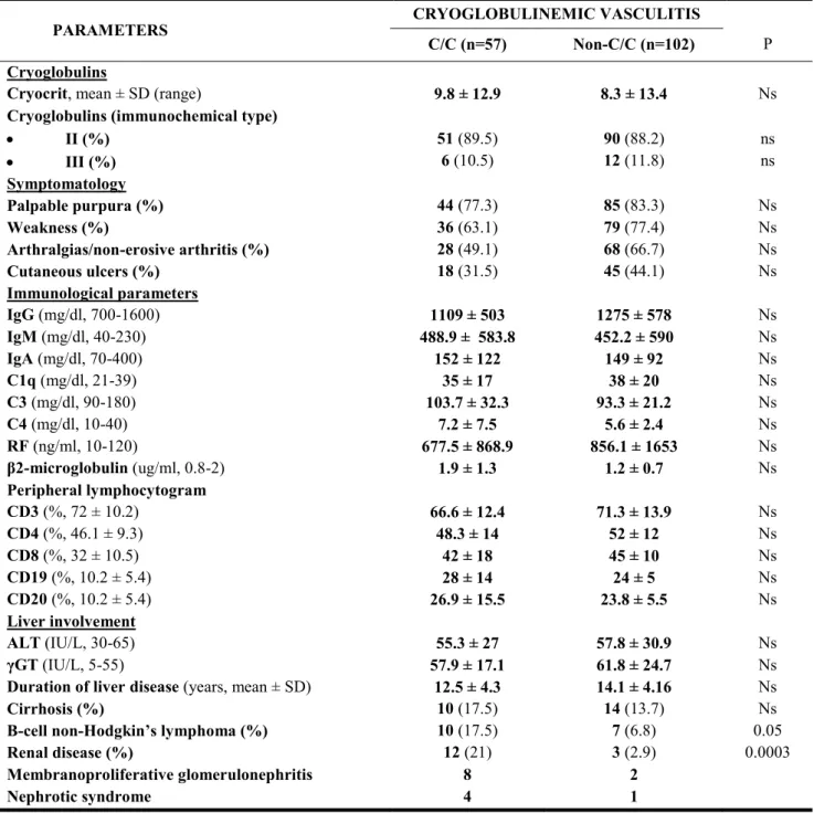
Table 2 summarizes clinical and immunological variables of cryoglobulinemic patients stratified for IL28B C and non-C alleles. There was no difference between the C/C and non-C/C subgroups of MC patients regarding the mean levels and the immunochemical type of cryoglobulins. By contrast, chronic renal damage, including glomerulonephritis and nephrotic syndrome, resulted more frequent in C/C than in non-C/C variants (21% vs. 2.9%, p=0.003).
Similarly, in the C/C variant there was an increased prevalence of B-cell non-Hodgkin’s lymphoma (B-NHL) (17.5% vs. 6.8%, p<0.05). Palpable purpura, weakness, arthralgias/non-erosive arthritis, or the occurrence of cutaneous torpid ulcers did not differ between the two groups, as well as the frequencies of liver diseases, including cirrhosis, and the mean duration of liver disease were roughly comparable.
28
TABLE 2. Clinical and immunological parameters in 159 patients with HCV-related cryoglobulinemic vasculitis,
stratified for IL-28B rs12979860 C- and non-C alleles
PARAMETERS CRYOGLOBULINEMIC VASCULITIS
C/C (n=57) Non-C/C (n=102) P
Cryoglobulins
Cryocrit, mean ± SD (range) 9.8 ± 12.9 8.3 ± 13.4 Ns
Cryoglobulins (immunochemical type)
II (%) III (%) 51 (89.5) 6 (10.5) 90 (88.2) 12 (11.8) ns ns Symptomatology Palpable purpura (%) 44 (77.3) 85 (83.3) Ns Weakness (%) 36 (63.1) 79 (77.4) Ns Arthralgias/non-erosive arthritis (%) 28 (49.1) 68 (66.7) Ns Cutaneous ulcers (%) 18 (31.5) 45 (44.1) Ns Immunological parameters IgG (mg/dl, 700-1600) 1109 ± 503 1275 ± 578 Ns IgM (mg/dl, 40-230) 488.9 ± 583.8 452.2 ± 590 Ns IgA (mg/dl, 70-400) 152 ± 122 149 ± 92 Ns C1q (mg/dl, 21-39) 35 ± 17 38 ± 20 Ns C3 (mg/dl, 90-180) 103.7 ± 32.3 93.3 ± 21.2 Ns C4 (mg/dl, 10-40) 7.2 ± 7.5 5.6 ± 2.4 Ns RF (ng/ml, 10-120) 677.5 ± 868.9 856.1 ± 1653 Ns β2-microglobulin (ug/ml, 0.8-2) 1.9 ± 1.3 1.2 ± 0.7 Ns Peripheral lymphocytogram CD3 (%, 72 ± 10.2) 66.6 ± 12.4 71.3 ± 13.9 Ns CD4 (%, 46.1 ± 9.3) 48.3 ± 14 52 ± 12 Ns CD8 (%, 32 ± 10.5) 42 ± 18 45 ± 10 Ns CD19 (%, 10.2 ± 5.4) 28 ± 14 24 ± 5 Ns CD20 (%, 10.2 ± 5.4) 26.9 ± 15.5 23.8 ± 5.5 Ns Liver involvement ALT (IU/L, 30-65) 55.3 ± 27 57.8 ± 30.9 Ns γGT (IU/L, 5-55) 57.9 ± 17.1 61.8 ± 24.7 Ns
Duration of liver disease (years, mean ± SD) 12.5 ± 4.3 14.1 ± 4.16 Ns
Cirrhosis (%) 10 (17.5) 14 (13.7) Ns
B-cell non-Hodgkin’s lymphoma (%) 10 (17.5) 7 (6.8) 0.05
Renal disease (%) 12 (21) 3 (2.9) 0.0003
Membranoproliferative glomerulonephritis 8 2
Nephrotic syndrome 4 1
No differences were detected for complement C3 and C4 and C1q concentrations and also in serum RF and IgM levels, in the distribution of peripheral lymphocytes subpopulations, and in mean serum ALT and γGT levels.
We also analyzed IgH VDJ gene rearrangement in circulating B cells and carried out in step with IL28B genotyping in 106 of the 159 (66.6%) MC patients with the aim to assess whether IL28B polymorphisms were associated with profiles of circulating B cell clonalities.
29
C/C patients showed a significantly higher frequency of expanded B-cell clonalities than their non-C/C counterparts. B-cell monoclonality and oligoclonality were recorded in 32 of 38 (84.2%) C/C and in 38 of 68 (55.9%) non-C/C patients (p=0.005), while a fully polyclonal pattern was detected in the remaining six (15.8%) C/C and 30 (44.1%) non-C/C patients (Table 3).
TABLE 3. Frequencies and profiles of IgH VDJ gene rearrangements in the
circulating B cells of 106 patients with HCV-related cryoglobulinemic vasculitis
CIRCULATING B CELLS IgH VDJ gene rearrangements SNP rs12979860 genotype Patients (n) Monoclonal/
Oligoclonal Polyclonal
CC, n (%) 38 32 (84.2) 6 (15.8)
Non-CC, n (%) 68 38 (55.9) 30 (44.1)
TOTAL 106 70 (66) 36 (34)
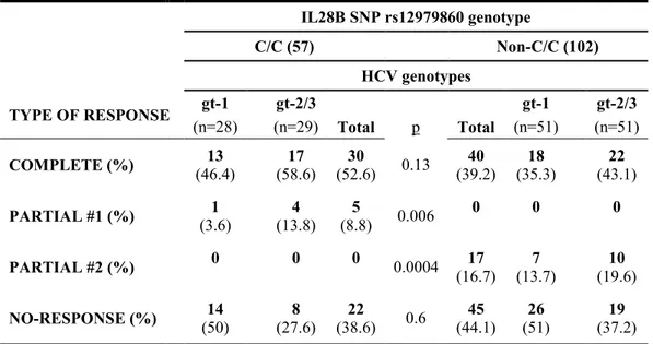
We evaluated virological, molecular and immunological responses to antiviral treatment as a function of viral and host genotypes, and the presence of variables associated with a complete response to pIFNα/RBV-based therapy in MC patients. The response to pIFN/RBV combined therapy is summarized in Table 4.
TABLE 4. Response to antiviral treatment as a function of viral and host genotypes. IL28B SNP rs12979860 genotype C/C (57) Non-C/C (102) HCV genotypes TYPE OF RESPONSE gt-1 gt-2/3 gt-1 gt-2/3 (n=28) (n=29) Total p Total (n=51) (n=51) COMPLETE (%) (46.4) 13 (58.6) 17 (52.6) 30 0.13 (39.2) 40 (35.3) 18 (43.1) 22 PARTIAL #1 (%) (3.6) 1 (13.8) 4 (8.8) 5 0.006 0 0 0 PARTIAL #2 (%) 0 0 0 0.0004 (16.7) 17 (13.7) 7 (19.6) 10 NO-RESPONSE (%) (50) 14 (27.6) 8 (38.6) 22 0.6 (44.1) 45 (51) 26 (37.2) 19
Complete response (SVR, MR, IR); Partial response -1 (SVR); Partial response -2 (MR, IR)
30
Sustained virological response (SVR) was defined as negative HCV RNA 24 weeks after the end of therapy, whereas molecular response (MR) was defined as disappearance of B cell clonalities and immunological response (IR) as absence of circulating cryoglobulins. A complete response (CR), defined as the association of SVR + MR + IR, was achieved more frequently in patients with C/C (52.6%) than in those with non-C/C variants (39.2%). HCV genotype 1 was identified in 13 C/C (46.4%) and 18 non-C/C (35.3%) patients, and HCV genotypes 2/3 in 17 (58.6%) and 22 (43.1%) patients, respectively. Overall, a SVR occurred in 35/57 (61.4%) patients with the IL28B C/C genotype and in 40/102 (39.2%) of those with the non-CC variant (p=0.008).
The presence of SVR without MR and IR was defined as partial response-1 (PR-1), whereas the occurrence of MR and IR in the absence of SVR was defined as partial response-2. PR-1 was confirmed in five of the 57 (8.8%) C/C patients but was not detected in any of the 102 patients with the non-C/C variant (p=0.006). Conversely, 17 (16.7%) patients with the non-C/C genotype but none of the 57 patients with the C/C genotype had a PR-2 (p=0.0004). In 22/57 (38.6%) and in 45/102 (44.1%) patients with the C/C and non-C/C variants, respectively, response to therapy was consistently absent.
Finally, we also compared virological, immunological and molecular responses between IL28 rs8099917 and rs12979860 polymorphisms. Results showed that IL-28B rs12979860 C/C and rs8099917 T/T variants seems to protect in terms of virological response, whereas the rs12979860 T/T and rs8099917 G/G variants significantly associated with immunological/molecular response (Figure 6).
31
Figure 6. Comparison of virological, immunological and molecular responses between IL-28B
rs12979860 and rs8099917 SNPs
DISCUSSION
Our data are consistent with a possible role of IL28B polymorphisms not only in HCV-related chronic hepatitis, but also in HCV-HCV-related MC, in which the immune system, genetic disposition and environmental factors are involved [104, 105].
Recently, genome-wide association studies identified single nucleotide polymorphisms near the IL28B region which were more frequent in responders to treatment. IL28B encodes interferon (IFN) λ3, a type III IFN involved in host antiviral immunity. Favorable variants of the two most widely studied IL28B polymorphisms, rs12979860 and rs8099917, are predictive factors of early viral clearance and sustained virological response (SVR) after treatment, in patients with genotype 1 HCV infection [106]. Moreover, IL28B was involved into the development of chronic HCV infection versus spontaneous resolution of acute infection and so it could play a role in host immunity against HCV.
32
The frequency of the T allele in IL28B rs12979860 haplotype resulted higher in patients with MC, whereas C/C genotype failed to protect against B-NHL development as well as renal damage, being associated to an higher risk of cryoglobulinemic nephropathy and higher frequency of B-cell malignancies. Our study was based on stringent criteria for the definition of MC and evaluation of response to treatment; on these bases, CC genotype characterized biologically the presence of restricted B cell response and clinically the increased occurrence of nephropathy and B cell malignancies. A possible interpretation of these results is that the activation and expansion of B-cell clonotypes contributes to the formation of harmful circulating immune complexes (including IgM RF molecules) that can mediate tissue damage, whereas the development of B-NHL belongs to an HCV-driven B-cell derangement that interrupts crucial cell regulatory mechanisms. These results are apparently in contrast with previous studies [102] in which there was no significant difference in IL28B polymorphisms in HCV positive patients with and without MC; a possible explanation of such differences in our opinion is due to the definition of more stringent diagnostic criteria, assessment of B cell clonalities and longer follow-up.
Our data showed that IL28B rs12979860 C/C genotype was a significant independent predictor of a virological response to antiviral therapy. We also demonstrated that the C/C genotype cannot predict treatment response in about 9% of MC patients who, despite achieving a SVR, had persisting B-cell expanded clones, cryoglobulin production, and vasculitic manifestations. Otherwise, HCV seems to act as an independent factor leading to the activation of the immune system and clonal B-cell expansions, which become autonomous because of the ongoing infection. Therefore, these patients are refractory to antiviral therapy for the persistence of B-cell clonalities and are characterized by persistent cryoprotein production. Previous studies showed that in HCV-related MC dominant B-cell clonotypes displayed identical rearrangements of the IgH VDJ gene, that sustained the antigen-independent nature of these cells [36].
On the other hand, 16.7% of our patients with non-C/C genotypes were unable to achieve a SVR but did develop both molecular and immunological response; the MC outcome was more
33
favorable despite ongoing HCV infection. In this subgroup, there was a regression of B-cell clonal expansions, although the patients remained viremic. These findings suggest that rs12979860 C and T alleles function as independent genetic protective factors, such as rs8099917 T and G alleles.
Multivariate analysis identified patients’ age and cryocrit levels as responsible for the discordance between IL28B genotype and the response to therapy. Younger age and lower cryocrit values resulted independently associated with the response to pIFN/RBV. It may be hypothesized that these factors influence disease duration, highlighting the importance of treating patients as soon as they are diagnosed.
Both the CC/rs12979860 and TT/rs8099917 alleles could be used as independent predictors of the virological response. The IL-28B polymorphisms can also contributed to explain the difference in response rate as regards immunological/molecular features, in that cryoproteins and peripheral B-cell clonotypes persisted in spite of sustained HCV RNA clearance. Both TT-rs12979860 and GG-rs8099917 were independently associated with disappearance of cryoglobulins in HCV-related MC.
Finally, although the IL28B variant was more frequently predictive of response in HCV genotype 1 than in HCV genotypes 2/3 infected patients, we could also evaluate the possible role of IL28B in the era of therapy with new direct-acting antiviral agents (DAAs) [107, 108]. Although the role of DAAs is not yet fully established for cryoglobulinemia, preliminary studies have evaluated the efficacy and safety of triple therapy with DDAs in HCV-associated MC [109, 110].
In addition, the recent availability of new and more effective antiviral drugs like sofosbuvir, simeprevir, etc, could further improve HCV eradication, also in patients with persistently viremia and immunological/molecular response.
In this changing scenario of HCV infection and its exthaheaptic manifestations, our work will lead to novel predictive factors that could improve the outcome of patients with HCV-related MC and lead to the selection of MC patients most likely to respond to antiviral therapy.
34
REFERENCES
[1] Choo QL, Richman KH, Han JH, Berger K, Lee C, Dong C, Gallegos C, Coit D, Medina-Selby R, Barr PJ, et al. Genetic organization and diversity of the hepatitis C virus. Proc Natl Acad Sci USA 1991; 88: 2451–2455.
[2] Hoofnagle JH. Course and outcome of hepatitis C. Hepatology 2002; 36: S21–S29.
[3] Moradpour D, Penin F. Hepatitis C virus proteins: from structure to function. Curr Top Microbiol Immunol 2013; 369: 113-142.
[4] Wintrobe MM, Buell MV. Hyperproteinemia associated with multiple myeloma. Bulletin of the Johns Hopkins Hospital 1933; 52: 156–165.
[5] Lerner AB, Watson CJ. Studies of cryoglobulins I: unusual purpura associated with the presence of a high concentration of cryoglobulin (cold precipitable serumglobulin). Am J Med Sci 1947; 214: 410–415.
[6] Meltzer M, Franklin EC. Cryoglobulinemia-A study of twenty-nine patients. I. IgG and IgM cryoglobulins and factors affecting cryoprecipitability. Am J Med 1966; 40: 828–836.
[7] Brouet JC, Clauvel JP, Danon F. Biologic and clinical significance of cryoglobulins. A report of 86 cases. Am J Med 1974; 57: 775-788.
[8] Dammacco F, Sansonno D, Piccoli C, Tucci FA, Racanelli V. The cryoglobulins: an overview. Eur J Clin Invest 2001; 31: 628-638.
[9] Lauletta G, Russi S, Conteduca V, Sansonno L. Hepatitis C virus infection and mixed cryoglobulinemia. Clin Dev Immunol 2012; 2012:502156. doi: 10.1155/2012/502156.
[10] Alpers CE, Smith KD. Cryoglobulinaemia and renal disease. Curr Opin Nephrol Hypertens 2008; 17: 243-249.
[11] Beddhu S, Bastacky S, Johnson JP. The clinical and morphologic spectrum of renal cryoglobulinemia. Medicine 2002; 81: 398-409.
[12] Roccatello D, Fornasieri A, Giachino O, et al. Multicenter study on hepatitis C virus-related cryoglobulinemic glomerulonephritis. Am J Kidney Dis 2007; 49: 69-82.
[13] Sansonno D, Gesualdo L, Manno C, Schena FP, Dammacco F. Hepatitis C virus-related proteins in kidney tissue from hepatitis C virus-infected patients with cryoglobulinemic membranoproliferative glomerulonephritis. Hepatology 1997; 25: 1237-1244.
[14] Sansonno D, Lauletta G, Montrone M, Grandaliano G, Schena FP, Dammacco F. Hepatitis C virus RNA and core protein in kidney glomerular and tubular structures isolated with laser capture microdissection. Clin Exp Immunol 2005; 140: 498-506
[15] Authier FJ, Pawlotsky JM, Viard JP, Guillevin L, Degos JD, Gherardi RK. High incidence of hepatitis C virus infection in patients with cryoglobulinemic neuropathy. Ann Neurol 1993; 34: 749-750.
[16] Authier FJ, Bassez G, Payan C, Guillevin L, Pawlotsky JM, Degos JD, Gherardi RK, Belec L. Detection of genomic viral RNA in nerve and muscle of patients with HCV neuropathy. Neurology 2003; 60: 808-812.
[17] Gemignani F, Melli G, Inglese C, Marbini A. Cryoglobulinemia is a frequent cause of peripheral neuropathy in undiagnosed referral patients. J Peripher Nerv Syst 2002; 7: 59-64.
35
[18] Cappellari A, Origgi L, Spina MF, Yiannopoulou KG, Meola G, Vanoli M, Ciammola A, Gregorini F, Scorza R, Bresolin N. Central nervous system involvement in HCV-related mixed cryoglobulinemia. Electromyogr Clin Neurophysiol 2006; 46: 149-158
[19] Dammacco F, Sansonno D. Antibodies to hepatitis C virus in essential mixed cryoglobulinemia. Clin Exp Immunol 1992; 87: 352-356.
[20] Terrier B, Saadoun D, Sène D, Scerra S, Musset L, Cacoub P. Presentation and outcome of gastrointestinal involvement in hepatitis C virus-related systemic vasculitis: a case-control study from a single-centre cohort of 163 patients. Gut 2010; 59: 1709-1715.
[21] Ramos-Casals M, Robles A, Brito-Zeron P, Nardi N, Nicolas JM, Forns X, Plaza J, Yague J, Sanchez-Tapias JM, Font J. Life-threatening cryoglobulinaemia: clinical and immunological characterization of 29 cases. Semin Arthritis Rheum 2006; 36: 189-196.
[22] Agnello V. The aetiology of mixed cryoglobulinaemia associated with hepatitis C virus infection. Scand J Immunol 1995; 42: 179–184.
[23] Agnello V, Chung RT, Kaplan LM. A role for hepatitis C virus infection in type II cryoglobulinemia. N Engl J Med 1992; 327: 1490-1495.
[24] Dammacco F, Sansonno D, Cornacchiulo V, Mennuni C, Carbone R, Lauletta G, Iacobelli AR, Rizzi R. Hepatitis C virus infection and mixed cryoglobulinemia: a striking association. Int J Clin Lab Res 1993; 23: 45-49.
[25] Sansonno D, Dammacco F. Hepatitis C virus, cryoglobulinaemia, and vasculitis: immune complex relations. Lancet Infect Dis 2005; 5: 227–236.
[26] Sansonno D, Carbone A, De Re V, Dammacco F. Hepatitis C virus infection, cryoglobulinaemia, and beyond. Rheumatology 2007; 46: 572-578.
[27] Rehermann B. Hepatitis C virus versus innate and adaptive immune responses: a tale of coevolution and coexistence. J Clin Invest 2009; 119: 1745–1754.
[28] Chen PP, Fong S, Goni F, Silverman GJ, Fox RI, Liu MF, Frangione B, Carson DA. Cross-reacting idiotypes on cryoprecipitating rheumatoid factor. Springer Semin Immunopathol; 1988: 10: 35–55.
[29] Gorevic PD, Frangione B. Mixed cryoglobulinemia cross-reactive idiotypes: implications for the relationship of MC to rheumatic and lymphoproliferative diseases. Semin Hematol 1991; 28: 79–94.
[30] Sansonno D, Iacobelli AR, Cornacchiulo V, Lauletta G, Distasi MA, Gatti P, Dammacco F. Immunochemical and biomolecular studies of circulating immune complexes isolated from patients with acute and chronic hepatitis C virus infection. Eur J Clin Invest 1996; 26: 465– 475.
[31] Sansonno D, Lauletta G, Nisi L, Gatti P, Pesola F, Pansini N, Dammacco F. Non-enveloped HCV core protein as constitutive antigen of cold-precipitable immune complexes in type II mixed cryoglobulinaemia. Clin Exp Immunol 2003; 133: 275–282.
[32] Curry MP, Golden-Mason L, Doherty DG, Deignan T, Norris S, Duffy M, Nolan N, Hall W, Hegarty JE, O’Farrelly C. Expansion of innate CD5pos B cells expressing high levels of CD81 in hepatitis C virus infected liver. J Hepatol 2003; 38: 642–650.
[33] Newkirk MM. Rheumatoid factors: host resistance or autoimmunity? Clin Immunol 2002; 104: 1–13.
36
[34] Tissot JD, Schifferli JA, Hochstrasser DF, Pasquali C, Spertini F, Clément F, Frutiger S, Paquet N, Hughes GJ, Schneider P. Two-dimensional polyacrylamide gel electrophoresis analysis of cryoglobulins and identification of an IgM-associated peptide. J Immunol Methods 1994; 173: 63–75.
[35] Charles ED, Green RM, Marukian S, Talal AH, Lake-Bakaar GV, Jacobson IM, Rice CM, Dustin LB. Clonal expansion of immunoglobulin M+CD27+ B cells in HCV-associated mixed cryoglobulinemia. Blood 2008; 111: 1344–1356.
[36] Dammacco F, Sansonno D, Piccoli C, Racanelli V, D’Amore FP, Lauletta G. The lymphoid system in hepatitis C virus infection: autoimmunity, mixed cryoglobulinemia, and overt B-cell malignancy. Semin Liver Dis 2000; 20: 143–157.
[37] Lindahl G, Sjobring U, Johnsson E. Human complement regulators: a major target for pathogenic macroorganisms. Curr Opin Immunol 2000; 12: 44–51.
[38] Kittlesen DJ, Chianese-Bullock KA, Yao ZQ, Braciale TJ, Hahn YS. Interaction between complement receptor gC1qR and hepatitis C virus core protein inhibits T-lymphocyte proliferation. J Clin Invest 2000; 106: 1239–1249.
[39] Yao ZQ, Prayther D, Trabue C, Dong ZP, Moorman J. Differential regulation of SOCS-1 signalling in B and T lymphocytes by hepatitis C virus core protein. Immunology 2008; 125: 197–207.
[40] Sansonno D, Tucci FA, Ghebrehiwet B, Lauletta G, Peerschke EI, Conteduca V, Russi S, Gatti P, Sansonno L, Dammacco F. Role of the receptor for the globular domain of C1q protein in the pathogenesis of hepatitis C virus-related cryoglobulin vascular damage. J Immunol 2009; 183: 6013–6020.
[41] Petracca R, Falugi F, Galli G, Norais N, Rosa D, Campagnoli S, Burgio V, Di Stasio E, Giardina B, Houghton M, Abrignani S, Grandi G. Structure-function analysis of hepatitis C virus envelope-CD81 binding. J Virol 2000; 74: 4824–4830.
[42] Sansonno D, Lauletta G, Montrone M, Tucci FA, Nisi L, Dammacco F. Virological analysis and phenotypic characterization of peripheral blood lymphocytes of hepatitis C virus-infected patients with and without mixed cryoglobulinaemia. Clin Exp Immunol 2006; 143: 288–296. [43] Zuckerman E, Kessel A, Slobodin G, Sabo E, Yeshurun D, Toubi E. Antiviral treatment
down-regulates peripheral B-cell CD81 expression and CD5 expansion in chronic hepatitis C virus infection. J Virol 2003; 77: 10432–10436.
[44] Kronenberger B, Sarrazin C, Hofmann WP, von Wagner M, Hermann E, Welsch C, Elez R, Ruster B, Piiper A, Zeuzem S. Mutations in the putative HCV-E2 CD81 binding regions and correlation with cell surface CD81 expression. J Viral Hepat 2004; 11: 310–318.
[45] Pal S, Sullivan DG, Kim S, Lai KK, Kae J, Cotler SJ, Carithers RL Jr, Wood BL, Perkins JD, Gretch DR. Productive replication of hepatitis C virus in perihepatic lymph nodes in vivo: implications of HCV lymphotropism. Gastroenterology 2006; 130: 1107–1116.