1
PhD Course
Tecnologie Innovative nelle malattie dello scheletro, della
cute e del distretto oro-cranio-facciale
_____________________________________________________________
PhD Thesis
Giulia Di Stefano, MD
Trigeminal Neuralgia: from clinical characteristics to
pathophysiological mechanisms
Academic advisors: Prof Giorgio Cruccu
Prof Andrea Truini
2
Frontpage: Trigger zone distribution. Data from 70
3
Acknowledgements
I sincerely thank Professor Cruccu, who trusted me since the very beginning, and for making me a researcher.
I wish to thank Professor Truini, supporting and guiding me with his example.
Special thanks to Silvia, Caterina, Antonella, Alessandra, Eleonora, Andrea, Giuseppe and Gianfranco, making pleasant and enjoyable these years of work and cooperation.
I would like to thank all the students, enhancing with their interest the worth of our research activities.
I truly wish to thank Serena, for her gentleness and amity, always being by my side, especially in the most demanding periods. I do thank my family: my mother, my father, Alessia and Andrea, who sustained me with their lovely patience and care.
A special acknowledge to Andrea, for his contribution in the development of the TZ Map software.
4
Preface
This thesis is based on work performed from 2014 to 2018 at the Department of Human Neuroscience, “Sapienza” University of Rome. Supervision was provided by Professor Giorgio Cruccu.
The research activity, in the field of neuropathic pain, especially focuses on Trigeminal Neuralgia. Below, the list of papers selected as the most relevant to the main research topic:
I. Triggering trigeminal neuralgia. Di Stefano G, Maarbjerg S,
Nurmikko T, Truini A, Cruccu G. Cephalalgia. 2018 May;38(6):1049-1056.
II. Current and Innovative Pharmacological Options to Treat Typical and Atypical Trigeminal Neuralgia. Di Stefano G,
Truini A, Cruccu G. Drugs. 2018 Sep;78(14):1433-1442.
III. Natural history and outcome of 200 outpatients with classical trigeminal neuralgia treated with carbamazepine or oxcarbazepine in a tertiary centre for neuropathic pain.
Di Stefano G, La Cesa S, Truini A, Cruccu G. J Headache Pain. 2014 Jun 9;15:34.
5
List of publications from 2014 to 2018
1. “Trigeminal isolated sensory neuropathy (TISN) and FOSMN syndrome. Despite a dissimilar disease course do they share common pathophysiological mechanisms?” Cruccu G, Pennisi EM, Antonini G, Biasiotta A, di Stefano G, La Cesa S, Leone C, Raffa S, Sommer C, Truini A. BMC Neurol. 2014 Dec 19;14(1):248. 2. “Small-fibre neuropathy related to bulbar and spinal-onset in patients with ALS”. Truini A, Biasiotta A, Onesti E, Di Stefano G, Ceccanti M, La Cesa S, Pepe A, Giordano C, Cruccu G, Inghilleri M. J Neurol. 2015;262(4):1014-8.
3. “Hyperexcitability in pain matrices in patients with fibromyalgia”. Truini A, Gerardi MC, Di Stefano G, La Cesa S, Iannuccelli C, Pepe A, Sarzi-Puttini P, Cruccu G, Di Franco M. Clin Exp Rheumatol. 2015 Jan-Feb;33(1 Suppl 88):S68-72.
4. “N-acetyl-cysteine, a drug that enhances the endogenous activation of group-II metabotropic glutamate receptors, inhibits nociceptive transmission in humans”. Truini A, Piroso S, Pasquale E, Notartomaso S, Di Stefano G, Lattanzi R, Battaglia G, Nicoletti F, Cruccu G. Mol Pain. 2015 Mar 20;11:14.
5. “Cutaneous silent period recordings in demyelinating and axonal polyneuropathies”. Lopergolo D, Isak B, Gabriele M, Onesti E, Ceccanti M, Capua G, Fionda L, Biasiotta A, Di Stefano G, La Cesa S, Frasca V, Inghilleri M. Clin Neurophysiol. 2015 Sep;126(9):1780-9.
6. “Central sensitization as the mechanism underlying pain in joint hypermobility syndrome/Ehlers–Danlos syndrome, hypermobility type”. Di Stefano G, Celletti C, Baron R, Castori M, Di Franco M, La Cesa S, Leone C, Pepe A, Cruccu G, Truini A, Camerota F. Eur J Pain. 2016 Sep;20(8):1319-25.
6
7. “An observational study assessing peripheral neuropathy related to multiple myeloma”. Leone C, Federico V, La Cesa S, Russo E, Di Stefano G, Finsinger P, Labriola R, Cruccu G, Petrucci MT, Truini A. Neurol Sci. 2016 Jul;37(7):1141-3.
8. “Nociceptive-Evoked Potentials Are Sensitive to Behaviorally Relevant Stimulus Displacements in Egocentric Coordinates”. Moayedi M, Di Stefano G, Stubbs MT, Djeugam B, Liang M, Iannetti GD. eNeuro. 2016 Jul 11;3(3).
9. “Pain-processing abnormalities in bipolar I disorder, bipolar II disorder, and schizophrenia: A novel trait marker for psychosis proneness and functional outcome?” Minichino A, Delle Chiaie R, Cruccu G, Piroso S, Di Stefano G, Francesconi M, Bersani FS, Biondi M, Truini A. Bipolar Disord. 2016 Nov;18(7):591-601. 10. “Trigeminal neuralgia - diagnosis and treatment”. Maarbjerg S,
Di Stefano G, Bendtsen L, Cruccu G. Cephalalgia. 2017 Jun;37(7):648-657.
11. “Diagnostic accuracy of laser evoked potentials in diabetic neuropathy”. Di Stefano G, La Cesa S, Leone C, Pepe A, Galosi E, Fiorelli M, Valeriani M, Lacerenza M, Pergolini M, Biasiotta A, Cruccu G, Truini A. Pain. 2017 Jun;158(6):1100-1107.
12. “Trigeminal neuralgia completely relieved after stent assisted coiling of a superior cerebellar artery aneurysm”. Di Stefano G, Limbucci N, Cruccu G, Truini A, Mangiafico S. World Neurosurgery. 2017 May; 101:812.e5-812.e9.
13. “Triggering trigeminal neuralgia”. Di Stefano G, Maarbjerg S, Nurmikko T, Truini A, Cruccu G. Cephalalgia. 2018 May;38(6):1049-1056.
7
14. “An unusual case of simultaneous bilateral trigeminal neuralgia due to multiple sclerosis”. Di Stefano G, Tinelli E, Truini A. J Oral Facial Pain Headache. 2017 31(4):e4-e6.
15. “Pharmacological treatment of trigeminal neuralgia”. Di Stefano G, Truini A. Expert Rev Neurother. 2017 Oct;17(10):1003-1011. 16. “Skin denervation does not alter cortical potentials to surface
concentric electrode stimulation: a comparison with laser evoked potentials and contact heat evoked potentials”. S La Cesa, G Di Stefano, C Leone, A Pepe, E Galosi, F Alu, A Fasolino, G Cruccu, M Valeriani, A Truini. Eur J Pain. 2018 Jan;22(1):161-169.
17. “Pain outside the body: Defensive peripersonal space deformation in trigeminal neuralgia”. Bufacchi, R.J., Sambo, C.F., Di Stefano, G., Cruccu, G., Iannetti, G.D. 2017. Sci Rep. 2017 Oct 2;7(1):12487.
18. Trigeminal neuralgia typical and atypical. A disease or two? Leone CM, Di Stefano G, Cruccu G, Truini A. Clinical Neurophysiology 128(12):e416.
19. L-Acetyl-carnitine in Patients with Carpal Tunnel Syndrome: Effects on Nerve Protection, Hand Function and Pain. Cruccu G, Di Stefano G, Fattaposta F, Jann S, Padua L, Schenone A, Truini A. CNS Drugs. 2017 Dec;31(12):1103-1111.
20. A pain in the skin. Regenerating nerve sprouts are distinctly associated with ongoing burning pain in patients with diabetes. Galosi E, La Cesa S, Di Stefano G, Karlsson P, Fasolino A, Leone C, Biasiotta A, Cruccu G, Truini A. Eur J Pain. 2018 Nov;22(10):1727-1734.
8
21. Characterising the short-term habituation of event-related evoked potentials. Mancini F, Pepe A, Bernacchia a, Di Stefano G, Mouraux A, Iannetti GD. eNeuro. 2018 Sep 28;5(5).
22. Current and Innovative Pharmacological Options to Treat Typical and Atypical Trigeminal Neuralgia. Di Stefano G, Truini A, Cruccu G. Drugs. 2018 Sep;78(14):1433-1442.
23. Challenges recruiting to a proof-of-concept pharmaceutical trial for a rare disease: the trigeminal neuralgia experience. Zakrzewska JM, Palmer J, Bendtsen L, Di Stefano G, Ettlin DA, Maarbjerg S, Obermann M, Morisset V, Steiner D, Tate S, Cruccu G. Trials. 2018 Dec 27;19(1):704.
9
Table Of Contents
Acknowledgements ... 3
Preface ... 4
List of publications from 2014 to 2018 ... 5
Table Of Contents ... 9
Abbreviations... 11
Background ... 12
Clinical characteristics and diagnostic criteria ... 12
Etiology ... 16
Pathophysiological mechanisms ... 18
Treatment of trigeminal neuralgia ... 19
Aims ... 23
Triggering Trigeminal Neuralgia ... 24
Introduction ... 24 Methods ... 25 Statistical analysis ... 28 Results ... 28 Trigger analysis... 30 Discussion ... 32 Conclusion ... 36
Natural history and outcome of 200 outpatients with classical trigeminal neuralgia treated with carbamazepine or oxcarbazepine in a tertiary centre for neuropathic pain ... 44
Introduction ... 44
10
Findings ... 47
Discussion ... 50
Current and Innovative Pharmacological Options to Treat Typical and Atypical Trigeminal Neuralgia ... 55
Introduction ... 55
Search Process... 58
Drugs in Classical or Idiopathic Trigeminal Neuralgia ... 59
First‑line Treatment ... 59
Alternative Treatments ... 60
Drugs Under Development ... 67
Presurgical Procedures ... 68
Drugs in Secondary Trigeminal Neuralgia ... 70
Drugs in Trigeminal Neuralgia with Concomitant Continuous Pain ... 71
Expert Opinion ... 72
Supplementary material... 79
General conclusions and future perspectives ... 81
11
Abbreviations
TN: trigeminal neuralgia MS: multiple sclerosis CBZ: carbamazepine OXC: oxcarbazepine NRS: numerical rating scale CCP: concomitant continuous pain NNT: number-needed-to-treat NNH: number-needed-to-harm BTX-A: botulinum toxin type A12
Background
Clinical characteristics and diagnostic criteria
Trigeminal Neuralgia (TN) is a unique neuropathic facial pain condition, characterized by unilateral paroxysmal pain most often described as stabbing or electric shock-like, restricted to the distribution of one or more divisions of the trigeminal nerve territory and triggered by innocuous stimuli.1 Some patients also suffer from
persistent, dull, tingling pain between the paroxysms. The distribution of persistent pain coincides with that of the paroxysmal pain. TN with persistent pain between the paroxysms has been described with several definitions, including atypical TN and TN type 2; the International Headache Society Classification defined this relatively uncommon type of TN as TN with concomitant continuous facial pain.
TN has an annual incidence of three to five per 100.000. It is more common in women than men (age adjusted ratio: 1.74:1) and in people aged 50 to 69 years.2
According to the new classification and diagnostic grading of TN issued by the International Association for the Study of Pain, TN is distinguished in classical, caused by vascular compression producing anatomical changes in the trigeminal nerve root, secondary, due to an identifiable underlying neurologic disease, and idiopathic, when
13
even after MRI or other investigation, the aetiology of TN remains unclear (Table 1, Figure 1).3
The starting points for a diagnosis of “possible TN” include unilateral facial pain, pain that cannot be felt outside the trigeminal territory, and pain that is paroxysmal. Bilateral TN is very rare, except for secondary TN in multiple sclerosis (MS) (Figure 2).
Stimulus dependence is one of the most striking features of TN, necessary for a diagnosis of “clinically established TN” (Figure 1). Although trigger factors constitute a hallmark sign of TN, very few studies to date have systematically investigated the role of triggers involved.
14
Table 1. Trigeminal Neuralgia diagnostic criteriaIASP criteria
Definition TN is orofacial pain restricted to one or more divisions of the trigeminal nerve. With the exception of TN caused by multiple sclerosis, the pain affects one side of the face. It is abrupt in onset and typically lasts only a few seconds (2 minutes at maximum). Patients may report their pain as arising spontaneously, but these pain paroxysms can always be triggered by innocuous mechanical stimuli or movements. Patients usually do not experience pain between paroxysms. If they do report additional continuous pain, in the same distribution and in the same periods as the paroxysmal pain, they are considered to have TN with continuous pain.
Classification Classical TN: caused by vascular compression of the trigeminal nerve root resulting in morphological changes of the root
Secondary TN: caused by major neurological disease, e.g., a tumor of the cerebellopontine angle or multiple sclerosis
Idiopathic TN: no apparent cause ICHD criteria
Criteria At least three attacks of unilateral facial pain fulfilling criteria B and C Occurring in one or more divisions of the trigeminal nerve, with no radiation beyond the trigeminal distribution
Pain has at least three of the following four characteristics: 1. recurring in paroxysmal attacks lasting from a fraction of a second to 2 minutes 2. severe intensity 3. electric shock-like, shooting, stabbing or sharp in quality 4. precipitated by innocuous stimuli to the affected side of the face
No clinically evident neurological deficit
Not better accounted for by another ICHD-3 diagnosis
Classification 13.1.1.1 Classical TN (Classical TN, purely paroxysmal; Classical TN with concomitant continuous pain)
13.1.1.2 Secondary TN (TN attributed to multiple sclerosis; TN attributed to space-occupying lesion; TN attributed to other cause
13.1.1.3 Idiopathic TN (Idiopathic TN, purely paroxysmal; Idiopathic TN with concomitant continuous pain
15
Figure 1.New classification and diagnostic grading system for trigeminal neuralgia.
16
Etiology
A diagnosis of “etiologically established TN” is based on identification of a cause for TN.
Classical TN is defined as a specific category of TN in which MRI demonstrates vascular compression with morphologic changes of the trigeminal nerve root. The degree of morphologic trigeminal root changes is therapeutically relevant. The long-term outcome after surgical correction of simple neurovascular contacts is poorer compared to the decompression of dislocated, distorted, or flattened nerve roots.4
Secondary TN is often related to MS or tumors, but other major neurologic diseases including aneurysms or rare trigeminal isolated sensory neuropathies should be considered (Figure 3-4). MS plaques are the most commonly identified abnormalities. Patients with MS have a 20-fold increased risk of developing TN; 1.9-4.9% of patients with MS suffer from this neuropathic pain condition.5-7 MRI is
routinely used for diagnosing MS and identifying TN secondary to MS. In patients with TN secondary to MS, T2-weighted MRI scans identify any linear plaques in the ventrolateral pons located between the trigeminal root entry zone and the trigeminal nuclei and involving the intrapontine part of primary afferents of the trigeminal nerve.8 A
17
revealed a significant association between neurovascular compression and TN secondary to MS, thus suggesting that a pontine plaque affecting the intra-axial primary afferents and neurovascular compression in concert might cause TN secondary to MS through a double-crush mechanism, involving inflammatory demyelination and mechanical demyelination, on the same first-order neurons9 (Figure
2).
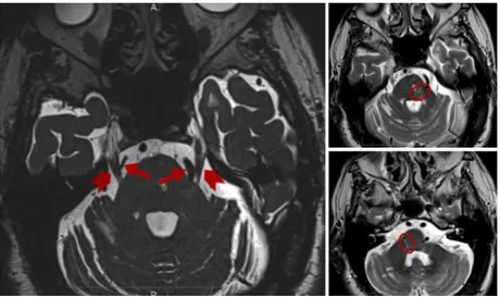
Figure 2The MRI 3D constructive interference in steady state (3D-CISS) image (a) shows the trigeminal nerve course (marked by arrowheads) and an arterial loop consisting of the superior cerebellar artery on the right side and the vertebral artery on the left side (arrows). The MRI TSE T2-weighted images show the demyelinating plaque (circle) involving (b) the trigeminal root entry zone and (c) the caudal and posterior pontine portion. Di Stefano et al, J Oral Facial Pain Headache, 2017.
18
Pathophysiological mechanisms
Both in classic and secondary TN, the primary mechanism is focal demyelination of primary afferents near the entry (extraaxial or intraaxial) of the trigeminal root into the pons. This area represents a locus minoris resistentiae because it is the site where Schwann cells are substituted by oligodendroglia in providing the myelin sheath. Focal demyelination makes the axons hyperexcitable and increases the susceptibility to ectopic excitation, ephaptic transmission, and high-frequency discharges. The consequences of the focal demyelination are not fully clarified, but it has been hypothesized that the focally demyelinated primary afferents become hyperexcitable when demyelination reaches such a level that ions can move in and out of the axon, also away from the Ranvier node zones, at which point the axons do not have enough energy to promptly re-establish the resting potential.4 Hence the axons tend towards a
depolarization level which makes them hyperexcitable. Ectopic impulses, which are generated either spontaneously along the sensory afferent or because of a local direct mechanical stimulus such as arterial pulsation, are probably also involved in the hyperexcitability. Moreover, supported by evidence in animal models of focal demyelination of the trigeminal root, ephaptic transmission, i.e. cross-talk from close, healthy nerve fibres, and the generation of
19
high-frequency discharges are also suggested to contribute to the hyperexcitable nervous state in TN.10,11 Finally, there is some
evidence suggesting that the hyperactivity of primary afferents secondarily induces central sensitization of wide-dynamic-range neurons in the spinal trigeminal nucleus or even more central changes.12
The mechanisms underlying continuous as opposed to paroxysmal pain are not fully understood. Continuous pain may develop as a result of progressive root damage after prolonged compression13 or
reflect central mechanisms.12
Treatment of trigeminal neuralgia
Carbamazepine (CBZ) and oxcarbazepine (OXC) are the first-choice medical treatment in TN. They have the same mechanism of action, the blockade of voltage gated sodium channel in a frequency dependent manner, resulting in the stabilization of hyperexcited neural membranes and in the inhibition of repetitive firing. The AAN-EFNS guidelines recommended that patients unresponsive or that cannot reach the therapeutic dosage of the drug because of adverse events should be made aware of the availability of surgery.14 Surgical
procedures include Gasserian ganglion percutaneous techniques, microvascular decompression in the posterior fossa, and gamma
20
knife radiosurgery. These procedures are extremely efficacious with relatively few complications. Microvascular decompression may be considered over other surgical techniques to provide the longest duration of pain freedom.4 According to the available evidence no
oral treatment is better than CBZ or OXC, but in case of refractory TN, among the non-surgical option, lamotrigine and botulinum toxin injections should be considered.4 Up to now, only few studies have
21
Figure 3. Magnetic resonance imaging, sagittal reconstruction of T2-weighted 3-dimensional (3D) CISS sequence (A) shows the conflict between the aneurysm (white arrow) and trigeminal nerve (dotted arrow); the black arrow indicates the Meckel cave. The axial image from the same sequence (B) shows the aneurysm (white arrow) of the lateral pontomesencephalic segment of the superior cerebellar artery in contact with the trigeminal nerve root. The volumetric reconstruction of the 3D TOF sequence (C) shows the aneurysm (arrow) and course of the artery.22
Figure 4. Right vertebral angiography shows the wide-necked distal aneurysm of the superior cerebellar artery (A). Image acquired during deployment of the low-profile stent after detachment of a single coil (B). Final angiography shows occlusion of the aneurysm and patency of the stent (C). Nine-month follow-up angiography shows persistent occlusion of the aneurysm (D).23
Aims
The overall aim is to provide new insight into TN pathophysiological mechanisms and treatment. Specifically, the thesis aims to:
1. Describe the clinical characteristics of TN in a large consecutive cohort of patients focusing on trigger factors. Investigate how frequently triggers are present, which manoeuvres activate them and where cutaneous and mucosal trigger zones are located.
2. Analyse the natural history of TN in a large cohort of patients, by focusing on the drug responsiveness, the side effects related to the first-line pharmacological treatment, the changes in pain characteristics along with the duration of the disease, such as duration and intensity of paroxysms, and the possible onset of sensory disturbances.
3. Performing a systematic search of relevant literature, in order to provide current, evidence-based, knowledge about the pharmacological treatment of typical and atypical TN, with a specific focus on drugs in development.
24
Triggering Trigeminal Neuralgia
Introduction
TN is a unique neuropathic pain condition characterized by unilateral paroxysmal pain, usually described as stabbing or electric shock-like, and restricted to the distribution of one or more divisions of the trigeminal nerve territory.1,3 In ICHD-3, the most used classification
of headache and facial pain disorders, the diagnostic criteria of classical TN, and those of MS-related TN (labelled painful trigeminal neuropathy) include provocation of paroxysmal pain from innocuous stimuli, but not as an essential condition (i.e., TN can be diagnosed without a trigger if three other pain characteristics are present). By contrast, the novel diagnostic grading system issued by the Special Interest Group on Neuropathic Pain of the International Association for the Study of Pain makes the presence of a trigger an essential criterion, without which the clinical diagnosis of TN cannot be established. The view was taken on the basis of a literature review that suggested the presence of trigger zones in a high percentage of patients diagnosed with TN on clinical grounds.3 In none of the
quoted papers, however, was the frequency or characterization of trigger zones the main purpose. It is therefore imperative that given the dominance of triggers in the new classification their frequency
25
and characteristics are defined as accurately as possible. Not only is this critical for the diagnosis of an individual patient, but also to support meaningful research on the pathophysiology and treatment of TN.
Provocation of paroxysmal facial pain by innocuous stimuli is very rare, except for TN. A very high percentage of triggers in patients who report all other pain qualities suggestive of TN (intense, short-lived pains of abrupt onset and cessation, coming in paroxysms) would strengthen the concept of TN as a unique pain, and an explanation for its pathophysiology would have to include generation of trigger zones. A low percentage would in turn suggest that TN can present with and without triggers and requiring separate pathophysiological explanations. In this study, we aimed to determine the frequency and nature of triggers as they pertain to patients with TN whose diagnosis is compatible with the ICDH-3 criteria.
Methods
We prospectively screened consecutive patients attending the Centre for Neuropathic Pain at Sapienza University from January 2015 to December 2016. Inclusion criteria were a diagnosis of TN according to the 3rd edition of the International Classification of Headache Disorders (ICHD-3, 2013), including 13.1. Classical
26
trigeminal neuralgia, 13.1.2.4 Painful trigeminal neuropathy attributed to MS plaque and 13.1.2.5 Painful trigeminal neuropathy attributed to space-occupying lesion. (In this classification, both latter diagnostic categories refer to patients who report facial pain with the characteristics of those in classical trigeminal neuralgia and are also called ‘secondary TN’.3 Exclusion criteria were cognitive
disturbances and diagnosis of other orofacial pain condition. We also excluded four patients with secondary TN from the present series because they were shown to have a benign tumour at the cerebellopontine angle verified by MRI, and were promptly referred for surgery; therefore, they were not available for the present study. Two patients were excluded because, even though they presented with one-sided paroxysmal attacks and had normal MRI scans, the trigeminal reflex testing showed bilateral abnormalities in the mouth area that were typical of trigeminal neuropathy. The total number of the patients prospectively enrolled was 70.
Each patient underwent a precise sensory profiling using bedside tools as indicated by the European guidelines on neuropathic pain assessment.15 Each patient also underwent both trigeminal reflex
testing14 and 3T MRI, with specifications optimized for identification
of the cause of TN.9
Three staff members were involved in the clinical examination and two in the neurophysiological testing. The diagnosis of TN was
27
confirmed by two clinicians. Clinical characteristics were systematically collected using a dedicated questionnaire, focusing on triggers. Patients provided a thorough description of all trigger manoeuvres and drew both the trigger zones and the evoked paroxysmal pain distribution on a facial map. The overlap profiling of the trigger zones was carried out with dedicated software that provided representation and sum of the trigger areas on a standard 3D model of face and mouth. The frequency distribution of each trigger zone was computed at pixel level by counting the number of times each pixel of the model fell within each trigger area.
Besides the prospective patient enrolment, we also collected patients retrospectively. A staff nurse selected from our database the names and medical records of the most recent 70 outpatients with a diagnosis of TN (according to ICHD-3), who had attended our centre up to the point of commencement of the above prospective study (i.e., December 2014). A further staff member analysed the records and the diagnostic investigations, to confirm the diagnosis and classify the patients in classical or secondary TN.
The principal investigator subsequently examined the individual records for information on pain and triggers.
All patients included in the prospective or retrospective series suffered from paroxysmal attacks of pain affecting one or more
28
divisions of the trigeminal nerve, regardless of the presence of concomitant continuous pain.3,16
For all patients included, we used as main outcome variables the side, distribution, and time course of pain, as well as manoeuvres triggering the paroxysms and distribution of trigger zones. For the prospective group we also collected information about the mean severity of paroxysms in the last month, as assessed with a numerical rating scale (NRS) ranging from 0 (no pain) to 10 (worst possible pain) and represented the trigger zones on the 3D-face model.
Statistical analysis
Descriptive statistics only was used for evaluation of the frequency of triggers. For comparisons between the prospective and retrospective groups we used Mann-Whitney Test, given that the main demographic and clinical data did not have a normal distribution. For comparisons of categorical data, we used Fisher exact test and Chi-squared tests.
Results
We included 140 patients in the analysis (54 M, 86 F, mean age 65.32 ± 12.01 years). Of these, 70 were collected retrospectively. Age and gender distribution did not differ between the prospective and
29
retrospective groups (P> 0.3). Of the 140 included patients, 124 had a classical TN, and 16, all having trigeminal reflex testing abnormalities, had secondary TN. MRI showed that 14 out of these 16 patients had a multiple sclerosis-related TN, while two patients had TN due to an aneurysm and a megadolichobasilar artery. No difference was found between the two patient groups in the frequency of classical and secondary TN (P= 0.4), patient age at the time of onset of pain (P= 0.3) or frequency of affected trigeminal divisions (Table 2).
In all 140 patients combined, pain was more often located on the right (70%) than left side (29%), and the second (V2) and the third (V3) trigeminal divisions were more frequently affected than the first trigeminal division (V1) (Table 2). Patients described paroxysmal pain as a very abrupt, short-lasting pain, stabbing or similar to an electric shock in quality. Although most patients reported only daytime pain, 27 patients in the prospective group (39%) reported paroxysmal pain also during the night. The intensity of paroxysmal pain was 8.5 ± 1.6 on NRS (0-10). The duration of paroxysms was less than one second in four patients, from one to two minutes in nine patients and had a mean duration of 7 seconds in the remaining patients. The mean number of paroxysms was 11 a day. Fiftythree percent of patients experienced pain remission periods (mean duration of 10.52 ± 10.18 months) during which external stimuli failed to provoke any attacks.
30
Thirty-three patients (24%; 28 with classical and 5 with secondary TN) experienced concomitant continuous pain in the same division affected by paroxysms. Continuous pain was described as dull, burning or tingling. Only in five patients this kind of pain was also evoked by trigger manoeuvres, including talking, eating, drinking, swallowing and gently touching the face. The mean pain intensity of continuous pain was 7.4 ± 2.5 in the prospective group. Concomitant pain was reported as being unrelenting day and night without pain-free intervals in 12 patients and lasting from five minutes to two hours in the remaining patients.
Whereas there was no difference in age at onset of TN between prospectively and retrospectively recruited patients, we found that patients with secondary TN had a lower age at onset compared to those with classical TN (median 51, 95% CI 46-57; median 61, 95% CI 57-62; P <0.02). On this point, however, there was a substantial overlap between the two populations as shown in Figure 5. The youngest age at onset was 31 and the oldest 89, both with classical TN.
Trigger analysis
Virtually all patients (136/140) reported trigger manoeuvres. The most frequent trigger manoeuvres were touching face (79%), talking
31
(54%), chewing (44%), and brushing teeth (31%), with no difference between the prospective and retrospective groups. Unusual trigger manoeuvres included flexing the trunk (5%), contact with hot or cold food/water (4%), speaking loudly (2%) and turning the eyes to the right (1%) (Table 3). Of the 140 patients, 110 (78.6%) had extraoral and 117 (83.6%) intraoral trigger zones. The 3D face model showed that although the trigger zones were widely distributed within the whole trigeminal territory, they were more frequently located in the nasal wing (22%), upper lip (17%), cheek (13%), lower lip (12%), chin (11%), alveolar gingiva (11%) and cheekbone (10%) (Table 4, Figure 6). Most patients had more than one trigger zone/manoeuvre. All prospective patients drew their paroxysmal pain as a line rather than a circle and some explained that they felt the pain radiating from a point to another. Almost all patients, however, drew or reported that the pain paroxysms were restricted to the same trigeminal division of the trigger zone. Only in four patients the trigeminal division of the paroxysm and that of the trigger zone did not coincide: in one patient light touch of the nasal wing evoked pain in the supraorbital region, in the second patient pressing the first molar of the lower dental arch evoked pain in the upper lip, in the third light touch of the chin evoked pain in the upper lip, and in the fourth light touch of the eyelid evoked pain in the mandibular region.
32
Of the four patients apparently without trigger manoeuvres (three in the prospective, one in the retrospective group), one suffered from multiple sclerosis-related TN. Of the three patients with classical TN, although being unaware of any specific trigger manoeuvre, one did report that to avoid the pain paroxysms she had to keep the face perfectly still, thus suggesting that a facial movement was the trigger.
Discussion
In this clinical study with a large sample of patients we show that in virtually all TN patients (97%) paroxysmal pain is associated with a trigger.
The frequency of triggers we found is higher (96% in the prospective study group) than that reported in a previous study (91%).16 This
difference probably reflects the dedicated questionnaire we used in our study for collecting information on triggers. We also hypothesize that in the few patients with no apparent trigger, the pain attacks are evoked by muscle movements they are unaware of, e.g. eye-blinking or facial mimicry. The observation that besides spontaneous paroxysmal pain, patients always invariably report triggered pain, further supports the use of triggers as a criterion for clinically established TN, and with that for probable neuropathic pain.3
33
As shown on our 3D face model, nearly all trigger zones were located within the central mask, most frequently in the perioral region. Although four of our patients felt the evoked paroxysmal pain in a trigeminal division different from that of the trigger zone, most often evoked paroxysms and trigger zones are located in the same trigeminal division. This finding, in line with previous studies,17 is
consistent with the mechanism of cross-excitation via ephaptic transmission from adjacent unaffected fibres within the trigeminal root.10,11 The trigger zones were variable in size, and while some were
no more than a pinpoint, most were much larger. Similar variability was mentioned in a previous study.18 Our method of zone drawing
has the advantage that it allows the patient to indicate the size directly on the facial map without relying on the estimate by an examiner. We hypothesize that the evoked paroxysmal pain might in some patients require a sequential activation of mechanoreceptors, resulting in an enlarged trigger area. This hypothesis might also explain the “towel’s sign”, i.e. some patients soon learn to dry their face by slowly pressing, rather than brushing, the affected side to avoid the painful paroxysms.
Unexpectedly, we found that a few patients (4%) reported that consuming hot or cold food/water could provoke paroxysms. This finding goes against the common notion that only innocuous, mechanical stimuli can evoke the paroxysmal pain.18,19 However,
34
while the specific qualification of a thermal component was volunteered by the patients, the muscular activity in the lips, tongue and pharynx during eating or drinking is certainly sufficient to act as a trigger, precluding the argument that heat or cold alone could do so.
Unlike the common belief that TN does not awake patients from sleep, we found that many patients (38% of our sample in the prospective group) have nightly painful awakenings. This finding is however in line with the only other studies that used a dedicated questionnaire for patients and their partners about painful awakenings in TN.20,16 Given that all our patients with nightly painful
awakenings had extra-oral triggers, we suggest that an innocuous contact of the patient’s skin with the sheet or the pillow acted as a triggering factor. We only found patients with nightly awakenings in the prospective group probably because we included a specific question about this in the questionnaire. It is worth pointing out that nightly attacks are less common than cluster headache where they are a prominent feature.
Besides the paroxysmal pain, several patients (24% of our sample) complained of concomitant continuous pain. All these patients, by definition, felt this type of pain in the same territory of paroxysmal pain. These findings, consistent with previous studies21-23 support the
35
Central mechanisms12,24 and progressive root damage due to
compression13 have been proposed as possible mechanisms
underlying this type of pain.
We found that age at onset of pain in patients with secondary TN was significantly lower than that of patients with classical TN, confirming the current long-held view. However, there was substantial age overlap between the two populations preventing this aspect to be used as an indicator of secondary TN, in line with previous guidelines on trigeminal neuralgia.14
The demographic and clinical variables in our cohort of patients are similar to those reported in previous studies.2,16 TN is more frequent
in women and more frequently affects the right side of the face, in the V2 and V3 divisions, probably because of the somatotopic distribution of sensory fibres in the trigeminal root.25
The main limitation of the study is reliance on both retrospective and prospective data. However, as the demographics, clinical features, frequency of triggers and the nature of trigger manoeuvres were very similar between the two groups, we believe it was justified to use the combined data. The information extracted from the medical records was facilitated by a longstanding interest of the study centre in TN and the established clinical practice of detailed collection of all clinical data.
36
Conclusion
In nearly all patients with the diagnosis of TN based on ICHD-3 a trigger capable of provoking a paroxysm can be identified. Trigger zones are seen almost exclusively in the central mask, most commonly in the perioral area, and are variable in size. In all the trigger manoeuvres listed by the patients a mechanical component (touch or muscle movement) is present. These findings will be of assistance in future studies on the pathophysiological mechanisms of trigeminal neuralgia.
37
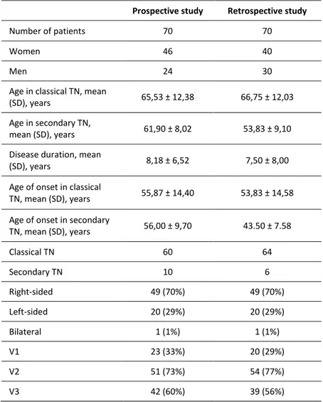
Table 2. Demographics, age at onset, duration of disease, side and
anatomical localization of pain.
Prospective study Retrospective study
Number of patients 70 70
Women 46 40
Men 24 30
Age in classical TN, mean
(SD), years 65,53 ± 12,38 66,75 ± 12,03
Age in secondary TN,
mean (SD), years 61,90 ± 8,02 53,83 ± 9,10
Disease duration, mean
(SD), years 8,18 ± 6,52 7,50 ± 8,00
Age of onset in classical
TN, mean (SD), years 55,87 ± 14,40 53,83 ± 14,58
Age of onset in secondary
TN, mean (SD), years 56,00 ± 9,70 43.50 ± 7.58 Classical TN 60 64 Secondary TN 10 6 Right-sided 49 (70%) 49 (70%) Left-sided 20 (29%) 20 (29%) Bilateral 1 (1%) 1 (1%) V1 23 (33%) 20 (29%) V2 51 (73%) 54 (77%) V3 42 (60%) 39 (56%)
38
Table 3. Frequency of trigger manoeuvres in trigeminal neuralgia. Trigger Manoeuvres N(%) prospective study N(%) retrospective
study
Gently touching the face 58 (83) 52 (74)
Talking 41 (59) 35 (50)
Chewing 29 (41) 32 (46)
Tooth brushing 25 (36) 19 (27)
Washing one’s face 19 (27) 20 (29)
Eating 16 (23) 19 (27)
Shaving 7 (10) 13 (19)
Drying one’s face 8 (11) 10 (14)
Swallowing 7 (10) 9 (13)
Drinking 6 (9) 7 (10)
Jaw movement 6 (9) 5 (7)
Blowing one’s nose 5 (7) 4 (6)
Flexing the trunk forward 4 (6) 3 (4)
Hot or cold food/water 2 (3) 4 (6)
Laughing 1 (1) 3 (4)
Raising own voice 2 (3) 1 (1)
Application of make-up 2 (3) 1 (1)
Yawning 1 (1) 2 (3)
Pronouncing labial letters 2 (3) -
39
Eye movement 1 (1) 1 (1)
Washing one’s hair 1 (1) 1 (1)
Head movements 2 (3) -
Tongue movement - 2 (3)
Sneezing 1 (1) 1 (1)
40
Table 4. Frequency of trigger zones in trigeminal neuralgia. Trigger Zones N% Prospective study N% Retrospective study
Nasal wing 18 (26) 13 (19) Upper lip 14 (20) 10 (14) Cheek 8 (11) 10 (14) Lower lip 7 (10) 10 (14) Chin 10 (14) 6 (9) Alveolar gingiva 7 (10) 8 (11) Nasolabial fold 8 (11) 6 (9) Cheekbone 9 (13) 5 (7) Jaw 7 (10) 5 (7) Supraorbital region 5 (7) 5 (7) Eyebrow 5 (7) 5 (7)
External eye side 4 (6) 2 (3)
Lower lateral incisor 1 (1) 3 (4)
Tongue - 3 (4)
Scalp 3 (4) -
Hard palate - 2 (3)
Conjunctival fornix 2 (3) -
Upper lateral incisor 2 (3) -
Upper molars 2 (3) -
Lower eyelid 1 (1) 1 (1)
41
Upper premolars 1 (1) -
Lower premolars 1 (1) 1 (1)
42
Figure 5. Histogram of age of onset. X-axis: age of onset. Y-axis: number of patients. CTN: classical trigeminal neuralgia. STN: secondary trigeminal neuralgia. Note that although STN patients are younger, their age of onset are intermingled with those of CTN patients.43
Figure 6. Trigger zone distribution. Data from 70 prospectively-enrolled patients with classical or secondary TN. Upper panel: extra-oral territories. Lower panel: intra-oral territories. Left column: trigger-zone contours. Right column: trigger-zone overlap profiling. The number of superimpositions ranges from 2 (cyan) to 15 (dark orange). The number of trigger zones in the intra-oral territory is smaller in comparison with the number of patients reporting talking or chewing as the main trigger manoeuvres, because of the patients’ difficulty in identifying a circumscribed trigger zone region within the mouth.44
Natural history and outcome of 200
outpatients with classical trigeminal neuralgia
treated with carbamazepine or oxcarbazepine
in a tertiary centre for neuropathic pain
Introduction
TN is a clinical condition characterized by a sudden, usually unilateral, brief, stabbing, recurrent pain with a distribution consistent with one or more divisions of the fifth cranial nerve. The paroxysmal attacks are stereotyped in the individual patient, last from a fraction of a second to 2 minutes and may be evoked by stimulating cutaneous or mucous trigeminal territories, the so-called trigger zones. The pain-free intervals may range from days to years TN may be distinguished in classical, namely without a cause other than a neurovascular compression, or secondary to a demonstrable lesion, including benign tumors of the cerebellopontine angle or multiple sclerosis. According to the symptom constellation, TN is categorized into typical and atypical form, the latter characterized by a constant and non-lancinating background pain and, sometimes, sensory disturbances in the affected division The annual age-adjusted
45
incidence is 5.9% for women and 3.4% for men.2 According to the
American Academy of Neurology (AAN), the European Federation of Neurological Societies (EFNS) and also other recent guidelines, carbamazepine (CBZ) and oxcarbazepine (OXC) are the first-line medical treatments for pain control in patients with TN.14,26 They
have the same mechanism of action, namely the blockade of voltage-gated sodium channels in a frequency-dependent manner. OXC may be preferred because of the minor risk for drug interactions and its better tolerability in comparison with CBZ.26 The AAN-EFNS
guidelines recommended that patients unresponsive or that cannot reach the therapeutic dosage of the drug because of adverse events should be made aware of the availability of surgery. Surgical procedures include Gasserian ganglion percutaneous techniques, microvascular decompression in the posterior fossa, and gamma knife radiosurgery. These procedures are extremely efficacious with relatively few complications. Microvascular decompression may be considered over other surgical techniques to provide the longest duration of pain freedom.14 According to the available evidence no
oral treatment is better than CBZ or OXC, but in case of refractory TN, among the non-surgical option, lamotrigine and botulinum toxin injections should be considered.27 Up to now, only few studies have
focused on the development of the clinical picture and the drug efficacy and tolerability in time. The aim of this retrospective study
46
was to analyse the natural history of TN in a large cohort of patients, by focusing on the drug responsiveness, the side effects related to the pharmacological treatment, the changes in pain characteristics along with the duration of the disease, such as duration and intensity of paroxysms, and the possible onset of sensory disturbances.
Methods
The staff nurse retrospectively selected the clinical notes of outpatients with a diagnosis of classical TN who had attended our centre for neuropathic pain from January 2000 to June 2013. One of us analysed the clinical notes and selected the last consecutive 100 patients who began treatment with CBZ and the last consecutive 100 who did it with OXC. All patients included in the analysis suffered from paroxysmal attacks of pain lasting from a fraction of a second to 2 minutes, affecting one or more divisions of the trigeminal nerve. Pain was described as intense, sharp, superficial or stabbing; paroxysms could be both spontaneous and precipitated from trigger areas or by trigger factors. The attacks were stereotyped in the individual patient. The clinical examination did not show any clinical neurological deficit. All patients had undergone MRI scans of the brain and trigeminal reflex testing in order to identify with certainty even patients with typical presentation but a possibly secondary
47
origin, including idiopathic sensory trigeminal neuropathy and nerve trauma. Patients were seen at least monthly until the target dosage and/or a significant pain reduction was reached. Then, follow-up visits were scheduled every six months, unless side effects occurred. Two staff members were involved in the clinical examination and two in the neurophysiological testing. The diagnosis of classical TN was confirmed by at least two clinicians. We focused our attention on the average onset age of TN, the number of responders to CBZ or OXC, the possible CBZ/OXC lost efficacy, the side effects that caused interruption of treatment or a dosage reduction to an unsatisfactory level and the latency for the side effect onset. Patients were considered as responders on the bases of their global satisfaction and the willingness to continue the drug. We also analysed the possible change in pain characteristics during the course of disease, including paroxysms duration and intensity. By definition, classical TN is a pain syndrome that arises without a clinically manifest sensory deficit: anyway, we wanted to test the likelihood of the onset of sensory disturbances during the disease course.
Findings
We considered an otherwise homogeneous group of 200 patients (68 M, 132 F, mean age 67.54 ± 12.11), with a mean follow-up period of
48
7.31 years. Among them, 22 patients with typical symptom constellation and a diagnosis of classical TN were excluded from the study because two of us considered the results of neurophysiological or neuroimaging investigations insufficient to exclude a secondary form with absolute certainty. The other 178 patients had a classical TN, without any evidence of a cause other than a neurovascular conflict at dedicated MRI scans. Ninety-five out of 178 patients were treated with CBZ and the remaining 83 with OXC. The average onset age of symptoms was 60 ± 11.6 years (range 35-80). The initial number of responders was 98% with CBZ at a median dosage of 600 mg (range 200–1200 mg), and of 94% with OXC at a median dosage of 1200 mg (range 600–1800 mg). Among responders to CBZ, 27% of patients incurred in adverse events that directly caused interruption of treatment by the physician or a dosage reduction to a level that was insufficient to control pain, thus causing discontinuation, after a mean period of 8.6 months. In a mean period of 13 months, physician- or patient-decided discontinuations occurred in 18% of patients initially responders to OXC. The causes of these discontinuations are plotted in Figure 7. The CNS disturbances were about triple in patients on CBZ than those on OXC. In detail, the CNS disturbances included somnolence (10 patients treated with CBZ and 5 with OXC), postural unbalance (6 with CBZ and 4 with OXC) and dizziness (6 with CBZ and 1 with OXC). Among patients under CBZ,
49
three had an increase of transaminases, one anemia, one leucopenia, and one thrombocytopenia. Among those under OXC, 5 had hyponatremia, one patient had thrombocytopenia. Allergic reactions (cutaneous rash) affected two patients on CBZ and two on OXC. The onset of side effects on the CNS occurred with a mean dosage of 600 mg for CBZ and 1200 mg for OXC. With CBZ, anaemia, leucopenia, and thrombocytopenia occurred within the first three weeks of treatment, with dosages of 600 mg, 600 mg, and 1000 mg, respectively. With OXC, thrombocytopenia occurred within the first two weeks of treatment, with a dosage of 1200 mg. Hyponatremia was observed within the first month, with dosages of 900 mg in one patient, 1200 mg in three patients and 1800 mg in the fifth patient. Allergic reactions occurred at the beginning of treatment, i.e. within the first two days. In this case the treatment was interrupted immediately. Among patients that had to interrupt CBZ because of adverse events, 16 were switched to OXC, and 5 to gabapentin. The new treatment was effective in 12 patients (10 with OXC and 2 with gabapentin), whereas in 7 patients (6 with OXC and 1 with gabapentin) the new treatment was unsuccessful. The remaining two patients were lost to follow up. Among patients that had to interrupt OXC because of adverse events, 3 were switched to CBZ, and 2 to gabapentin. The new treatment was effective in 4 patients (3 with CBZ and 1 with gabapentin), whereas in the remaining patient the
50
new treatment was unsuccessful. Eight patients were lost to follow up. Eventually, 13 patients out of 178 were referred for surgery (5 patients treated with OXC and 8 with CBZ). Among patients who had a good initial response, three patients with CBZ and two with OXC developed late resistance after 24–76 months. The intensity of paroxysms worsened in six patients and their duration in four. Such a worsening occurred in a mean time of 55 months. Constant pain developed in 5 patients, after an average duration of disease of 7 years. We did not observe the development of sensory disturbances with time in any patient suffering from classical TN.
Discussion
In this retrospective study in a large cohort of patients, we investigated the natural history of classical TN, focusing our attention on the efficacy of CBZ or OXC, the possible onset of a late resistance and the side effects that eventually caused either interruption of treatment or a dosage reduction to an unsatisfactory level. The possible modifications in pain characteristics during the course of disease, including paroxysms duration and intensity, were also examined. We found that the worsening of pain with time and the development of late resistance only occurred in a very small minority of patients. CBZ and OXC were confirmed to be efficacious in a large
51
majority of patients, but the side effects caused the withdrawal from treatment in an important percentage of patients. Demographics matched those observed in previous studies,2 with a higher
frequency in women (65.17%) and an average onset age at 60 years. We found changes in pain characteristics only in an extremely small sub-set of patients, compared to the total number. Such changes included the increase of both paroxysms’ duration and intensity. Unlike other reports,19 no sensory deficit was observed since the
beginning of the disease. It is generally agreed that the first line therapy of TN is pharmacological and based on the use of sodium channels blockers, CBZ and OXC. Four placebo-controlled trials demonstrated the efficacy of CBZ28-31 with a number needed to treat
to obtain important pain relief of 1.7-1.8.32 This efficacy is however
compromised by the tolerability, with a numbers needed to harm of 3.4 for minor and of 24 for severe adverse events.33,34 OXC has a
comparable efficacy to that of CBZ but a greater tolerability and a lower potential for drug interaction.35-37 This study confirmed that
CBZ and OXC are efficacious in a great majority of patients and that OXC is more tolerated in comparison with CBZ. If compared with other reports, the percentage of non-responders was somewhat lower in our sample. Because CBZ and OXC are extremely efficacious in increasing the refractory period of action potentials, they are bound to be most active on the high-frequency discharges that
52
characterize the paroxysms of TN. Naturally, if the patient selection is not very strict, and concedes the recruitment of a few patients that also have some ongoing pain, then the efficacy of CBZ/ OXC may drop. Indeed, the diagnostic accuracy has always been a problem in studies in TN. Adverse events may cause withdrawal from treatment. This occurred in a significant number of patients, 27% of those with CBZ and 18% of those with OXC, who were initially responders. The most frequent adverse effects involved the CNS, and included somnolence, dizziness and postural unbalance. CBZ had a higher percentage of discontinuations for all kinds of side effect, except for sodium depletion, which only occurred with OXC (Figure 7). Although in our centre we are well aware of the great efficacy of surgical interventions for TN and we always offer this chance to patients resistant to CBZ/OXC, only 7% of this large cohort of patients was eventually sent for surgery, a proportion decidedly low if compared to those reported by neurosurgical centres. To explain the low proportion of patients sent for surgery, we may think of this main explanation: the local population is not so keen on undergoing surgery unless they really cannot manage with medical treatment and the number of patients resistant to CBZ/OXC was very low. In conclusion, the failure of the treatment with CBZ/OXC, most of the times, is not due to the inefficacy of the drug, but rather to undesired effects to a level that causes interruption of treatment or a dosage
53
reduction to an insufficient level. These results suggest the opportunity to develop a better tolerated drug.
54
Figure 7. Tolerability of Carbamazepine (CBZ) and Oxcarbazepine (OXC). Y-axis: number of patients that discontinued first treatment because of adverse events. X-axis: causes of discontinuation. Note that CNS disturbances affected far more frequently the patients on CBZ, whereas hyponatremia only affected patients on OXC. The sum of patients reporting somnolence, postural unbalance, and dizziness is higher than the total CNS disturbances because many patients complained of more than one CNS disturbance.55
Current and Innovative Pharmacological
Options to Treat Typical and Atypical
Trigeminal Neuralgia
Introduction
TN is a representative neuropathic facial pain condition, characterized by unilateral paroxysmal pain described as stabbing or electric shock-like, in the distribution territory of one or more divisions of the trigeminal nerve and triggered by innocuous stimuli.1
According to the new classification and diagnostic grading of TN issued by the International Association for the Study of Pain (IASP), TN is distinguished in three diagnostic categories-classical, caused by vascular compression producing anatomical changes in the trigeminal nerve root-secondary, due to an identifiable underlying neurologic disease,-idiopathic, when even after MRI or other investigation, the aetiology of TN remains unclear (Table 1).3
Regardless of the aetiology, the primary mechanism of paroxysmal pain is the same, i.e. a focal demyelination of primary trigeminal afferents near the entry of the trigeminal root into the pons, making the axons hyper-excitable and increasing the susceptibility to ectopic excitation, ephaptic transmission, and high-frequency discharges.10
56
TN has an annual incidence of three to five per 100,000. It is more common in women than men (age adjusted ratio: 1.74:1) and in people aged 50–69 years.2 In virtually the entire population of
patients with TN, at least one trigger capable of provoking a paroxysm can be identified. In a recent study, provocation of paroxysmal pain by various trigger manoeuvres was reported by 136 of the 140 patients. The most frequent manoeuvres were gentle touching of the face and talking. Trigger zones were predominantly reported in the perioral and nasal region and were variable in size. These data are coherent with the use of trigger factor as an essential diagnostic feature for a clinically established diagnosis of TN.3
Patients with TN may suffer from different types of pain, ranging from single attacks to a series of prolonged attacks, and it was suggested that these pain characteristics can vary over time.38
Traditionally, autonomic symptoms such as tearing and rhinorrhea have not been associated with TN. However, it is now known that a large proportion of TN patients have autonomic symptoms.16 A
subgroup of patients with TN also suffer from concomitant continuous pain (CCP), described as dull, burning or aching.23 This
condition has been described with several definitions, including atypical TN and TN type 2; the International Headache Society Classification (ICHD) defined this relatively uncommon type of TN as TN with concomitant continuous facial pain. The presence of
57
continuous pain is not related to aetiology and may occur in idiopathic, classic, or secondary TN. Background pain distribution coincides with that of the paroxysmal pain, and fluctuations of its intensity parallel in time those of the paroxysmal pain.21,22 A
prevalence three times higher in women than in men was reported.23
In a cohort of 158 patients with TN, continuous pain developed within a mean period of 1.5 years since the disease onset, thus suggesting that this kind of pain is not a consequence of a long duration of stabbing pain.23 The mechanisms underlying continuous pain, as
opposed to paroxysmal pain, are not fully understood, with implications for treatment. There is the evidence that continuous and paroxysmal pain may improve differently after microvascular decompression, thus supporting the hypothesis that the mechanisms responsible for the two pain components may be different.4 Central
mechanisms12 and progressive root damage due to compression13
have been proposed as possible factors. CBZ and OXC are the first-choice medical treatment in TN. They have the same mechanism of action, the blockade of voltage gated sodium channel in a frequency dependent manner, resulting in the stabilization of hyperexcited neural membranes and in the inhibition of repetitive firing. In patients with purely paroxysmal pain, CBZ and OXC are effective in virtually the entire patient population. However, they produce side effects to a level that cause interruption of treatment or a dosage
58
reduction in 23% of patients, making necessary the development of new, more selective sodium channel blockers. Conversely in patients with CCP, the efficacy of CBZ and OXC may drop, thus suggesting the opportunity to test the efficacy of different drug categories. A wide range of drugs has been investigated in TN, but the scientific literature highlighted the need of high-quality clinical trials in TN. The aim of this review, based on a systematic search of relevant literature, is to provide current, evidence-based, knowledge about the pharmacological treatment of typical and atypical TN, with a specific focus on drugs in development, such as botulinum toxin A and new, more selective sodium channel blockers.
Search Process
We searched for relevant papers within the PubMed, EMBASE and the Cochrane Database of Systematic Reviews, taking into account publications up to February 2018. All searches used the following synonyms for TN: trigeminal neuralgia and tic douloureux. The primary search was supplemented by a secondary search using the bibliographies of the retrieved articles. Only full-length, original communications including open-label studies were considered, and the search was limited to English language publications. Clinical trials database (ClinicalTrial.gov) has been checked in order to include in
59
the analysis studies currently in progress. The review process was carried out independently by two reviewers and only publications independently approved by the two authors were taken into account (Figure 8). The authors independently assessed the quality of the individual trials during data extraction. Inclusion criteria were the following: trials including patients with a diagnosis of typical or atypical TN, including classical, idiopathic and secondary TN, and a minimum sample of 10 patients.
Drugs in Classical or Idiopathic Trigeminal Neuralgia
First‑line Treatment
CBZ and OXC are the first-line treatment in TN. Their effect is related to the blockade of voltage-sensitive sodium channels in a frequency dependent manner, resulting in the stabilization of hyperexcited neural membranes and inhibition of repetitive firing. Systematic reviews and randomised controlled trials, including 147 patients,28-31
demonstrated the efficacy of CBZ compared to placebo, with a number-needed-to-treat (NNT) to obtain pain relief of 1.7–1.8.32
However, CBZ showed a number-needed-to-harm (NNH) of 3.4 for minor and 24 for severe adverse events.34 In the study of Killian and
Fromm,29 with a maximum daily dose of 1000 mg, 19 of 27
60
compared with minimal or no response with placebo on a 5-day treatment. Nicol,30 using a cross-over design and a maximum daily
dose of 2400 mg, reported that 15 of 20 participants randomised to initial CBZ had a good or excellent response after 14 days’ treatment, compared with 6 of 24 reporting good or excellent response who started on placebo. Superiority of CBZ was also reported by Rocklif and Davis31 in a small sample of patients with three days’ treatment
with CBZ. In the study from Rasmussen and Riishede,39 after 5 days’
treatment, 46 of 55 patients with TN had good effect on CBZ, compared with 8 of 55 on placebo. Campbell and colleagues28
reported a mean fall in maximum pain intensity of 58% after 2 weeks treatment with CBZ 400–800 mg daily compared to 26% with placebo (Supplementary Material). Compared to CBZ, OXC showed a similar efficacy in reducing pain attacks but a greater tolerability and a lower potential for drug interaction.37 However, data from full randomised
controlled trials are not available, thus precluding the NNT and NNH calculation. A pilot study on OXC with extended-release formulation is currently in progress (ClinicalTrials.gov Identifer: NCT03374709).
Alternative Treatments
Lamotrigine
Lamotrigine acts at level of the voltage-sensitive sodium channels, stabilizes neural membranes and inhibits the release of excitatory
61
neurotransmitters. Two systematic reviews40,41 identified a small
double-blind crossover randomised controlled trial comparing lamotrigine versus placebo in 14 patients receiving CBZ or phenytoin. Patients continued to take a steady dose of CBZ or phenytoin throughout the trial over a 31-day period. Each arm of the trial lasted 2 weeks with an intervening 3-day washout period. The maintenance dose of lamotrigine was 400 mg. This study showed that lamotrigine in combination with CBZ or phenytoin was slightly more effective than placebo. The adverse reactions with both lamotrigine and placebo were predominantly dose-dependent effects on the central nervous system (CNS). One patient withdrew from the study due to severe pain during the placebo arm of the trial. A crossover study involving 21 patients with TN compared lamotrigine (400 mg) with CBZ (1200 mg).42 CBZ reduced pain in 90.5% (19/21) and lamotrigine
in 62% (13/21) of the patients using both visual analogue scale (VAS) and verbal rating scale. The reported side effects were headache, dizziness and skin rash.
Baclofen
Two studies tested the efficacy of baclofen in TN.43,44 This drug is a
GABAB receptor agonist and depresses excitatory neurotransmission. Baclofen was superior to placebo in reducing the
62
number of painful paroxysms in a randomised controlled trial including ten participants; baclofen significantly decreased the number of painful paroxysms in seven patients.43 A double-blind
crossover trial in 15 patients showed that l-baclofen was more effective than five times as much racemic baclofen in nine patients. Six of these nine patients have continued pain-free on l-baclofen for 4–17 months (mean, 10 months). l-baclofen was much better tolerated than racemic baclofen.44 However, these studies showed
several limitations, such as the small sample of patients and the short duration of treatment, so the results must be interpreted with caution.
Pimozide, Tizanidine and Tocainide
One systematic review45 identified three randomised controlled trials
comparing pimozide,46 tizanidine47 and tocainide48 with CBZ.
Pimozide was more effective than CBZ in a double-blind crossover 24-week trial including 48 patients suffering from refractory TN but significant side effects of this neuroleptic drug, including CNS disturbances, hand tremors and memory impairment were reported. The effect of tizanidine, a centrally acting alpha-adrenergic agonist, in comparison with CBZ was tested in a very small sample of patients. After individual titration of tizanidine and CBZ, the maximum daily doses were 18 mg and 900 mg, respectively, and the difference was