Trivandrum Kerala, India
Recent Advances in Pharmaceutical Sciences VIII, 2018: 19-41 ISBN: 978-81-308-0579-5 Editors: Diego Muñoz-Torrero, Yolanda Cajal and Joan Maria Llobet
2. An overview on the modulation of the
intestinal barrier and immune response by
membrane vesicles secreted by the probiotic
Escherichia coli Nissle 1917
Josefa Badia, Maria-José Fábrega, María-Alexandra Cañas Laura Aguilera, Carina-Shianya Alvarez, Rosa Giménez andLaura Baldomà
Secció de Bioquímica i Biologia Molecular, Departament de Bioquímica I Fisiologia Facultat de Farmàcia i Ciències de l’Alimentació, Universitat de Barcelona
Institut de Biomedicina de la Universitat de Barcelona Institut de Recerca Sant Joan de Deu (SJD) Barcelona, Spain
Abstract. Probiotic Escherichia coli Nissle 1917 (EcN) is a good
colonizer of the human gut and its efficacy in the inflammatory process undergone in ulcerative colitis has been demonstrated. The probiotic action is mainly through the modulation of intestinal epithelial tight junctions and immune system. Here we review the role of outer membrane vesicles (OMVs) released by this probiotic strain on the modulation of intestinal homeostasis. EcN OMVs enter into host epithelial cells via clathrin-mediated endocytosis and are sorted to lysosomes via endocytic compartments. In cellular models of intestinal barrier, EcN OMVs stimulate the underlying immune system through the intestinal epithelium, triggering immune and defense responses. Thus, the use of probiotic derived OMVs could be a safe probiotic-derived strategy targeting intestinal inflammatory processes.
Correspondence/Reprint request: Dr. Josefa Badia, Departament de Bioquímica i Fisiologia, Universitat de Barcelona, E-08028 Barcelona, Spain. E-mail: [email protected]
Introduction
In the mammalian gut, bacteria are not in direct contact with the epithelium due to the mucin layer that covers the intestinal mucosa. In fact, a compact inner mucin layer, which is impenetrable to bacteria, physically separates microbiota and host epithelial cells. Thus, the crosstalk between microbiota and the host mainly relies on secreted factors that can go through the mucus layer, reach the epithelium and activate cell signalling pathways that modulate host immune and defence responses such as secretion of cytokines or antimicrobial peptides, mucus production or regulation of intercellular intestinal junctions [1,2]. Bacterial secreted factors can be released into the extracellular milieu as free-soluble compounds or included in membrane vesicles.
All bacteria, either commensal or pathogenic strains, have evolved different systems to contact and communicate with host cells. One strategy is the formation of membrane vesicles that can deliver the cargo to distant targets in the host. In fact, membrane vesicles are considered intercellular communicasomes.
Here we give a view of the intestinal microbiota and especially in the vesicles released by Gram-negative bacteria, which are known as outer membrane vesicles (OMVs). We review the modulation of the intestinal barrier and the immune response provided by membrane vesicles secreted by the gram-negative probiotic Escherichia coli Nissle 1917.
1. Gut microbiota
The mammalian intestine is one of the most densely colonized ecosystems. The whole of the microorganisms that inhabit the intestine constitute the so-called intestinal microbiota, which is formed by between 500 and 1000 microbial species, mostly anaerobic and anaerobic facultative bacteria. Human gut microbiota constitutes a biomass of 1.5 kg shaped by 1014 microorganisms [3], thus outnumbering the total host cells by an estimated 10-fold. The microbiota has an enormous metabolic capacity and carries a repertory of genes that exceeds by more than 100 times the genetic information of the host. The concentration and the variety of microorganisms increase throughout the gastrointestinal tract, being the colon the most densely colonized section [4].
Gut microbiota plays important roles that are crucial for the function of the intestine and host survival. Therefore, it has been considered as an organ that acts synergistically with the host [5]. In this context, microbiota plays metabolic functions mainly associated with energy recovery, synthesis
of vitamins and fermentation of non-digestible compounds, which metabolism produces short chain fatty acids that have a positive influence on the differentiation and proliferation of the intestinal epithelium. Gut microbiota also provide important protective functions related with displacement of pathogens, either by competing for nutrients and specific receptors, or through expression of anti-microbial factors. Finally, gut microbiota has relevant structural functions that control the integrity of the intestinal barrier and it is essential for the development and normal function of the host immune system [6].
2. Gut microbiota-host communication
The communication between the microbiota and the host occurs at the intestinal mucosa, which is complex and structured in three components: the mucus layer, the epithelial cell barrier and the intestinal immune system.
The mucus layer that covers the gastrointestinal tract is organized in two layers at the colon level. The inner layer (50-200 μm thick) is firmly attached to the epithelium and consists of a compact structure formed by stacked mucin polymers in which bacteria cannot penetrate. The outer mucus layer is looser and is the habitat for commensal bacteria.
Epithelial cells that form a monolayer that separates the external environment from the host internal medium constitute the second layer of defence. This epithelium is mainly formed by enterocyte-type cells, which are the absorptive cells, and by other specialized cell types such as goblet cells, responsible for mucus secretion. M cells detect bacteria that have surpassed the internal mucus layer, phagocyte and translocate them to the lamina propria where a local immune response is triggered in order to eliminate the microorganism or induce tolerance. Paneth cells are specialized epithelial cells that secrete peptides with antimicrobial activity. This layer of epithelial cells constitutes a dynamic physical barrier. Adjacent cells in the monolayer are interconnected by different types of intercellular junctions; of special interest are the tight junctions (TJ), which regulate the permeability of the intestine helping to maintain a strict and regulated separation between the organism and the intestinal lumen. In addition, the intestinal epithelial cells must signal to the underlying immune system in response to luminal bacteria.
The third component of the intestinal mucosa is the immune system formed by the lymphoid tissue associated to the intestine. Specifically, the immune cells of the lamina propria coordinate the development of innate and adaptive responses that allow a state of tolerance to food antigens and
commensal microbiota and trigger specific mechanisms to eradicate pathogens [7].
The interaction between the microbiota, the intestinal epithelium and the host immune system is essential for host homeostasis. Changes in microbiota population can have harmful impact on human health. Many factors (infections, genetics, environment, diet, reduced physical activity) may lead to imbalances or shifts in microbiota composition, a concept known as dysbiosis. This condition has been linked to gastrointestinal disorders caused by bacterial or viral infections, and also to metabolic (obesity, insulin resistance, hepatic steatosis) and immunological diseases (allergies, autoimmune diseases or inflammatory diseases) [8,9].
3. Probiotics in gut homeostasis
The administration of probiotics is one of the approaches in the treatment of intestinal disorders, especially those associated with microbiota imbalance and homeostasis alteration. These approaches are aimed to restore the initial equilibrium and to regain the integrity of the intestinal barrier and the health status of the individual.
Probiotics are defined as “live microorganisms that, when administered in adequate amounts, confer a health benefit on the host” (WHO and FAO 2006). To be considered a probiotic, the strain should not have human pathogenic effects, be able to survive during the intestinal transit, be a good colonizer of the gut mucosa, diminish pathogen adherence to cell surface and invasion, and display antimicrobial properties against pathogens. Besides all these characteristics, the probiotic must be accompanied by beneficial effects on the host at different levels such as on the intestinal barrier function, intestinal immune system and on the microbiota itself [8]. Concerning intestinal barrier function, the action can be directed to several targets. One mechanism is the strengthening of the epithelial barrier through the regulation of the expression of TJ proteins or their redistribution inside the cell. Other mechanisms are related with the expression of molecules that interfere with pathogens such as adhesins that allow adhesion of the probiotic to intestinal mucosa diminishing pathogen adhesion by competitive exclusion, or secreting anti-microbial peptides that inhibit pathogen growth or survival. A probiotic may also modulate the host immune system.
However, it is important to note that not all these properties are found in all probiotics. Thus, each probiotic has individual mechanisms of action, which differ from those of other strains and confer its specific characteristics. This explains why a probiotic strain is useful to treat a specific pathology but not others. However, it has been proposed that some
mechanisms could be highly widespread among the probiotic genera while others are strain-specific.
Most of the widely used probiotics are Gram-positive bacteria from the genus Lactibacillus or Bifidobacterium. Besides, the Gram-negative strain
Escherichia coli Nissle 1917 (EcN) is a well-studied probiotic used in the
treatment of intestinal diseases.
3.1. Escherichia coli Nissle 1917 (EcN)
This Gram-negative bacterium belongs to the Enterobacteriacea family and is included in the E. coli B2 phylogenetic subgroup. This strain was isolated by Alfred Nissle in 1917 from the stool of a soldier who survived a shigellosis outbreak during the First World War. It is currently commercialized as a probiotic for the treatment of dysbiosis and intestinal inflammatory bowel diseases in Germany with the trade name of Mutaflor® and in Italy as EcN®. The use of this probiotic has been particularly recommended for the prevention of diarrheal diseases caused by pathogens such as Shigella, Salmonella or E. coli, and for the treatment of ulcerative colitis. In fact, various clinical trials have proved its therapeutic benefits in inducing and maintaining remission of this inflammatory disease [10].
A number of studies have been carried out to characterize the properties of EcN, both at the phenotypic and genetic levels. This probiotic is a good colonizer of the human gut and positively affects gastrointestinal homeostasis and microbiota balance, as it promotes anti-inflammatory modulation of the immune response and reinforces the function of epithelial barrier through the positive regulation and redistribution of the TJ proteins. EcN has GRAS status (generally recognized as safe). Several factors contribute to its safety, such as the expression of a semi-rough lipopolysaccharide (LPS) with shortened O6 side chains that confers this probiotic sensitivity to serum. This probiotic does not produce known toxins, but several adhesins and fimbriae that allow this probiotic to effectively compete with pathogens for the binding sites in the host intestine. The EcN genome has been sequenced (genome size 5,441,200 bp) and is estimated to contain 5,324 coding sequences, being about 108 genes strain-specific [11,12]. Comparative genomic analyses indicate that EcN arises from of an uropathogenic ancestor like E. coli strain CFT073 and that through evolutionary processes has lost virulence factors and gained genes that confer to this strain the probiotic properties [13]. In this context, EcN expresses a wide repertoire of fitness factors that promote its competitiveness, a fact that can probably explain its success as a probiotic.
Within the fitness factors there are microcins, iron uptake systems, adhesins and proteases that help intestinal colonization [14,15]. EcN encodes six different iron transport systems that are advantageous for competing with other intestinal bacteria for the acquisition of iron within the gastrointestinal tract, in which this metal is limiting. In addition, this probiotic strain expresses several adhesins such as FimA, F1C and curli that help biofilm formation and contribute to EcN adhesion to intestinal mucosa and, therefore, the colonization and persistence in the intestinal tract [16].
Regarding the immunomodulatory effects, EcN modulates the host immune system by promoting a decrease in proinflammatory cytokines IL-2, TNF-α, IFNγ and an increase in anti-inflammatory cytokines like IL-10. This probiotic can reduce intestinal inflammation by downregulating expansion of newly recruited T cells into the intestinal mucosa. In this situation, resident activated intestinal T cells allow elimination of deleterious antigens, and hence contribute to maintaining immunological homeostasis [17]. On the other hand, the structural protein of the EcN flagellum, flagellin H1, is recognized by the immune receptor Toll-like receptor-5 (TLR5) on the epithelial host cell membrane. This interaction activates the downstream signaling pathway that triggers secretion of interleukin (IL) -8 (IL-8), a relevant cytokine that acts as a chemoattractant of neutrophils, ensuring the phagocytosis of pathogens in situ. Flagellin H1 also activates the host defense response to counteract adhesion and invasion of pathogens by increasing the synthesis of the inducible antimicrobial peptide β-defensin-2 (hBD-2) [18]. This antimicrobial peptide has a large spectrum of action being active against Gram-negative and Gram-positive bacteria, yeasts and virus. Additionally, EcN expresses on its cell surface a capsular polysaccharide, named K5 antigen, typical of pathogenic E. coli strains of the urinary tract and other strains causing extraintestinal infections. This capsule mediates the interaction of EcN with enterocytes and induces the expression of various chemokines such as the monocyte chemoattractant protein-1 (MCP-1), CCL5 (or RANTES) that recruits T cells, eosinophils and basophils, the macrophage inflammatory proteins 2-alpha and 2-beta (MIP- 2α and MIP-2β) and interferon-γ (IFN-γ). The loss of this capsular polysaccharide drastically reduces the level of chemokine induction after interaction with the host [19].
Within the modulatory effects, this probiotic is able to improve the intestinal epithelial barrier by strengthening TJs between adjacent epithelial cells. In vivo and in vitro studies have revealed that EcN promotes increased expression of the zonula occludens (ZO)-1 and ZO-2 proteins [20,21], although the microbial factors mediating these effects are not still known. More recently, it has been described that EcN mediates positive regulation of
the TJ protein claudin-14, and this effect has been attributed to the secreted TcpC protein [22].
The probiotic effect of EcN has been attributed to the combination of several properties that include particular fitness factors, interference factors and immunomodulatory properties [23]. However, despite the successful therapeutic applications of EcN the bacterial effectors and molecular mechanisms responsible for its beneficial effects are not always well-known.
4. Bacterial vesicles in host-microbiota cross-talk
OMVs are spherical membranous structures with an average diameter between 20 to 300 nm in size, which bud and detach from the cell during active growth. They contain a big range of bacterial molecules and compounds such as LPS, outer membrane proteins, periplasmic and cytosolic proteins, virulence factors, DNA, RNA and lipids. Thus, unlike other secretion systems, OMVs allow transfer of a diverse range of biochemically active molecules to proximal cells in a protected form. In the mammalian gut, OMVs are able to diffuse through the mucin layer, reach the intestinal epithelium and modulate its function and also control the innate and adaptive immune responses [24,25,26,27].
OMVs are released during all stages of bacterial growth in numerous media and conditions including natural environments such host fluids and tissues. They are originated from the outer membrane bacterial envelope, whose architecture is basic to understand vesicle biogenesis. The envelope of Gram-negative bacteria is formed by two membranes, the outer membrane and the cytoplasmic membrane, separated by the periplasmic space. These structures contain proteins that have key roles, such as nutrient acquisition, bacterial adhesion, secretion, signalling and protection against the external environment [28]. The mechanisms that lead to OMVs biogenesis are still poorly understood, although it is known that multiple processes seem to be involved, being destabilization of the membrane integrity a crucial factor [29]. All models proposed for OMVs biogenesis depend on an initial decoupling of the outer membrane from the peptidoglycan (PG) layer by disruption of the crosslinks between PG and the lipoproteins Pal and Lpp. This promotes the curvature of the outer membrane, leading to vesicle formation. Vesiculation can be also altered by factors such as temperature, nutrient availability, oxidation, quorum sensing and antibiotics [28]. During biogenesis, OMVs acquire a vast number of compounds, which are important for bacterial survival and for the interaction with the host [27]. However, the pathways by which OMVs include their cargo remain unknown [28,30].
Many studies carried out with Gram-negative pathogens showed that OMVs are internalized into host cells and contribute to virulence by delivering cytotoxic factors and mediators that interfere with the immune system [24,31,32]. Nowadays, microbiota-derived vesicles are seen as key players in signaling processes in the intestinal mucosa [33,34]. However, studies in this field are still limited, being the first reports focuse d on
Bacteroides fragilis [35,36], a main Gram-negative group in the
gastrointestinal tract of mammals. OMVs from this commensal promote immunomodulatory effects and ameliorate colitis in mouse models. The beneficial effects are in part mediated by the capsular polysaccharide A (PSA) through TLR-2, although in dendritic cells OMVs from this microbiota strain trigger changes in gene expression that are PSA-independent. Other studies reported the ability of Akkermansia muciniphila OMVs to protect the progression of induced colitis in mice [37]. Regarding Gram-positive bacteria, studies performed with Bifidobacterium bifidum LMG13195 showed that membrane vesicles from this probiotic activate the maturation of dendritic cells, triggering a regulatory T cells response [38].
5. OMVs released by the probiotic EcN modulate intestinal
immune and defense responses
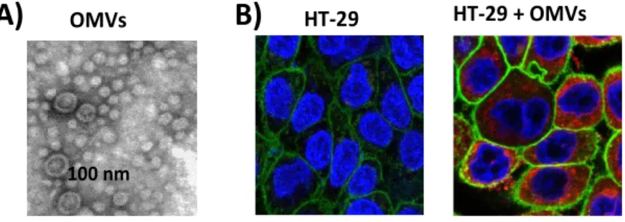
In the field of microbiota-derived vesicles, studies have been focused on OVMs released by the probiotic strain EcN and by commensal E. coli strains. Bacterial OMVs are isolated from culture supernatants and examined by transmission electron microscopy after negative staining (Fig. 1A).
5.1. Proteome of EcN OMVs
We approached proteomic analysis to identify the protein content of EcN OMVs [39]. This study was the first report of the vesicular proteome
of a probiotic strain. By means of 1D SDS–PAGE and highly sensitive LC–MS/MS analysis, 192 EcN vesicular proteins were identified with high
confidence in three independent experiments. In general, the identified proteins provide functions related with bacterial survival and interaction with the host, similarly to other bacterial vesicles [40]. Interestingly, of the 192 proteins, 18 were encoded by strain-linked genes and around 21% had not been previously described in bacterial vesicles. The EcN specific proteins are involved in the adhesion to the host tissues (fimbriae), immune modulation or bacterial survival in host niches (proteases, iron uptake
systems). Thus, these proteins may facilitate the colonization of the human gut. For instance, the presence of components of multiple iron uptake systems in EcN OMVs may allow the probiotic strain to outgrow commensal bacteria and compete with pathogenic strains that use similar siderophores for iron uptake. Concerning the identified proteases Pic and Sat, these proteins are typically associated to pathogens. Although the specific role in the probiotic strain has not been elucidated, it seems that they may act as fitness factors rather than virulence factors [41]. In this sense, Pic has been shown to promote intestinal colonization, hypersecretion of mucus and modulation of the host immune response [42].
In addition to the strain-related proteins, other identified proteins may contribute to the beneficial effects of this probiotic. These are proteins also found in OMVs isolated from Gram-negative pathogens (57 common proteins out of 192 total identified proteins). These common proteins may allow bacterial establishment and persistence in host tissues, which is an essential issue for the activity of both probiotic and pathogens. This common protein group contains fitness factors that allow bacterial survival in the host, including periplasmic proteins that help nutrient sensing (transport systems for amino acids, carbohydrates or inorganic ions), antimicrobial enzymes to kill competing bacteria (murein hydrolases), surface proteins that promote adhesion to host tissues, and factors able to modulate the host immune response (FlgE and FlgK). Interestingly, this common group is mainly formed by cytoplasmic proteins, most of them metabolic enzymes. They are moonlighting proteins that, depending on the cell location, can play different roles. Thus, in addition to the metabolic role, the secreted proteins play a function that contributes to gut colonization or modulation of the host immune response. A well-studied moonlighting protein is glyceraldehyde-3-phosphate dehydrogenase [43,44,45,46].
Regarding the proteins identified for the first time in bacterial vesicles, the in silico functional analysis revealed that they may also contribute to OMVs functions that are essential in the context of this probiotic strain. An
example is the porin NanC, whose expression is induced by
N-acetylneuraminic acid, one of the most abundant sialic acids on the
eukaryote cell membrane. The ability to use sialic acids as carbon and nitrogen sources may help EcN to colonize and persist in the intestinal tract. This proteomic study [39] revealed that EcN OMVs are equipped with a wide variety of proteins related with adhesion, immune modulation and bacterial survival in host niches. Therefore, these proteins may contribute to vesicle targeting to particular host tissues and mediate the probiotic beneficial effects on intestinal function.
5.2. Internalization of EcN OMVs by intestinal epithelial cells
Uptake of OMVs by epithelial host cells is mainly mediated by endocytosis. The endocytic pathway depends on the composition and cargo of the vesicles. The two main pathways involved in OMVs internalization are clathrin-mediated endocytosis and lipid raft-dependent endocytosis. These pathways create endosomal compartments with different surfaces that allow the delivery of vesicle components to various subcellular destinations.
Many studies dealing with internalization of bacterial OMVs have been carried out with vesicles from Gram-negative pathogens. However, regarding microbiota or probiotic-derived OMVs, contribution to this field is restricted to studies performed with the probiotic EcN [47]. In this study EcN OMVs were labelled with rhodamine isothiocyanate B-R18. Fluorescence of this fluorochrome is quenched when inserted in bilayer membranes at high concentration, but when vesicles are fused with the host cell, the fluorescence is emitted and can be monitored using a microplate fluorescence reader. In this case, the intensity of fluorescence emitted is proportional to the amount of internalized vesicles. Following this experimental approach, kinetics studies performed in several intestinal epithelial cells (Caco-2, HT-29, HT-29-MTX) incubated with rhodamine B-R18-labeled OMVs showed a time-dependent increase in fluorescence, consistent with internalization of EcN OMVs. The presence of intracellular vesicles was confirmed by confocal fluorescence microscopy (Fig. 1B).
fluorescence microscopy (Fig. 1B).
Figure 1. A) Image of EcN OMVs visualized by electron microscope after
negative staining. B) Visualization of internalized EcN OMVs in HT-29 epithelial cells by fluorescence microscopy. Cell membrane was stained in green and nuclei in
OMVs
100 nm
HT-29
HT-29 + OMVs
Figure 1. A) Image of EcN OMVs visualized by electron microscope after negative
staining. B) Visualization of internalized EcN OMVs in HT-29 epithelial cells by fluorescence microscopy. Cell membrane was stained in green and nuclei in blue. Internalized rhodamine B-R-18 labeled OMVs were visualized in red. (Images have been provided by Alexandra Cañas).
Experiments performed in the presence of specific endocytosis inhibitors allowed the identification of the pathway responsible for the uptake of EcN OMVs. The inhibitors tested were the cholesterol-sequestering drugs nystatin and filipin III that disrupt lipid rafts, and chlorpromazine that inhibit clathrin-mediated endocytosis. OMVs entry was inhibited by chlorpromazine but not by nystatin or filipin III. Therefore, EcN OMVs are internalized by intestinal epithelial cells via clathrin-mediated endocytosis. It is well known that vesicles internalized through this pathway are sorted to lysosomal compartments. Accordingly, co-localization of EcN OMVs with clathrin and specific markers of endosomes and lysosomes was confirmed by confocal fluorescence microscopy [47]. No detection of OMVs in nuclei or mitochondria was observed, thus EcN vesicles are likely degraded inside lysosomes. In the acidic endo-lysosomal compartments some specific proteins or factors are dissociated from the vesicles and targeted to other subcellular locations to trigger specific actions and cell responses [48]. Although for EcN OMVs, the specific function and intracellular trafficking of vesicle-derived factors to other organelles have not been studied so far, flow cytometry analysis of phosphorylated γH2AX in the nucleus of cells incubated with EcN OMVs showed that components released from internalized vesicles promote double strand breaks in the cell DNA. This effect on DNA was also
confirmed by the formation of DNA tails in the Comet assay. The vesicle-associated factor responsible for this genotoxic activity has not
been identified. Interestingly, the probiotic EcN produces colibacin, a non-ribosomal peptide-polyketide that induces double strand breaks in DNA. This peptide is synthesized by enzyme activities encoded in the pks island. Studies performed with an EcN mutant deficient in colibactin synthesis showed that, besides its genotoxic activity, colibactin is required for the in vivo anti-inflammatory effects of this probiotic [49]. Although the mechanism by which colibactin is delivered into the host cell remains unknown, the effects triggered by EcN OMVs on DNA suggest that colibactin could be delivered to mammalian cells by OMVs.
Despite EcN OMVs promote double strand breaks on the cell DNA, these vesicles do not reduce cell viability nor cause oxidative damage [47].
5.3. EcN OMVs reinforce the intestinal epithelial barrier
The intestinal epithelial layer forms a physical and biochemical barrier that maintains the segregation between host and intestinal microbiota. The function and integrity of this epithelial barrier depend on several factors, including the production of a mucin layer that covers the epithelial surface
and avoids the intimate contact with intestinal microbes, the secretion of antimicrobial peptides, and the formation of TJs that seal the paracellular space between adjacent epithelial cells. In addition, intestinal epithelial cells are essential to integrate microbial signals and coordinate the immune cell responses.
Recent studies have shown the ability of OMVs released by the probiotic EcN to reinforce the intestinal epithelial barrier by modulating the expression of genes encoding proteins relevant for the barrier function, particularly those involved in antimicrobial defense mechanisms [50] and the establishment of TJs [51].
Experiments performed with human colonic explants showed that EcN vesicles promote upregulation of the antimicrobial peptide hBD-2. Epithelial hBD-2 plays an important role in intestinal barrier function. Its expression is known to be induced by some probiotics, including EcN, as a mechanism involved in their beneficial action [52]. The induction of hBD-2 elicited by EcN OMVs might be attributed to flagella-associated proteins present in these vesicles [50]. In contrast, EcN OMVs do not significantly modify expression of β-defensin-1 (hBD-1), an antimicrobial peptide that acts as a component of host innate defenses at the intestinal surface. The
lack of vesicle-mediated effects on hBD-1 is consistent with the non-regulated, high constitutive expression of this antimicrobial peptide in
colon epithelial cells.
Regarding the modulation of mucus production, the probiotic EcN has been shown to upregulate the expression of several mucins in intestinal epithelial cell lines [53]. In the gut mucin-2 (MUC-2), which is secreted by globet cells, is of special relevance since it is the main component of the intestinal mucus layer. However, up-regulation of MUC-2 by EcN seems not to be mediated by released OMVs [50].
Another mechanism by which microbiota, and especially probiotic strains, help to strengthen the integrity of the intestinal barrier is through the regulation of TJ proteins. TJs are multiprotein complexes that regulate the paracellular trafficking of macromolecules. They seal the adjacent epithelial cells in a selective permeable form and determine the apical and basolateral parts of the membrane and thus, cell polarity. TJs are formed by four types of transmembrane proteins: occludin, claudins, junctional adhesion molecules, and tricellulin. TJs also contain proteins ZO-1, ZO-2 and ZO-3, which bind to claudins and act as scaffolds anchoring the TJ transmembrane proteins to the actin cytoskeleton. Association of the ZO-proteins to TJ multiprotein complexes is regulated by phosphorylation
mechanisms. Specific kinases control the subcellular distribution of the TJ proteins and by end the strength of the epithelial barrier. Claudins constitute a large family of TJ proteins that regulate cell permeability and cohesion. In humans, this family includes 27 members. Some claudins have a sealing function, while others act as selective channels for small charged molecules. The pore- forming protein claudin-2 belongs to this last group and greatly contributes to the transepithelial water secretion. In fact, stimuli that raise claudin-2 levels result in increased barrier permeability.
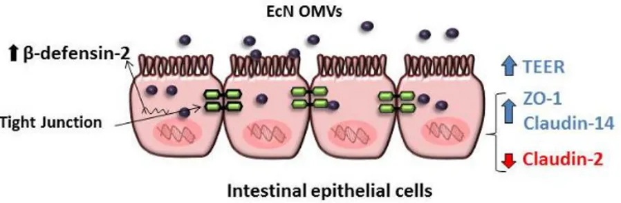
Several in vitro and in vivo studies performed with live EcN bacteria showed that this probiotic positively modulates the intestinal epithelial barrier through the positive regulation and redistribution of TJ proteins ZO-1, ZO-2 and claudin-14 [20,21,22]. Upregulation of claudin-14 was attributed to the secreted protein TcpC [22]. These studies, however, did not reveal whether extracellular vesicles could mediate these effects. In addition, the TcpC secretion mechanism was unknown. With this information different experimental approaches were set up to assess the ability of EcN OMVs to strengthen the epithelial barrier in different epithelial cell lines [50]. To distinguish between TcpC and vesicle effects, a TcpC deficient mutant was derived from EcN, and OMVs were isolated from both the wild-type and the mutant EcN strains. The effect of OMVs on the epithelial barrier was confirmed by measuring the increase in transepithelial electrical resistance (TER) of T-84 cell monolayers at 24 h post-incubation. The vesicle-reinforcement effect was independent of TcpC expression. The positive TcpC activity on the TER was associated with the soluble secreted fraction. These results prove that TcpC is not secreted by OMVs.
The regulatory role of EcN OMVs on the expression of TJ proteins was analyzed measuring the mRNA (RT-qPCR) and protein levels (Western blot and confocal fluorescence microscopy) of ZO-1, ZO-2 and claudin-14 (known to be regulated by the probiotic EcN), and also the leaky protein claudin-2. EcN OMVs promote up-regulation of ZO-1 and
claudin-14, and down-regulation of claudin-2. These effects are TcpC-independent (Fig. 2). Interestingly, soluble secreted TcpC also
positively regulates the expression of ZO-1 and claudin-14, but this protein has no effect on the transcriptional regulation of claudin-2. Concerning ZO-2, results showed that vesicles released by this probiotic do not regulate its expression. Thus, the EcN-mediated upregulation of ZO-2 does not rely on secreted factors but probably depends on bacteria-associated components.
Figure 2. Modulation of the intestinal epithelial barrier by EcN OMVs in an intact
epithelial barrier model: EcN OMVs reinforce the epithelial barrier through the increase of TER and positive regulation of the ZO-1 and claudin-14 proteins, as well as the negative regulation of claudin-2. In addition, EcN OMVs increase the expression of the antimicrobial peptide β-defensin-2.
Thus, contribution of EcN OMVs on the reinforcement of the intestinal epithelial barrier integrity, through several mechanisms, has been proved [51]. As stated, EcN OMVs directly modulate transcriptional regulation of TJ proteins, but also elicit additional responses that indirectly contribute to strengthen the epithelial barrier. In this sense, we have reported that OMVs released by this probiotic induce IL-22 expression in colonic explants [50]. This cytokine, mainly expressed by immune cells, targets epithelial cells and reinforces the intestinal barrier, thus limiting the access of microbial compounds and allergens to the systemic circulation.
5.4. Immunomodulatory role of EcN OMVs
As commented above, EcN is a good colonizer of the human gut and positively affects gastrointestinal homeostasis and microbiota balance. In addition to its beneficial effects on intestinal epithelial barrier, this probiotic modulates the host immune response towards an anti-inflammatory balance. The EcN-mediated effects have been mainly evidenced from a great number of in vitro and in vivo experiments performed with live probiotic suspensions [17,18,20,21,54]. Nonetheless, the bacterial factors that mediate these effects are not always known.
To define whether the immune modulation effects promoted by EcN are mediated through released OMVs, several in vitro and ex vivo cell models were set up to analyze the cytokine/chemokine response elicited by EcN OMVs in gut epithelial and immune cells [50].
(i) Direct stimulation of peripheral blood mononuclear cells (PBMCs) was used as an in vitro model of intestinal inflammation and barrier disruption. Stimulation of these immune cells with EcN OMVs activates the expression and secretion of IL-10, MIP1α, TNF-α, IL-6 and IL-8. In this model of inflamed barrier, bacterial lysates also trigger activation of these cytokines, even to a higher extent.
(ii) Apical stimulation of Caco-2/PBMCs co-cultures in Transwell permeable supports. This in vitro model that mimics intact intestinal barrier allows evaluating the crosstalk between OMVs, intestinal epithelial cells and the underlying immune cells [55,56]. In this model, OMVs are not in direct contact with the immune cells. Therefore, upon apical stimulation OMVs are internalized by epithelial cells, and signaling between epithelial and immune cells is produced through the release of soluble mediators. Apical stimulation with EcN OMVs results in the activation of immunomodulatory responses in the underlying immunocompetent cells, leading to increased secretion of IL-10, MIP1α, TNF-α, IL-6 and IL-8 in the basolateral compartment. In this model of intact intestinal barrier, bacterial lysates do not produce any activation effect. Parallel experiments performed in Caco-2 monolayers without underlying PBMCs showed that polarized Caco-2 cells are almost unresponsive to bacterial factors in the absence of crosstalk with immune cells. Overall results from this model indicate that released EcN OMVs can mediate the probiotic immunomodulatory effects in the intact intestinal mucosa. This mechanism may also apply to microbiota-derived vesicles.
(iii) Stimulation of human colonic explants, an ex vivo model closer to the
in vivo gut conditions. Results in this model confirmed the great
potential of EcN OMVs to modulate the immune response in intact intestinal mucosa, being the expression profile for the cytokines analyzed similar to that from the in vitro co-culture model. The genes encoding IL-10, MIP1α, TNF-α, IL-6 and IL-8 were upregulated in colonic explants incubated with EcN OMVs. In addition, vesicles from this probiotic diminished the expression of the pro-inflammatory cytokine IL-12.
The study on the immunomodulatory effects of EcN OMVs [50] proved the ability of probiotic vesicles to mediate signaling events to the immune system through the intestinal epithelial barrier. Thus, the beneficial effects of the probiotic EcN on gut homeostasis, especially modulation of the immune
response and barrier function, can be mediated by released OMVs. Our results with probiotic vesicles can be extrapolated to the microbiota-host crosstalk. Release of extracellular vesicles allows microbiota to communicate with intestinal mucosa cells, promoting the delivery of mediators that trigger host immune and defense responses.
6. OMVs in the treatment of intestinal inflammatory
diseases
Inflammatory bowel diseases (IBD) are chronic gut inflammatory disorders that have an increasing incidence worldwide, particularly ulcerative colitis (UC) and Crohn’s disease. They are multifactorial diseases that involve a dysregulated immune response against commensal gut microbes in genetically susceptible individuals with altered intestinal epithelial integrity [57]. Conventional IBD therapies including salicylates, corticoids, immunosuppressants and biological agents do not always yield good results [58]. As a common trait, IBD patients show an altered intestinal microbiota balance, a condition known as dysbiosis. For this reason several studies have evaluated the therapeutic effect of commensal and probiotic strains in clinical trials [59,60] or in animal models of colitis [35,37,61,62,63,64].
Concerning EcN, various clinical trials have evidenced its therapeutic benefits in inducing and maintaining remission of UC, showing similar anti-inflammatory effects as the standard treatment with aminosalicylate mesalazine [65,66]. Several mechanisms contribute to the therapeutic action of EcN against IBD, including the modulation of the host immune response toward an anti-inflammatory balance, the ability to reinforce the intestinal epithelial barrier [20,21,22], production of antimicrobial factors such as microcins and the induction of epithelial hBD-2 [18].
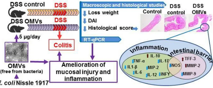
It has been reported that EcN OMVs could mediate the anti-inflammatory and barrier protection effects reported for this probiotic
in experimental colitis [67]. In this study, oral administration of EcN OMVs was evaluated in the dextran sodium sulphate (DSS)-induced
colitis mouse model. The experimental design included pre-treatment with EcN OMVs for ten days before DSS intake and a final five-day recovery period. The OMVs-treated group received EcN vesicles during all the experimental period (20 days). Administration of purified EcN OMVs significantly reduced DSS-induced weight loss and ameliorated clinical symptoms and histological damage. In addition, OMVs treatment counteracted the altered expression of cytokines and markers of intestinal
barrier function, thus proving the ability of EcN OMVs to ameliorate mucosal injury and gut inflammation.
Concerning microscopic analysis of colonic samples, the OMVs-treated mice showed preserved goblet cells replenished with their mucin content, and reduced mucosal infiltration areas, ulceration and oedema. Consistently, values for the colonic weight/length ratio were significantly smaller in this treated group than in the colitic control group. All these observations are consistent with an improvement in the mucosal barrier integrity in mice that received EcN OMVs.
The OMV-mediated anti-inflammatory effects were clearly evidenced by the improvement in the exacerbated immune response associated to colonic damage in DSS-treated mice. This treatment activates several cells of the innate immune response including epithelial cells, macrophages and dendritic cells, which leads to upregulated expression of pro-inflammatory cytokines such as IL-1β, TNF-α, and IL-6, thus triggering an imbalance in T-regulatory cells and Th1/Th17 cell responses. This regulatory network facilitates sustained inflammation through activation of other cytokines such as IFN-γ, IL-12 and IL-17 and also reduction of the anti-inflammatory cytokine IL-10 [68]. This altered cytokine expression profile was counteracted by EcN OMVs treatment by increasing IL-10 expression and diminishing expression of the anti-inflammatory cytokines [67].
One of the first events in intestinal inflammation is the impairment of the epithelial barrier function. This alteration enables the access of luminal antigens that trigger the exacerbated immune response. Therefore, promotion of mucosal healing is fundamental to prevent intestinal inflammation. One
mechanism used by EcN OMVs to protect the intestinal barrier in DSS-colitic mice is downregulation of MMP-9, a metalloprotease that
disrupt gut epithelial TJs, thus leading to increased intestinal permeability [69]. In addition, this treatment preserves expression of the MMP-2, which displays an opposite role to MMP-9 [70].
Another factor that contributes to mucosal barrier function is the protein trefoil factor 3 (TFF-3). In conditions of intestinal inflammation, such as DSS-induced colitis, expression of TFF-3 is downregulated [71]. Interestingly, EcN OMVs were able to restore the mRNA levels of TFF-3 to values similar to those of healthy mice. Thus, upregulation of this peptide by EcN OMVs contributes to epithelial protection and repair, preserving the cell structure of the colonic mucosa in DSS-treated mice [67]. It is known that TFF-3 reinforces TJs by promoting the redistribution of ZO-1 from the cytosol to the intercellular junctions of intestinal epithelial cells, where
strongly interacts with occludin, without altering ZO-1 expression [72]. Although EcN OMVs upregulate ZO-1 expression in several intestinal epithelial cell lines [51] they do not compensate the reduced ZO-1 expression in the DSS-experimental colitis model. This may reflect that EcN OMVs activate different regulatory mechanisms in the presence of highly expressed inflammatory mediators, including those that modulate ZO-1 location at the TJ structures, such as TFF-3. In addition to the beneficial effects on epithelial barrier repair, upregulation of TFF-3 may also contribute to ameliorate the inflammatory response in DSS-treated mice, since overexpression of this peptide abolishes the IL-1β mediated upregulation of certain pro-inflammatory cytokines [73].
Other markers of tissue damage and inflammation upregulated in DSS-induced colitis are cyclooxygenase-2 (COX-2) and inducible oxide
nitric synthase (iNOS). These enzymes confer protection against tissue injury, inflammation and infection and are highly expressed at damaged sites. Although iNOS upregulation is part of the intestinal antibacterial response that results on nitric oxide (NO) production, excess NO has been associated with intestinal inflammation in IBD patients [74], being the infiltrating macrophages in intestinal mucosa the main source of upregulated iNOS [75,76]. In these conditions, high levels of NO may cause tissue injury and the mucosal lesions observed in both human IBD and mouse experimental colitis. In DSS-treated mice, administration of EcN OMVs downregulates the altered expression of both enzymes, thus contributing to mucosal healing [67].
Oral administration of OMVs isolated from the probiotic EcN significantly reduces colonic damage in colitic mice [67]. These vesicles mediate anti-inflammatory and barrier protection effects, similarly to what has been reported for this probiotic in experimental colitis. The components of EcN OMVs responsible for these effects remain unknown. The polyketide colibactin could be one of these mediators. It is known that colibactin is required for the in vivo anti-inflammatory effects of EcN [49]. Moreover, in the DSS-induced colitis mouse model, administration of an EcN mutant deficient in colibactin synthesis results in exacerbated colitis. Although the
colibactin secretion mechanism has not been described so far, the anti-inflammatory effects of EcN OMVs in experimental colitis suggest that
colibactin could be delivered to the intestinal mucosa, at least in part, through released vesicles. However, the contribution of other specific probiotic OMV-associated factors to the modulation of mucosal healing markers cannot be ruled out.
Figure 3. Antiinflammatory and barrier protection effects of EcN OMVs in
DSS-induced experimental colitis in mice.
7. Concluding remarks
The influence of microbiota in human health is well known. Nowadays, microbiota imbalances (dysbiosis) have been associated with great variety of inflammatory and metabolic diseases. A therapeutic strategy to modulate microbiota composition is the administration of probiotics. This therapy is essentially an attempt to harness the beneficial effects of commensal microbiota. However, translation of probiotics or microbiota-based drugs to human healthcare requires knowledge of the molecular mechanisms involved in probiotic-host interactions. Moreover, although probiotics are Generally Regarded As Safe, some concerns about the potential risk associated with their use should be considered, especially in immunocompromised individuals and neonates.
Extracellular vesicles released by microbiota have a key role in bacteria-host cross-talk at the intestinal mucosa, as they act as a secretion and delivery pathway for selected bacterial proteins and active mediators directly to the host cells. Studies performed with the probiotic EcN prove the ability of the released vesicles to mediate signalling events to the immune system through the intestinal epithelial barrier, thus triggering appropriate host immune and defence responses. In addition, EcN OMVs mediates intestinal anti-inflammatory and barrier protective effects in the DSS experimental model of colitis.
Interestingly, isolated OMVs are free from bacteria and can elicit the same effect on gastrointestinal health as the probiotic itself. Thereby, EcN OMVs (either alone or in combination with secreted soluble factors) could represent a new therapeutic treatment with a high bio-safety level to
prevent and ameliorate inflammatory conditions associated with intestinal disorders. This potential formulation fits into the term postbiotic, a group of new potential treatments based on probiotic or microbiota products that is nowadays receiving great interest.
Acknowledgments
We thank Dr. F. J. Pérez-Cano for critical revision of the review.
References
1. Sánchez, B., Bressollier, P., Urdaci, M. C. 2008, FEMS Immunol. Med.
Microbiol., 54, 1.
2. Sánchez B, Urdaci MC, Margolles A. 2010, Microbiology. 156, 3232. 3. Moore, W. E., Holdeman, L.V. 1974, Am. J. Clin. Nutr., 27, 1450.
4. Tiihonen, K., Ouwehand, A. C., Rautonen, N. 2010, Ageing Res. Rev., 9, 107. 5. Scaldaferri, F., Gerardi, V., Lopetuso, L. R., Del Zompo, F., Mangiola, F.,
Boškoski, I., Bruno, G., Petito, V., Laterza, L., Cammarota, G., Gaetani, E., Sgambato, A., Gasbarrini, A.2013, Biomed. Res. Int., 2013, 435268.
6. O'Hara, A. M., Shanahan, F. 2006, EMBO reports, 7, 688. 7. Bevins, C. L., Salzman, N. H. 2011, Nat. Rev. Microbiol., 9, 356.
8. Ghoshal, U.C., Gwee, K. A., Holtmann, G., Li, Y., Park, S. L., Simadibrata, M., Sugano, K., Wu, K., Quigley, E. M. M., Cohen, H. 2018, J. Gastroenterol.
Hepatol., 33, 57.
9. Guinane, C. M., Cotter, P. D. 2013, Therap. Adv. Gastroenterol., 6, 295. 10. Scaldaferri, F., Gerardi, V., Mangiola, F., Lopetuso, L. R., Pizzoferrato, M.,
Petito, V., Papa, A., Stojanovic, J., Poscia, A., Cammarota, G., Gasbarrini, A. 2016, World J. Gastroenterol,. 22, 5505.
11. Cress, B.F., Linhardt, R.J., Koffas, M.A.G. 2013, Genome Announc., 1, e0004713.
12. Reister, M., Hoffmeier, K., Krezdorn, N., Rotter, B., Liang, C., Rund, S., Dandekar, T., Sonnenborn, U., Oelschlaeger, T.A. 2014, Biotechnol., 187, 106. 13. Vejborg, R. M., Friis, C., Hancock, V., Schembri, M. A., Klemm, P. Mol. Genet.
Genomics, 2010, 283, 469.
14. Grozdanov, L., Raasch, C., Schulze, J., Sonnenborn, U., Gottschalk, G., Hacker, J., Dobrindt, U. 2004, J. Bacteriol., 186, 5432
15. Sun, J., Gunzer, F., Westendorf, A. M., Buer, J., Scharfe, M., Jarek, M., Gosling, F., Blocker, H., Ping, A. Z. 2005, J. Biotechnol., 117, 147.
16. Lasaro, M. A., Salinger, N., Zhang, J., Wang, Y., Zhong, Z., Goulian, M., Zhu, J. 2009, Microbiol., 75, 246.
17. Sturm, A., Rilling, K., Baumgart, D. C., Gargas, K., Abou-Ghazalé, T., Raupach, B., Eckert, J., Schumann, R. R., Enders, C., Sonnenborn, U., Wiedenmann, B., Dignass, A. U. 2005, Infect. Immun., 73, 1452.
18. Schlee, M., Wehkamp, J., Altenhoefer, A., Oelschlaeger, T. A., Stange, E. F., Fellermann, K. 2007, Infect. Immun., 75, 2399.
19. Hafez, M., Hayes, K., Goldrick, M., Warhurst, G., Grencis, R., Roberts, I.S. 2009, Infect. Immun., 77, 2995.
20. Ukena, S.N., Singh, A., Dringenberg, U., Engelhardt, R., Seidler, U., Hansen, W., Bleich, A., Bruder, D., Franzke, A., Rogler, G., Suerbaum, S., Buer, J., Gunzer, F., Westendorf, A. M. 2007, PLoS ONE, 2, e1308.
21. Zyrek, A.A., Cichon, C., Helms, S., Enders, C., Sonnenborn, U., and Schmidt, M.A. 2007, Cell. Microbiol., 9, 804.
22. Hering, N., Richter, J.F., Fromm, A., Wieser, A., Hartmann, S., Gunzel, D., Bucker, R., Fromm, M., Schulzke, J.D., and Troeger, H. 2014, Mucosal
Immunol., 7, 369.
23. Sonnenborn, U., Schulze, J. 2009, Microb. Ecol. Health. Dis., 21, 122. 24. Ellis, T. N., Kuehn, M. J. 2010, Microbio. Mol. Biol. Rev., 74, 81. 25. Donoghue, E. J. O, Krachler, A. M. 2016, Cell Microbiol., 18, 1508. 26. Kuehn, M. J, Kesty, N. C. 2005, Genes Develop., 1999, 2645. 27. Kulp, A., Kuehn, M. J. 2010, Annu. Rev. Microbiol., 64, 163. 28. Schwechheimer, C., Kuehn, M. J. 2015, Nat. Rev. Microbiol., 13, 605. 29. Pathirana, R. D., Kaparakis-Liaskos, M. 2016, Cell. Microbiol., 18, 1518. 30. Kim, Y.S., Ho, S.B. 2010, Curr. Gastroenterol. Rep., 12, 319.
31. Schertzer, J. W., Whiteley, M. 2013, J. Mol. Microbiol. Biotechnol., 23, 118. 32. Chatterjee, D., Chaudhuri, K. 2013, J. Biol. Chem., 288, 4299.
33. Kaparakis-Liaskos, M., Ferrero, R. L. 2015. Nat. Rev. Immunol., 15, 375. 34. Olsen, I., Amano, A.J. 2015, Oral Microbiol., 7, 27468.
35. Shen, Y, Giardino Torchia M. L., Lawson, G. W., Karp, C. L., Ashwell, J. D., Mazmanian, S. K. 2012, Cell Host Microbe., 12, 509.
36. Stentz, R., Horn, N., Cross, K., Salt, L., Brearley, C., Livermore, D. M., Carding, S. R. 2015, J. Antimicrob. Chemother., 70, 701.
37. Kang, C. S., Ban, M., Choi, E. J., Moon, H. G., Jeon, J. S., Kim, D. K., Park, S. K., Jeon, S. G., Roh, T. Y., Myung, S. J., Gho, Y. S,, Kim, J. G., Kim, Y. K. 2013,
PLoS One, 8, e76520.
38. López, P., González-Rodríguez, I., Sánchez, B., Gueimonde, M., Margolles, A., Suárez, A. 2012, Vaccine, 30, 825.
39. Aguilera, L., Toloza, L., Giménez, R., Odena, A., Oliveira, E., Aguilar J., Badia, J., Baldoma, L. 2014, Proteomics, 14, 222.
40. Lee, E.Y., Choi, D. S., Kim, K. P., Gho, Y. S. 2008, Mass Spectrom. Rev., 27, 535.
41. Toloza, L., Giménez, R., Fábrega, M. J., Alvarez, C. S., Aguilera, L., Cañas, M. A., Martín-Venegas, R., Badia, J., Baldomà, L. 2015, BMC Microbiol., 15, 250. 42. Navarro-Garcia, F., Gutierrez-Jimenez, J., Garcia-Tovar, C., Castro, L. A.,
Salazar-Gonzalez, H., Cordova, V. 2010, Infect. Immun., 78, 4101.
43. Ferreira, E., Giménez, R., Cañas, M. A., Aguilera, L., Aguilar, J., Badia, J., Baldomà, L. 2015, Int. J. Biochem. Cell Biol., 60, 202.
44. Ferreira, E., Giménez, R., Aguilera, L., Guzmán, K., Aguilar, J., Badia, J., Baldomà, L. 2013, Res. Microbiol., 164, 145.
45. Aguilera, L., Ferreira, E., Giménez, R., Fernández, F. J., Taulés, M., Aguilar, J., Vega, M. C., Badia, J., Baldomà, L. 2012, Int. J. Biochem. Cell Biol., 44, 955. 46. Egea, L., Aguilera, L,, Giménez, R., Sorolla, M. A., Aguilar, J., Badía, J.,
Baldoma, L. 2007, Int. J. Biochem. Cell Biol., 39, 1190.
47. Cañas, M. A., Giménez, R., Fábrega, M. J., Toloza, L., Baldomà, L., Badia, J. 2016, PLoS ONE, 11, e0160374
48. Bielaszewska, M., Rüter, C., Kunsmann, L., Greune, L., Bauwens, A., Zhang, W., Kuczius, T, Kim, K. S., Mellmann, A., Schmidt, M. A., Karch, H. 2013, PLoS
Pathog., 9, e1003797.
49. Olier, M., Marcq, I., Salvador-Cartier, C., Secher, T., Dobrindt, U., Boury, M., Bacquié, V., Pénary, M., Gaultier, E., Nougayrède, J. P., Fioramonti, J., Oswald, E. 2012, Gut Microbes, 3, 501.
50. Fábrega, M. J., Aguilera, L., Giménez, R., Varela, E., Cañas M. A., Antolín, M., Badía, J., Baldomà, L. 2016, Front. Microbiol., 7, 705.
51. Alvarez, C. S., Badia, J., Bosch, M., Giménez, R., Baldomà, L. 2016, Front
Microbiol., 7, 1981.
52. Möndel, M., Schroeder, B. O., Zimmermann, K., Huber, H., Nuding, S., Beisner, J., Fellermann, K., Stange, E. F., Wehkamp, J. 2009, Mucosal Immunol., 2, 166. 53. Hafez, M. M. 2012, Probiotics Antimicrob. Proteins, 4, 67.
54. Arribas, B., Rodríguez-Cabezas, M. E., Camuesco, D., Comalada, M., Bailón, E., Utrilla, P., Nieto, A., Concha, A., Zarzuelo, A., Gálvez, J. 2009, Br. J.
Pharmacol., 157, 1024.
55. Fang, H. W., Fang, S. B., Chiang-Chiau, J. S., Yeung, C. Y., Chan, W. T., Jiang, C. B., Cheng, M. L., Lee, H. C. 2010, J. Med. Microbiol., 59, 573. 56. Pozo-Rubio, T., Mujico, J. R., Marcos, A., Puertollano, E., Nadal, I., Sanz, Y.,
Nova, E. 2011, Br. J. Nutr. 106, 1216.
57. Zhang, Y. Z., Li, Y. Y. 2014, World J. Gastroenterol., 20, 91. 58. Ko, J. K., Auyeung, K. K. 2014, Curr. Pharm., 20, 1082. 59. Fedorak, R. N. 2010, Gastroenterol. Hepatol., 6, 688.
60. Wasilewski, A., Zielinska, M., Storr, M., and Fichna, J. 2015, Inflamm. Bowel
Dis., 21, 1674.
61. Ewaschuk, J. B., Diaz, H., Meddings, L., Diederichs, B., Dmytrash, A., Backer, J., Looijer-van Langen, M., Madsen, K. L. 2008, Am. J. Physiol. Gastrointest. Liver
Physiol., 295, G1025.
62. Garrido-Mesa, N., Utrilla, P., Comalada, M., Zorrilla, P., Garrido-Mesa, J., Zarzuelo, A., Rodríguez-Cabezas, M. E, Gálvez, J. 2011, Biochem. Pharmacol. 82, 1891.
63. Martín, R., Miquel, S., Chain, F., Natividad, J. M., Jury, J., Lu, J., Sokol, H, Theodorou, V., Bercik, P., Verdu, E. F., Langella, P., Bermúdez-Humarán, L. G. 2015, BMC Microbiol. 15, 67.
64. Souza, B. M., Preisser, T. M., Pereira, V. B., Zurita-Turk, M., de Castro, C. P., da Cunha, V. P., de Oliveira, R. P., Gomes-Santos, A. C., de Faria, A. M., Machado, D. C., Chatel, J. M., Azevedo, V. A., Langella, P., Miyoshi, A. 2016,
65. Losurdo, G., Iannone, A., Contaldo, A., Ierardi, E., Di Leo, A., Principi, M. 2015, J. Gastrointestin. Liver Dis., 24, 499.
66. Floch, M. H., Walker, W.A., Madsen, K., Sanders, M. E., Macfarlane, G. T., Flint, H. J., Dieleman, L. A., Ringel, Y., Guandalini, S., Kelly, C. P., Brandt, L. J. 2011, J. Clin. Gastroenterol., 45, S168.
67. Fábrega, M. J., Rodríguez-Nogales, A., Garrido-Mesa, J., Algieri, F., Badía, J., Giménez, R., Gálvez, J., Baldomà, L. 2017, Front. Microbiol., 8,1274.
68. Neurath, M. F. 2014, Nat. Rev. Immunol., 14, 329.
69. Nighot, P., Al-Sadi, R., Rawat, M., Guo, S., Watterson, D.M., Ma, T. 2015,
Am. J. Physiol. Gastrointest. Liver Physiol., 309, G988.
70. Garg, P., Vijay-Kumar, M., Wang, L., Gewirtz, A. T., Merlin, D., Sitaraman, S. V. 2009, Am. J. Physiol. Gastrointest. Liver Physiol., 296, G175.
71. Lozano-Pérez, A. A., Ortiz-cullera, V., Zorrilla, P., Rodriguez-Cabezas, M. E., Cenis, J. L. 2014, Int. J. Nanomed., 9, 4507.
72. Buda, A., Jepson, M. A., Pignatelli, M. 2012, Cell Commun. Adhes., 19, 63. 73. Lin, N., Xu, L. F., Sun, M. 2013, Biochem. Biophys. Res. Commun., 440, 143. 74. Kolios, G., Valatas, V., Ward, S. G. 2004, Immunology, 113, 427.
75. Rachmilewitz, D., Stamler, J. S., Bachwich, D., Karmeli, F., Ackerman, Z., Podolsky, D. K. 1995, Gut, 36,718–723.
76. Palatka, K., Serfozo, Z., Veréb, Z., Hargitay, Z., Lontay, B., Erdodi, F., Bánfalvi, G., Nemes, Z., Udvardy, M., Altorjay, I. 2005, Scand. J.