Annals of theRheumatic Diseases 1996; 55: 30-33
Analysis
of
the association
between
chondrocalcinosis
and osteoarthritis:
a
community
based study
RaimonSanmarti, Eduardo Kanterewicz, Manel Pladevall, Domingo Pafiella, JoanBrugue's Tarradellas,JoseMufioz Gomez
Abstract
Objectives-To analyse the association between chondrocalcinosis and
osteo-arthritis (OA)ofthehands andknees inan
unselectedelderly rural population. Methods-A community based cross sectional study was performed in indi-vidualsrandomlyselected from aprevious epidemiologicalsurvey on the prevalence ofchondrocalcinosis in people older than
60 years from Osona county, Catalonia, northeastern Spain. Radiological OA (grade 2 or more of
Keligren's
classifica-tion) was evaluated in 26individuals with chondrocalcinosis and in 104 controls. A total of 18 articular areas of both knees (medialand lateraltibiofemoralcompart-ments) and hands (first, second and third metacarpophalangeal (MCP), first
carpo-metacarpal, trapezium-scaphoid, radio-carpal and distal radioulnar joints) were
studied.
Results-Radiological changes of OA in
theknees were more commonin subjects with chondrocalcinosis than in those withoutit, withanodds ratioadjustedfor
age and gender (aOR) of 4-3 (95% confi-dence interval (CI)
1P6
to 11-8, p=0005).OA was also more frequent in almost all
areas of the hands in individuals with chondrocalcinosis, though the difference reachedstatistical significance only in the MCP joints (aOR 3-1; 95% CI
1.1
to 8*8;p=0.033).
However, taking into accountthe side and the different joint compart-ments analysed, the association between chondrocalcinosisandOAwassignificant only in the lateral tibiofemoral compart-mentand the left MCPjoints.
Conclusions-Inanelderly population
un-selected for their rheumatic complaints,
there was a realassociation between OA and chondrocalcinosis. This association
was particularly relevant in the lateral tibiofemoralcompartmentofthe knee and
inthe first threeleft MCPjoints. (AnnRheum Dis1996; 55:30-33)
Chondrocalcinosis, defined by its
character-istic radiological calcifications that result
usually from the articulardepositionof calcium pyrophosphate dihydrate
crystals,'
has beenfound to affect from 8 to 10 percent of indi-viduals aged 60 and older.23 Since its first
description, a subchondral arthropathy resembling that of idiopathic osteoarthritis (OA) has been recognised in patients with chondrocalcinosis.4 5Some authors have found that, in addition to the radiological features, the distribution of joints affected in pyrophos-phate arthropathydiffers fromthatobserved in
primary OA;`8 however, other investigators havenotfoundimportantdifferences between thetwo arthropathies.9 Furthermore, it is not
unusualindaily practicetodiagnose,by radio-graphic examination, isolated chondro-calcinosis that lacks clinical
expression.3'4
Thusdiscrepancies remain regardingthe fre-quency andnature ofthe association between chondrocalcinosis and OA. Most investigators concludethat the frequencyof OA is increased in individuals with
chondrocalcinosis,2
8-13 whereasothers havenotconfirmed thisassoci-ation.'4
'5
However, the variable methodology and criteria used to evaluate the presence of OA, in addition to the different methods of selection of thepopulations studied, make the results of previously published work in-conclusive.Theaim of thisstudywas to investigate the association between chondrocalcinosis and osteoarthritis. Acaseseriesof chondrocalcino-sis was selected from a previous community basedcrosssectionalstudyand, asareference
group, a sample ofsubjects without chondro-calcinosis was assembled from the same study.3
Patients and methods
In 1993, an epidemiological survey to deter-mine theprevalence ofchondrocalcinosis was
performedin Osona,a ruralareaof Catalonia
(northeastern
Spain). The study methodology has been describedpreviously.3
Briefly, 31 family physicians each selected randomly 10 patients aged 60 years or older during a one weekperiod, regardlessof thepatients'reasons forseekingmedicalcare.Theparticipationratewas 84%, resulting in a study population of
261 individuals. A questionnairewith clinical anddemographic informationwascompleted,
in addition to knee (anteroposterior without
weight bearing) and hand-wrist(palmarview) radiographs. Onlythe presence orabsence of
chondrocalcinosis was evaluated. Twenty
seven individuals
(10%/)
had typical calcifi-cations (26 in the knees, 14 in the carpal triangular ligament, seven in othercarpal
Hospital Clinici Provincial de Barcelona, Servei de Reumatologia, Barcelona,Spain RSanmarti JMGomez Hospital General deVic, Serveide Medicina Interna(Unitat de Reumatologia), Vic(Barcelona),Spain
D Pafiella EKanterewicz JBTarradellas Unitat d'Epidemiologia Clinica MPladevall Correspondenceto: DrEduardoKanterewicz,
HospitalGeneral deVic, C/.Francesc Pla
'ELVigati' 1, 08500 Vic(Barcelona),
Spain.
Acceptedforpublication 31August1995 30
Associationbetween chondrocalcinosisand osteoarthritis
locations, and three in the metacarpophalan-gealjoints).3
In the present study, conducted two years afterthe aforementioned cross sectional study was completed, the original radiographs were examined for the presence of OA. Becauseone
of theradiographs was not available, 26 cases of chondrocalcinosis were included in our
study and 104 individuals without chondro-calcinosis wererandomly selected as controls. Ineach of the cases andcontrols, radiographic presenceofOA was evaluated for thefollowing joints: first, second, and third metacarpo-phalangeal, first carpometacarpal, trapezium-scaphoid, radiocarpal, and inferior radioulnar of bothhands, in addition to the medial and lateral tibiofemoral compartnents of both knees. Kellgren and Lawrence's16 graded criteriawereused to define thepresence of OA. Grade II(thefindingofanunequivocal
osteo-phyte) was considered OA, while grades III and IV were consideredtobe severeOA. The radiographs were read independently by two observers (RS, EK) whowere ignorant of the data from the questionnaires. If there was a discrepancybetween theconclusionsdrawn by the observersregarding any radiograph, itwas
re-examinedandaconsensusagreed.
STATISTICAL ANALYSIS
Interobserver agreement for each grade in Kellgren and Lawrence's classification criteria of OA was assessed using the weighted K statistic.'7 Odds ratios with 95% confidence intervals (CI) were used to evaluate the degree of the association between OA and chondro-calcinosis. The x2 test was usedto determine statistical significance. When an expected frequencyof less than five was found in one of the cells, Fischer's exact method was used as
thetest ofstatistical significance. Because age
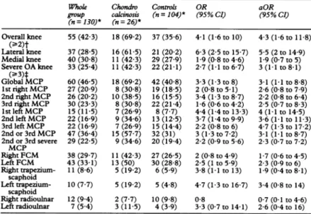
Table1 Presenceofosteoarthritis(OA)inthejointsanalysed:numberofpatients diagnosedwithOAin the whole group, distributionof OAamongpatientswith chondrocalcinosis and amongcontrols,and crude(OR)and age andgenderadjusted (aOR)odds ratios with95%confidenceintervals(CI)
Whole Chondro Controls OR aOR
group calcinosis (n=104)* (95%CI) (95%CI) (n=130)* (n=26)* Overall knee 55(42-3) 18(69 2) 37(35-6) 4-1(1-6to10) 4-3(1-6to11-8) (>2)t Lateralknee 37(28 5) 16(61-5) 21(202) 6-3(2-5to15-7) 5-5(2to14-9) Medialknee 40(30 8) 11(42 3) 29(27-9) 1-9(0-8to4-6) 1 9(07to5) Severe OAknee 33(25 4) 11(42 3) 22(21-1) 2-7(1l1to67) 3(11to8-1) (_-3)t Global MCP 60(46 5) 18(69-2) 42(408) 3-3(1-3to8) 3-1(11to 88) 1strightMCP 27(20 9) 8(30 8) 19(18-5) 2(0-8to5-1) 2-6(0-8 to7-9) 2ndrightMCP 26(20 2) 10(38-5) 16(15-5) 3-4(1-3 to 87) 2-2(0-8 to 6-4) 3rdrightMCP 30(23-3) 8(308) 22(21-4) 1-6(0-6 to 4 2) 2-5(0-7 to 8-3) 1stleftMCP 15(11-5) 7(26-9) 8(7-7) 4-4(1-4to13-3) 4(11 to14-5) 2ndleft MCP 22(16-9) 9(34 6) 13(12-5) 3-7(1-4 to9-9) 3-6(1l1to 11-3) 3rd left MCP 22(16-9) 7(26-9) 15(14-4) 2-2(0-8 to 6) 4-7 (1-3to17-2) 2nd or 3rd MCP 47(36-4) 15(57-7) 32(31) 3(1-3to 72) 3-1(11to8-7) 2nd or 3rd severe 29(22 5) 9(34-6) 20(19-4) 2-2(0-9 to 5 6) 2-3(0-7 to 7-2) MCP RightFCM 38(29-7) 11(42-3) 27(265) 2(0-8to49) 1-7(0-6to4-5) LeftFCM 43(33 1) 13(50) 30(28-8) 2-5(1to59) 2-3(0-9to6) Right trapezium- 11(8-6) 5(19 2) 6(5-9) 3-8(1-1to13) 1-9(04to8-1) scaphoid Lefttrapezium- 10(7 7) 5(19-2) 5(4 8) 4-7(1-3to16-7) 3-4(0-8to14) scaphoid Rightradioulnar 12(9 4) 2(7-7) 10(9 8) 0-8 0-7(0-1to 46) Leftradioulnar 7(5 4) 3(11-5) 4(3 9) 3-3(0-7to14-1) 2-6(0-4to16) *Valuesinparenthesesarepercentages of the value ofnfor thecolumn.
tGrade2orsuperioraccordingtoKeUlgren'scriteria;tgrade3orsuperioraccordingtoKeUgren's
criteria.
MCP=metacarpophalangeal;FCM=firstcarpometacarpal.
and gender are variables that influence the
frequency of chondrocalcinosis and OA, an
unconditional logistic regression model was
used to obtain estimates of the adjusted odds ratios(aOR) forageandgender. The Hosmer-Lemeshow test was used to test thegoodness
of fit of themodel;first orderinteractions were tested also.'8 A probability level of less than 005 wasconsideredstatistically significantfor all tests of significance in the analysis. All statistical calculations were performed using
theprogramSTATA.19
Results
Individuals with chondrocalcinosis (19 women/
sevenmen) had anaverage ageof 73 (SD 5-9) years, while that of the control group (50 women/54 men) was 67-7 (6 9) years
(p<0-001).Theproportionofindividualswho
consulted their primary care physician for rheumatic problems was similar among cases andcontrols (15 4%v 125-/5%).
The degree of agreement among the two observers in thediagnosisof OA washigh,with a weighted K estimatorgreater than 0-6 in all the joints studied, with the exception of the first leftmetacarpophalangeal, for whichKwas
0 53. The highest rate of agreement was
obtained for the third right metacarpophalan-geal joint (K082).
Table 1 shows thefrequency of OA in each of the joints studied, for both patients with chondrocalcinosis and controls, together with crude oddsratios (OR) and thoseadjusted for
ageandgender (aOR). Knee OA (radiographic changes of grade 2 or greater in one or more
of the fourcompartments analysed)wasmore
frequentlyfound in individuals with
chondro-calcinosis than in controls (aOR 4-3; 95% CI
1 6 to 118). Ifonly severeOA (grade3 or 4)
was considered, the association was weaker, although still significant (aOR 3; 95% CI 1-1 to8-1). When the association between OA and chondrocalcinosis was analysed separately for thetwotibiofemoral compartments,apositive significant association was observed for the lateral compartment (aOR 5-5; 95% CI 2 to
14.9), butnot themedial compartment (aOR
1 9;95% CI07 to5). Inaddition, the
involve-ment of the lateral compartment without involvement of the medial side was more
frequentin individuals withchondrocalcinosis than inthe controls (aOR4'8; 95% CI 1-3 to
17-4). When all the evaluatedcompartmentsof the kneewereincluded,anincreasedfrequency of OA was observed among those with chondrocalcinosis (table 2).
Table 2 Frequency ofkneeosteoarthritis(OA) in the medial andlateral compartments
Chondrocalinosis Controls Subjects 26 104 No ofcompartments 104 416 OA in any 38-5%(40/104) 15-8%(66/416) compartment OA in lateral 46%(24/52) 13%(27/208) compartment OA in medial 30%(16/52) 19%(39/208) compartment 31
Sanmarti, Kanterewicz, PladevaU,PanieUa,TarradeUlas, Gomez
Of the 104 femorotibial compartments
analysed in the individuals with chondro-calcinosis, 69 showed evidence of
chondro-calcinosis. Table 3 shows that OA was as
frequentin thosecomparunentswith
chondro-calcinosisasinthosewithout it.
Individuals withchondrocalcinosis hadmore
OA than the controls in almost all articular
areas of the hand (table 1). However, the
difference was statistically significant only for
the metacarpophalangeal joints.
Metacarpo-phalangeal OA (definedasthepresenceof OA
inatleastoneof the sixmetacarpophalangeals
studied in each case) was significantly more
frequent in subjects who had chondrocalcino-sisthan in those who did not (aOR3*1; 950/O CI 1H1 to 88). This association was also
evident when the first metacarpophalangeal joint of both hands was excluded-that is to
say, when the presence of OA was analysed
onlyfor thesecondorthird
metacarpophalan-geal (aOR 3-1; 95% CI 1 1 to 8-7). Both associations were also observed when the
relation between OA and chondrocalcinosis
was evaluated regarding the number of
meta-carpophalangeal joints analysed (data not
shown). Severe OA of the second or third
metacarpophalangeal jointswasmorefrequent amongpeoplewithchondrocalcinosis, though
the differencewasnotstatisticallysignificant.
Whenaseparateanalysiswasmade foreach
of themetacarpophalangeal joints,thepositive associationbetweenOAandchondrocalcinosis
waspresentonlyinthelefthand(table 1). OA
in the trapezium-scaphoid, first
carpometa-carpal and distal radioulnarjoints wasslightly more frequent in the group with
chondro-calcinosis,butthedifferenceswerenot signifi-cant. Only two subjects had radiocarpal OA; neitherofthetwohadchondrocalcinosis.
Discussion
Theresults ofthis study showthatindividuals
withchondrocalcinosishadmoreosteoarthritic
changes in their hands and knees than those without chondrocalcinosis. However, the
magnitude ofthe association between OA and
chondrocalcinosis varied depending on the
articular areas analysed. The association was
particularly stronginthelateral compartment of the knee, and in the first three
metacarpo-phalangeal jointsof the left hand.
The relation between OA and chondro-calcinosis has been studied previously. Inone
study of 338 biopsy specimens of the knee, chondrocalcinosis wassix timesmorefrequent
in osteoarthritic knees thaninthosethatwere
non-osteoarthritic, after adjusting for age and
gender.12Almost all theradiographicstudies7-11
Table3 Relatiownshipbetween thepresenceofosteoarthritis
(OA)andradiographicchondrocalcinosisinthe
femorotibialcompartmentsofsubjectswith chondrocalcinosis
(26subjects,104compartments)
Compartmentswith Compartmentswithout chonXdrcalcinosis chondrocalcinosis (n=69) (n =35)
OA 26(38%) 14(40%/6)
NoOA 43(62%) 21(60%)
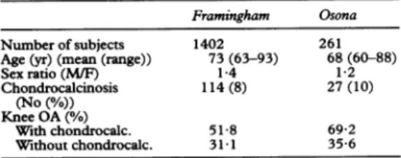
agree that OA is more frequent in those indi-viduals that have chondrocalcinosis than in those that do not.However,homogeneity does not exist between studies regarding the articular areas evaluated, the criteria used to define OA, or the statistical methods used. Moreover, these studies compared the frequency of OA between a group of patients withchondrocalcinosis (previously selected for their rheumatic complaints) with a non-rheumatic 'control' population without chondrocalcinosis-a circumstance that could result in abias of the realassociation between both entities. In this respect, we are aware of only one previous study on the association between OA and chondrocalcinosis of the knees, carried out in arepresentative sample of the general population of
Framingham.2
This large study included more than 1400 indi-viduals older than 63 years, and a weak but significant positive association was observed between the two diseases (relativerisk=1 5). Using the same diagnostic criteria and with a population having characteristics similar to those of theFramingham population (table 4), wehave alsofound anassociation between OA of the knee and chondrocalcinosis (aOR 4 3). Had we used the relative risk as the measure of association, the estimate of risk would have been similarto that of the Framingham study (relative risk of 2). Notably in our study, as in the Framinghamsurvey,2
the association betweenchondrocalcinosisand severe OAwasnot stronger than that observed between chondrocalcinosisand mild OA.
When we analysed the association of OA and chondrocalcinosis in both tibiofemoral compartments (not evaluated in the previous studies), we found that such association was stronger in the lateral compartment. This finding has not been observed in idiopathic OA, where the medialcompartment is affected commonly,and theisolated involvement of the lateralcompartment is
rare.20
Although Resnick et
al8
concluded that in patients with chondrocalcinosis the medial compartment was the area most affected by OA, two radiographic studies comparing the knees ofsubjectswith chondrocalcinosis with thoseofsubjects withidiopathicOA9
21showed that the involvement of the medialcompart-ment in patients with chondrocalcinosis was
less evident than that of patients with OA. Moreover, in one of the
studies21
osteophyteswere more frequently identified in the lateral compartment than in the medial side. More recently,in a large study ofknee radiographic
patterns
of OApatients,itwas found that the involvement of thelateraltibiofemoral side hadTable4 Comparison ofpopulationcharacteristicsof
OsonaandFramingham
Framingham Osona
Numberofsubjects 1402 261
Age(yr)(mean (range)) 73(63-93) 68(60-88)
Sexratio(M/F) 1-4 1i2 Chondrocalcinosis 114(8) 27(10) (No(%/)) KneeOA(%/6) Withchondrocalc. 518 69-2 Withoutchondrocalc. 31-1 35-6 32
Association betweenchondrocalcinosis and osteoarthnitis
a greater degree of association with
chondro-calcinosis.22
This is the first study in which the
asso-ciationbetween OA and chondrocalcinosishas
been analysed in several articular areas ofthe hand in an unselected population. As in
previousstudies,6-9wehavefoundastatistically significant association between
metacarpo-phalangeal OA and chondrocalcinosis,
es-pecially for the second and third joints. The
fact that the association was significant only when the lefthandwasanalysed iscuriousand unexpected, although this finding could be explained by the population included in our
study. Metacarpophalangeal OA is frequent in
the rural Spanish population,23 and is most
probably related to repetitive manual labour.23 24 As this type of lesion is
predomi-nant inthe right hand,2325 the degree of asso-ciation between OA and chondrocalcinosis could be masked by this phenomenon and therefore be less evident than in theleftor
non-dominant hand.
The present study has several limitations. First, as the 'cases' were pre-existing we can
establish anassociation, but cannotbe certain aboutitsdirectionality: that is tosay, it is not
clear whether the pyrophosphate deposits are
the predisposing factor that triggers the articular lesion, or the consequence of the degenerating process in the articular cartilage.26 Second, lateral knee radiographs were nottaken. Theseprojectionsarerequired to evaluate the patellofemoral compartment
that is saidtobeaffected, characteristicallyand
in isolation, in chondrocalcinosis,8 21 though this was not confirmed in a recent study.27 Third, analysis of the femorotibial
compart-ments without radiographs during weight bearing makes it more difficult to detect definite joint space narrowing; however, evaluation of the less involved compartment
is problematic in the weight bearing
pro-jection.20
27Webelieve,nevertheless, that these limitations have not influenced the nature of the results ofourstudy. The absence of radio-carpalOA in thepresentpatients is somewhat surprising, especially as Resnick eta128 found ittobecharacteristic of chondrocalcinosis. Our findingsmaybe explained by the fact thatthe presenceoftypicalosteophyteswasrequired inourdefinition ofOA,whilejointspace
narrow-ing and subchondral sclerosis are the most
frequent radiological signsofradiocarpal OA.28 As Felson et
a12
suggested, an overdiagnosis of OA in subjects with chondrocalcinosis (information bias) cannot be discarded; however, as the K estimator was high and theobservers assessed thejointsindependently,we
believe that information bias was unlikely inr
this study. Finally, the sample size was
relatively small, thus there was insufficient
powertostudytheassociationin severaljoints
orin
subgroups
ofsubjects.We conclude that there was an association between osteoarthritis and chondrocalcinosis
in an elderly population sample selected
in-dependently
of rheumatic complaints. Peoplewith chondrocalcinosis had a greater preva-lence ofosteoarthritis in the knees, especially
in the lateral compartment. Inaddition, osteo-arthritis in the first three
metacarpophalangeal
joints of the left or non-dominant hand was more frequent in people with chondrocalcino-sis than in those without it.
We dedicate this article to the late Dr MariaAntonia Branc6s.
1 Resnick D, Niwayama G, eds. Calcium pyrophosphate
dihydrate(CPPD) crystal depositiondisease.Diagnosisof
bone andjoint disorders, 2nd edn. Philadelphia: W B
Saunders, 1988; 1673-732.
2 Felson D T, Anderson J J, Naimark A, Kannel W,
Meenan R F. Theprevalenceof chondrocalcinosis in the
elderly and its association with knee osteoarthritis: the
Framingham study.JRheumatol1989;16:1241-5. 3 Sanmarti R,PaflellaD,Brancos MA,CanelaJ,ColladoA,
Brugues J. Prevalence of articular chondrocalcinosis in
elderlysubjectsinaruralareaof Catalonia. Ann Rheum
Dis1993;52:418-22.
4 McCarty D J. Calcium pyrophosphate dihydrate crystal deposition disease-1975. Arthritis Rheum 1976; 19
(suppl):275-86.
5 ZimanD, Sit'aj S. Chondrocalcinosis articularis. Clinical and radiological study. Ann Rheum Dis 1963; 22: 142-52.
6 MartelW,ChampionCK, Thompson G R, CarterT L. A roentgenologically distinctive arthropathy in some
patientswith thepseudogoutsyndrome.AmJfRoentgenol 1970;109: 587-605.
7Martel W, McCarter D K, Solsky M A, et al. Further observations on the arthropathy of calcium pyro-phosphate crystal deposition disease. Radiology 1981; 141: 1-15.
8 Resnick D, Niwayama G, GoergenT G, etal. Clinical,
radiographic and pathologic abnormalities in calcium
pyrophosphate dihydrate deposition disease (CPPD): pseudogout. Radiology1977; 122: 1-15.
9 RiestraJ L,SanchezA,Rodriguez-ValverdeV, CastilloE,
Calderon J. Roentgenographic features of the
arthro-pathy associated withCPPD crystal deposition disease.
A comparative study with primary osteoarthritis.
JfRheumatol1985;12:1154-8.
10Wilkins E, Dieppe P, Maddison P, Evison G.Osteoarthritis and articular chondrocalcinosis in the elderly. Ann Rheum Dis 1983; 42: 280-4.
11 Gordon T P, Smith M, Ebert B, McCredie M, Brooks P M. Articularchondrocalcinosis inahospital population: an Australian experience. Aust N ZJfMed 1984; 14: 655-9.
12Sokoloff L, Varma A A. Chondrocalcinosis in surgically resectedjoint.Arthritis Rheum 1988; 31: 750-6. 13 Bensasson M,Dorfmann H, Perez-Busquier M, et al. Etude
radiographique de la main dans 50 cas de chondro-calcinose articular primitive avec une serie de 100 temoins.Rev RhumMalOsteoartic 1975; 42: 3-11. 14Leonard A, Solnica J, Cauvin M, et al. La chondrocalcinose:
etude de sa frequence radiologique et de ses rapports avec l'arthrose. Etude du taux de la parathormone. Rev Rhum MalOsteoartic1977; 44: 559-64.
15 Hochberg M C. Chondrocalcinosis articularis of the knee: prevalenceand association with osteoarthritis of the knee. Arthritis Rheum 1984; 27: S49.
16 The epidemiology ofchronic rheumatism. Atlas of standard radiographs, Vol 2. Oxford: Blackwell Scientific, 1963. 17Fleiss JL. Statistical methods for rates and proportions, 2nd
edn. New York:WileyandSons,1981.
18Hosmer D W, Lemeshow S.Applied logisticregression. New York:Wileyand Sons, 1989.
19StataReferenceManual. Release 3, 5th edn. SantaMonica, CA, USA: Computing Resource Center, 1992. 20Thomas R H, Resnick D, Alazraki N P, Daniel D,
GreenfieldR. Compartmental evaluation of osteoarthritis ofthe knee. Radiology 1975; 116:585-94.
21 Hansen S E, Heming M. A comparative study of radiographic changes in knee joint in chondrocalcino-sis, osteoarthrosis and rheumatoid arthritis. Scand J Rheumatol 1984; 13: 85-92.
22Ledingham J, Regan M, Jones A, et al. Radiographic
patternsandassociationsofosteoarthritis of the knees in patients referred to hospital. Ann RheumDis 1993; 52: 520-6.
21TorneroMolina J, Diez Andres M L, Vidal Fuentes J. La artrosis de las articulaciones metacarpofalangicas es un
hallazgo habituel en los trabajadores manuales de la
Espafiarural. Rev Esp Reum1992;19:430-5.
24Williams W V, Cope R, Gaunt W D, et al. Meta-carpophalangeal arthropathy associated with manual labor (Missouri metacarpalsyndrome). ArthritisRheum
1987;30: 1362-71.
25 GenizF, Cisnal del Mazo A, Caracuel M A,MartinezF G. Artrosis de articulaciones metacarpofalangicas. Estudio clinico de 35pacientes. Rev Esp Reum 1993; 20: 9-12. 26 Schumacher H R, Gibilisco P, Reginato A, et al.
Impli-cationsof crystal deposition inosteoarthritis. J Rheumatol 1983; 10 (suppl 9):40-1.
27 DohertyM, Dieppe P, Watt I. Pyrophosphate arthropathy: Aprospectivestudy.BrJRheumatol1993; 32: 189-96. 28 Resnick D, Utsinger P D. The wrist arthropathy of
'pseudogout' occurring with and without chondro-calcinosis.Radiology1974; 113: 633-41.
doi: 10.1136/ard.55.1.30
1996 55: 30-33
Ann Rheum Dis
R Sanmarti, E Kanterewicz, M Pladevall, et al.
community based study.
chondrocalcinosis and osteoarthritis: a
Analysis of the association between
http://ard.bmj.com/content/55/1/30
Updated information and services can be found at:
These include:
References
http://ard.bmj.com/content/55/1/30#related-urls
Article cited in:
service Email alerting
the box at the top right corner of the online article.
Receive free email alerts when new articles cite this article. Sign up in
Notes
http://group.bmj.com/group/rights-licensing/permissions
To request permissions go to:
http://journals.bmj.com/cgi/reprintform
To order reprints go to:
http://group.bmj.com/subscribe/