Tutor
PhD Student
Prof. Bice Avallone
Dr. Monica Tizzano
Academic Year 2017/2018
UNIVERSITY OF NAPLES FEDERICO IIGenetic Dissection Of Hoxb1 Function In
The Developing Mouse Auditory And
Vestibular System Rhombomere 4-Derived
DEPARTMENT OF BIOLOGY
PhD in Biology
XXXI cycle
Index
Abstract ... pag.3 Riassunto ... pag.5 Chapter 1: Introduction ... pag.10 1.1 Inner ear ... pag.11 1.2 Cochlea ... pag.11 1.3 Vestibular system ... pag.18 1.4 Hox genes ... pag.26 1.5 Structure and expression of the Hoxb1 gene ... pag.27 1.6 Rhombomere 4 and Hox genes ... pag.28 Chapter 2: Aim of the research ... pag.31 Chapter 3: Materials and Methods ... pag.37. 3.1 Generation of Hox mouse mutant lines and matings ... pag.38 3.2 Auditory brainstem response (ABR) ... pag.38 3.3 Tissue preparation ... pag.40 3.4 Transmission electron microscopy (TEM) ... pag.40 3.5 Scanning electron microscopy (SEM)... pag.41 3.6 Immunohistochemistry ... pag.41 Chapter 4: Results: Auditory system ... pag.42
4.1 Auditory response of the mutant mice in sensory domain of
rhombomere 4 ... pag.43 4.2 Localization of YFP in Hoxb1 mutants’ cochleae ... pag.45 4.3 Ultrastructural analysis of hair cell morphology in Hoxb1
mutant mice ... pag.49 4.4 Innervation of medial olivocochlear neurons in Hoxb1 mutant
mice ... pag.51 Chapter 5: Results: Vestibular system ... pag.53
5.1 Innervation of vestibular efferent neurons in 3 months old Hoxb1
mutant mice ... pag.54 5.2 Innervation of vestibular efferent neurons in P8 Hoxb1mutant
mice ... pag.59 Chapter 6: Discussion and conclusion ... pag.61 6.1 Auditory system ... pag.62 6.2 Vestibular system ... pag.65
Chapter 7: References ... pag.67 Chapter 8: Abbreviations ... pag.74
Abstract
Rhombomere (r4) and Hox associated genes Hoxb1 and Hoxb2 contribute to the formation of specific auditory and vestibular subcircuits. In particular, sensory and motor components of the sound transmission pathway, sensorimotor reflex circuits, as well as the hindlimb vestibule-spinal reflex, derive from r4 and are strongly affected in Hoxb1 mutants (Di Bonito
et al., 2013). Inner ear efferent (IEE) neurons also originate from r4 and form vestibular
(VEN) and cochlear (CEN) efferent neuron subpopulations. The CEN is further subdivided into medial (MOC) and lateral (LOC) olivo-cochlear motoneurons. MOC neurons inhibit the motility of outer hair cells (OHCs), which amplify low intensity sounds, while LOCs innervate the afferent terminations on the inner hair cells (IHC) modulating the excitability of the cochlear nerve, thus protecting the cochlea from acoustic damage.
Hoxb1null mutants lack MOC and LOC efferent neurons leading to defects in OHCs and in cochlear amplification, and mice have increasing auditory thresholds (Di Bonito et al., 2013). Our hypothesis is that MOC neuron endings play a trophic function on OHCs and that the physical interaction between MOC efferents and OHCs is essential for proper maturation and functioning of OHCs. To exclude a possible contribute of sensory auditory neurons at this phenotype, we performed analysis of conditional Hoxb1 mutants (Ptf1acre Hoxb1 flox/flox, Atoh1cre Hoxb1 flox/flox) where Hoxb1 expression were eliminated in the dorsal region of rhombomere 4 and the sensory structures involved in the acoustic pathway, were selective affected. Our data show that when the ventral domain, where the motoneurons MOC and LOC develop, normally expresses Hoxb1 the OHCs show a regular morphology, even if Hoxb1 expression is affected in the dorsal sensory auditory neurons. So, our data suggest that MOCs could play an important postnatal role but to confirm this hypothesis further
investigations are needed using a Cre specific line that selectively eliminate the Hoxb1 expression in the ventral domain of motoneurons.
Regarding the vestibular system, Hoxb1null mutant mice also fail to form the VEN at early developmental stages (Di Bonito et al., 2015). However, transmission electron microscopy (TEM) in adult mice reveals the presence of both afferent and efferent neuronal endings on receptor cells. To understand whether projections are missing at birth and new connections gradually appear during the first month by compensatory plasticity mechanisms, we analyzed newborn mutant pups for the presence of efferent endings on hair cells by TEM. Our analysis showed that this mutant fail to develop efferent terminations corroborating our hypothesis that these endings derive from other district and could act as a compensatory mechanism. So, to further confirm this hypothesis we aim to use retrograde labelling in 3-month old mutant mice to assess their eventual presence and identify their origin.
Riassunto
Il romboencefalo nei mammiferi comprende cervelletto, ponte e bulbo. Durante le prime fasi embrionali il romboencefalo è suddiviso lungo l’asse antero-posteriore (AP) in unità segmentali ripetute dette rombomeri, ognuna delle quali è nettamente separata dall’unità adiacente e darà origine, durante le successive fasi di sviluppo, a specifiche e differenti linee cellulari. Grazie alla presenza di segnali induttivi e schemi di trascrizione specifici per i diversi rombomeri i progenitori neurali avranno un differente destino e target assonale in base alla loro relativa posizione lungo gli assi antero-posteriore (AP) e dorso-ventrale (DV). I geni Hox sono necessari per il corretto conferimento dell’identità AP durante la segmentazione del rombencefalo e contribuiscono anche a regolare la specificazione dei sottotipi neuronali lungo l'asse DV. Nella zona rostrale del romboencefalo i geni Hox dei gruppi paraloghi da 1 a 4 presentano schemi di espressione sovrapposti, tranne che nel rombomero 1 (r1). In particolare, Hoxb1 è espresso fino al confine tra i rombomeri 3 e 4 (r3/r4) nelle cellule progenitrici durante lo sviluppo precoce del romboencefalo. Una volta che i rombomeri sono pienamente formati, l’espressione di Hoxb1 viene mantenuta soltanto in r4 da un meccanismo di autoregolazione e dalla espressione di Hoxb2 (Di Bonito et al.,
2013).
Di particolare rilievo risulta essere la formazione dei sistemi uditivo e vestibolare che originano da r4 sotto il diretto controllo di Hoxb1. Nel rombomero 4 hanno origine i corpi assonali delle efferenti otiche a partire dai neuroni IEE (inner ear efferent) che al giorno E13.5 migrano dorso-ventralmente e, a partire dal giorno E14.5, si separano in una sottopopolazione vestibolare (VEN) e una cocleare (CEN). Nella regione ventrale del
rombomero 4 quindi si differenziano i motoneuroni efferenti olivo-cocleari (OC) e i motoneuroni efferenti vestibolari. I motoneuroni OC modulano le informazioni uditive sensoriali afferenti in entrata, proteggendo così le cellule ciliate sensoriali da danni dovuti ad eccessiva stimolazione.
Il percorso uditivo centrale è formato da proiezioni sensoriali che trasmettono informazioni ascendenti acustiche e proiezioni efferenti che modulano le risposte primarie afferenti. Le cellule ciliate interne ed esterne nell'organo di Corti della coclea reagiscono entrambe agli stimoli sonori, ma mentre le cellule ciliate interne (IHC) sono i veri sensori degli stimoli uditivi, le cellule ciliate esterne (OHC) mediano il controllo efferente e amplificano suoni a bassa intensità aumentando l'ampiezza e la selettività di frequenza delle vibrazioni della membrana basilare attraverso il processo di amplificazione cocleare (Fettiplace e Hackney,
2006, Dallos, 2008).
La corretta funzione uditiva dipende dal controllo efferente delle IHC da parte dei motoneuroni olivocochleari laterali (LOC). I motoneuroni LOC innervano i terminali sinaptici dei neuroni sensoriali afferenti sulle IHC, modulando l'eccitabilità del nervo cocleare e proteggendo la coclea da lesioni acustiche (Simmons, 2002; Darrow et al., 2007). Il controllo efferente delle OHC è mediato dai motoneuroni olivocochleari mediali (MOC). I MOC sono innervati a loro volta dai neuroni del nucleo cocleare postero-ventrale (PVCN) (Brown MC and Levine J L. 2008; de Venecia et al., 2005) e modulano "l'amplificazione cochleare" attraverso il riflesso MOC inibendo la vibrazione delle OHC (Guinan, 2006). È nota la necessità di una corretta interazione tra i motoneuroni efferenti cocleari ed i loro bersagli postsinaptici già durante le prime fasi di vita postnatale al fine di garantire il normale sviluppo e funzionalità della coclea (Walsh et al., 1998; Simmons, 2002; Di Bonito et al.,
2013). È stato dimostrato come in mancanza di Hoxb1 nei topi mutanti vengono ad essere
perse le terminazioni efferenti LOC e MOC oltre ad una degenerazione delle OHC, il che porta ad una perdita della trasmissione degli stimoli a bassa intensità e conseguenti danni uditivi (Di Bonito et al., 2013). La degenerazione delle OHC e, di conseguenza, l'amplificazione cocleare anomala, potrebbero essere causate dall'assenza di stimolazione sinaptica/trofica delle OHC da parte delle fibre MOC efferenti durante un periodo critico postnatale, cruciale per la maturazione delle OHC (Walsh et al., 1998).
Per quanto riguarda il sistema vestibolare è noto che esso deriva in parte da r4 e consiste in una serie di nuclei sensoriali che integrano e rilasciano informazioni per il coordinamento dei movimenti degli occhi, dell'equilibrio e della postura. È stato dimostrato come il tratto spinale vestibolo laterale (LVST) ed il gruppo di neuroni vestibolari efferenti (VEN) vengano ad essere persi nei topi mutanti Hoxb1. L'analisi di marcatura retrograda sugli embrioni mutanti Hoxb1 e sui postnatali ha confermato la specifica assenza del LVST e dei neuroni efferenti vestibolari (VEN), in accordo con la perdita dell'identità r4 e della produzione ectopica dei neuroni r3 (Di Bonito et al., 2015).
Il mio obiettivo è stato quindi quello di andare a studiare in maniera approfondita le conseguenze della perdita di Hoxb1 nei sistemi uditivo e vestibolare.
Per quanto concerne il sistema uditivo siamo andati a valutare la reale funzione delle fibre efferenti MOC nel corretto sviluppo delle OHC, la cui degenerazione porta alla perdita di udito nei mutanti. A tale scopo abbiamo creato una serie di linee mutanti nelle quali il gene Hoxb1 viene selettivamente inattivato nel dominio sensoriale, lasciando inalterato il dominio
motorio da cui derivano le fibre efferenti MOC e LOC ed escludere un eventuale contributo di questo dominio al severo fenotipo :
•R4cre/+ YFP/+
•R4cre/+ Hoxb1null/null YFP/+ •Atoh1cre/+ YFP/+
•Atoh1cre/+ Hoxb1null/null YFP/+ •Ptf1cre/+ YFP/+
•Ptf1cre/+ Hoxb1null/null YFP/+
Su questi campioni ho effettuato analisi di:
•Microscopia elettronica a trasmissione: al fine di valutare lo stato delle terminazioni efferenti MOC e LOC.
•Microscopia elettronica a scansione: al fine di valutare lo stato delle OHC. •Test ABR: al fine di valutare la corretta funzionalità uditiva.
•Immunoistochimica: al fine di valutare eventuali differenze nelle cellule sensoriali del Corti
I miei risultati hanno messo in luce che i mutanti nel dominio sensoriale non mostrano differenze nei threshold uditivi, la morfologia delle cellule si mantiene inalterata rispetto ai WT e controlli e le terminazioni MOC sono presenti senza nessuna variazione di aspetto e numero. Quindi è plausibile ipotizzare che i motoneuroni MOC potrebbero svolgere un importante ruolo postnatale, ma per confermare questa ipotesi saranno necessarie ulteriori
indagini usando una linea specifica Cre che elimini selettivamente l'espressione di Hoxb1 nel dominio ventrale dei motoneuroni.
Passando al sistema vestibolare è stato dimostrato che in assenza di Hoxb1 vengono persi sia i neuroni vestibolari efferenti (VEN) sia il tratto vestibolo spinale laterale (LVST). I miei esperimenti di microscopia elettronica a trasmissione hanno mostrato invece la presenza di terminazioni efferenti agli organi vestibolari quali ampolla, sacculo e utricolo in mutanti Hoxb1 di 3 mesi. L’ipotesi è che queste terminazioni si stabiliscano come una sorta di compensazione per la perdita di quelle che derivano dal VEN. Abbiamo quindi pensato di analizzare mutanti a P8 per capire se queste terminazioni sono già presenti alla nascita o si stabiliscono in un secondo momento. Dai dati ottenuti è emerso che queste risultano assenti alla nascita, ma per poter capire l’esatta provenienza saranno effettuati esperimenti di marcatura retrograda (Dil) in mutanti a 3 mesi.
1.1 Inner ear
The inner ear is the innermost part of the vertebrate ear that derives from the otic placode. The development and morphogenesis of the inner ear starts with the formation of an ectodermal thickening, the otic placode, that arise on the surface at the level of the hindbrain through temporally and spatially coordinated gene expression. Each placode invaginates, separates from the surface, and gives rise to a small saccule called the otocyst. With continued growth, it develops into the membranous labyrinth of the inner ear. Then, coiling of the otocyst, accompanied by a series of complex constrictions and outgrowths, gives rise to the saccule, utricle, three semicircular canals, and cochlea. For this reason, the inner ear can be functionally divided in two main parts: the auditory organ, which contains the coiled cochlea that senses sound, and the vestibular system, organ of equilibrium consisting of semicircular channels, saccule and utricle. (S. Chatterjee et al., 2010).
1.2 Cochlea
In mice the cochlea makes 2.5 turns around the modiolo, a central bone pyramid, and is about 5-6 mm long; it is divided into three compartments (Fig. 1): The cochlear structures include:
• the vestibular duct or scala vestibuli (containing perilymph), • the tympanic duct or scala tympani (containing perilymph), • the cochlear duct or scala media (containing endolymph).
The cochlear duct divides the cochlea longitudinally. The wall of the membranous labyrinth that delimits the vestibular scale is a thin membrane (the Reissner or vestibular membrane) that separates the cochlear duct and the vestibular duct. Between the scala media and the
scala tympani there is a structure the organ of Corti, the receptive organ of the inner ear. The gelatinous tectorial membrane of the organ of Corti is produced by the support cells, covers the apical stereocilia of the inner and outer hair cells, and is surrounded by the endolymph of the cochlear duct.
Fig1. Cochlea.
Source:
https://anatomyofdiagram.com/organ-of-corti-anatomy/organ-of-corti-anatomy-inner-ear
Organ of Corti. Lying on the basilar membrane, it is composed of hair cells, which have a
complex organization, with several types of columnar supporting cells. Hair cells are arranged segmentally in two groups on the sides of an inner tunnel. They represent the sensory cells of the cochlea and are arranged in a group of inner hair cells (IHC) and one of outer hair cells (OHC) (Fig.2).
IHCs are pear shaped cells with a round centrally located nucleus. One row of inner hair cells runs along the cochlear duct. The cells are in contact with the inner pillar cell and the phalangeal cells (Y. Raphael and R. A. Altschuler, 2003).
OHCs instead are more cylindrical, the nucleus is round and located in the basal portion of the cell. The apical domain includes the stereocilia while the basal end rests on the Deiters cells. There are three rows of OHCs, but in some cases a fourth and even fifth row are found, especially in the apical turn of mammals specialized in low frequency hearing. Both types of hair cells have numerous apical stereocilia that allow the detection of the vibrations of the endolymph produced by the perceived sound waves through the movements of the basilar membrane.
These two groups of sensitive cells have distinct roles in the sound reception, the differences in their structure reflect their functional differences. While the IHCs are the true sensory detectors of the auditory stimuli, OHCs amplify low-intensity sounds by increasing the selectivity, amplitude and frequency of basal membrane vibrations through the “cochlear amplification” process (Fettplace and Hackney, 2006; Dallos, 2008).
Synapsing with these bases of both types of cells are afferent and efferent nerve terminals of cranial nerve VIII.
Fig.2 Organ of Corti. Source: Treuting P, Dintzis S, Montine K S. (2012).
https://doi.org/10.1016/S0361-9230(03)00047-9
Afferent innervation: The deflection of the stereocilia is the direct stimulus that determines
the excitation of the hair cells of the cochlea. Such deflection results in the onset of a potential receptor. As can be expected from the directional sensitivity of the cells, by their orientation within the organ of Corti an upward movement of the basilar membrane leads to a depolarization of the cells, while a downward deflection leads to hyperpolarization. Due to the tonotopic arrangement of the basilar membrane, each hair cell has a particularly sensitive to a frequency. The acoustic information is then transmitted from the hair cells to the neurons that have the cell bodies in the cochlear ganglion. The afferent fibers of these sensory neurons join together to form the cochlear nerve which subsequently fuses with the vestibular nerve to form the olivo-cochlear nerve and finally the signal is transmitted to the auditory branch of the eighth cranial nerve. These impulses are then transmitted from the VIII cranial nerve to the nuclear cochlear complex (CN), which is the primary transmission station for
central auditory processing, and they reach the superior olivary complex. The axons that come from the nuclei of the superior olivary complex are the main components of the lateral lemniscus (VLL) and reach the inferior colliculus (IC) of the midbrain and the medial geniculate body (MG) of the thalamus. Finally, from the thalamus the information arrives to the primary auditory cortex (Di Bonito et al., 2013).
The CN is a very complex cell assembly, which can be divided into two subregions, the ventral and dorsal cochlear nuclei (VCN, further subdivide in anterior, AVCN, and posterior, PVCN, and DCN), that differ in structure and features. Because of its importance in sound perception, the CN has been intensely studied from anatomical, physiological and histochemical points of view (Osen, 1969; Ryugo and Willard, 1985; Hackney et al., 1990). It plays pivotal roles in processing and relaying auditory information to the brain. Two basic helix-loop-helix transcription factors play crucial roles in specifying neuron subtypes in the CN. Pancreatic transcription factor 1a (Ptf1a) and atonal homolog 1 (Atoh1) were found to be expressed in discrete dorsolateral regions of the embryonic neuroepithelia of the middle hindbrain (rhombomeres 2-5) and it was found that inhibitory (GABAergic and glycinergic) or excitatory (glutamatergic) neurons of both DCN and VCN are derived from the Ptf1a- and Atoh1-expressing neuroepithelial regions, respectively (Fujiyama T et al., 2009). Moreover, absence of Atoh1 results in apoptosis of this specific group of cells and in the failure to form the cochlear sensory epithelium (Ben-Arie et al., 2000; Bermingham et al., 1999; Chen et al.,
2002; Woods et al., 2004). Forced expression of Atoh1 leads to the ectopic formation of hair
cells and supporting cells in non-sensory regions of the cochlea (Woods et al., 2004) or in the formation of additional hair cells (Gubbels et al., 2008; Izumikawa et al., 2005; Kawamoto et
al., 2003; Zheng and Gao, 2000). Thus, Atoh1 is necessary and sufficient to direct hair cell
differentiation.
Fig. 3 Auditory pathway. Source: heritage.me
Efferent innervation: Efferent terminals have the function of modulating the input of
auditory sensory information in order to protect the cochlea from cellular damage that may be caused by acute auditory stimuli (Treuting and Dintzis, 2012). Most of the afferent fibers are related to the IHCs, while few are the afferent terminals that reach OHCs. On the other hand, efferent innervation seems to be the opposite: in fact, while the IHCs receive a modest efferent innervation, the OHCs show direct connections with the efferent fibers (Kandel,
2003). The efferent motor connections, therefore, modulate the afferent auditory information.
subpopulation of efferent neurons of the inner ear (IEE), whose name derives from their origin in the superior olivary complex (Di Bonito et al., 2013). The group of olivocochlear efferences consists of two components that originate respectively in the contralateral and homolateral superior olivary regions and end in the organ of Corti (Iurato, 1967) (Fig. 4). The lateral efferences (LOC) derive from a small group of neurons inside and close to the lateral superior olivary nucleus and originate mainly on the same side. LOC motoneurons innervate both the afferent fibers that synapse with the IHCs, modulating the excitability of the cochlear nerve and protecting the cochlea from acoustic damage (Simmons, 2002;
Darrow et al., 2007). LOC responds to sounds and seems to modify the transmission through
their postsynaptic action on the afferences to the IHCs, therefore the correct auditory function depends on the efferent control of IHCs by LOC motoneurons even if their role is less well known than that of medial efferences (Seidman MD., 2010). The medial efferences (MOC) arise from larger neurons, near the medial superior olivary nucleus and, for the most part, originate contralaterally. They are myelinated and cross the Corti tunnel to contract synapses with OHCs, mainly through direct contact with their bases, even if some take synapses with the afferent terminations (Sobkowicz HM et al., 2004). The MOC motoneurons are innervated by the neurons of the postero-ventral cochlear nucleus (PVCN) (Brown et al.,
2003; de Venecia et al., 2005) and regulate the OHCs, modulating in this way the “cochlear
amplification” process through the “MOC reflex”, a feedback mechanism that acts on the auditory periphery in order to modulate the incoming acoustic stimuli (Di Bonito et al, 2013) and inhibit the vibrating OHC (Guinan, 2006). The activity of the medial efferences therefore inhibits the cochlear responses to the sounds modulating the micromechanics of the cochlea by modifying the mechanical responses of the outer hair cells, changing their contribution to
selectivity and frequency sensitivity (Standring, 2009). OHCs receive an average of nine MOC terminations (Liberman et al. 1990).
Fig.4 Afferent and efferent innervation to IHCs and OHCs.
Source: Maison S.F.et al. 2012
1.3 Vestibular system
The vestibular system provides information on the position and movement of the head and the body in the space, it regulates the balance and helps to coordinate the movements of the head and eyes and to adjust the body posture.
The vestibular apparatus is composed of 5 organs: the utricle, the saccule, the lateral, superior, and posterior semicircular canals. The utricle and saccule are located in the
vestibule (Fig.5). The semicircular ducts are contained in the bony semicircular canals (S.
Khana, and R. Chang, 2013). In particular, only the otolithic organs (saccule and utricle)
participates in the fine postural regulation (influencing the muscular tone), while the semicircular system is involved exclusively in the dynamic equilibrium. There are five sensory receptor regions associated with the vestibular system: one in the utricle, one in the saccule, and one for each of the three semicircular canals. The balance receptors, cristae ampullaris and maculae, are located within the vestibular membranous (Piper M. et al.,
2012). The vestibular system has two types of sensory neuroepithelium, the macula and the
crista ampullaris. Both structures contain sensory mechanoreceptors named hair cells. These receptor cells are embedded in a membrane of neuroepithelium.
Fig. 5 Vestibular organs. Source: Encyclopedia Britannica, Inc.
The basic structure of the hair cell of the vestibular system includes a single large kinocilium and approximately 70–100 stereocilia on its apical end (Oghalai, J. S. et al, 2012), stereocilia are organized in rows that range from the tallest, which are the closest to the kinocilium, and progressively decrease in size to the shortest stereocilia, which are the farthest from the kinocilium.
Utricle and saccule. The utricle and saccule represent the otolithic organs of the
membranous labyrinth. The oval-shaped utricle is located backwards and is connected to the semicircular canals by five openings. The saccule lies more forward on the medial wall of the vestibule in a spherical recess and communicates with the cochlea through the reunent duct. Each otolithic organ has a macula, made of sensory and supporting cells.
The saccule and the utricle respond to the linear acceleration thanks to the presence of sensory epithelia, acoustic macules, innervated, respectively, by the saccular and utricular branches of the vestibular nerve.
The vestibular hair cells in both the utricle and saccule transduce the linear components of the movements tangent to the axis of the ciliary apparatus. The receptors in the macula are specular arranged around a central line, the striola. In the utricle the kinocilium is facing the striola, while in the saccule it is the opposite.
The stimuli directed towards the kinocilium are excitatory, depolarizing, while those directed in the opposite direction are inhibitors, hyperpolarizing. Since the receptors are specular arranged around the striola, each movement results in two equal signals, but with opposite result: increasing the difference between the signals increases the accuracy of the sensation. In the macular epithelium there are the support cells and the sensory hair cells; the cilia of the sensory cells surrounded by a gelatin and on its free surface there are numerous crystals of calcium carbonate, the otoliths, which amplify the vibrations transmitted by the endolymph.
Semicircular canals and ampullary crests. The semicircular canals can be classified in
anterior, posterior and lateral; they have an ampullary and non-ampullary end. The sensory epithelium of the canals is located on the transverse side of the ampulla. The ampullary crests are similar to the macules, unlike them, however, they don’t have otoliths but only a
gelatinous cupola that incorporates the hair cells; the cupola in turn is immersed in the endolymph and follows the flows thus detecting the movements of the head in the space. In the mouse the anterior and posterior crests are apparently similar, both have a central non-sensory area called septum cruciatum, absent in the lateral cristae. As in the macules of the saccule and the utricle, the sensory epithelium of the cristae ampullaris is composed of two main cell types: supporting cells and sensory hair cells.
Also in the vestibular organs two types of sensory cells can be detected: hair cells type I (HCI) and II (HCII).
HCIs show a calyx shape with a thin neck and a flattened apical end. Each cell, sometimes even two or three together, is entirely surrounded by the afferent termination. On the afferent termination the efferences synapse. They are the cell type most represented in the central area of the sensory epithelium. From the apical portion of each cell the stereocilia and the kinocilium protrude.
HCIIs have a cylindrical shape, the cytoplasm is not well organized as the cytoplasm of type I cells, for this reason they seem to be less differentiated. Both the afferent and efferent synapses are established in the basal portion of the cell (Goldberg J.M. et al., 2000).
Afferent innervation: According to their morphology there are 3 types of afferent
terminations: calyx, (innervate the type I hair cells); button, (innervate the type II ciliated cells); dimorphic (innervate both types of cells). There are about 60 calyx-type afferents for each vestibular organ and their terminal calyx can be defined as single or complex depending on whether they contact a single sensory cell or several sensory cells (Eatock e Songer,
there is a synapse, characterized by a less electrondense space between the two membranes that contact each other.
The bodies of the afferent vestibular bipolar neurons are located in the vestibular ganglion located in the internal acoustic meatus. From the ganglion the fibers are directed towards the complex of the ventral nuclei entering the encephalic trunk through the ponto-bulbar junction inside the vestibular branch of the VIII cranial nerve, the acoustic nerve. From here they ventrally proceed below the dorsal cochlear nucleus, then move dorsally until they reach the vestibular nucleus (VNC) complex where they separate into a thin ascending branch and into a descending branch. The VNC is located rostrally to the obex, at the level of the fourth ventricle. Nerve fibers start from the nucleus and rise in the medial longitudinal fasciculus to then pass into the thalamus and from here originate thalamo-cortical fibers for the vestibular area of the cerebral cortex which is located in the temporal lobe and in the ascending parietal gyrus of the cerebral hemisphere.
In mice, as in other mammals, VNC consists of four main divisions (Fig. 6): - superior vestibular nucleus (SuVe);
- medial vestibular nucleus (Mve); - lateral vestibular nucleus (Lve);
- spinal vestibular nucleus (also called descending or inferior – SpVe).
The development of VNC neurons is gradual and starts from the rhombomeric region r1-r6. In the mouse, the neurons of the Lve (E10.5) develop first, subsequently the neurons of the SuVe (E11.5), then those of the SpVe E12.5) and finally those of the Mve
(E11.5-E12.5) (Pasqualetti et al., 2007). Lastly the primary afferent connections between the final organs and the VNC neurons establish. SuVe is the most rostral of the vestibular nuclei and extends from the abducent nucleus to the caudal termination of the trigeminal motor nucleus (Newlands and Perachio, 2003). Projections related to the SuVe come from the three ampullary crests (Maklad and Fritzsch, 2002) and from the otolithic macules. In addition, the SuVe in the mouse receives input from the reticular formation and from the flocculus. In rodents, this nucleus projects to oculomotor nuclei, to the thalamus, reticular formation, cerebellum, ipsi- and contro-lateral VNCs, and in particular to the contro-lateral SuVE (Highstein and Holstein, 2006). So SuVe plays an important role in controlling eye movement (Vidal and Sans, 2004).
The Lve is located ventrally and laterally to the SuVe. It includes small and large neurons and the typical large (> 40μm in diameter), the main projection neurons and the origin of the lateral spinal vestibular tract. This region receives primary afferent innervations from the posterior and anterior ampullae, from the saccule and, with minor innervations, from the utricle. The main output of the Lve is the lateral vestibulo-spinal tract (LVST) headed to through the spinal cord to the cervical and lumbar region. Lve neurons drive the antigravity extensor muscles of the neck and trunk, so it can be said that this nucleus is mainly involved in maintaining the posture of the head and the body.
The Mve is the largest of the four nuclei and is located laterally and dorsally to the floor of the fourth ventricle and extends caudally towards the obex. Projections from the MveP include: ascending projections to extraocular motor nuclei through the medial longitudinal fasciculus, projections descending to the cervical spinal cord through the medial spinal vestibular tract (MVST). The Mve projects to the abducent, oculomotor and trochlear nuclei,
the thalamus, the cerebellum and the spinal cord and participates in the vestibulo-ocular reflex (VOR). Together with the input from the proprioceptors of the neck, the Mve helps to control the expression and the posture.
The SpVe is located under the Lve and extends caudally to the obex. This nucleus receives afferences from the macules and the ampullary crests. SpVe efferents project to the spinal cord, the thalamus, the contralateral vestibular nuclei, the cerebellar vermis and the cerebellar nuclei suggesting the involvement of this nucleus in the control of the position of the body.
Fig.6 Vestibular nuclei
Source: neuroscience/auditory-and-vestibular-systems-sensory-system
Efferent innervation: Vestibular efferent neurons are located as small sets of about 200-300
neurons ventrally and medially to the vestibular nuclei of the encephalic trunk. These group of neurons is defined as efferent vestibular nucleus (VEN) (Paxinos and Franklin, 2001).
The axonal bodies of the otic efferents originate in the rhombomere 4 from IEE (inner ear efferent) neurons together with the motoneurons of the facial nucleus; they subsequently migrate away to day E13.5 and, starting from day E14.5, the remaining efferents divide into vestibular (VEN) and cochlear (CEN) group. In mice, the interaction of the efferent fibers to the hair cells occurs before birth (Bruce et al., 1997). These fibers run together with the cochlear olive bundle up to the Scarpa ganglion – in the internal acoustic meatus – where they separate from the olivo-cochlear fibers and join the vestibular bundle, thus reaching the terminal organs. The vestibular efferent axons not only branch within the individual vestibular organs but a single neuron can innervate more than one organ and some of them even innervate the organs of both sides (Fig.7). The efferent motor neurons have the dual function of modulating the primary afferent stimuli, going to regulate the discharge times of the membrane potential and decreasing its sensitivity, even if their role is still not so clear.
Fig.7 Efferent endings on HCI and HCII.
1.4 Hox genes
The Hox genes encode for transcriptional factors that, through their interaction with specific areas of DNA, regulate the formation of body planes during the development, playing a crucial role in the definition of neural subpopulations and their respective targets (Fig.8). They are homeotic genes, featured by a DNA sequence of about 180 bp which takes the name of homeobox. This DNA sequence encodes for the homeodomain, a DNA-binding protein domain made of about 60 amino acids, which allows these factors to recognize the target nuclear sequences and express their function in a targeted and precise manner. In the vertebrates the Hox genes are 39 and they are organized into four clusters (A, B, C and D). Each cluster contains up to 13 homologous genes which are located on 4 different chromosomes; the genes that occupy the same position of the clusters on different chromosomes are defined paralogs. These gene clusters control the polarity of the embryo, the formation of the antero-posterior axis of the body and the segmentation during embryogenesis in all species, including humans. The organization of Hox genes on chromosomes reflects their antero-posterior expression in the body and the order in which the same genes are activated during embryogenesis according the Hox code. In other words, the order in which they are placed along the DNA molecule in 3’-5’ reflects their spatial and temporal expression in embryonic tissues. The Hox genes are all involved in similar development processes; for example, they are involved in the segmentation of the hindbrain in rhombomeres, the temporary structures from which the cranial nerves develop. The function of these genes is to give different features in the different segments in which they are expressed, for this reason if the expression domains are impaired by mutations or
overexpression, the identity of the segments will be altered in a predictable way (Studer et al.
1998.)
Fig.8 Hox genes.
Source: Gaurang S. and Daftary Hugh S. Taylor, 2006
1.5 Structure and expression of the Hoxb1 gene
Hoxb1 is a homeobox gene for a transcription factor. This gene in the mouse is located on chromosome 11 and consists of two exons and an intron.
The Hoxb1 gene is regulated by two different enhancers located one at the 5 ‘end and the other at the 3’ end. The first regulate the late expression of Hoxb1 in r4; the second regulate the early expression in the mesoderm and neuroectoderm (Studer M. et al., 1998).
Furthermore, there are two consensus sequences for the retinoic acid reagent element (RARE), necessary for the early expression of Hoxb1 at the neural ectoderm level (Studer M
et al., 1994). During early neurulation (E 7.75-E 8), the expression of Hoxb1 becomes
detectable in the neuroectoderm. At E8.5 Hoxb1 is expressed along the entire A/ P axis up to the boundaries between r3-r4 and uniformly in the rhombomere 4. At E 9.5 the expression of Hoxb1 is restricted to the r4 region: in particular, two columns corresponding to the basal plate (ventral) and the alar plate (dorsal) of the r4 can be distinguished.
At 10.5 three different columns can be detected: the ventral, the intermediate and the dorsal along the DV axis, while further longitudinal columns expressing Hoxb1 appear in E 11.5 (Frohman et al., 1990).
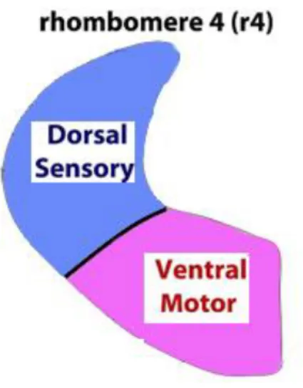
1.6 Rhombomere 4 and Hox genes
The hindbrain in mammals is made of cerebellum, pons and bulb. During the early embryonic phases, the hindbrain is subdivided along the anterior-posterior axis (AP) into repeated segmental units called rhombomeres, each one is separated from the adjacent unit and will give rise, during the subsequent development phases, to specific and different cell lines.
R4 contributes primarily to the generation and specification of auditory nuclei involved in sound transmission and amplification, as well as in the establishment of specific sensorimotor auditory circuitry during development. Previous studies showed that ventral r4 is responsible for the specification of distinct subtypes of motor neurons, such as facial branchiomotor and inner ear efferent neurons. Moreover, it was showed that dorsal r4
contributes to alar-plate-derived sensory components, such as the VLL, PVCN and DCN. In particular, cells migrating rostrally from r4 along the lateral lemniscus tract form the VLL nucleus, whereas dorsal sensory cells remaining within r4 contribute massively to the CN complex (PVCN and DCN), as well as to the vestibular and trigeminal sensory columns. R4-derived cochlear sensory neurons are responsible of two distinct auditory sensorimotor feedback sub-circuits essential for proper hearing: MOC and MEM (middle-ear-muscles) reflex. Some PVCN neurons together with auditory efferent MOC neurons contribute to the MOC reflex, which terminates directly on the OHCs of the cochlea and modulates the cochlear amplification process. VCN neurons are also involved in the MEM reflex through the action of the facial motor nucleus, originated in ventral r4 (Di Bonito et al., 2013).
Hox genes play a crucial role in the segmentation of the hindbrain in rhombomeres. Thanks their colinearity feature there is a direct correlation between the linear position that Hox genes occupy along the chromosome and the spatial and temporal limits of expression along the anteroposterior axis during the development. For example, Hoxb1 is expressed up to the border between the rhombomere 3 and 4 (r3/r4) in progenitor cells during the early development of the hindbrain. Once the rhombomeres are fully formed, Hoxb1 expression is maintained only in r4 by a self-regulating mechanism and by the expression of Hoxb2 (Di Bonito et al., 2013).
So, there is a direct correlation between Hox genes and rhombomeres. Indeed, it was showed that sensory and motor components of the sound transmission pathway, sensorimotor reflex circuits, as well as the hind limb vestibulo-spinal reflex derive from r4 are strongly affected in Hoxb1 mutants (Di Bonito et al. 2013, 2015). It was found that in absence of Hoxb1 expression in the rhombomere 4 a change of r4 to r3 identity occurs. Furthermore, inner ear
efferent (IEE) neurons that also originate from r4 and form vestibular (VEN) and cochlear (CEN) efferent neuron subpopulations fail to develop in Hoxb1 null mutant mice.
In summary, rhombomere 4 and the associated Hoxb1 genes are involved in establishing and maintaining two major functional circuits in the central auditory system. Hoxb1 indeed appear to regulate primarily the assembly of r4-derived neuron populations involved in the main perceptual pathway of sound transmission and the sensorimotor reflex circuits crucial for the cochlear amplification and protection (Di Bonito et al., 2013).
Starting from the results of a previous work in collaboration with the research group of Prof. Michèle Studer of the IBV in Nice (Di Bonito M., Y. Narita, B. Avallone, L. Sequino, M. Mancuso, G. Andolfi, A. M. Franze`, L. Puelles, F. M. Rijli, M. Studer (2013) “Assembly of the auditory circuitry by a Hox genetic network in the mouse brainstem”) concerning the proper development of the auditory system related to the presence of Hoxb1 gene my PhD study starts.
In that work it was showed that Hoxb1 loss strongly affects the proper development of the auditory system. As a matter of a fact Hoxb1 mutants show an increased auditory threshold that leads to severe hearing impairments. It is known from the literature that this phenotype is often associated with affected CN function and/ or alterations in the cochlear amplification mechanism executed by the OHCs. Indeed, defects in the CN complex and moreover strong morphological damage of the OHCs were found. A direct role of Hoxb1 on hair cell development was excluded, since it is not expressed in presumptive hair cells. It was also excluded that a defect in satellite glial cells, surrounding the spiral ganglion neurons and originating from r4- derived neural crest, can affect OHC development and/or contribute to the altered auditory threshold. Even if a decrease of double YFP+/Sox10+ cells in Hoxb1null cochleae was observed, spiral ganglion neurons seem to differentiate properly and appropriately express Gata3. Moreover, type II ganglion fibers innervating the OHCs are unmyelinated, differently from type I fibers innervating the IHCs, indicating that the r4- neural crest-derived YFP+ are mainly type I Schwann cells. Finally, an involvement of LOC efferent neurons was also excluded, which, even if affected in the mutants of the study, appear to have no direct effect on cochlear thresholds measured by ABR. Although the major structure responsible for the increased auditory threshold cannot be ascertained, it was
proposed that abnormal development of MOC neurons, which are required for proper postnatal survival and functioning of OHCs during the hearing process, are involved in the hearing impairments of mutants. In support of this, the strongest morphological hair cell abnormalities were found towards the apical region of the cochlea, where normally low frequency sounds are perceived. Furthermore, early development of hair cells proceeds normally in the absence of efferent neurons but become affected at later stages when OHCs are dependent on proper MOC innervation. Hence, degeneration of OHCs and consequently, altered hearing thresholds, might be caused by the absence of synaptic/ trophic stimulation of cochlear hair cells from the MOC fibers during a postnatal critical period, which is essential for accurate maturation of OHCs. More support comes from the observations that persistence of some MOC neurons innervating the OHCs in Hoxb1lateCKO and Hoxb2DKO mutant cochleae is sufficient to partially ‘‘rescue’’ the auditory threshold and OHC morphological defects. In addition, no differences in the latencies of the evoked responses are found in the ABR analysis, indicating that the auditory stimuli, when perceived, can travel normally along the successive nuclei of the central auditory pathway, even in the presence of abnormal CN and VLL specification. Finally, altered hearing thresholds were found already in one-month-old Hoxb1null mice that were acoustically isolated at birth, and thus not exposed to noise. This rules out that the observed increased threshold is due to reduced function of the MOC and MEM reflexes, as well as LOC neurons that cannot protect the organ of Corti from noise-induced hearing damage. Nevertheless, this does not exclude that deficiencies in the efferent feedback systems are involved in the progressive age-related degeneration of hearing, which is more pronounced in mutant mice than in WT. Hence, the minor increase of threshold with age observed in the Hoxb1lateCKO compared to Hoxb1null might be due to
the presence of a few efferent neurons in the conditional mice. Thus, these data suggest that efferent innervations play an important postnatal role in the hearing impairment observed in Hoxb1 mutants, although it cannot completely rule out that altered development of other auditory structures might also contribute (Di Bonito et al., 2013).
So, starting from these findings the aim of my work was to obtain and analyze selective mutants in which Hoxb1 expression was selectively abolished in the dorsal region of rhombomere 4 (Ptf1a cre Hoxb1 flox/flox; Atoh1 cre Hoxb1 flox/flox). The dorsal region of the rhombomere 4 is the region in which sensory auditory neurons origin. So to exclude a possible contribute of sensory auditory neurons at the OHCs degeneration and to test our hypothesis of a possible synaptic/trophic role of MOC terminal, that origin from the ventral domain of R4, we used Auditory brainstem response (ABR), Immunohistochemistry, Scanning electron microscopy (SEM) and Transmission electron microscopy (TEM) in Hoxb1 mutants in which the gene function is abolished exclusively in the sensory domain of the rhombomere 4 (Fig. 9).
Fig. 9 Rhombomere 4
Concerning the vestibular system my work starts from the findings of the work: “Loss of
Projections, Functional Compensation, and Residual Deficits in the Mammalian Vestibulospinal System of Hoxb1-Deficient Mice”, M. Di Bonito, J. L. Boulland, W. Krezel,
E. Setti, M. Studer and J. C. Glover. ENEURO.0096-15.2015.
In this work it was found that vestibular efferent neurons, such as cochlear efferent neurons, originate from ventral r4 and that fail to develop in absence of Hoxb1. They express high levels of Gata2 an Gata3 but no Gata3 expression was detected in the vestibular efferent neuron domain in E14.5 Hoxb1-null mice, commensurate with an absence of inner ear efferent neurons (Di Bonito et al., 2013a). To confirm the absence of vestibular efferents at postnatal stages when connectivity is established, Dil crystals were inserted into the inner ears of wild-type mice and Hoxb1-null mice at P8. No efferent neurons were retrogradely labeled at this stage in the mutant mice, confirming their absence and excluding the presence of any other ectopic source of efferent input to the inner ear. Thus, vestibular projection neurons and vestibular efferent neurons derived from r4 are critically dependent on the expression and function of Hoxb1 protein (Di Bonito et al., 2015).
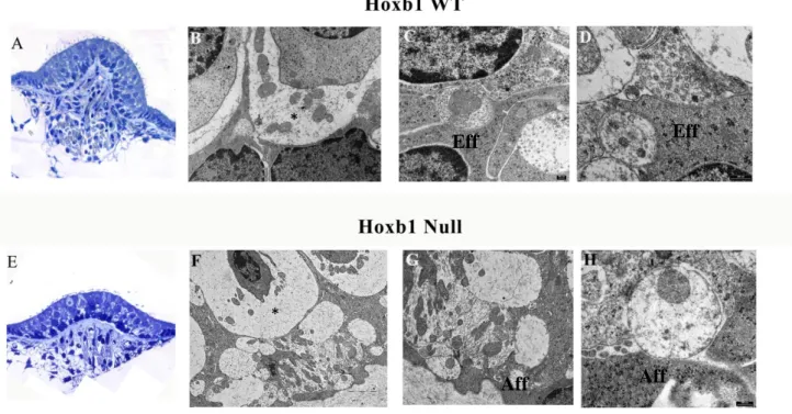
So, based on the fact that p8 mutants have shown no sign of efferent terminations the aim of my work was to study trough transmission electron microscopy the presence or absence of these efferent neuronal endings on receptor cells in 3-months old mutants. Moreover, to understand whether projections are missing at, we also tested newborn mutant pups for the presence of efferent endings on hair cells in different Hoxb1 mutants.
3.1 Generation of Hox mouse mutant lines and matings
According to Di Bonito et al, 2013 the b1r4-Cre mice were crossed with the ROSA26YY reporter line to obtain double heterozygous b1Cre/YFP mice in which r4 and r4-derivatives are selectively labeled. Hoxb1null mice were mated to the b1r4-Cre/YFP line to permanently label r4 and r4-derivatives in a null background. Similarly, Hoxb1flox mice were mated to the b1r4-Cre or b1r4-Cre/YFP transgenic lines to obtain Hoxb1lateCKO mutants in which Hoxb1 is inactivated exclusively in r4 at around E9.5. Atoh1cre/+ e Ptf1acre/+ were crossed with Hoxb1 flox/+ YFP/+ in order to obtain Hoxb1flox/+ YFP/+ Ptf1acre/+ and Hoxb1flox/+YFP/+ Atoh1cre/+. Both mutants were crossed with Hoxb1flox/+ to obtain Hoxb1flox/flox YFP/+ Ptf1acre/+ and Hoxb1flox/flox YFP/+ Atoh1cre/+ in order to eliminate the Hoxb1 expression in the sensory region of rhombomere 4. All experiments were conducted following guidelines of the Institutional Animal Care and Use Committee of the University of Nice Sophia-Antipolis, Nice.
3.2 Auditory brainstem response (ABR)
The tests were performed by Anne-Gabrielle Harrus of the Jean-Luc group Puel in Montpellier. Briefly as described in Di Bonito et al., 2013 control and mutant animals were sedated with medetomidine chlorhydrate (1mg/kg) and ketamine (100mg/kg) via intraperitoneal injection, and sedation was reversed with atipamezole (medetomidin antagonist) (1mg/kg) after the ABR measurement. Stimulation, averaging and analysis were carried out using an Amplaid MK12 system (Amplifon, Italy). Monaural stimuli consisted of alternating clicks lasting 0,1 ms at a repetition rate of 11/s. ABRs were recorded in both ears
via subcutaneously placed tungsten needle electrodes (diameter 0,25mm). The active electrode was inserted in the vertex, the reference electrode was inserted in the posterior bulla region of the left or right ear, and the ground electrode was inserted in the lower back. Ipsilateral stimuli and contralateral masking white noise were presented with ear probes (Madsen Electronics, Kobenhavn, Denmark). The ear masker level was set at 50dB HL, while the click level was set at 110 dB SPL and was decreased in 10dB steps until the detection threshold was approached. Filter settings ranged from 100Hz to 2.5kHz. The analysis time was of 12ms, the amplifier sensibility was of 5nV/division and potentials were amplified 105 X. The ABR signals were calibrated as specified by ISO (International Organisation for Standardisation) 389 and ANSI (American National Standards Institute) S3.6 directives. The detection of the threshold was defined on the basis of 2-3 repeated measurements close to threshold. A threshold value is obtained by the mean between the lowest intensity at which the response is revealed and the highest intensity at which a response is absent. The ABR patterns were examined by two independent observers and accepted if both observers agreed. In the mouse, the sequence of five positive ABR waves (PI-PV) are localized to: the cochlea and/or the compound action potential of the VIII nerve (PI), the cochlear nuclei (PII), the contralateral superior olive complex or SOC (PIII), the lateral lemniscus complex (PIV), and the contralateral inferior colliculus or IC (PV).
3.3 Tissue preparation
According to Di Bonito et al. 2013 adult and P8 mice were perfused with 4% paraformaldehyde (PFA). The inner ear was fixed overnight in 4% paraformaldehyde (PFA) in phosphate-buffered saline, pH 7.4 (PBS). Tissues were cryoprotected with 10, 20 and 30% sucrose in PBS and frozen in OCT embedding matrix (Kaltek) and sectioned at 20 mm.
3.4 Transmission electron microscopy (TEM)
According to Di Bonito et al. 2013, for TEM analysis, the inner ear was fixed in 2.5% glutaraldehyde in 0.1 M phosphate-buffered saline (PBS) pH 7.4 for 4 h at 4ºC and rinsed in PBS overnight. The organs of Corti and the vestibular organs (ampullae, sacculus and utriculum) were isolated, rinsed in PBS and postfixed in 1% osmium tetroxide solution (Fluka) in a 0.05 M PBS at pH 7.4. Specimens were dehydrated with ethanol and then, with propylene oxide and embedded in Epon 812 resin (Fluka). The blocks were cut using a Super Nova Leica Ultratome. Semithin sections at 2 mm thickness were studied with a light microscope (Polivar Reichert-Jung) after staining with 1% toluidine blue (Carlo Erba). Ultrathin sections (80 nm) were stained with 2% uranyl acetate (Electron Microscopy Sciences) for 10 min at room temperature and 2.66% lead citrate (Electron Microscopy Sciences) for 8 min at room temperature. Grids were examined by using a Philips EM 208 S transmission electron microscope (Philips) operating at 80 kV.
3.5 Scanning electron microscopy (SEM)
In accordance with Di Bonito et al. 2013, for SEM analysis, cochleae were fixed in 2.5% glutaraldehyde in 0.1 M phosphate-buffered saline (PBS) pH 7.4 (19 ml of 0.2 M sodium phosphate monobasic NaH2PO4 and 81 ml of 0.2 M sodium phosphate bibasic Na2HPO4) for 4 h at 4ºC and rinsed in PBS overnight. The organs of Corti were isolated, rinsed in PBS and post-fixed in 1% OsO4 in the same buffer for 1 h at 4ºC. After several rinses in PBS, the samples were dehydrated, and a critical point drying was performed. The samples were mounted on aluminum stubs and sputter coated with gold. The processed specimens were investigated and photographed using a JEOL 6700F SEM operated at 5 kV and at a 8.3 mm working distance. SEM images were collected digitally.
3.6 Immunohistochemistry
According to Di Bonito et al. 2013, we proceeded to the inactivation of endogenous peroxidase with 0.5% H2O2, tissue cryosections were blocked in blocking buffer (0.05% Tween 20, 20% newborn calf serum, NBCS, in PBS) and incubated overnight at 4ºC with primary antibodies diluted in hybridization buffer (0.05% Tween 20, 5% NBCS in PBS): anti-GFP rabbit polyclonal antibody (1:500, Molecular Probes). Sections were washed in blocking buffer and incubated for an hour and a half at room temperature with secondary antibodies: biotinylated rabbit anti-goat (1:300), goat anti-rabbit or goat anti-mouse (1:200). The Vectastain Elite ABC kit and DAB substrate kit for peroxidase (Vector) were used for immunohistochemical staining.
4.1 Auditory response of the mutant mice in sensory domain of rhombomere 4
This first experiment carried out aimed to understand the auditory response of the mutant mice in sensory domain of rhombomere 4 (Hoxb1flox/flox Atoh1/+, Hoxb1 flox/flox Ptf1a/+), in order to assess if the mutation could cause some sort of auditory impairment. For this reason, the auditory brainstem response (ABR) was measured. This test consists in a series of electrical potentials evoked by auditory stimuli ranging from 4 to 32 dB SPL (Sound Pressure Level), determining the lowest decibel level, or threshold, at which a response peak is reproducibly present.
We first analyzed the ABR response in WT (N=5), Hoxb1null (N=5), Atoh1 cre/+ (N=4), Hoxb1 flox/flox Atoh1/+ (N=6), Ptf1a cre/+ (N=4), Hoxb1 flox/flox Ptf1a/+ (N=7) mice. We used null mice as positive controls and the Atoh1cre/+ and Ptf1a cre/+ to compare the floxed mice with them. In Hoxb1WT 3-month-old mice a normal threshold where detected while in Hoxb1null 3-month-old mice the threshold is elevated, resulting in a higher stimulus to evoke a response. Both Hoxb1flox/flox Atoh1/+ and Hoxb1 flox/flox Ptf1a/+ show a normal threshold compared to WT and Cre individuals, showing that this kind of mutation does not affect the hearing of the mice.
Fig 10. Representative ABR measurements of 3-month-old WT, Hoxb1null (KO), Atoh1
cre/+, Hoxb1flox/flox Atoh1cre/+, Hoxb1 flox/flox Ptf1acre/+, Ptf1a cre/+. The threshold, the lowest intensity of sound at which the response is present, is higher in Hoxb1null than WT mice, while no significative differences are observed for Atoh1 and Ptf1a mutants compared to the WT. Ptf1a cre and Atoh1cre where used in order to asses any interferences of the cre recombinase.
4.2 Localization of YFP in Hoxb1 mutants’ cochleae
In order to understand if the proper development of the cochlea is affected by the absence of expression of Hoxb1 in the sensory area of rhombomere 4, particularly in the domains that express Atoh1 and Ptf1a, we performed immunohistochemistry analysis in Hoxb1 r4Cre/+ YFP/+, used as control, Hoxb1 null/null r4Cre/+ YFP/+, in which Hoxb1 is not expressed in the entire rhombomere4, Ptf1acre/+ YFP/+, in which the YFP label Ptf1a derivatives in the cochlea, Ptf1acre/+ YFP/+ Hoxb1 flox/flox, in which Hoxb1 expression is abolished in the sensory domain that express Ptf1a, Atoh1cre/+ YFP/+, in which the YFP label Atoh1 derivates and Atoh1cre/+ YFP/+ Hoxb1 flox/flox in which Hoxb1 expression is abolished in the sensory domain that express Atoh1.
We used an anti-GFP antibody that cross-reacts with the YFP protein and amplify the endogenous signal of the yellow fluorescent protein.
For what concerns r4 the fibers of the cochlear nerve seem to be labeled. The same label disappears when Hoxb1 is not expressed in the entire rhombomere (Fig.11 A-B).
Ptf1a staining on the other hand shows that not only the sensory cells of the Organ of Corti (IHC and OHC) are labeled but also the cochlear nerve seems labeled even if in a different way respect to the r4 staining, it appears as cell bodies. In the same way as the r4 they disappear in Hoxb1 mutants (Fig.11 C-D).
Finally, Atoh1 sections show no differences between the mutant and the control. The staining indeed highlights in both samples the label of the organ of Corti, so Hoxb1 seems not have a rule in the specification of the Atoh1 derivatives for what concerns rhombomere 4 (Fig.11 E-F).
Such as for the organ of Corti, Ptf1a and Atoh1 are also involved in the proper specification of the hair cells that compose the vestibular organs. In Fig.12 an ampullary section is showed and no significative differences can be detected even when Hoxb1 is not expressed in the sensory region of rhombomere 4.
Fig.11 Serial sections of cochlea labeled with anti-GFP. A and B show respectively r4
derivatives and r4 derivatives in absence of Hoxb1. It is evident that when Hoxb1 is lost also the positivity decrease. C and D: label of Ptf1a derivatives in presence or absence of Hoxb1 in r4. In both the case the sensory cells of the organ of Corti are labeled, while in absence of Hoxb1 in D a decrease of the positivityof the neurons bodies of the cochlear nerve is evident. E and F show the labeling of sensory cells in presence or absence of Hoxb1 showing no significant changes beetween the two groups.
A B C D E F
Fig.12 Ampullary sections indicating the YFP+ Atoh1and Ptf1a –derived. Both mutants
show the positivity in the sensory epithelia without evident differences beetween mutant and control.
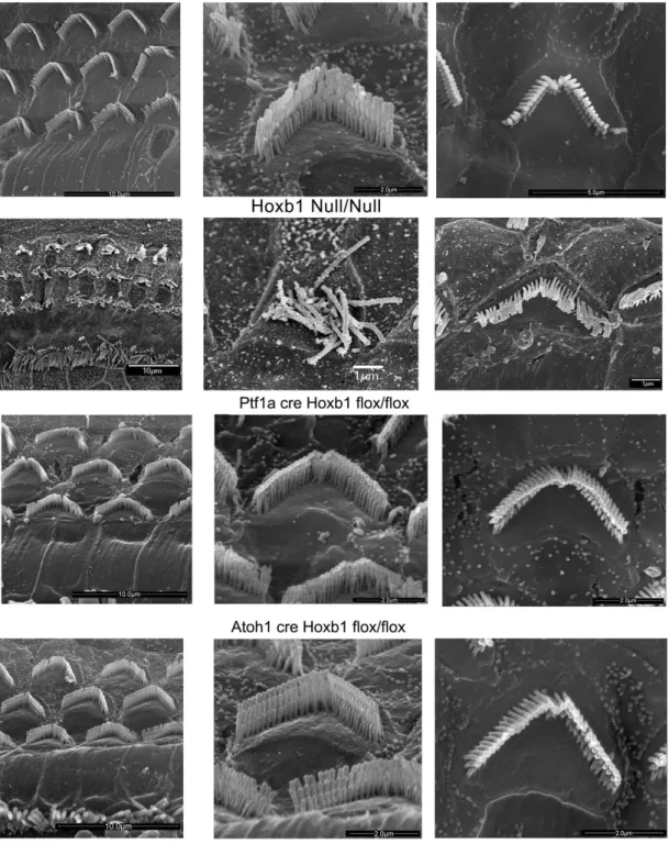
4.3 Ultrastructural analysis of hair cell morphology in Hoxb1 mutant mice
To assess hair cell morphology in Hox mutant cochleae, we used scanning electron
microscopy on the apical and basal turns of WT, Hoxb1 null/null (used as a positive control), Hoxb1flox/flox Atoh1cre/+ and Hoxb1 flox/flox Ptf1acre/+ 3 months old mutant mice, in order to understand if the sensory domain of the rhombomere 4 is somehow involved in the proper development of these cells.
Usually, three rows of OHCs and one row of IHCs are orderly arranged along the entire organ of Corti. Figure 13 points out the three rows of stereocilia of increasing height arranged in the characteristic V-shaped morphology; the ones located at the basal turn appear wider than the one’s at the apical turn. Also a difference in the lenght of the stereocilia can be observed: the cells located in the apical turn show longer stereocilia respect to the cells located in the basal turn. Their morphology reflects the cell specialization and indeed the OHCs of the apical region perceive low-frequency sounds.
Hoxb1null adult mice show severely disorganized OHC rows with occasional loss of hair cells in the apical turn. Furthermore, close observation of individual OHCs in Hoxb1null cochleae indicates that most stereocilia lose their typical V-shaped arrangement, as well as their organized structure and characteristic differences in ciliar length.
No obvious differences in the shape and organization of OHCs at the apical and basal cochlear turns where detected in all Hoxb1flox/flox Ptf1acre/+ and Hoxb1 flox/flox Atoh1cre/+ mutants analyzed excluding then an involvement of the sensory region of the rhombomere 4 in their development.
Fig. 13 Overview of the apical turns of WT, Hoxb1null, Hoxb1 flox/flox Ptf1a cre/+ and
Hoxb1 flox/flox Atoh1 cre/+ cochleae showing three orderly arrayed rows of outer hair cells (OHCs). Representative high magnification images illustrate stereocilia of hair bundles of single OHCs arranged according to their different lengths. Shape and organization of OHCs in both apical and basal regions are normal at this stage in WT, Hoxb1 flox/flox Ptf1acre/+ and Hoxb1flox/flox Atoh1cre/+ compared to Hoxb1 null mutant in which a degeneration of the apical OHCs is evident.
4.4 Innervation of medial olivocochlear neurons in Hoxb1 mutant mice
Transmission electron microscopy let us to evaluate the state of the efferent MOC endings, in order to confirm their presence and proper development since the Hoxb1 gene inactivation in the dorsal domain should not affect these nerve endings.
The MOC neurons support OHC maturation at early postnatal stages and regulate the vibration of OHCs in the cochlea and from E18.5 onwards, MOC motor axons reach the contralateral cochlea.
TEM investigations highlight in WT mice the presence of efferent nerve endings that contract synapses with outer hair cells. In WT mice, the typical arrangement in several rows of outer hair cells with their nucleus located in the basal portion of the cell is observed. MOCs show the typical organization with a more electrondense cytoplasm (Fig.14, B-C). From the observations of the samples Ptf1a cre/+ Hoxb1flox/ flox (Fig.14, E-F) and Atoh1cre/+ Hoxb1flox/flox (Fig.14, H-I), no evidence of efferent nerve endings alteration that take contact with the OHCs is detected.
Indeed, the efferent fibers have the typical characteristics observed in the WT, characterized by the presence of the sub-synaptic cistern, the synaptic vesicles and the mitochondria that appear predominantly localized in the area opposite the synaptic contact.
Fig.14 Transmission electron microscopy of OHCs in adults WT, Ptf1acre/+ Hoxb1flox/flox
and Atoh1cre/+ Hoxb1flox/flox cochleae. In high magnification views, MOC terminals synapse on OHCs in WT and mutants without differences in shape and morphology.
5. Results
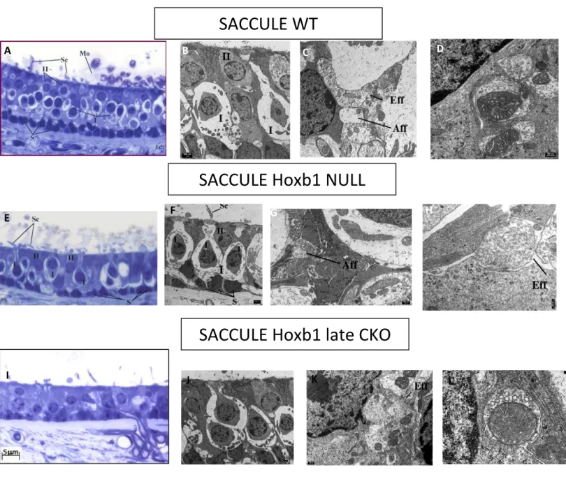
5.1 Innervation of vestibular efferent neurons in 3 months old Hoxb1 mutant mice
TEM investigations were used to understand the state of efferent ending that innervate the vestibular organs. We started from the data that retrograde labeling showed the absence of the vestibular efferent neurons (VEN) in Hoxb1 Null mutants and a caudal label of few neurons in Hoxb1 late CKO, in which the gene is inactivated exclusively in r4 at around E9.5.
Light and transmission microscopy investigations in 3-month old mutants allowed to
evaluate the general morphology of the ampullary crest, macula of saccule and utricle with a focus on quantity and quality of efferent terminations.
The general morphology of the vestibular organs appears without substantial variations in all the samples analyzed, both wild type (WT) and mutants. Any apparent differences are to be charged to the different cutting plans (Fig. 15,16,17 A-E-I).
In these sections the basal layer of support cells resting on the basal lamina is clearly visible. In the apical area of the epithelium the type I and II sensory cells and the relative stereocillia are recognizable (Fig. 15,16,17 A-E-I).
For what concerns transmission electron microscopy, the Figures 15,16,17 B-F-J show the general organization of the ampullary sensory region and macula of saccule and utricle with focus on the two types of sensory cells that allow the perception of the stimulus.
The presence of both afferent and efferent endings in ampullae, saccule and utricle of all the mutants analyzed, both Hoxb1 null, even if with a less frequency, and Hoxb1 late CKO, is always detected without any change in appearance (Fig.15,16,17 D-H-L).
Particularly, afferent endings, bottoniform or calyx, are characterized by a clearly less electrondense cytoplasm, lots of mitochondria and a small amount of vesicles. In fig. 15D a detail of a synaptic bar is clearly visible. Synaptic bar is a feature of only HCI, it origins in the contact point between the cell and the afferent ending and it is surrounded by small vesicles.
Typical of the efferent terminations are the more electrondense aspect of the cytoplasm and the larger presence of synaptic vesicles. Moreover, one of the features of the efferent endings is the presence of a subsynaptic cistern. This structure is recognizable in Fig. 16H.
Fig.15 Ampullary crest sections of WT, Hoxb1 null and Hoxb1 late CKO (A-E-I) observed
by light microscopy. Staining with toluidine blue. In C and G HCI are surrounded by the afferent calyx. In all the samples analyzed afferent (Aff) and efferent (Eff) terminations were detected. In D a synaptic bar is highlighted. *=afferent calyx.
A
Aff Eff
AMPULLA WT
AMPULLA Hoxb1 NULL
AMPULLA Hoxb1 late CKO
B C D E F G H I J K L C H Eff * * *