Short-term synaptic plasticity
in chronic migraine with medication overuse
Facoltà di Medicina e Chirurgia Dipartimento di Neuroscienze
Dottorato di Ricerca in Neuroscienze clinico-sperimentali e Psichiatria – Ciclo XXXI
Dott.ssa Francesca Cortese Matricola 941455
Relatore Correlatore
Chiar.mo Prof. Francesco Pierelli Chiar.mo Prof. Gianluca Coppola
1. INTRODUCTION 3
1.1.MIGRAINE: EPIDEMIOLOGY AND PATHOPHYSIOLOGY 3
1.2.HABITUATION AND SENSITIZATION IN MIGRAINE 5
1.3.MOTOR CORTEX EXCITABILITY IN MIGRAINE 8
2. CHRONIC MIGRAINE 10
2.1.CLINICAL ASPECTS OF CHRONIC MIGRAINE 10
2.2.CORTICAL PLASTICITY IN CHRONIC MIGRAINE 15
3. SHORT-TERM SYNAPTIC PLASTICITY IN MEDICATION OVERUSE HEADACHE 19
3.1.RATIONALE OF THE STUDY 19
3.2.WITHDRAWAL FROM ACUTE MEDICATION NORMALISES SHORT-TERM CORTICAL SYNAPTIC
POTENTIATION IN MEDICATION OVERUSE HEADACHE 20
4. DISCUSSION AND FUTURE PERSPECTIVES 32
5. REFERENCES 36
6. APPENDIX: OTHER SCIENTIFIC WORKS ON MIGRAINE 44
A.ANODAL TRANSCRANIAL DIRECT CURRENT STIMULATION OVER THE LEFT TEMPORAL POLE RESTORES
NORMAL VISUAL EVOKED POTENTIAL HABITUATION IN INTERICTAL MIGRAINEURS 45
B.EXCITABILITY OF THE MOTOR CORTEX IN PATIENTS WITH MIGRAINE CHANGES WITH THE TIME ELAPSED
FROM THE LAST ATTACK 62
C.SHORT-TERM CORTICAL SYNAPTIC DEPRESSION/POTENTIATION MECHANISMS IN CHRONIC MIGRAINE
1.
I
NTRODUCTION1.1. Migraine: epidemiology and pathophysiology
Headaches are the most common painful syndromes in the young-middle age, affecting people’s quality of life and causing a significant economic impact.
Among primary headaches, migraine is the most common and disabling one, affecting about 15% of the population of North America and Western Europe.
Since migraine was first included in the Global Burden of Disease study (GBD), which represents the most comprehensive worldwide observational epidemiological study to date, it has ascended the ranks of top causes of disability worldwide.
In the recent publication of 2016, in the age group of 15–49 years, migraine is the top cause of disability.
Even though a comprehensive knowledge of migraine pathophysiological mechanism is still lacking, many efforts have been done, in the last decades, to disentangle the complicated puzzle of migraine pathophysiology with the help of new research tools, as neurophysiological and modern neuroimaging technologies.
Clinical neurophysiology methods are non-invasive techniques that allow in vivo measurements of cortical excitability and electrocortical responses to various sensory stimuli and to deepen our knowledge on cortical plasticity in healthy subjects and in migraine patients.
Progresses in headache research were also favoured by the introduction of the International Classification of Headache Disorders (ICHD) and its revisions, because detailed diagnostic criteria allowed to perform better comparison of clinical and neurophysiological data between headache centres.
Migraine is considered a neurovascular disorder, where the trigemino-vascular system plays a central role. Neurogenic inflammation in meningeal trigeminal afferents that innervate the dural vasculature is characterized by the release of neuropeptides (substance P, calcitonin gene related peptide), leading to vasodilation, plasma extravasation and mast cell degranulation. These sensitized trigeminal
afferents, with their cell bodies located in the trigeminal ganglion, project to the trigeminal nucleus caudalis in the brainstem, which in turn projects to higher brain centers.
Indeed, together with the trigemino-vascular system, the brainstem, the thalamus and the cerebral cortex are also involved in migraine pathophysiology as main actors. In particular, abnormal thalamic pacemaker rhythmic activity, namely “thalamo-cortical dysrhythmia” could be responsible for a low level of cortical preactivation in the sensory cortices.
Since episodic migraine is characterized by recurrent clinical attacks separated by variable-length headache-free intervals, several studies have focused on ictal versus interictal electrophysiological abnormalities. They showed that migraine brain exhibits, if compared to healthy subjects, interictal dysfunctions of the central nervous system. Such dysfunctions probably represent the neurophysiological substrate of the clinical predisposition to attack recurrence.
This predisposition is the “core” of migraine disease and is genetically determined, with environmental factors that may act as triggers.
Furthermore, chronic migraine, defined as headache occurring on at least 15 days per month for more than 3 months, behaves from a neurophysiological point of view, like a “never ending migraine attack”. Consequently, the exploration of neurophysiological disfunctions in migraine could help defining and understanding the mechanisms involved in migraine pathophysiology and chronification.
1.2. Habituation and sensitization in migraine
Since migraine is characterized by a dysfunction in sensorial information processing, studying the cortical responses to sensory stimulation, particularly in interictal phase, could help defining the neurophysiological markers of migraine brain, and probably understanding the mechanisms underlying predisposition to attack recurrence and chronification.
Habituation is defined as a decremental response to repeated sensory stimulations (Harris, 1943) and represents a physiological response observed in a wide range of neuronal circuits, allowing to control the signal-to-noise ratio generated by sensory stimuli and to orientate human response to environmental modifications.
According to the “dual-process” theory (proposed in the 70's by Groves and Thompson), during a sequence of repetitive stimuli, two opposing processes compete to define the final response: sensitization and habituation. The first one is prevalent in the initial part of the stimulus session, causing a transitory increase in response amplitude, while the second one occurs in the following phase and accounts for the delayed response decrement.
Habituation is a physiological response that protects the cortex against the risk of inward information overflow, while preparing brain networks to adequately respond to subsequent relevant stimuli.
Since habituation may be considered a basic form of learning (Thomson et al, 1966; Chung et al, 2002), the phenomenon of habituation is useful for studying the mechanisms of information processing and learning within the central nervous system and, ultimately, the neuronal substrates of behaviour.
Habituation of the evoked potentials can be assessed by averaging successive blocks of responses. Migraineurs are interictally characterized by a “deficient habituation”, meaning that they show, instead of a physiological decrease (habituation), an amplitude increase of scalp-evoked potentials to repeated stereotyped stimuli.
This phenomenon was reported in migraine patients for almost all sensory modalities: visual (Schoenen et al, 1995), auditory (Ambrosini et al, 2009),
somatosensory (Ozkul et al, 2002) and painful stimuli (Valeriani et al, 2003; de Tommaso et al, 2005). Nevertheless, it was also reported for cognitive stimulations (Kropp et al, 1995).
Interestingly, the habituation deficit may have a familiar character and was proposed as a neurophysiological marker for migraineurs (Siniatchkin et al, 2001) and asymptomatic subjects at risk of developing the disorder (Di Clemente et al, 2007).
Besides the lack of habituation, evoked potentials in migraineurs are characterized by a low amplitude of the first block of averagings (for a review see Ambrosini et al, 2003), ruling out the theory that the habituation deficit in migraine could be due to cortex hyperexcitability. These data support the hypothesis of an interictal reduced pre-activation level of sensory cortices, possibly due to insufficient activation by aminergic projections from the upper brainstem (Schoenen et al, 1996) and, consequently, a dysfunction in thalamo-cortical drive, known as “talamo-cortical dysrhythmia” (Llinas et al, 1999; Coppola et al, 2007).
Therefore, the heightened response to repeated stimuli (or habituation deficit) in migraineurs is the consequence of sensory cortices “hyperresponsivity”, which results in an exaggerated energy demand and, possibly, in subtle cognitive dysfunctions (Magis et al, 2007).
However, habituation is not a static phenomenon but fluctuates over time in relation to the migraine cycle. In particular, the habituation deficit reaches its maximum during the days preceding the attack, while increases and normalizes immediately before and during the attack, when the thalamo-cortical drive also increases (Coppola et al, 2005). Such fluctuation is probably related to changes in serotonin transmission, which is low interictally (Panconesi et al, 2008) and may increase ictally.
On the other side, sensitization,defined as facilitation occurring at the beginning of the stimulus presentation, was evidenced during the attack, especially with somatosensory stimuli (as reflected by a significant increase in SSEP 1st N20-P25 block amplitude), while disappeared between attacks (Coppola et al, 2010).
Sensitization was evidenced also in chronic migraine with and without medication overuse. However, in chronic migraine without drug overuse a normal habituation
was found for visual evoked responses (Chen et al, 2011; Schoenen et al, 2011), while in medication overuse headache (MOH) a deficient habituation was found for somatosensory evoked potentials (Coppola et al, 2010).
Therefore, cortical sensitization and hyperresponsiveness (Coppola et al, 2010; Currà et al, 2011) could be considered neurophysiological markers of MOH.
The neural network underlying habituation is poorly understood. Consequently, the deficient habituation, even representing the most reproducible abnormality of evoked potentials detectable during the pain-free interval in migraineurs, still lacks a conclusive interpretation.
Relevant information on the pathophysiology of the interictal dysfunction in migraine came from studies of the high-frequency oscillations (HFOs) embedded in somatosensory and visual evoked potentials. Early somatosensory HFOs, reflecting spike activity in thalamo-cortical drives, were shown to decrease in interictal migraineurs and to normalize during the attack, while a significant habituation deficit of the late visual HFOs was observed in the interictal phase, demonstrating a dysfunction in cortical oscillatory networks, reflected by an abnormal thalamic rhythmic activity, named “thalamo-cortical dysrhythmia” (Coppola et al, 2007).
Several biochemical and neuroimaging studies suggested that the habituation deficit could be related to modifications in serotonin transmission, which fluctuate during the migraine cycle (Ferrari et al, 1993, Evers et al, 1999), reflecting dysfunctions in monoaminergic nuclei activity and activation of the pontomesencephalic areas of the brainstem during migraine attacks (Weiller et al, 1995; Bahra et al, 2001).
Futhermore, the serotonergic system is presumably affected by the chronic use of medications, determining neuronal hyperexcitability and trigeminal activation in patients affected by MOH (Srikiatkhachorn et al, 2000; Dobson et al, 2004).
1.3. Motor cortex excitability in migraine
Impairment of mechanisms regulating the responsivity to various stimuli is a hallmark of migraine brain. Although the mechanisms underpinning brain “dysexcitability” are still debated, there is general agreement that such abnormalities widely affect subcortical areas and likely the whole cerebral cortex.
Therefore, besides studying sensory processing in migraine patients, great effort was also made to characterize motor cortex excitability in migraine.
Cortical excitability of the motor cortex can be examined using transcranial magnetic stimulation (TMS) over the motor cortex and then recording the evoked peripheral activity from a muscle, namely the motor-evoked potentials (MEPs). TMS is a non-invasive technique that is used worldwide both in clinical practise, to assess the conduction of the descending cortico-nuclear and cortico-spinal pathways, and for neuroscientific purposes. Indeed, changes in motor activation and excitability can be easily assessed by recording MEPs (for a review see Rossini et al, 2015). Neurophysiological measures, such as corticomotor threshold (MT), MEP amplitude and latency, Cortical Silent Period (CSP) duration, Central Motor Conduction Time (CMCT) can be used to provide evidence of pathological changes in motor cortical control or corticospinal output in patients.
Corticospinal excitability can be estimated by measuring the cortical motor threshold (or resting motor threshold, RMT), which is the minimal intensity of motor cortex stimulation required to elicit a MEP of minimal amplitude in a relaxed target muscle. The MEP size can be estimated by measuring the peak-to-peak amplitude after setting the stimulation intensity at 115-125% of the individual’s RMT.
Intestingly, studies regarding motor cortex excitability in migraine patients reported controversial findings. In particular, resting motor threshold in interictal migraine were reported to be normal (Werhahn et al, 2000), increased (Afra et al, 1998) or reduced (van der Kamp et al, 1996).
A recent neurophysiological study didn’t find any difference in RMT between interictal migraineurs and controls but, by exploring the effect of a first conditioning stimulus on the motor evoked potential (MEP) elicited by a second test stimulus,
according to the migraine cycle (ictal, interictal, pre-ictal) were disclosed. Indeed, they found decreased short-interval intracortical inhibition (SICI) in interictal migraineurs when compared to healthy controls, a shortened CSP only in female interictal migraineurs and a decreased ICF in pre-ictal compared to interictal migraineurs (Neverdahl et al, 2017).
Furthermore, intracortical excitability was found to be variable in relation to the intensity of stimulation, indicating that different neuronal circuits can show different activation and inhibition thresholds: an increased ICF was found in migraineurs, as compared to the healthy subjects, only by using a 110% intensity of the test stimulus (Cosentino, 2018). Anyway, neither in this case, the authors found any differences between interictal migraineurs and controls as regards RMT (Cosentino et al, 2018).
That probably happened because motor cortex excitability is not a static parameter. As happens for habituation in sensory cortices, motor cortex excitability may fluctuate according to the migraine cycle and, within interictal phase, according to the time interval from the last ictal phase.
Indeed, we recently found that motor cortex excitability (MEP threshold and amplitude) in interictal migraineurs varies on the basis of the time elapsed since the last attack: RMT is lower when long time interval has passed after an attack and is higher when measured close to an attack (Cortese et al, 2017). Such dynamic RMT variations in relation to the migraine cycle represent time-dependent plastic changes in brain excitability that resemble those occurring for visual and somatosensory evoked potentials.
Several neurophysiological studies failed to disclose significant differences in motor cortex excitability (in terms of RMT, latency and first MEP size) between chronic migraine patients, healthy subjects and episodic migraineurs (Cosentino et al, 2014; Cortese et al, 2018; Ozturk et al, 2002). That means that basal MEP amplitude in chronic migraine is not different from healthy subjects, but it doesn’t exclude dysfunctions in motor cortex plasticity, as will be discussed later.
2.
C
HRONIC MIGRAINE2.1. Clinical aspects of chronic migraine
Chronic migraine is a disease characterized by a deep impact on patients’ life (see May et al, 2016), with considerable disability rates and burden of disease. Chronic migraine, if compared to episodic migraine, has a more profound impact on socioeconomic functioning and quality of life (Buse et al, 2012; Blumenfeld et al, 2011).
Impressively, about 25% of patients with chronic migraine report a very severe headache-related disability, as defined by the Migraine Disability Assessment Scale (also known as MIDAS). That brings to reduced household and family activities and high direct costs (related to healthcare and therapies) and indirect costs (due to absenteeism from work and reduced productivity) (Bigal et al, 2008; Munakata et al, 2009).
According to the current diagnostic criteria of the International Classification of Headache Disorders (ICHD‑3 beta), chronic migraine is defined as headache occurring on at least 15 days per month for more than 3 months, fulfilling, at least 8 headache days per month, the criteria for migraine headache (Headache Classification Committee of the HIS, 2013). Noticeably, in contrast to earlier classification editions, analgesic overuse is no longer an exclusion criterion for the diagnosis of chronic migraine. Consequently, according the new criteria, patients with medication overuse should be considered as affected by both chronic migraine and medication overuse headache (MOH).
The prevalence of chronic migraine is about 1–2% in the general population, with three times higher prevalence in women than in men (Buse et al, 2012).
Primary chronic migraine is rare; usually chronic migraine usually evolves from episodic migraine with an annual progression rate of about 3% (Scher et al, 2003). Risk factors for migraine chronification are: age, female sex and low educational status (among the non-modifiable risk factors) and overuse of acute migraine
medication (Bigal et al, 2008), ineffective acute treatments (Lipton et al, 2015), obesity and insulin-resistance (Peterlin et al, 2010; Fava et al, 2014), depression (Ashina et al, 2012), and stressful life events (Scher et al, 2003) (among the potentially modifiable factors).
Probably the most important risk factor for migraine chronification is the overuse of acute migraine medication. Medication overuse headache (MOH) represents a relevant social burden, affecting around 63 million people worldwide (Kristoffersen et al, 2014). The prevalence of MOH in general population is between 1 and 2% (ranging from 0,5% and 7,2% in different countries), affecting mostly middle-aged adults from age of 30 to 50 years, with higher prevalence in studies from headache specialist centers, ranging from 30% to 50% of patients (Westergaard et al, 2014; Munksgaard et al, 2014). The majority of studies reports higher incidence in females with a male-to-female ratio of around 1 to 3–4 (Kristoffersen et al, 2014).
MOH consists of a complication of a pre-existing headache syndrome and is characterized by overuse of one or several types of acute painkilling medications as simple analgesics, combination-analgesics, ergots, triptans and opioids. The diagnosis is based on headache frequency (equal to or greater than 15 days/a month) and overuse of headache medications on more than 10 or 15 days per month, depending on the drug class, for more than 3 months. Noticeably, migraine is the most common pre-existing headache disorder.
Medication overuse discontinuation leads to reduction of headache frequency, facilitating prophylactic therapy effectiveness.
Even though MOH usually resolves once the overuse is stopped (Manzoni et al, 2015), it is no longer a requirement for the diagnosis to be made.
The risk of chronification depends on the type of used drug, with lower risk for triptans and ergotamine, if compared to analgesics and opioids (Thorlund et al, 2016). The most important risk factors associated to the development of MOH are: regular use of benzodiazepines, depression, physical inactivity, smoking, age younger than 50, female gender and low level of education (Hagen et al, 2011). Furthermore, an increased risk of developing MOH was detected if a family history of MOH or other substance abuse was present (Cevoli et al, 2009).
Indeed, genetic polymorphic variants in genes of the dopaminergic system and genes related to drug-dependence pathways have been described as potential risk factors for excessive use of acute medications and consequent development of MOH (Cargnin et al, 2017).
Patients affected by MOH often show multiple psychiatric comorbidities. Anxiety and depression are the most frequently described (Lampl et al, 2016). An association was also found between MOH and greater susceptibility to drug dependency and with clinically relevant obsessive-compulsive disorder (Sarchielli et al, 2016). A high prevalence of sleep complaints, including insomnia, daytime sleepiness, and snoring was also reported (Sancisi et al, 2010) in MOH patients.
Various respiratory and cardiovascular conditions more likely coexist with chronic migraine than with episodic migraine (Buse et al, 2010).
Anyway, protective factors, such as physical exercise, stress management, and preventive medications, may help reducing the frequency of migraine attacks, thereby reducing the risk of migraine chronification.
Withdrawal of acute painkilling drugs is the first-line approach for the management of MOH patients.
A recent randomized clinical trial showed that complete discontinuation of acute medications is the most effective strategy (if compared to restricted medications intake) (Carlsen et al, 2018).
Withdrawal can be quite difficult for some patients because of the frequent appearance of withdrawal symptoms as headache, nausea, vomiting, anxiety, sleep disturbances, that usually last for 2–10 days and can be very disturbing.
The choice of the setting for withdrawal (inpatient or outpatient withdrawal) should consider several factors, including the type of overused medications, the duration of the overuse, the possible history of previous detoxification failures or psychiatric comorbidities.
In clinics, a standardized therapeutic protocol for medication withdrawal is lacking.
Several strategies are commonly used such as intravenous hydration, rescue medications (different from overused drugs), antiemetics, benzodiazepines, and sometimes corticosteroids.
Special reference needs to be made about prophylactic treatment.
Discussion between the three main options is still going on: some authors advocate withdrawal of acute medication alone, others suggest early prophylaxis alone, and a third group stands for withdrawal in combination with early prophylaxis (Rossi et al, 2009).
Although evidence-based recommendation for MOH treatment is not possible for the lack of randomized controlled trials, there is currently more evidence for discontinuation of acute medication overuse, or tapering plus early prophylaxis, than for withdrawal alone (Chiang et al, 2016).
Anyway, what we know for sure is that a successful detoxification leads to a better response for preventive treatments, even in patients with little improvement in headache frequency after withdrawal (Zeeberg et al, 2006).
There are various prophylactic treatment options for chronic migraine. Standard pharmacological treatment includes topiramate, which has been investigated in more than one double-blinded RCTs (Diener, 2007; Silberstein, 2009), but also candesartan (Stovner et al, 2013), amitriptyline (Magalhães et al, 2010), sodium valproate (Yurekli et al, 2008), gabapentin (Spira et al, 2003) and tizanidine (Saper et al, 2002).
Botulinum neurotoxin A (BoNT-A) is specifically approved for chronic migraine. In two large-scale phase III RCTs, called Phase III Research Evaluating Migraine Prophylaxis Therapy (PREEMPT) 1 and 2, BoNT‑A at a dose of 155-195 U, was shown to reduce the number of headache days in chronic migraine patients with or without acute medication overuse (Aurora et al, 2010; Diener et al, 2010).
Non-pharmacological therapies include biofeedback, manual therapy, stress management, neuromodulatory techniques. Neuromodulatory methods can target peripheral nerves or specific areas in the central nervous system. Among peripheral neuromodulation methods, the most frequently used are: pharmacological blockade of the greater occipital nerve (Saracco et al, 2010) and electrical stimulation of
occipital nerves (Dodick et al, 2015), supraorbital nerves (Schoenen et al, 2013; Hann et al, 2013); or the vagal nerve (Straube et al, 2015; Kinfe et al, 2015). Transcranial magnetic stimulation (TMS) (Shehata et al, 2016) and transcranial direct current stimulation (tDCS) (Antal et al, 2011; Andrade et al, 2017) are used to achieve central neuromodulation.
2.2. Cortical plasticity in chronic migraine
Animal studies, genetic studies, structural and functional neuroimaging, and neurophysiological examinations have been carried on to disclose some aspects of chronic migraine pathophysiology, which is far from being completely understood.
Dysfunction of the descending pain-modulating network (Bigal et al, 2008) and central sensitization probably play a central role in migraine chronification.
Some evidences support the hypothesis of a persistent dysfunction in the periacqueductal gray (PAG) in chronic migraine, likely caused by repeated migraine attacks (Welch et al, 2001).
Accordingly, disrupted functional connectivity between the PAG and brain regions primarily involved in nociception, somatosensory processing, emotion processing, and pain modulation was shown in animal models (Zihihua et al, 2017). Furthermore, atypical resting state functional connectivity of affective pain processing brain regions was evidenced in patients affected by chronic migraine (Todd et al, 2013).
Overall, neurophysiological and imaging studies seem to indicate that chronic migraine behaves like a “never ending migraine attack”. Indeed, the pattern of brainstem activation was found to be similar to that observed during an attack in episodic migraine (Aurora et al, 2007) and evoked potentials studies indicate an increased cortical excitability of somatosensory and visual cortex (Coppola et al, 2010; Chen et al, 2011). Accordingly, using a method called magnetic suppression of perceptual accuracy (MSPA), decreased activity of inhibitory cortical interneurons, reflected in the smallest suppression index, was found in chronic migraine patients, if compared to episodic migraineurs and healthy controls (Aurora et al, 2007).
Increased evoked responses were found also after noxious stimulations: increased laser-evoked potential (LEP) amplitude (de Tommaso et al, 2003) and pain-related evoked potentials (PREPs) amplitude after electrical cephalic and extracephalic stimulation (Ayzenberg et al, 2006) were observed in chronic migraine patients.
Hence, chronic migraine is characterized by sensitization of sensory cortices, as reflected by an increased response amplitude to single or low numbers of non-noxious and non-noxious stimuli.
The clinical manifestation of central sensitization is probably represented by cutaneous allodynia, that occurs transiently during migraine attacks in episodic migraine, while is more persistent in chronic migraine (Lovati et al, 2008).
Interestingly, habituation (defined as a decrease in average response amplitude after high numbers of stimuli) was found to be normal in chronic migraine in the visual cortex (Chen et al, 2011), while a deficient habituation was shown in patients with MOH, at least in the somatosensory cortex (Coppola et al, 2010).
The differences in habituation between the two groups of chronic migraineurs (with and without medication overuse) are probably related to the different mechanism of migraine chronification.
In MOH patients, migraine chronification is the consequence of the effects of the prolonged overuse of drugs on the brain. Indeed, the increased SSEP amplitude in MOH was found to be proportional to the duration of headache chronification (Coppola et al, 2010).
The serotonergic system is presumably affected by the chronic use of medications, resulting in neuronal hyperexcitability, enhanced cortical spreading depression and trigeminal activation (Srikiatkhachorn et al, 2000; Dobson et al, 2004). Interestingly, chronic migraine and MOH patients exhibit similar phenotype, similar response to single or low number of stimuli (cortical sensitization), but different adaptation to repetitive stimuli (normal habituation for CM and deficient habituation for MOH).
Furthermore, a progressive normalization of sensory processing after detoxification during follow-up was demonstrated (Munksgaard et al, 2013). This adds to the importance of detoxification to favour not only a clinical improvement but also the reversal of electrophysiological abnormalities of MOH.
Since sensory cortices are strictly interconnected with other cortical and subcortical structures, several neurophysiological and neurofunctional imaging studies tried to shed light on the complex balance between excitatory and inhibitory networks in the whole brain in chronic migraineurs.
A magnetoencephalography (MEG) study on a pediatric population showed an aberrant brain activation during a simple motor task (Leiken et al, 2016). The authors
found significantly prolonged latencies of movement-elicited magnetic fields in chronic migraine and relevant spatio-temporal and spectral differences between chronic and acute migraine, with a significant increase of brain activation in chronic migraine also in the ipsilateral sensori-motor cortices and deep brain areas. This finding indicate that chronic migraine is characterized by the recruitment of an abnormally large neural network for a basic motor task, indicating aberrant neural activation in both cortical and subcortical structures.
A PET study showed that MOH patients exhibit significant metabolic reductions in thalamus and an increased metabolism in middle temporal gyrus and insula relative to chronic migraineurs without medication overuse (Di et al, 2013).
Accordingly, a hypometabolism of the bilateral thalamus, orbitofrontal cortex (OFC), anterior cingulate gyrus, insula/ventral striatum and right inferior parietal lobule was found in MOH, with a following recovery to normal metabolism after withdrawal of analgesics, for all dysmetabolic areas except the OFC (Fumal et al, 2006).
Hence, several regions involved in pain processing networks were hypometabolic during medication overuse but recovered to normal metabolism after painkilling medications withdrawal, except for the OFC, whose dysfunction is linked with drug dependence and addiction. This region remained hypometabolic after successful detoxification, thus implying a potential causal role (Fumal et al, 2006).
Exploring motor cortex excitability Ozturk and coworkers found no differences in thresholds, latencies and amplitudes of motor evoked potentials between chronic, episodic migraine and controls, while, exploring cortical inhibitory circuits by measuring the TMS-induced cortical silent period (CSP), they observed longer duration of the cortical silent period (CSP) in CM patients, being significantly different from both other groups (Ozturk et al, 2002).
Another neurophysiological study on MOH patients revealed a normal CSP duration of the facial muscles in the whole group of MOH patients. Nevertheless, a subgroup analysis revealed that CSP duration was different according to the headache medication overused, with longer duration for patients overusing NSAIDs (Currà et al, 2011).
This finding of different neurophysiological effects depending on the overused drug, support the hypothesis of a direct effect of the overused medication in promoting plastic modifications in brain networks that may facilitate migraine chronification.
Taken together, the previous evidences reveal that chronic migraine and MOH patients, even similar from a clinical point of view, exhibit metabolic and neurophysiological differences that may suggest a different mechanism of migraine chronification.
Furthermore, even though motor cortex excitability (in terms of RMT and MEP amplitude) seems to be within normal limits, some evidences point toward dysfunctional plastic responses.
Indeed, studying motor cortex plasticity, a paradoxical inhibitory response was found after facilitatory high-frequency repetitive transcranial magnetic stimulation of the motor cortex in chronic migraine (Cosentino et al, 2014). The author hypothesized that in conditions of increased cortical excitability the rTMS trains induce paradoxical responses, mediated by cortical homeostatic mechanisms.
Hypothesizing that MOH and CM, despite exhibiting a similar clinical phenotype, could show different plastic behaviour, probably related to different pathophysiological mechanisms of migraine chronification, we recently performed a detailed examination of short-term plasticity mechanisms of the primary motor cortex in CM and MOH patients, using both low- and high- frequency rTMS over the motor cortex. We evidenced a dysfunction in brain plasticity in patients affected by MOH, showing a paradoxical inhibitory response to facilitatory trains of rTMS (Cortese et al, 2018), thus identifying distinctive neurophysiological mechanisms underpinning learning and plasticity in patients with CM or MOH.
3.
S
HORT-
TERM SYNAPTIC PLASTICITY IN MEDICATION OVERUSE HEADACHE3.1. Rationale of the study
Withdrawal from acute medication is the first-choice strategy in the management of MOH patients, but the mechanisms involved in clinical improvement after detoxification are not clear, even though numerous structural and functional neuroimaging studies showed that detoxification is associated to normalization of gray matter volume and connectivity of several brain areas involved in pain processing, cognition and planning strategies.
Since we previously found that patients affected by chronic migraine with medication overuse show a maladaptive plasticity of the motor cortex, with a paradoxical inhibitory response to facilitatory trains of rTMS (Cortese et al, 2018), we carried on a neurophysiological study to understand the effects of detoxication on motor cortex plasticity.
In particular, we performed an rTMS study to compare short-term plasticity mechanisms in MOH patients before and after withdrawal from acute medications. We found that the dysfunctions in short term potentiation mechanisms in MOH are fully reversible after withdrawal, indicating that this strategy may achieve clinical improvement by restoring the physiological brain plasticity. This finding adds to the importance of starting a withdrawal treatment as early as possible in patients with MOH in order to facilitate normalisation of brain plasticity mechanisms.
This study has been recently submitted for possible publication to “Neurological Sciences”.
3.2. Withdrawal from acute medication normalises short-term cortical
synaptic potentiation in medication overuse headache
FRANCESCA CORTESE1, MD; FRANCESCO PIERELLI1,2, MD, PHD; FLAVIA PAURI3, MD, PHD;
CHERUBINO DI LORENZO4,MD,PHD;CHIARA LEPRE3,MD,PHD;GIULIA MALAVOLTA3,MD,PHD;
CHIARA MERLUZZO3, MD; VINCENZO PARISI5, MD; ANNA AMBROSINI2, MD, PHD; MARIANO
SERRAO1,MD,PHD;GIANLUCA COPPOLA5,MD,PHD
1. “Sapienza” University of Rome Polo Pontino, Department of Medico-Surgical Sciences and Biotechnologies, Latina, Italy
2. IRCCS Neuromed, Pozzilli (IS), Italy
3. “Sapienza” University of Rome, Department of Medico-surgical Sciences and Biotechnologies, Neurology Section, Rome, Italy
4. Don Carlo Gnocchi Foundation IRCCS, Milan, Italy
5. IRCCS – Fondazione Bietti, Research Unit of Neurophysiology of Vision and Neurophthalmology, Rome, Italy
Introduction
The International Classification of Headache Disorders (ICHD 3) [1] defines medication overuse headache (MOH) as headaches occurring ≥ 15 days per month for a period of at least 3 months as the result of excessive intake of acute medications such as non-steroidal analgesic drugs (NSAIDs) and triptans. Several electrophysiological studies have investigated the pathophysiology of MOH and demonstrated that patients with MOH exhibit characteristic neurophysiological abnormalities. For example, patients with MOH show response sensitisation of the somatosensory cortex in response to different repetitive sensorial stimulations, demonstrated by an initial increase in the amplitude of evoked potentials [2]. Patients with MOH also exhibit impaired amplitude habituation, defined as the absence of a decrease in amplitude in response to repeated stimulation [2–4]. Since habituation is a basic form of learning [5], these findings suggested that patients with MOH experience alterations in neural plasticity and learning processes.
We recently assessed neural plasticity in the motor cortex of chronic migraineurs with and without medication overuse using low- and high-frequency repetitive transcranial magnetic stimulation (rTMS). We found that, depending on the duration of overuse headache, patients did not show short-term potentiation of motor evoked
potentials in response to facilitatory trains of rTMS [6]. In contrast, chronic migraineurs without medication overuse showed normal responses to inhibitory/facilitatory trains of rTMS. These observations led us to hypothesise that medication overuse induces a dysfunctional state of brain plasticity. On this premise, we further speculated that medication-induced alterations in short-term plasticity would normalise after the discontinuation of medication overuse.
The aim of this study was to examine responses of patients with MOH to both low- and high-frequency rTMS over the motor cortex before and after drug withdrawal in comparison to normal subjects in order to understand the characteristics of short-term plasticity dysfunction in MOH.
Material and Methods
Subjects
We recruited 16 patients with de novo MOH (according to the International Classification of Headache Disorders III [1]) from our headache clinic. Of these, 3 patients were excluded because they did not meet the primary inclusion criteria (see below). We previously published the results of rTMS studies performed on the initial 8 patients [6] and have combined these data with data from 5 additional patients in order to verify the observed effect of acute medication withdrawal. Participants were included if they were between 18 and 65 years of age, had at least a 1-year clinical history of migraine, and had never completed a detoxification program before their first screening visit. The inclusion criteria were restricted to patients with MOH as a result of NSAID use only (IHCD-III code 8.2.3.2) based on a previous study demonstrating that these patients exhibit more pronounced sensorimotor abnormalities than patients overusing acute migraine medications such as triptans [2, 7]. Participants were excluded from the study if they had been taking regular medications in the previous 3 months (e.g., antibiotics, corticosteroids, antidepressants, benzodiazepines, or prophylactic migraine medication; contraceptive pills were allowed) or if they had a history of other neurological disorders, systemic hypertension, diabetes or other metabolic or autoimmune disease, or any other type of primary or secondary headache. All participants
received a comprehensive description of the study and provided written informed consent prior to participation. The study was approved by the local ethics review board and was conducted in accordance with the Declaration of Helsinki.
After application of the inclusion/exclusion criteria, the final dataset comprised 13 patients. All patients had a clear history of episodic migraine without aura (ICHD-III code 1.1) prior to the development of MOH. With the exception of 2 patients who indicated the presence of mild headache (mean visual analogue scale score, 4/10), all patients underwent MEP recordings in a pain-free state. Recordings were performed at least 3 hours after the last dose of medication; because patients with MOH self-administer acute medication in a compulsive manner, we were unable to prevent patients from taking medication on the day of recording. For comparison, we also recruited 16 healthy volunteers (HVs) with comparable distributions of age and sex and no personal or familial history (first- or second-degree relatives) of migraine or other health conditions. Neurophysiological data for these HVs were previously published elsewhere [6]. To avoid hormonal interference, female participants completed the experimental protocol between menstrual periods. All participants were right-handed.
Patients with MOH underwent a 3-week standard acute medication withdrawal program without any prophylactic medication. After the withdrawal period, patients were re-evaluated using the same experimental TMS protocol. We ensured that the post-withdrawal recording session occurred at least 3 days before and after a migraine attack, as verified by telephone or email interview.
TMS procedures
During the TMS procedure, patients were seated in a comfortable armchair and instructed to relax with their eyes closed. TMS was delivered through a high-frequency biphasic magnetic stimulator (MagstimRapid, The Magstim Company Ltd., Whitland, South West Wales, United Kingdom) connected to a figure-of-eight coil with a maximal output of 1.2 T. We first determined the optimal orientation and position of the coil (i.e., “hot spot”) over the left motor area for stimulating the first right dorsal interosseous (FDI) muscle. Thereafter, the resting motor threshold (RMT)
was identified using single TMS pulses. The RMT was defined as the minimal intensity required to elicit an electromyographic (EMG) response of at least 50 μV with 50% probability in a fully relaxed muscle. Complete relaxation of the FDI muscle was verified by the absence of EMG signals as determined by visual (on a monitor) and acoustic feedback. Because all participants were right-handed and because patients did not always experience the headaches on the same side, rTMS trains were delivered exclusively over the left motor cortex. EMG activity in the right FDI muscle was recorded with surface electrodes. Thereafter, 10 consecutive trains of 10 single pulses of TMS (stimulus intensity, 120% of the RMT; inter-train interval, 1 min) were delivered at a frequency of 1 or 5 Hz in 2 separate sessions (with an intersession interval of at least 1 week) in a randomised order. The resulting EMG signal was filtered (20 Hz–1 kHz) and stored on a personal computer. All recordings were collected during a 3-hour period in the morning between 09:00 and 12:00 by 2 investigators (C.L. and C.C.). The 10 trains of 10 stimuli were averaged and analysed off-line in a blind manner by a single investigator (F.C.). Peak-to-peak MEP amplitudes (µV) were measured for each of the 10 responses within the train of 10 stimuli.
Statistical analysis
All data were analysed in a blinded manner by a single investigator (G.C.) using Statistica version 8.0 (StatSoft Inc., Tulsa, USA) for Windows (Microsoft Corporation, Redmond, WA, USA).
We first checked the normality of the data distribution using the Kolmogorov-Smirnov test. A preliminary descriptive analysis revealed that some the peak-to-peak MEP amplitudes within individual rTMS trains had non-normal distributions. After log transformation (log10[x]), all data satisfied a normal distribution
(Kolmogorov-Smirnov test, p > 0.05).
In order to compare the baseline findings in patients with MOH (MOH-b) with those of HVs, we performed a repeated measures analysis of variance (rm-ANOVA) with “group” as the between-subject factor (HV, MOH-b) and “stimuli” as the within-subject factor (n = 10). Moreover, as previously described [6], we calculated the slope of the linear regression line for all 10 stimuli using normalised data in order to quickly
evaluate MEP amplitude trends within trains of rTMS stimuli. Baseline slope values were compared using independent Student’s t-tests. Relative changes (RC) in mean monthly headache days and in the slope of the linear regression were assessed using the following formula: RC = 100 − ([MOH-a × 100] ÷ MOH-b), where MOH-a represents findings obtained after medication withdrawal. Electrophysiological and clinical variables before and after the 3-week acute medication withdrawal program were compared using paired Student’s t-tests. The threshold for statistical significance was P < 0.05.
Results
Basic clinical and neurophysiological parameters
Complete rTMS trains of MEPs were obtained for all study participants. Baseline neurophysiological parameters (RMT and the 1st MEP amplitude) were not
significantly different between groups for either condition (1 and 5 Hz rTMS) or after 3-week withdrawal from acute medication in patients with MOH.
Effects of rTMS on baseline neurophysiological parameters
In a rm-ANOVA model using the rTMS 1 Hz MEP peak-to-peak amplitude as the dependent variable, there was a borderline significant main effect of stimuli (F9,243 =
1.867, p = 0.057), but not of group (F1,27 = 0.0255, p = 0.874) or the group × stimuli
interaction effect (F9,243 = 0.340, p = 0.961) (Figure 1, left panel). The slope of the
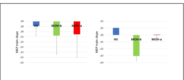
linear regression of MEP amplitudes over all stimuli was not significantly different between groups (t = 0.490, p = 0.628) (Figure 2, left panel).
In a rm-ANOVA model using the rTMS 5 Hz MEP peak-to-peak amplitude as the dependent variable, there was a significant main effect of stimuli (F9,243 = 2.367, p =
0.014) and the group × stimuli interaction (F9,243 = 3.714, p = 0.0002) but not group
(F1,27 = 1.029, p = 0.319) (Figure 1, right panel). The slope of the linear regression of
MEP amplitudes over all stimuli was significantly different between groups (t = 3.803, p = 0.0007) (Figure 2, right panel).
Effects of drug withdrawal on neurophysiological and clinical parameters
There was no significant difference in the mean slope of the linear regression of MEP amplitudes over all stimuli obtained in response to 1Hz rTMS before and after the 3-week drug withdrawal period in patients with MOH (t = −0.810, p = 0.937) (Table 2). In contrast, there was a significant difference in the mean slope of the linear regression of MEP amplitudes recorded in response to 5Hz rTMS between before and after drug withdrawal (t = -2,831, p = 0.015). Of note, the mean slope of MOH-a data was not significantly different from that for HVs (t = 0.854, p = 0.400).
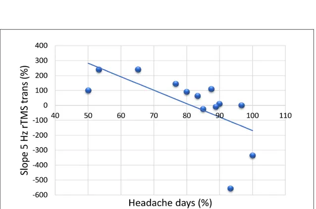
Mean days with headache per month and the mean number of tablets taken per month were also significantly decreased 1 month after withdrawal compared to baseline in patients with MOH (t = 12.338, p < 0.001; t = 5.252, p < 0.001 respectively) (Table 1). Moreover, there was significant negative correlation between the percentage reduction of days with headache at 1-month after withdrawal and the relative variation of the slope of the linear regression of MEP amplitudes recorded in response to 5 Hz rTMS (r = −0.637, p = 0.019) (Figure 3).
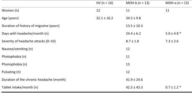
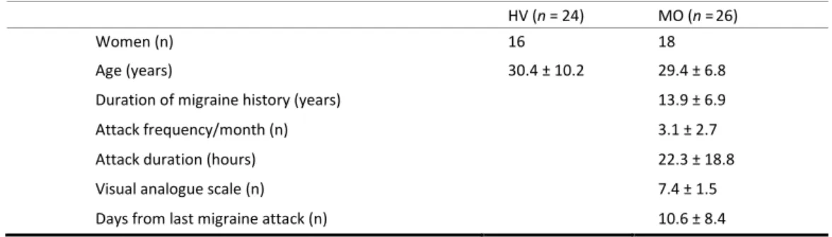
Table 1. Demographics characteristics of study participants and headache profiles of patients. Data
expressed as mean ± SD. HV healthy volunteers; MOH medication overuse headache patients before (MOH-b) and after (MOH-a) acute medication withdrawal; N number of subjects.
HV (n = 16) MOH-b (n = 13) MOH-a (n = 13)
Women (n) 12 11 11
Age (years) 32.1 ± 10.2 34.5 ± 9.8
Duration of history of migraine (years) 13.5 ± 10.3
Days with headache/month (n) 24.4 ± 6.2 5.0 ± 4.8 *
Severity of headache attacks (0–10) 8.7 ± 1.8 7.3 ± 2.6
Nausea/vomiting (n) 12
Photophobia (n) 11
Phonophobia (n) 13
Pulsating (n) 12
Duration of the chronic headache (month) 41.9 ± 24.6
Tablet intake/month (n) 42.5 ± 43.3 0.7 ± 1.2 *
Table 2. Transcranial magnetic stimulation (TMS) resting motor thresholds (RMT) and motor evoked
potential (MEP) 1st amplitude (Log transformed) and slope of the linear regression line from the 1st to the 10th stimulus of the train in MOH subgroup (n = 13) before and after 3 weeks of drug withdrawal. Data expressed as mean ± SD. HV healthy volunteers; CM chronic migraine patients; MOH medication overuse headache patients; N number of subjects; § p < 0.05 v. HV.
HV (n = 16) MOH-b (n = 13) MOH-a (n = 13) 1 Hz repetitive TMS train RMT (%) 54.9 ± 11.3 60.0 ± 11.4 61.2 ± 8.8 1st MEP amplitude 2.2 ± 0.3 2.3 ± 0.5 2.2 ± 0.5 MEP slope - 0.0020 ± 0.0151 - 0.0056 ± 0.0286 - 0.0050 ± 0.0320 5 Hz repetitive TMS train RMT (%) 54.6 ± 11.4 59.2 ± 9.0 58.5 ± 6.6 1st MEP amplitude 2.3 ± 0.3 2.4 ± 0.3 2.2 ± 0.5 MEP slope 0.0104 ± 0.0309 - 0.0303 ± 0.0255 § 0.0006 ± 0.0300 *
*p < 0.05 vs. MOH before withdrawal
Figure 1. Motor evoked potentials (MEP) elicited by repetitive transcranial magnetic stimulation trains delivered
at 1 Hz [left panel] and 5 Hz [right panel] at 120% resting motor threshold in healthy volunteers (HV), in medication overuse headache (MOH) patients before (MOH-b) and after (MOH-a) 3-week drug withdrawal.
Figure 2. Bar charts representing the motor evoked potential (MEP) amplitude slope of the linear regression line
from the 1st to the 10th stimulus of the 1 Hz [left panel] and 5 Hz [right panel] train of transcranial magnetic
stimulations in healthy volunteers (HV) and medication overuse headache patients before (MOH-b) and after (MOH-a) 3-week drug withdrawal.
Figure 3. Percentage changes in the mean monthly headache days and the slope of the linear regression after
patients with medication overuse headache completed a 3-week medication withdrawal program.
Discussion
The main finding of the present study was that a standard withdrawal program for patients overusing medication restored normal short-term synaptic potentiation in
the primary motor cortex of patients with MOH. Several neurobiological factors can account for these results.
In healthy subjects, trains of rTMS alter MEP amplitudes during and immediately after stimulation depending on the frequency and intensity of stimulation. When applied over the motor cortex at suprathreshold intensity (120% RTM), high frequency (5 Hz) rTMS increases MEP amplitudes [8], whereas low frequency stimulation (1 Hz) diminishes MEP amplitudes. Therefore, rTMS produces plastic changes in motor cortex excitability that outlast the period of stimulation for a period of minutes to hours [8–10].
The results of this study confirm our previous finding of dysfunctional short-term synaptic potentiation in patients with MOH [6]; in this study, trains of high-frequency rTMS induced a paradoxical decrease in amplitude in patients with MOH prior to medication withdrawal. This neurophysiological dysfunction may reflect a general alteration in plasticity and learning processes in the MOH brain. Moreover, the absence of these abnormalities in another group of patients with chronic migraine patients without MOH suggests that these findings are specifically related to medication overuse. In our previous study, this conclusion was underscored by the observation that longer durations of medication overuse were associated with more pronounced dysfunction of short term potentiation in the motor cortex [6], as previously demonstrated for the somatosensory cortex [2].
The present results expand on our previous findings by demonstrating that complete medication withdrawal restores normal short-term potentiation mechanisms within the motor cortex of patients with MOH. Moreover, withdrawal-related normalisation of rTMS responses corresponded to a change from a chronic migraine to episodic migraine as indicated by a relative reduction in the number of monthly headache days.
Since the primary motor cortex is involved in several aspects of pain integration and modulation, likely influencing affective or sensory components of pain or by top-down activation of descending antinociceptive systems [11, 12], drug withdrawal may induce the normalisation of a complex network involving brain areas that participate in pain modulation and control such as M1. Consistent with this idea,
previous studies have associated the discontinuation of medication overuse with the normalisation of several neurophysiological parameters and morphological features in brain areas of the salient network (also known as the “pain matrix”) [13].
Pain-related cortical potentials [4, 14] and spinal noxious flexion reflex responses [15] are sensitised in patients with MOH. These abnormal responses normalise after withdrawal treatment [4, 14, 15]. Perrotta and colleagues found that at the spinal level, the sensitisation process in MOH was related at least in part to insufficient descending inhibition from the brainstem, subserving the counterirritation phenomenon activated by heterotopic pain stimulation to suppress incoming nociceptive information [15]. The supraspinal antinociceptive structures include the periaqueductal grey, rostral ventromedial medulla, thalamus, nucleus raphe magnus, and nucleus reticularis gigantocellularis [16]. Altered structural integrity and functional connectivity of descending pain modulatory areas such as the periaqueductal grey [17–20] and thalamic nuclei [21] has been repeatedly identified in patients with MOH. These structures are all interconnected with areas belonging to the salient network such as the sensorimotor cortex and orbitofrontal and anterior cingulate cortices [13].
A voxel-based morphometry study identified significant increases in grey matter volume in the midbrain (including periaqueductal grey matter) of patients with MOH and subsequent decreases in volume after the discontinuation of medication overuse. Of note, low grey matter volume in the orbitofrontal cortex before withdrawal was associated with a poor response to drug discontinuation in a previous study [17]. In another study, the orbitofrontal cortex was less connected both metabolically [22] and functionally to nociceptive input regions such as spinal trigeminal nucleus and cerebellum [23] in patients with MOH before drug withdrawal, whereas these connections were normalized after drug withdrawal [22, 23]. Taken together, these data support the hypothesis that medication overuse promotes maladaptive neurophysiological and morphological changes in the brain.
Conclusion
In conclusion, we demonstrate that the dysfunction of short-term plasticity mechanisms in patients with MOH are alleviated by the discontinuation of medication overuse. On this premise, clinical improvements associated with withdrawal treatment may be related to the restoration of physiological brain plasticity. Our findings underscore the importance of initiating withdrawal treatment as early as possible in patients with MOH in order to facilitate normalisation of brain plasticity mechanisms. Future studies in a larger cohort of patients are necessary to determine the exact relationships between neurophysiological changes and clinical variables in patients with MOH, and whether the normalisation of such brain processes allow patients to regain clinical efficacy from acute and prophylactic migraine medications.
References
1. Headache Classification Committee of the International Headache Society (IHS) The International Classification of Headache Disorders, 3rd edition. Cephalalgia 2018; 38: 1–211.
2. Coppola G, Currà A, Di Lorenzo C, et al. Abnormal cortical responses to somatosensory stimulation in medication-overuse headache. BMC Neurol 2010; 10: 126.
3. Siniatchkin M, Gerber WD, Kropp P, et al. Contingent negative variation in patients with chronic daily headache. Cephalalgia 1998; 18: 565–9; discussion 531.
4. Ferraro D, Vollono C, Miliucci R, et al. Habituation to pain in ‘medication overuse headache’: a CO2 laser-evoked potential study. Headache 2012; 52: 792–807.
5. Rankin CH, Abrams T, Barry RJ, et al. Habituation revisited: an updated and revised description of the behavioral characteristics of habituation. Neurobiol Learn Mem 2009; 92: 135–138.
6. Cortese F, Pierelli F, Pauri F, et al. Short-term cortical synaptic depression/potentiation mechanisms in chronic migraine patients with or without medication overuse. Cephalalgia 2018; doi: 10.1177/0333102418784747.
7. Currà A, Coppola G, Gorini M, et al. Drug-induced changes in cortical inhibition in medication overuse headache. Cephalalgia 2011; 31: 1282–1290.
8. Pascual-Leone A, Valls-Solé J, Wassermann EM, et al. Responses to rapid-rate transcranial magnetic stimulation of the human motor cortex. Brain 1994; 117 ( Pt 4): 847–58.
9. Pascual-Leone A, Tormos JM, Keenan J, et al. Study and modulation of human cortical excitability with transcranial magnetic stimulation. J Clin Neurophysiol 1998; 15: 333–43.
10. Chen R, Classen J, Gerloff C, et al. Depression of motor cortex excitability by low-frequency transcranial magnetic stimulation. Neurology 1997; 48: 1398–1403.
11. García-Larrea L, Peyron R, Mertens P, et al. Electrical stimulation of motor cortex for pain control: a combined PET-scan and electrophysiological study. Pain 1999; 83: 259–73.
12. Pagano RL, Fonoff ET, Dale CS, et al. Motor cortex stimulation inhibits thalamic sensory neurons and enhances activity of PAG neurons: possible pathways for antinociception. Pain 2012; 153: 2359–69. 13. Uddin LQ. Anatomy of the Salience Network. In: Salience Network of the Human Brain. Elsevier, pp. 5–10. 14. Ayzenberg I, Obermann M, Nyhuis P, et al. Central sensitization of the trigeminal and somatic nociceptive
systems in medication overuse headache mainly involves cerebral supraspinal structures. Cephalalgia 2006; 26: 1106–14.
15. Perrotta A, Serrao M, Sandrini G, et al. Sensitisation of spinal cord pain processing in medication overuse headache involves supraspinal pain control. Cephalalgia 2010; 30: 272–284.
16. Peyron R, García-Larrea L, Grégoire MC, et al. Haemodynamic brain responses to acute pain in humans: sensory and attentional networks. Brain 1999; 1765–1780.
17. Riederer F, Gantenbein AR, Marti M, et al. Decrease of gray matter volume in the midbrain is associated with treatment response in medication-overuse headache: possible influence of orbitofrontal cortex. J Neurosci 2013; 33: 15343–15349.
18. Chen Z, Chen X, Liu M, et al. Disrupted functional connectivity of periaqueductal gray subregions in episodic migraine. J Headache Pain 2017; 18: 36.
19. Chen Z, Chen X, Liu M, et al. Texture features of periaqueductal gray in the patients with medication-overuse headache. J Headache Pain 2017; 18: 14.
20. Michels L, Christidi F, Steiger VR, et al. Pain modulation is affected differently in medication-overuse headache and chronic myofascial pain – A multimodal MRI study. Cephalalgia 2017; 37: 764–779.
21. Chen Z, Jia Z, Chen X, et al. Volumetric abnormalities of thalamic subnuclei in medication-overuse headache.
J Headache Pain 2017; 18: 82.
22. Fumal A, Laureys S, Di Clemente L, et al. Orbitofrontal cortex involvement in chronic analgesic-overuse headache evolving from episodic migraine. Brain 2006; 129: 543–550.
23. Mehnert J, Hebestreit J, May A. Cortical and Subcortical Alterations in Medication Overuse Headache. Front
4. D
ISCUSSION AND FUTURE PERSPECTIVESMigraine pathophysiology represents a complicated puzzle, which has not been completely disentangled.
Overall, the scientific evidences highlight the concept that both episodic and chronic migraine are characterized by neurophysiological dysfunctions in sensory processing and motor cortex plasticity, that probably represent permissive factors predisposing the brain to migraine attacks and pain chronification. The understanding of such dysfunctions can be useful not only to shed light on the complex mosaic of migraine pathophysiology, but also to set targets for neuromodulatory therapeutic strategies to prevent migraine attacks and interfere with the mechanisms involved in migraine chronification.
Chronic migraine is characterized by a maladaptive plasticity. From an electrophysiological point of view, sensory cortices in chronic migraine show abnormalities that have also been reported in episodic migraineurs during attacks, as if chronic migraine was a “never ending migraine attack”. Indeed, sensory cortices in chronic migraine are sensitized and exhibit normal habituation. Contrarily, in medication overuse headache (MOH) patients, sensitization and deficient habituation were demonstrated in the sensory cortices (for a review see Coppola et al, 2013).
Even though no differences were found in motor cortex excitability between chronic migraine, episodic migraine and healthy subjects, some studies revealed alteration in motor cortex plasticity.
Indeed, a paradoxical inhibitory response was found after facilitatory high-frequency repetitive transcranial magnetic stimulation of the motor cortex in chronic migraine (Cosentino et al, 2014)
The interest in studying motor cortex plasticity in chronic pain syndrome, came from the results of several neurostimulation studies showing that both invasive and non-invasive neuromodulatory techniques applied on the motor cortex could achieve an analgesic effect in various kinds of chronic pain (Leufaucheur et al, 2001; Fregni et
thalamocortical pathways or favouring opioids release (see Dos Santos et al, 2016). Furthermore, chronic pain can induce a reorganization of the motor cortex, whose extension is positively associated to pain intensity (Lotze et al, 1999).
Comparing motor cortex responses to trains of facilitating (high-frequency) and inhibiting (low-frequency) TMS in patients affected by MOH with those affected by chronic migraine (without medication overuse) and with healthy subjects, we showed that in MOH patients, rTMS-5 Hz depressed instead of potentiating MEP amplitudes with a significantly different response from that in HVs and CM patients (Cortese et al, 2018).
This finding suggests that CM and MOH patients, although exhibiting a similar phenotypic expression, represent distinct pathological conditions, characterized by different pathophysiological mechanisms of migraine chronification.
Furthermore, we found that the slope of the linear regression of MEP amplitudes was negatively correlated with the duration of overuse headache in MOH patients.
That means that medication overuse itself may probably promote plastic modifications in the motor cortex. This hypothesis is also supported by the finding of different CSP duration in MOH patients according to the different overused drug.
Studies about the relationship between chronic migraine and motor cortex plasticity could be interesting, not only to disclose the neurophysiological mechanisms underpinning learning processes and plastic behaviour in chronic migraine, but also to develop future therapeutic targets and interventions.
Interestingly we found that a 3-week pharmacological wash-out program restored a normal short-term synaptic potentiation in the primary motor cortex of patients with medication overuse headache. This finding has important pathophysiological implications. Firstly, a direct effect of medication overuse on the brain, causing short-term plasticity dysfunctions, may be hypothesized. Secondly, since such dysfunctions are reversible after drug discontinuation, it’s conceivable that the restoration of physiological brain plasticity could be the neurophysiological underpinning of the clinical improvement.
The presence of brain dysfunctions in MOH patients that can be reverted after detoxification was also described using metabolic and functional neuroimaging
techniques. These findings support the importance of early medication withdrawal in MOH patients also to prevent the development of more pronounced alterations in brain plasticity.
Probably drug withdrawal is able to induce the normalization of a complex network involving areas participating in pain modulation, including the primary motor cortex. Indeed, this area is known to be involved in several aspects of pain integration and modulation, likely influencing affective or sensory components of pain or by top-down activation of descending antinociceptive systems.
Our findings have important implications in neurorehabilitation.
Since neurorehabilitation includes all the approaches aimed to aid recovery from a nervous system injury or dysfunction and reduce disability, drug withdrawal in medication overuse headache could be completely considered a neurorehabilitation strategy. Indeed, its objective is brain recovery both from an electrophysiological point of view, with the restoration of physiological cortical plasticity, and from a clinical point of view, reducing the disability caused by chronic migraine and inducing the conversion from chronic to episodic migraine.
Even though, sometimes, simple information and advice may be enough to achieve headache improvement, for several patients drug discontinuation could be quite hard. Patients need to be guided by the physician during the process since withdrawal symptoms (headache, nausea, vomiting, arterial hypotension, tachycardia, sleep disturbances), lasting generally for 2–10 days, could complicate the discontinuation phase and induce patients to fall back into medication overuse.
The normalization of brain plasticity after medication discontinuation underscores the importance of initiating withdrawal treatment as early as possible in patients with medication overuse headache in order to induce the restoration of physiological brain plasticity and prevent the development of more pronounced alterations in brain plasticity and learning processes.
An interesting future perspective could be to use neuromodulatory strategies in order to normalize brain plasticity in medication overuse headache patients, thus helping them in the withdrawal treatment.
Remarkably, the response to high frequency stimulation could be used as a biomarker during the discontinuation process and to distinguish between chronic migraine patients with or without medication overuse.
5. R
EFERENCES1. Afra J, Mascia A, Gérard P, et al. Interictal cortical excitability in migraine: a study using transcranial magnetic stimulation of motor and visual cortices. Ann Neurol. 1998;44:209–215. 2. Ambrosini A, de Noordhout AM, Sándor PS, Schoenen J. Electrophysiological studies in
migraine: a comprehensive review of their interest and limitations. Cephalalgia 2003; 23 (Suppl. 1):13– 31.
3. Ambrosini A, Rossi P, De Pasqua V, Pierelli F, Schoenen J. Lack of habituation causes high intensity dependence of auditory evoked cortical potentials in migraine. Brain 2003; 126:2009– 15.
4. Andrade SM, de Brito Aranha REL, de Oliveira EA, de Mendonça CTPL, Martins WKN, Alves NT, Fernández-Calvo B. Transcranial direct current stimulation over the primary motor vs prefrontal cortex in refractory chronic migraine: A pilot randomized controlled trial. J Neurol Sci. 2017 Jul 15;378:225-232.
5. Antal, A., Kriener, N., Lang, N., Boros, K. & Paulus, W. Cathodal transcranial direct current stimulation of the visual cortex in the prophylactic treatment of migraine. Cephalalgia, 2011; 31, 820–828.
6. Ashina, S. et al. Depression and risk of transformation of episodic to chronic migraine. J. Headache Pain, 2012; 13, 615–624
7. Aurora S, Barrodale P, Tipton R, Khodavirdi A. Brainstem dysfunction in chronic migraine as evidenced by neurophysiological and positron emission tomography studies. Headache. 2007;47 (7):996–1003.
8. Aurora, S. K. et al. OnabotulinumtoxinA for treatment of chronic migraine: results from the double-blind, randomized, placebo-controlled phase of the PREEMPT 1 trial. Cephalalgia, 2010 30, 793–803.
9. Ayzenberg I, Obermann M, Nyhuis P, et al. Central sensitization of the trigeminal and somatic nociceptive systems in medication overuse headache mainly involves cerebral supraspinal structures. Cephalalgia. 2006;26(9):1106–14.
10. Bahra A, Matharu MS, Buchel C, et al. Brainstem activation specific to migraine headache. Lancet. 2001;357(9261):1016–7.
11. Bigal, M. E. & Lipton, R. B. Clinical course in migraine: conceptualizing migraine transformation. Neurology, 2008; 71, 848–855.
12. Bigal, M. E. et al. Acute migraine medications and evolution from episodic to chronic migraine: a longitudinal population-based study. Headache, 2008; 48, 1157–1168.
13. Bigal, M. E., Serrano, D., Reed, M. & Lipton, R. B. Chronic migraine in the population: burden, diagnosis, and satisfaction with treatment. Neurology, 2008; 71, 559–566
14. Blumenfeld, A. M. et al. Disability, HRQoL and resource use among chronic and episodic migraineurs: results from the International Burden of Migraine Study (IBMS). Cephalalgia 31, 301–315 (2011).
15. Buse, D. C. et al. Chronic migraine prevalence, disability, and sociodemographic factors: results from the American Migraine Prevalence and Prevention Study. Headache 2012; 52, 1456–1470. 16. Buse, D. C., Manack, A., Serrano, D., Turkel, C. & Lipton, R. B. Sociodemographic and
comorbidity profiles of chronic migraine and episodic migraine sufferers. J. Neurol. Neurosurg. Psychiatry, 2010; 81, 428–432.
17. Cargnin S, Viana M, Sances G et al (2017) A systematic review and critical appraisal of gene polymorphism association studies in medication-overuse headache. Cephalalgia 0:1–13. 18. Carlsen LN, Munksgaard SB, Jensen RH, Bendtsen L (2018) Complete detoxification is the most
effective treatment of medication-overuse headache: a randomized controlled open-label trial. Cephalalgia 38:225–236.
19. Cevoli S, Sancisi E, Grimaldi D et al (2009) Family history for chronic headache and drug overuse as a risk factor for headache chronification. Headache 49:412–418.
20. Chen WT, Wang SJ, Fuh JL, et al: Persistent ictal-like visual cortical excitability in chronicmigraine. Pain. 2011, 152(2):254–258.
21. Chen WT, Wang SJ, Fuh JL, et al: Persistent ictal-like visual cortical excitability in chronicmigraine. Pain. 2011, 152(2):254–258.
22. Chiang, C.‑C., Schwedt, T. J., Wang, S.‑J. & Dodick, D. W. Treatment of medication-overuse headache: a systematic review. Cephalalgia 36, 371–386 (2016).
23. Chung S, Li X, Nelson SB. Short-term depression at thalamocortical synapses contributes to rapid adaptation of cortical sensory responses in vivo Neuron 2002; 34:437–46.
24. Coppola G, Ambrosini A, Di Clemente L, Magis D, Fumal A, Gérard P, et al. (2007) Interictal abnormalities of gamma band activity in visual evoked responses in migraine: an indication of thalamocortical dysrhythmia? Cephalalgia 27:1360–1367
25. Coppola G, Curra A, Di Lorenzo C et al (2010) Abnormal cortical responses to somatosensory stimulation in medication-overuse headache. BMC Neurol 10:126.
26. Coppola G, Currà A, Di Lorenzo C, et al: Abnormal cortical responses to somatosensory stimulation in medication-overuse headache. BMC Neurol 2010, 10:126.
27. Coppola G, Currà A, Di Lorenzo C, et al: Abnormal cortical responses to somatosensory stimulation in medication-overuse headache. BMC Neurol 2010, 10:126.
28. Coppola G, De P, Pierelli F, Schoenen J (2012) Effects of repetitive transcranial magnetic stimulation on somatosensory evoked potentials and high frequency oscillations in migraine. Cephalalgia 32:700–709
29. Coppola G, Di Renzo A, Tinelli E, et al (2015) Evidence for brain morphometric changes during the migraine cycle: A magnetic resonance-based morphometry study. Cephalalgia 35:783–791. 30. Coppola G, Vandenheede M, Di Clemente L, Ambrosini A, Fumal A, De Pasqua V et al.
Somatosensory evoked high-frequency oscillations reflecting thalamo-cortical activity are decreased in migraine patients between attacks. Brain 2005; 128:98–103.