UNIVERSITÀ DEGLI STUDI DI SASSARI
International Ph.D School in Life Sciences and Biotechnology Curriculum: Biochemistry, Physiology and Molecular Biology
Coordinator: Prof. Leonardo Antonio Sechi XXIX Ciclo
A STUDY ON THE CARDIO - METABOLIC RISK FACTORS
IN VIETNAMESE FEMALES WITH LONG -TERM VEGAN DIET
Coordinator Prof. Leonardo Antonio Sechi
Tutor/Supervisor Professor Marilena Formato
Co-Tutor/Supervisor Professor Nguyen Hai Thuy
Co-tutor Dott Antonio Junior Lepedda, PhD
Table of contents List of abbreviation
ABSTRACT ... 1
1. INTRODUCTION ... 4
1.1. History of the metabolic syndrome ... 4
1.2. Global cardiovascular risk and mortality ... 6
1.3. The prevalance of diabetes and mortality ... 8
1.4. The cardio-metabolic risk factors ... 8
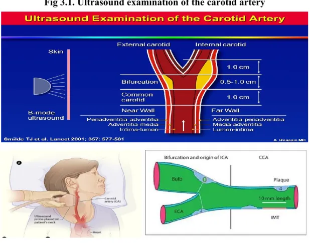
1.5. Role of ultrasound echography in detection of atherosclerosis ... 20
1.6. Vegan diet ... 21
2. RESEARCH OBJECTIVES ... 26
3. MATERIALS AND METHODS ... 28
3.1. Materials ... 28
3.2. Methods ... 28
3.3. Study parameters... 29
3.4. Data analysis ... 42
4. RESULTS IN STUDY ... 44
4.1. The relationship of cardio-metabolic risk factors and long-term vegan group with risk predictions in study groups ... 44
4.2. The cardio-metabolic risk factors of study groups ... 44
4.3. The relationship of age with the cardio-metabolic risk factors and risk prediction in study groups………..49
4.4. The relationship of cardio metabolic risk factors with the duration of vegan diet and risk prediction in vegan groups………..65
5. DISCUSSION ... 75
5.1. The effective of cardio-metabolic risk factors on long –term of vegan diet group and the non-vegan group………75
5.2. The relationship between the age and the vegan duration with the cardio metabolic risk factors in study groups……….82
CONCLUSIONS ... 88
STUDY IN ITALY ... 92 REFERENCES
ATTESTATION OF AUTHORSHIP ACKNOWLEGEMENT
Table 1.1. Intakes of Protein, Fat, Carbohydrates, Cholesterol, and Fiber .... 22
Table 3.1. World Health Organization cut-off points and risk of metabolic complications Indicator Cut-off points Risk of metabolic complications ... 31
Table 3.2. Waist –to-Hip Ratio (WHR) for males and females ... 32
Table 3.3. Assess obesity based on BMI with standard of WHO for the Asians countries following ... 32
Table 3.4. International Diabetes Federation criteria for ethnic or country-specific values for WC ... 32
Table 3.5. The WHO/ISH and American heart association of classification ABP ... 34
Table 3.6. The following information summarizes the meaning of other test results ... 38
Table 3.7. ATP III Classification of LDL, Total, and HDL Cholesterol (mg/dL) (2001) ... 42
Table 4.1.1. Age of study groups ... 44
Table 4.1.2. Duration of vegan diet ... 44
Table 4.2.1. BMI of study groups ... 44
Table 4.2.2. WC of study groups ... 45
Table 4.2.3. Blood Pressure of study groups ... 45
Table 4.2.4. IMTc of study groups ... 46
Table 4.2.5. hsCRP of study groups ... 46
Table 4.2.6. Ischemic heart disease of vegan group detected by ECG ... 46
Table 4.2.7.1. Fasting glucose of study groups ... 47
Table 4.2.7.2. HbA1c of study groups ... 47
Table 4.2.8.1. Fasting Insulin of study groups ... 47
Table 4.2.8.2. HOMA-IR of study groups ... 48
Table 4.2.9: Total lipid of study groups ... 48
Table 4.2.10. Atherogenic index ... 49
Table 4.2.11. Prevalence of MS in study subjects ... 50
Table 4.3.1. Relationship between BMI and age in the study groups ... 50
Table 4.3.3.2. Relationship between DBP and age in study groups ... 52
Table 4.3.4. Relationship between Age and IMTc in study groups ... 53
Table 4.3.5. Relationship between hsCRP and age in study groups ... 54
Table 4.3.6. Relationship between Age and IHD (ECG+) in study groups ... 54
Table 4.3.7.1. Relationship between Age and fasting glucose in study groups ... 55
Table 4.3.7.2. Relationship between HbA1c and age in study groups ... 55
Table 4.3.8.1. Relationship between fasting insulin level and age in study groups .... 57
Table 4.3.8.2. Relationship between HOMA-IR and age in study groups ... 58
Table 4.3.9.1. Relationship between lipid profile and age in vegan group ... 58
Table 4.3.9.2. Relationship between lipid profile and age in control group ... 59
Table 4.3.9.3. Relationship between atherogenic indices and age in vegan group. ... 61
Table 4.3.9.4. Relationship between atherogenic indices and age in control group .. 61
Table 4.3.10.1. Proportion of MS in study subjects ... 62
Table 4.3.10.2. Age of MS groups ... 63
Table 4.3.11. Proportion of cardiometabolic risk factors in the study groups .... 63
Table 4.3.12. Age cutoff values for cardio metabolic risk factors in study groups .. 64
Table 4.4.1. Relationship between duration of vegan diet with BMI and WC ... 65
Table 4.4.2. Relationship between duration of vegan diet and blood pressure .... 65
Table 4.4.3. Relationship between duration of vegan diet and hsCRP and IMTc .. 66
Table 4.4.4. Duration of vegetarian diet and CHO metabolism ... 67
Table 4.4.5. Relationship between the duration of vegan diet and insulin resistance indices ... 68
Table 4.4.6. Relationship between duration of vegan diet and total lipid profile ... 69
Table 4.4.7. Relationship between duration of vegan diet and atherogenic indices ... 71
Table 4.4.8. Duration of vegan diet and MS ... 72
Table 4.4.9. Correlation between the duration of vegan diet and cardio metabolic risk factors ... 72
Table 4.4.10. Multivariate regression analysis independent predictor between the HbA1c and the risk factors ... 73
Table 4.4.11. Age and duration of vegan diet cutoff point for cardio metabolic risk factors in vegan group ... 74
AUC Area under the curve
ROC Receiver operating characteristic DM Diabetes mellitus
NIDDM Non-insulin-dependent diabetes mellitus MS Metabolic syndrome
CMS Cardio metabolic syndrome CVD Cardiovascular disease
ASCVD Atherosclerotic cardiovascular disease IAA Intra-abdominal adiposity
FFAs Free fatty acids BMI Body mass index WC Waist circumference ABP Arterial blood pressure SBP Systolic plood pressure DBP Diastolic blood pressure
ISH International Society of Hypertension hsCRP High-sensitivity C-reactive protein. IMTc Intima-media thickness of carotid artery CCA Common carotid artery
ESC European Society of Hypertension ESH European Cardiovascular Society HbA1c Hemoglobin A1c
FBG Fasting blood glucose TC Total cholesterol
LDL.C Low-density lipoprotein (LDL) cholesterol HDL.C High-density lipoprotein (HDL) cholesterol STC Serum total cholesterol
TG Triglycerides
WHR Waist-to-hip ratio
HOMA-IR Homeostatic model assessment – insulin resistance SD Standard deviation
SDS-PAGE Sodium dodecyl sulfate-polyacrylamide gel electrophoresis
WB Western blot
WHO World Health Organisation
NHANES National Health and Nutrition Examination Survey NCDs Non communicable diseases
WHF The World Heart Federation IHD Ischemic heart disease PWV Pulse wave velocity FMD Flow-mediated dilation
ABSTRACT
A study of the cardio- metabolic risk factors in Vietnamese females with vegan diet.
Background
Numerous studies have shown that vegan diet has beneficial effects on the prevention of cardiovascular diseases. However, the effects of vegan diet on cardio-metabolic risk factors and the association between duration of vegan diet and those risk factors, are still unclear.
Objectives
The present study aims to investigate the prevalence and influence of duration of vegan diet on cardio- metabolic risk factors.
Materials and Methods
144 Buddhist nuns aged 20-75 years with duration of vegan diet ranged 10-70 years, were screened for cardio-metabolic risk factors. They were compared with 68 age-matched women 22-84 years of age on non-vegan diet.
Cardio-metabolic risk factors were assessed, including BMI, WC, blood pressure, fasting glucose, HbA1c, fasting insulin, HOMA-IR, plasma concentration of TC, LDL.C, HDL.C, TG, non-HDL.C, TC/HDL.C, LDL.C/HDL.C, TG/HDL.C, hsCRP, IMT of carotid artery and ischemic heart disease detected by ECG.
Results
1. Cardiovascular disease risk factors of female in vegan group
There was no significant difference in the mean BMI between vegan and control group (21.9 ± 3.1 vs 21.09 ± 2.50, p > 0.05). The prevalence of overweight (BMI ≥ 23) in vegan group was significantly higher than in control group (34.7% vs 10.3%, p < 0.05).
There was significant difference in the mean WC between vegan and control group (81.2 ±13.0 vs 74.18 ± 7.14cm, p < 0.05). The prevalence of android obesity (WC ≥ 80 cm) in vegan group was higher than in control group (53.5% vs 20.6%, p < 0.05).
The prevalence of hyper ABP (SBP and/or DBP) in vegan group was higher than in control group (26.45% vs 11.8 %, p < 0.05). The average SBP in vegan group was higher than that in control group (120.9±19.50 vs 115.59 ± 17.22 mmHg, p < 0.05)
The prevalence of ABP ≥ 130/85 mmHg (metabolic syndrome) in Vegan group was higher than in control group (34.03 % vs 26.47 %, p < 0.05).
The average fasting glucose in Vegan group was higher than in control group (5.00 ±1.4 vs 4.67 ± 0.98 mmol/l, p<0.05). The prevalence of hyperglycemia (based on fasting glucose) in Vegan group was higher than in control group (13.2% vs 10.3%, p < 0.05).
There were significant differences in HbA1c levels between two groups. The average HbA1c in Vegan group was higher than in control group (5.9 ±0.9 vs 4.3 ± 0.90 %, p < 0.05).
The prevalence of hyperglycemia (based on HbA1c) in Vegan group was higher than in control group (45.1% vs 13.2%, p < 0.05); prediabetes was 34% in Vegan group and 10.3% in control group.
The average fasting insulinemia in Vegan group was higher than that in control group (6.9 ± 4.3 vs 5.55 ± 2.13 µU/ml, p < 0.05). The proportion of fasting insulin ≥ 12 µU/ml in Vegan group was 7.6%.
The average HOMA-IR index in Vegan group was higher than in control group (1.67±1.62 vs 1.16 ± 0.55, p < 0.05). The proportion of HOMA-IR ≥ 2.6 in Vegan group was higher than control group (9.7% vs 1.5%, p < 0.05).
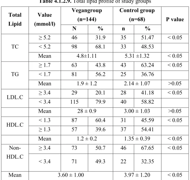
The mean TC in vegan group was significantly lower than in control group (4.8±1.11 vs 5.31±1.32 mmol/l, p < 0.05). The proportion of TG ( ≥ 1.7 mmol/l) in Vegan group was significantly lower than in control group (43.8% vs 63.2%, p < 0.05). The proportion of LDL.C (≥ 3.4 mmol/L) in Vegan group was significantly lower than in control group (20.1% vs 41.1. p < 0.05).
The average HDL.C in Vegan group was significantly lower than in control group (1.2 ± 0.2 vs 1.35 ± 0.39 mmol/l, p < 0.05). The proportion of HDL-C (< 1.3 mmol/L) in Vegan group was significantly higher than in control group (60.4 % vs 45.59%, p < 0.05).
The mean non-HDL.C in Vegan group was significantly lower than in the control group (3.6 ± 1.00 vs 3.97 ± 1.20 mmol/l, p < 0.05). The proportion of non-HDL.C (≥ 3.4 mmol/L) in Vegan group was significantly lower than in control group (50.7% vs 67.65 % p < 0.05).
The average IMTc in Vegan group was thinner than in control group (0.64 ± 0.39 mm vs 0.73 ± 0.11 mm, p < 0.05).
The prevalence of MS (+) in Vegan group was significantly higher than in controls (31.35% vs 2.9%, p < 0.001).
2. The prediction of age appeared the cardio-metabolic risk factors in study groups.
Benefits of Vegan diet with respect to the prevalence of cardio-metabolic risk factors were studied by using the ROC curves for predicting the age cut-off points between Vegan group and control group to; BP (58 vs 52 years), TC (61 vs 44 years), LDL.C (62 vs 44 years), non-HDL.C (46 vs 35 years), LDL.C/HDL.C (46 vs 39 years), CIMT (61 vs 56 years), respectively. Vegan diet seems to be disadvantageous towards prediabetes (43 vs 49 years), HOMA-IR (44 vs 68 years), TG (43 vs 53 years), hsCRP (50 vs 57 years) and MS (44 vs 68 years).
3. The relationship between duration of vegan diet and the cardio-metabolic risk factors with predicted values in Vegan females.
BMI was 20 yrs, WC was 30 yrs, SPB was 40 yrs, Hyper SBP and / SDP was 41 yrs, IMTc was 40 yrs, IHD (+) was 28 yrs, CRP was 49 yrs.
Prediabetes was 18 yrs and diabetes was 42 yrs, IR was 22 yrs.
Dyslipidemia: TC was 29 yrs, TC was 27 yrs, decrease HDL.C was 27 yrs, increase LDL.C was 44 yrs and atheroclerosis was 18 yrs.
MS (+) was 30 yrs.
There were correlations between duration of vegan diet and cardio- metabolic risk factors including BMI (r = 0.374), WC (r = 0.411), SBP (r = 0.539), FG (r = 0.312), HbA1c (r = 0.403), lipid profile (r = 0.307 - 0.525), hsCRP (r = 0.486) and IMTc (r = 0.463), in which the duration of vegan diet was considered as an independent risk factor for hyperglycemia.
Conclusions: A decrease in multiple cardio-metabolic risk factors such as
BP, TG, LDL.C, non-HDL.C, LDL.C/HDL.C and cIMT… was associated with vegetarian diet in female subjects. However, a long-term Vegan diet could increase metabolic syndrome (obesity, hyperglycemia, hypertension, insulin resistance, decreased HDL) in this population. These problems required an urgent need for greater public awareness on risk factors that correlated with the duration of vegan diet.
1. INTRODUCTION
1.1. HISTORY OF THE METABOLIC SYNDROME
According to a group of researchers [11]:
- In 1920s - Kylin first described the clustering of hypertension, hyperglycemia. Nicolae Paulescu, speaking about obesity and diabetes, said “most frequently, the obese people become glycosuria as if the two affections (obesity and fat diabetes) represent two consequent phases of the same pathological process” [4].
- In 1927, Maranon, in Spain, explicitly described the fact that the arterial hypertension is a pre-diabetical stage and this concept is similarly applied to obesity [15].
- In 1947,Vague also drew the attention to upper body adiposity (android or male-type obesity) as a metabolic abnormality commonly associated with type 2 diabetes and cardiovascular diseases [16] [17].
- In 1965, Yalow and Berson developed insulin assay and correlated insulin levels and glucose lowering effects in resistant and non-resistant individuals [21]. - In 1965, Vogaro et al., and then Haller et al in 1977 the frequent simultaneous presence of obesity, hypertension, diabetes and hyperlipidemia in association with atherosclerosis [11].
- Ten years later, in 1988. Reaven suggested that insulin resistance was a fundamental “disorder” associated with a set of metabolic abnormalities which not only increased the risk of type 2 diabetes but also contributed to the development of cardiovascular disease before the appearance of hyperglycemia [18-19]. He emphasized that insulin resistance was at the centre of a cluster of metabolic abnormalities, which include hypertriglyceridemia, low LDL.C level, increased glycemia, and elevated BP [13]. A later key conceptual advance was the recognition of the central role of abdominal obesity [20] in the diagnosis of the metabolic syndrome, and its introduction as a clinically easy-measurable entity.
1.1.1. Definition of metabolic syndrome and cardio metabolic syndrome
Metabolic syndrome was called by other names such as: Syndrome X, Cardiometabolic Syndrome, Cardiovascular Dysmetabolic Syndrome, Insulin-Resistance Syndrome, Metabolic Syndrome, Beer Belly Syndrome, and Reaven’s Syndrome.
1.1.1.1.Metabolic syndrome (MetS)
Metabolic syndrome is a name for a group of risk factors and combination of medical disorders that can increase your chance of developing heart disease, diabetes, and stroke. These risk factors include: Elevated blood pressure, Impaired glucose tolerance, Insulin resistance, Abdominal obesity, elevated triglyceride levels and low LDL.Cs. Each of the associated conditions has an independent effect, but clustering together they become synergistic, making the risk of developing cardiovascular disease (CVD) greater. This article reviews the evidence that demonstrates that individuals with the MetS are at increased risk for CVD incidence and mortality and discusses these debated issues [23].
1.1.1.2.Cardiometabolic syndrome
Cardiometabolic syndrome (CMS), also known as insulin resistance syndrome or metabolic syndrome X, a combination of metabolic disorders or risk factors that essentially includes a combination of diabetes mellitus, systemic arterial hypertension, central obesity and hyperlipidemia. Common to these diseases of metabolism is associated development of atherosclerotic cardiovascular disease (ASCVD). Studies have shown a strong link between CMS and increased prevalence of peripheral vascular diseases, coronary artery disease and myocardial infarctions as well as cerebro-vascular arterial diseases and stroke [5,22].
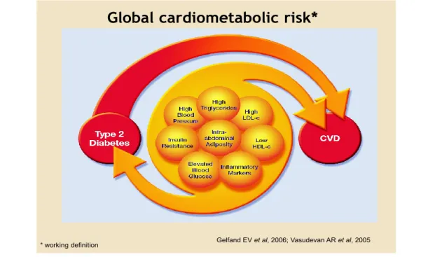
Fig 1.1.Global Cardiometabolic risks
Global cardiometabolic risk*
Gelfand EV et al, 2006; Vasudevan AR et al, 2005 * working definition
We have understood for decades the roles of ‘classical’ risk factors - elevated LDL.Cl, hypertension, elevated blood glucose and smoking in the pathogenesis of cardiovascular disease. Abdominal obesity is associated with multiple cardiometabolic risk factors such as atherogenic elevated blood glucose (hypertriglyceridaemia and low HDL.C), elevated blood glucose and inflammation, which are major drivers of cardiovascular disease and type 2 diabetes. In addition, atherosclerosis is increasingly regarded as an inflammatory condition.
Evaluation criteria of metabolic syndrome according to IDF 2006 (International Diabetes Federation) [85].
Three or more of the following:
1. Abdominal obesity: waist > 40” for men, >35” for women (Asia: WC male ≥ 90 cm and female ≥ 80 cm) 2. High triglycerides: >150 mg/dL (1.7 mmol/l)
3. Low HLD.C:
< 40 mg/dL male (1.03 mmol/l), < 50 mg/dL women (1.29 mmol/l) 4. High blood pressure: >130 systolic or > 85 diastolic (mmHg)
5. High fasting plasma glucose: >100 mg/dL (5.6 mmol/l)
Risk factor for cardiovascular disease and glucose intolerance
Malik and colleagues demonstrated that the cardio metabolic risk factors associated with metabolic syndrome increase the CVD mortality rate. Relative to an individual with no metabolic syndrome risk factors, having 1 to 2 risk factors increased a patient’s hazard ratio by more than 70%. Persons with metabolic syndrome (having ≥ 3 of the 5 risk factors) were found to have a hazard ratio of 2.71. The ratio increased with the onset of type 2 diabetes, CVD, and was greatest in persons with existing CVD and T2DM [1].
1.2. GLOBAL CARDIOVASCULAR RISK FACTORS AND MORTALITY
CVD are an emerging public health problem in developing countries that CVD has been an important health issue in developed countries for some decades, while in developing countries it has often not been seen as a major problem compared with communicable diseases and malnutrition [29]. The CVD epidemic is decreasing as a result of major efforts to identify risk factors and implement interventions [30]. Meanwhile, in many developing countries, CVD and related
risk factors are emerging as increasingly important public health problems [31-39]. CVD is the term used by the scientific community to embrace not just conditions of the heart (coronary artery, valvular, muscular, and congenital disease), but also hypertension and conditions involving the cerebral, carotid and peripheral circulation [40].
1.2.1. The prevalence of CVD in United States
Heart disease has been the leading cause of death in the United States for the past 80 years and is a major cause of disability.
- In 1995: the leading cause of death in US women was heart disease, which has been the leading cause of death for the past 50 years [24].
- In 1998: estimated direct and indirect costs of heart disease were $95.6 billion, and 53.3 million adults had elevated LDL-C and warrant intervention (1994 NHANES data); 22.3 million qualified for drug therapy and 5.5 million received therapy [25].
- In 2005: cardiovascular heart disease (CHD) was the single largest killer of men and women (Monthly Vital Statistics Report: MV). Each year 1.1 million people have myocardial infarction (MI); 370.000 die of MI, 250.000 die within 1 hr. By age 60, every 5th man and 17th woman develops CHD [26].
1.2.2. Cardiovascular disease epidemiology in Asia
CVD is the leading cause of death in the world and half of the cases of CVD are estimated to occur in Asia. Compared with Western countries, most Asian countries, except for Japan, South Korea, Singapore and Thailand, have higher age-adjusted mortality from CVD. Hypertension and smoking are the most notable risk factors for stroke and coronary artery disease, whereas dyslipidemia and DM are risk factors for IHD and ischemic stroke [27].
1.2.3. In Viet Nam
The probability of dying between ages 30 and 70 years from the 4 main NCDs is 17%. Adult risk factors included raised blood pressure (2008) affecting 20.5% females in total 23.1%, and obesity (2008) 2.1% females in total 1.7%.
The World Health Organization (WHO) defines CVD as disorders of the
heart and blood vessels, including hypertension, heart failure, stroke, myocardial infarction, coronary artery disease and atherosclerosis.
The World Heart Federation (WHF) forecast that up to 20 percent of
Vietnam’s population will suffer heath problems caused by cardiovascular diseases and hypertension by 2017. In addition to this, a recently conducted study [42] found that the prevalence of overweight and obesity in adults is 6.4% in Ho Chi Minh City.
1.3. THE PREVALANCE OF DIABETES AND MORTALITY 1.3.1. In United States
By the year 2025, it is estimated that nearly 22 million adults in the United States will have diabetes. The most common form - type 2 diabetes - accounts for 90% to 95% of all diagnosed cases, whereas type 1 diabetes accounts for 5% to 10% of all diagnosed cases. In some studies, in nearly 40% of women who had gestational diabetes, diabetes later developed [43].
1.3.2. In the South – East Asia
Close to one-fifth of all adults with diabetes in the world live in the South-East Asia Region. Current estimates indicate that 8.3% of the adult population, or 71.4 million people, have diabetes in 2011. 61.3 million of whom are in India. The number of people with diabetes in India, Bangladesh and Sri Lanka makes up 99% of the total for the region
1.3.3. The prevalence of mortality in Asia
The region has the second highest number of deaths attributable to diabetes of any of the seven IDF. 16 regions with 1 million deaths in 2011. This represents 14.5% of all deaths for the region among adults. More than half (55%) of these deaths occur in people under the age of 60 and almost a third (27%) under the age of 50. India is the largest contributor to regional mortality with 983.000 deaths attributable to diabetes [44].
1.4. THE CARDIO-METABOLIC RISK FACTORS 1.4.1. Overweight and Obesity
All of us love to have a well-built bodies, or at least not to be obese, and we have right to think like that, because obesity is not just a cosmetic concern, it is also a risk for some health problems, such as heart disease, diabetes and the high blood pressure and others. That is, one of the most common problems related to lifestyle today is being overweight. Obesity and overweight are serious problems that pose a huge and growing financial burden on national resources.
1.4.1.1. Definition of Overweight and Obesity by WHO [50]
Overweight and obesity are defined as abnormal or excessive fat accumulation in the fat tissues (adipose tissue) of body that presents a risk to health.
Body weight depends on the balance between food intake and utilization of food in the body [54]. Obesity may result when food intake exceeds the utilization of energy.
BMI: the most commonly used measure for overweight and obesity is BMI - a simple index to classify overweight and obesity in adults. The range that is considered "normal" or healthy weight depends upon a person's height. It is natural for taller people to weigh more. BMI stands for Body Mass Index. It is a number that is calculated based on both the height and weight to determine if the weight is high or low compared with what you would expect for that height. BMI is an indicator of body fatness, and is used as a screening tool for weight issues.
It is defined as the weight in kilograms divided by the square of the height in meters (kg/m2).
BMI = Weight (kg) / [Height sq. (m2)]
1.4.1.2. Differences between overweight and obesity
The term overweight is generally used to indicate the excess weight while obese refers to excess fat. Being overweight means having more body weight than is considered normal or healthy for one‘s age or build. On the other hand, Obesity is the condition of being obese, i. e., excess amount of body fat. While an overweight person will carry excess weight, he may or may not have excess accumulation of fat.
1.4.1.3. Causes of overweight and obesity
Our daily bodies activities need energy come from food we eat and more exercise and activities burn more calories that we get from food. And not just the activities need to burn calories also many metabolic reactions in the body need, such as to warm up in cold weather and to sweat in hot days. But when our food calories amount exceeds the body need, they will be stored in the body as fatty. The major causes of obesity are excessive food energy intake and lack of physical activity. A limited number of cases are due primarily to genetics, medical reasons, or psychiatric illness [52]. In contrast, increasing rates of obesity at a societal level are felt to be due to an easily accessible and palatable diet [53].
The causes of obesity is a combination of stated factors that work together to store more fat in our bodies and these factors include:
1. Inactivity: without activity you do not burn as much calories.
2. Diets: some bad eating habits like high calories diet epically in the night, or skipping breakfast healthy and replace it by junk fast food, all of that increase the body fat.
3. Pregnancy at a later age
4. Insufficient sleep: this cause disturbances in the body hormones and increase the appetite.
5. Increase in the use of medications that can cause weight gain (e.g., a typical antipsychotics)
6. Endocrine disruptors (environmental pollutants that interfere with lipid metabolism),
7. Proportional increases in ethnic and age groups that tend to be heavier 8. Genetics, environmental and social, as well as several other factors can all contribute to obesity and family lifestyle.
9. Proportional increases in ethnic and age groups that tend to be heavier: could occur at any age, and when we get age we lose more amount of muscles built. More amount of muscles gives higher rate of metabolism and calories burning. When we lose them we will reduce the calories burning and tend to fill the body with fat. And there is a decrease in energy expenditure, particularly in the 50- to 65-year-old age group. In those 65 years of age and older, hormonal changes that occur during aging may cause the accumulation of fat. Aging is associated with a decrease in growth hormone secretions, reduced responsiveness to thyroid.
1.4.1.4. The prevalence of overweight and obesity in some countries - In USA:
Urbanization and economic development have led to a nutritional transition characterized by a shift to diet of higher energy content and reduction of physical activities, resulting in changes individual body composition [46]. Over 1.6 billon adults worldwide are overweight, of which 400 million are obese with higher rates among women than in men. Obesity also increases with age at least up to 50 or 60 years [48]. The World Health
Organization (WHO) projects that more than 700 million adults will be obese by 2015. The prevalence of overweight and obesity is rapidly increasing in developing as well as industrialised countries [47].
- In Asia:
Prevalence rates of overweight and obesity in Asia Pacific countries are rising, compared to Australia, UK, New Zealand and USA, 2008. For example, between 1980 and 2013.China’s overweight and obesity prevalence in adults rose from 11.3% to 27.9% and in individuals below age 20 from 5.7 % to 18.8 % [51].
1.4.2. Waist circumference (Central obesity)
WHO Expert Consultation on WC and WHR was held in Geneva, Switzerland on 8–11 December 2008 and chose WC to be used as an indicator of abdominal obesity and is associated with the cardiovascular diseases and type 2 diabetes [76a].
1.4.2.1. WC is Pathophysiology of Metabolic Syndrome
Abdominal obesity and MS: contribution to Global Cardio metabolic Risk factors. In central obesity excess macronutrients are ingested and energy is always in over-supply. Not only are lipids unable to be accommodated and oxidised in a timely manner in the body’s cells, but glucose uptake is also compromised. The pancreatic β-cells increase the tonic insulin production to increase the transport of glucose into cells as glycogen storage molecules in the liver.
Excess intra-abdominal adipose (IAA) typically is accompanied by elevated levels of CRP and FFAs, as well as decreased levels of adiponectin. Abdominal obesity has been shown to be associated with the inflammation cascade, with adipose tissue expressing a number of inflammatory cytokines. Inflammation is now believed to play a role in the development of atherosclerosis and type 2 diabetes. There is substantial evidence of sex and age variations in WC and WHR and some evidence for ethnic differences. It also highlighted the need for other indicators to complement the measurement of BMI, to identify individuals at increased risk of obesity-related morbidity due to accumulation of abdominal fat [75]. WHR (i.e. the waist circumference divided by the hip circumference) was suggested as an additional measure of body fat distribution. The ratio can be measured more precisely than skin folds, and it provides an index of both subcutaneous and IAA [73]. A way to measure fat distribution is the circumference of the waist [2]. WC is unrelated to height and provides a simple and practical method of identifying overweight people who are at increased risk of obesity-related conditions. If waist circumference is greater than 94-102 cm for men and 80-88 cm for women, it means they have excess abdominal fat, which puts them at greater risk of health problems, even if their BMI is about right [64].
1.4.2.2. Relationships between IAA and increased cardio metabolic risk
IAA is a major contributor to increased cardio metabolic risk facors. The classical CVD risk and waist markers of the MetS are very useful concepts which have made clinicians and researchers aware of the importance of central obesity. The prevalence of insulin resistance in obese with abdominal fat accumulation was higher (p < 0.05) compared to obese with global fat (52.6 vs 28.6 %), respectively [63].
1.4.3. Insulin resistance and type 2 diabetes
1.4.3.1. Definitions of Diabetes and Impaired Fasting Glucose
Diabetes is defined as group of diseases characterized by high blood glucose concentrations resulting from defects in insulin secretion, insulin action, or both. It can be defined as an impaired response to the physiological effects of insulin, including those on glucose, lipid, and protein metabolism, and the effects on vascular endothelial function [77].
Diabetes Type 2 Pathophysiology
Results from a combination of insulin resistance and β-cell failure Insulin resistance: decreased tissue sensitivity or responsiveness to insulin
Endogenous insulin levels may be normal, depressed, or elevated, but
inadequate to overcome insulin resistance
Diabetes Type 2 Risk Factors
Family history of diabetes, Older age, Obesity, particularly intra-abdominal obesity, Physical inactivity, Prior history of gestational diabetes, Impaired glucose homeostasis, Race or ethnicity
Prediabetes (Impaired Glucose Homeostasis) [82]
Impaired fasting glucose (IFG)
Fasting plasma glucose (FPG) above normal (>100 mg/dl and < 126 mg/dl) Impaired glucose tolerance (IGT)
Plasma glucose elevated after 75 g glucose load (>140 and < 200 mg/dl) • New ADA definition (1998) defines fasting blood sugar of > 126 mg/dl as diabetes, casual blood glucose > 200 mg/dl. Impaired fasting glucose is 110-125 mg/dl
• Diabetic control generally defined as HbA1c < 8%.
• BP recommended < 130/80 mmHg, LDL.C goal < 100 mg/dl
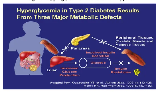
1.4.3.2. Hyperglycemia in type 2
The macronutrients are composed of (1) CHO subgroups, starches and sugars (2) lipids (fats), and (3) protein; of which the latter is not usually ingested primarily for energy.
Three major metabolic defects contribute to hyperglycemia in patients with type 2 diabetes: increased hepatic glucose production, impaired pancreatic insulin secretion, and peripheral tissue insulin resistance.
After eating a meal or ingesting glucose, insulin is secreted, hepatic glucose output is suppressed, and insulin-dependent glucose uptake by peripheral tissues is stimulated. In type 2 diabetes, insulin resistance and impaired insulin secretion inhibit normal suppression of hepatic glucose output. As a consequence, the liver continues to release glucose into the circulation.
This finding supports the idea of Stern and [62] which reported that central obesity has been associated with hyperinsulinemia and it has been suggested that the over production of insulin may act as an energy conserving mechanism under conditions of periodic famine and low energy intake.
The health consequences of obesity and overweight are many and varied, ranging from an increased risk of premature death to several non-fatal but debilitating and psychological complaints that can have an adverse effect on quality of life [55].
Women who are obese are more than 12 times more likely to develop Type 2 diabetes than women of healthy weight. The risk of Type 2 diabetes increases with BMI, especially in those with a family history of diabetes, and decreases with weight loss [56].
There is an age-related increase in total body fat and visceral adiposity until age 65 that is often accompanied by diabetes or impaired glucose intolerance (Wilson et al, 2007). In the Framingham Study 30-40% of people over 65 were found to have diabetes or glucose intolerance.
1.4.3.3. Insulin resistance
Insulin is a hormone made by your pancreas. It allows your cells to use glucose (sugar) for energy. People with insulin resistance have cells that don’t use insulin effectively. This means the cells have trouble absorbing glucose, which causes a buildup of sugar in the blood. If your blood glucose levels are higher than normal, but not high enough to be considered type 2 diabetes, you have a condition called prediabetes. Insulin resistance may also damage your blood vessels without you realizing it. This can increase your risk of heart disease and stroke. Insulin resistance is a primary defect in type 2 diabetes. As reported in a recent study by Haffner and colleagues, 92% of patients with type 2 diabetes have insulin resistance. Overweight and Obesity are known Risk Factors For Major Diseases.
1.4.3.4. Relationship between WC and IR
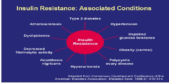
In addition to type 2 diabetes, IR is associated with the development of a broad spectrum of clinical conditions. These include hypertension, atherosclerosis, dyslipidemia, decreased fibrinolytic activity, impaired glucose tolerance, acanthosis nigricans, hyperuricemia, polycystic ovary disease, and obesity. The high prevalence of IR among women mainly those aged 40 - 49 years as shown in this study can be explained by the role of central obesity, a high percentage of fat and the strong sedentary lifestyle of women in Cameroon. Central obesity is associated with insulin resistance and elevated levels of FFAs. As illustrated, FFAs can reduce insulin-mediated glucose disposal under experimental conditions. This enzyme catalyzes the removal of lipids from LDL and HDL, which makes them smaller and more dense. In turn, these effects lead to hypertriglyceridemia, production of small, dense LDL particles, and reduced HDL2-cholesterol levels. This dyslipidemic pattern, which has been termed the atherogenic lipoprotein phenotype, is also characteristic of that found in type 2 diabetes [79]
Excess IAA increases overall cardio metabolic risk partially through alterations in the secretion of a series of biologically active molecules (adipokines). These include increased secretion of free fatty acids which can induce insulin resistance in muscle and ß-cell toxicity in the pancreas, inflammatory mediators, such as TNF, IL-6.resistin and PAI-1. and decreased secretion of the cardioprotective adipokine, leptin that contribute to insulin resistance [49].
Obesity predisposes an individual to a number of cardiovascular risk factors, including hypertension and elevated blood cholesterol. In women, obesity is the third most powerful predictor of CVD after age and blood pressure [57].
In addition to type 2 diabetes, insulin resistance is associated with the development of a broad spectrum of clinical conditions. These include hypertension, atherosclerosis, dyslipidemia, decreased fibrinolytic activity, impaired glucose tolerance, acanthosis nigricans, hyperuricemia, polycystic ovary disease, and obesity [80].
Fig 1.5.Atherosclerosis Mechanism
The Metabolic Syndrome
Insulin Resistance Hypertension Type 2 Diabetes Disordered Fibrinolysis Complex Dyslipidemia TG, LDL HDL Endothelial
Dysfunction InflammationSystemic
Athero-sclerosis
Visceral Obesity
Adapted from the ADA. Diabetes Care. 1998;21:310-314; Pradhan AD et al. JAMA. 2001;286:327-334.
Insulin resistance is a precursor to a variety of metabolic abnormalities, including systemic inflammation, visceral obesity, and type 2 diabetes. Insulin resistance is also a risk factor for cardiovascular abnormalities, including hypertension, dyslipidemia (increased triglycerides and LDL and decreased HDL), disordered fibrinolysis, and endothelial dysfunction. All of these aberrations contribute to the atherosclerotic process [81].
1.4.4. High cholesterol and triglycerides
The relevance of plasma triglyceride levels as a CHD risk marker has been debated for decades [87] although some recent studies have suggested that no fasting triglycerides concentrations may be a useful marker of risk [88].
Obese individuals are more likely to have elevated blood triglycerides (blood fats), low LDL.C ("bad cholesterol") and decreased high HDL.C (“good cholesterol”). A 10 kg weight loss can produce a 15% decrease in LDL cholesterol levels and an 8% increase in HDL cholesterol [58].
1.4.4.1. Elevated FFA levels: play a significant role in the cause of IR and β-cell
dysfunction. Adiponectin is an adipose tissue-specific circulating protein which is involved in the regulation of lipid and glucose metabolism. Adiponectin has been shown to be reduced in adults with obesity and type 2 diabetes. In non-diabetics, hypertriglyceridemia and low HDL-cholesterol have been shown to be associated with low plasma adiponectin concentrations. All of these components help to explain why excess abdominal adiposity is considered to be a great threat to cardiovascular and metabolic health. A proxy marker for which is waist circumference (waist), has been shown to increase risk for CVD. Hypertension or raised systolic and diastolic blood pressure (S/DBP), serum dyslipidemia, namely decreased high density lipoprotein cholesterol (HDL.C) and raised triglyceride (TG), and impaired fasting plasma glucose (FPG) or FPG in the type II diabetes mellitus (TIIDM) range, are conditions which commonly occur together.
1.4.4.2. Dysregulation of Lipid and Glucose Metabolism
The abundance of stored fat is required for survival during nutritionally deprived states such as starvation. However, very efficient fat storage results in
the excessive storage of fat, eventually resulting in obesity (Speigelman et al., 2001; Ravussin et al., 1989; Seeley et al., 2003). The release of these excessive free fatty acids then incites lipotoxicity, as lipids and their metabolites create oxidant stress to the endoplasmic reticulum and mitochondria. This affects adipose as well as nonadipose tissue, accounting for its pathophysiology in many organs, such as the liver and pancreas, and in the metabolic syndrome [59].
The FFAs released from excessively stored triacylglycerol deposits also inhibit lipogenesis, preventing adequate clearance of serum triacylglycerol levels that contribute to hypertriglyceridemia. Release of FFAs by endothelial lipoprotein lipase from increased serum triglycerides within elevated β lipoproteins causes lipotoxicity that results in insulin-receptor dysfunction. The consequent insulin-resistant state creates hyperglycemia with compensated hepatic gluconeogenesis. The latter increases hepatic glucose production, further accentuating the hyperglycemia caused by insulin resistance. Free fatty acids also decrease utilization of insulin-stimulated muscle glucose, contributing further to hyperglycemia (Pan et al., 1997; Boden et al., 1994). Lipotoxicity from excessive free fatty acids also decreases secretion of pancreatic β-cell insulin, which eventually results in β-cell exhaustion (Unger et al., 1995).
1.4.4.3. Atherosclerosis
As lipids are transported in the blood as lipoproteins, among them, serum HDL is responsible for reverse Transportation, specifically for carrying cholesterol from tissues back to the liver [2] and thus, acts as an anti-atherosclerotic factor. On the other hand, LDL, the chief pathogenic factor for atherosclerosis, drives cholesterol to the peripheral tissues. Since vegetable diets contain less saturated fat and cholesterol, and greater amounts of dietary fiber, their consumptions help to lower the level of serum cholesterol. Results of our study suggest that vegetable-based diets lower both atherogenic lipoproteins as LDLcholesterol and non-atherogenic lipoproteins as HDL lipoprotein, with potentials to reduce the risk of developing microvascular diseases. Vegetarians consume whole grains, soybeans, and nuts [3], all of which have significant cardio-protective effects, and reduce overall incidence of stroke, as well as risk of deaths from stroke and ischemic heart disease [7]. Elevated serum atherogenic lipoproteins and their higher ratios due to various vasoprotective lipoproteins are primary risk factors for atherosclerotic
changes with increased risk of micro vascular diseases [8]. Higher TC, LDL.C and TG, and higher ratio of LDL: HDL and lower serum HDL.C increase the risk of coronary heart disease, and are considered major risk factors beside smoking and hypertension [9].
1.4.5. C-reactive protein (CRP)
Excess IAA typically is accompanied by elevated levels of C-reactive protein (CRP) and free fatty acids (FFAs), as well as decreased levels of adiponectin. Elevated CRP is an indicator of inflammation. Abdominal obesity has been shown to be associated with the inflammation cascade, with adipose tissue expressing a number of inflammatory cytokines. Inflammation is now believed to play a role in the development of atherosclerosis and type 2 diabetes. CRP are considered to be predictive of cardiovascular disease and insulin resistance.
The progressive pro-inflammatory state resulting from increased obesity that promotes IR also perpetuates atherogenesis throughout its development, from early endothelial fatty streaks to late-plaque formation, rupture, and thrombosis. Endothelial modulators such as vasoactive endothelial growth factor (Ferrara et al., 1997), angiotensinogen, renin, and angiotensin II are secreted by white fat cells, in particular by perivascular fat tissues that contribute to vasomotor dysfunction and cause hypertension and endothelial injury. This activity causes atheroma cap thinning and plaque rupture that precipitates release of the tissue factor, also promoting intravascular thrombosis.
1.4.6. Cardiovascular disease, obesity and hypertension risk factors
Cardiovascular disease (CVD) includes coronary heart disease (CHD), stroke and peripheral vascular disease. These diseases account for a large proportion (up to one third) of deaths in men and women in most industrialised countries and their incidence is increasing in developing countries.
Hypertension or high blood pressure. Hypertension can lead to premature atherosclerosis, coronary artery disease, heart attacks, abnormally large hearts and strokes. Obesity may blunt certain actions of insulin that open blood vessels and may cause structural changes in the kidney and abnormal handling of sodium.
of the metabolic syndrome (in the aggregate often shown to be a cardiovascular risk factor), such as insulin resistance and glucose intolerance. The hypertension effect of blood lipid levels not effectively mitigated by drugs can contribute to a higher mortality rate from CHD. Furthermore, obesity as a significant risk factor for diabetes also increases cardiovascular risk through diabetes.
The prevalence of hypertension in overweight individuals is nearly three times higher than in non-overweight adults and the risk in overweight individuals aged 20-44 years of hypertension is nearly six times greater than in no overweight adults.
1.5. ROLE OF ULTRASOUND ECHOGRAPHY IN DETECTION OF ATHEROSCLEROSIS
Ultrasound is widely used to evaluate atherosclerosis, for the assessment the risk of cardiovascular disease in healthy persons and in persons with cardiovascular risk factors. [72] Three markers of vascular disease may help to a better evaluation of vascular dysfunction in type 2 DM and IMTc, arterial stiffness, assessed by PWV, and endothelial function, evaluated through the brachial artery (FMD). [86,51,61] Among these parameters, IMTc is considered a marker of structural vessel wall properties, and arterial stiffness reflects functional wall properties. Endothelial dysfunction is a systemic process who appear in the coronary circulation as in the systemic circulation. IMTc and brachial artery FMD are affected in diabetes mellitus, and they are risk marker of atherosclerotic cardiovascular disease. IMTc incresed in patients with type 2 DM and other independent risk factors, such as: age, hyperlipidaemia, and duration of DM. The patients with DM have shown increased arterial stiffness, with reductions in FMD which has already been reported to be inversely and strongly related to the extent of hyperglycemia [86].
In the early stage of atherosclerosis, the functional parameters, as FMD are helpful in the assessment of cardiovascular risk to the patients with cardiovascular risk factors [41]. The increased IMTc and the altered FMD are related with the presence and the degree of coronary atherosclerosis [71].
1.6. VEGAN DIET
1.6.1. Introduction of vegetarian diets
Nowadays, the movement vegetarian diet is very popular not only in Viet Nam but also over the world because there are health benefits such as better for heart, decrease obesity and hypertension, anticancer diseases, lower cholesterol levels, diabetes. A diet high in saturated fats (e. g. cheese) and trans fats (often used in cakes, cookies and fast food) leads to high levels of cholesterol.
Excess insulin is the second problem. Insulin is a major hormone in the body, and is released in high levels anytime you ingest what would be considered a “simple” carbohydrate which would include, but not be limited to: fruit juice, white bread, most “wheat” bread (basically white bread with a little extra fiber), white rice, baked white potato, bagels, croissants, pretzels, graham crackers, vanilla wafers, waffles, corn chips, cornflakes, cake, jelly beans, sugary drinks, Gatorade, beer, and anything that has high fructose corn syrup on the nutritional label. Two actions occur when the insulin levels are spiked. First, the body’s fat burning process is shut down so that the sugar that has just been ingested can be immediately used for energy. Then, insulin takes all that sugar and puts it into your muscles. Well, not quite! Actually, most of us, except those random Ironman triathletes and 8000-calories-per-day exercisers, walk around with fairly full energy stores in the muscles. As soon as the muscles energy stores are full, the excess sugars are converted to fat and, just like the fatty acids released from the liver, stored as adipose tissue on our waistline.
1.6.3. Classification of vegetarian diet
Vegetarian is a broad term, used to describe a person who refrains from eating any form of meat, poultry or seafood. There are different kinds of vegetarian diets but we divided into two main groups:
Vegetarian diet (no meat or fish or any product made using any part of any animal, including fish and sea-creatures, but products derived from live animals are acceptable so dairy products such as milk, cream, cheese and eggs are included in the diet). This type of vegetarian diet is also called ovo-lacto-vegetarian - because eggs and milk products are acceptable.
Vegan diet: (no meat or fish or any product made using any part of any animal, including fish and sea-creatures, and also excluding any and all products derived from animals - so dairy products such as milk, cream, cheese and eggs are not eaten and other products produced by animals e. g. honey - because that is made by bees - are also unacceptable.)
1.6.4. Health concerns
Some studies have shown that there is not a large margin of difference between the health of vegán and non-vegetarians. In fact, certain studies have shown that vegan or vegetarians tend to be healthier than non-vegetarians, with lower levels of obesity, lower cholesterol levels, and a reduced risk of heart disease. These findings are attributed to the consumption of larger amounts of vegetables, whole grains, nuts and fruits, which substitute animal meat.
Table 1.1: Intakes of Protein, Fat, Carbohydrates, Cholesterol, and Fiber
Nutrient No vegetarian Lacto-ovo
vegetarian Vegan Fat (% total calories) 34-38 30-36 28-33 Cholesterol (total grams) 300-500 150-300 0 Carbohydrate (% total calories) < 50 50-55 50-65
Dietary fiber (total grams)/day 10-12 20-35 25-50 Protein (% total calories) 14-18 12-14 10-12 Animal protein (% total calories) 60-70 40-60 0
Total fat not too varied though non-vegetarians consume more saturated fat Fiber consumption higher in vegetarians. The recommended daily amount of fiber is 25 grams for women and 38 grams for men
Vegan diet: consumed a mount of high carbohydrate. Some chronic diseases can be affected by a vegetarian diet such as obesity, heart diseases, cancer…
1.6.4.1. Health Benefits of Vegetarian Diet: obesity is a chronic disease and
treatment requires long-term lifestyle changes body defends itself against weight loss. Thyroid hormone concentrations (BMR) drop during weight loss and make it more difficult to lose weight and activity of lipoprotein lipase increases making it more efficient at taking up fat for storage. Aim to prevention of obesity is easier than curingbalance energy in (take) with energy out (put), focus on improving food habits and focus on increased physical activities. So many people choose vegan diet or vegetarian diet. Because they bring benefits health. It can decrease Cardiovascular Hypertension, Cancer, Diabetes, Obesity, Kidney disease/ renal stones, Gallstones, Diverticular disease.
Cardiovascular:
Death from ischemic heart disease lower in vegetarians Heart disease lowest in vegans
Lacto-ovo and vegans lower mean blood cholesterol
Vegetarian diets not low fat but lower in saturated fat, higher fiber, higher consumption of soy protein, higher intakes of antioxidants
Hypertension:
Lower blood pressure (systolic and diastolic), Lower rates of hypertension.
Possible collective effect of beneficial compounds from plant foods.
Cancer:
Vegetarians have higher fiber intake; higher intake of phytochemicals and iso-flavones that have anticancer effects.
1.6.2. Vegan diet situation in Viet Nam
Vegetarian diet is one of the popular diet in Vietnam in particular and the European countries with Buddhist beliefs in general. There are several types of vegetarian diet while a vegan diet is one of them. This is a strict vegetarian diet consisting of foods of plant origin, does not contain lipid, protid of animals, dairy products eggs [65]. Their food consists of whole plant: tofu, vegetables, sprouts, fruits, cereals. Thus energy by bringing vegetarian diet mainly sugars and plant derived proteins. For many years, the world has had many works as Patricia K Johnston, Ricardo Trespidi, Harman SK, Mezzano, EllaH. Haddad, Milton G. Crane, Lisa Ann Rauma, Krajcovicova studies on vegetarian diet in short recorded some good results [66-69]. Especially, in the summit of the WDF (World Diabetes
Foundation) in Hanoi [70] announced in India with a vegetarian diet that is common in people with diabetes in the country leads the world with 31.7 million (2000) plans to increase 79.4 million (2030).
In Viet Nam, most of people often choose vegan diet and especially the Buddhist nuns are living at the pagodas. They like vegan diet because of their religious reasons than keeping their benefits health. However, with the long-term diet depending on the manner and the duration of vegan diet also have undesired effects in the process of health protection. Indeed on these objects if the vegan diet can cause the opposite effect to dyslipidemia is hypertriglyceridemia, the increase in this substance is the source of insulin resistance and CVD. Before previous two views about vegetarian, so I need to continue studying. Subjects previously studied only in a short time vegetarian diet. There has been extensive research into the nutritional adequacy of vegetarian diets, but less is known about the long-term health of vegetarians and vegans.
SOME RESEARCH VEGETARIANS
Although more recent studies of different diets. It is recognized in surplus or deficit are nutrients that affect your health, but do not have a study on how a research project about the vegetarian diet a complete disaster
- Research and body mass index:
Some studies have recognized this index in vegetarians than non vegetarians, while Harman SK [71], Alix E [78] found that a BMI between two groups no crucial differences.
- The study of blood pressure:
Some studies recognized vegetarians effectively reduce blood pressure, reduce cholesterol. According to SK Harman's systolic blood pressure did not differ vegetarians than non-vegetarians, but diastolic blood pressure of vegetarians is higher than that of non-vegetarians
-Pham Thi Lich (1995) in Ho Chi Minh City to study some indicators Lipids on vegetarian Buddhists in 5 years showed a lowering of plasma lipid components of these objects.
2. RESEARCH OBJECTIVES
The term “Cardio-metabolic Syndrome” is used to indicate a clinical entity of substantial heterogeneity, represented by the co-occurrence of hypertension, impaired glucose tolerance, atherogenic dyslipidemia, central fat accumulation, insulin resistance, as well as prothrombotic and inflammatory states [114]. This multiple metabolic and cardiovascular disorders clusters together in the same individual more often than might be expected by chance, leading to an increased probability of suffering from cardiovascular disease and type 2 DM [115, 116, 118,119,120,121].
Abdominal obesity is associated with multiple cardiometabolic risk factors such as hypertriglyceridaemia, low HDL.C, elevated blood glucose and inflammation, which are major drivers of cardiovascular disease and type 2 diabetes [114,115,116]. This number is set to increase rapidly, fuelled by the increase in obesity and diabetes epidemics [124]. The pathogenesis of the metabolic syndrome is complex and so far incompletely understood but the interaction of obesity, sedentary lifestyle, dietary, environmental and genetic factors are known to contribute to its development [111,112, 113]. One important justification cited for the utility of the syndrome is that it changed medical perspective from a single-risk factor to the multiple-risk factors paradigm [122,123]. The new IDF definition focuses on abdominal obesity rather than insulin resistance. The relationships between intra-abdominal adiposity (IAA) and increased cardiometabolic risk. Intra-abdominal adiposity drives the progression of multiple risk factors directly, through the secretion of excess free fatty acids and inflammatory adipokines, and decreased secretion of adiponectin. t has been suggested that elevated FFAs. Adiponectin has been shown to be reduced in adults with obesity and type 2 diabetes.
Many people choose vegetarian diets to reduce risk factors. In the last year of my Ph.D. project, I tried to understand if long term vegan diet, that is a popular diet in Viet Nam, could contribute in preventing cardiovascular diseases and diabetes mellitus. The previous studies improved that vegetarian diets were benefit healths and can prevent CVD risk factors. However, an article published in Food Technology in October 2012 explained that plant-based diets either
minimize or completely eliminate people's genetic propensity to developing chronic diseases, such as diabetes type 2. cardiovascular disease, and cancer. This paper reviews data on the effects of dietary carbohydrates on body fatness. Especially, participants in my studywere vegans. They do not consume any foods of animal origin, not even by-products such as eggs, dairy products and honey because of a part of the reason is a belief in religion. They think that whole grains, vegetables, fruits, and legumes contain no cholesterol and are low in fat, especially saturated fats and also high in fiber and other nutrients will not obesity. There are several plant based foods that are good sources of protein, such as beans, peanuts, and soys. But if vegan diet with a long term vegan diet that they were benefit healths or not? Does the composition of the diet as related to carbohydrates affect the like hood of passive over-consumption and long-term weight change? Therefore, our study has been carried out to predict the risk factor of cardiometabolic syndrome on vegan diet with following objectives.
2.1. To assess the cardio-metabolic risk factors in the Vietnamese females
with long- term vegan diet and the non-vegan females.
2.2. To evaluate the relationship of age with the cardio-metabolic risk
factors and to predict the age appearing the cardio-metabolic risk factors through cut-off points by a ROC curve.
2.3. To define the correlation of vegan duration with the cardio-metabolic
risk factors and risk prediction through cut-off points by a ROC curve.
2.4. To identify plasma biomarkers useful for elucidating the biochemical
mechanisms underlying the strong associations between diabetes and atherosclerosis, by differential proteomic analysis of plasma samples from diabetic and non-diabetic atherosclerotic patients.
3. MATERIALS AND METHODS
3.1. MATERIALS 3.1.1. Study sites
This was a cross-sectional and prospective cohort study and was conducted from May 2014 to November 2016.at following settings:
- Hue University of Medicine and Pharmacy, Viet Nam
- Department of Biomedical Sciences – University of Sassari, Italy. - The pagodas in Hue province, Viet Nam.
- Central laboratory in Hue Centre General Hospital.
3.1.2. Study population
All of these participants are living in Hue. In that, vegan group are the Buddhist nuns living in some pagodas in Hue province, north center Viet nam. Hue is also an important center of Buddhism.
All individuals were provided information and signed informed consent on study procedure.
3.2. METHODS
3.2.1. Sample collection
Cohort, cross sectional, and case-control studies are collectively referred to as observational studies. Cohort studies are used to study incidence, causes, and prognosis.
Cross sectional studies are used to determine prevalence. They are relatively quick and easy but do not permit distinction between cause and effect. Case controlled studies compare groups retrospectively.
They seek to identify possible predictors of outcome and are useful for studying rare diseases or outcomes.
A total of 212 volunteers were divided into two groups:
♣Vegan group (pure vegetarians): 144 vegans were for more than 10
years (Min:10 years diet and Max: 70 years diet). The age was from 20-75 years old (mean: 48.19±17.3 yrs).
♣Control group: 68 non vegetarians were chosen randomly. The age was
3.2.2. Inclusion and exclusion criteria
The investigators must specify inclusion and exclusion criteria for participation in the study.
- Must be checked clinical symptoms carefully before participated in studying. - No signs or symptoms of opportunistic infections to remove the influence of specific confounding variables.
- Non smoking and no use stimulant and drugs.
- Past medical history and family history: blood sugar disorders, diabetes, obesity, hypertension, heart diseases which were surveyed through structured interview questionnaires.
- They were excluded if they self-reported to have clinician-diagnosed diseases that related to study.
3.2.3. Slide preparation
Contact the Unified Buddhist of Vietnam, Thua Thien Hue province for permission to conduct and support research projects.
Send invitations and letters of recommendation to the nuns at the pagoda in Hue City, the location and time of appointment to conduct research.
Establish the protocol: the questionnaire included sections on illness, diet, physical activity, demographics, age, the duration of vegan diet, arterial blood pressue, height and weight ect.
Clinical examination.
Total lipid profiles survey done by a random selection.
Surveys in the relevant risk factors such as gender, age, hyperglycemia, hypertension, body mass index (BMI), waist circumference (WC), CRP, IMTc, lipid profiles, Atherogenic indices, glucose, insulin resistance index.
3.3. STUDY PARAMETERS
Preparing subjects:
These subjects are contacted before the procedure and given instructions before conducting study.
Trained staff performed all anthropometric measurements and at the same meeting. Before starting the evaluation, they should remove all metal accessories and wear light clothing. They should empty the bladder and avoid the practice of rigorous physical activity in the previous 12 hours and consumption of alcohol in the 24 hours prior to the assessment of achievement.
Study parameters and methods:
1. Age: based on age of the calendar (at least 22 years old) 2. Duration of vegan diet: at least 10 years
3. BMI (Body Mass Index): is a physical measurement used to assess an
individual’s total amount of body fat. A BMI measurement is a statistical measurement using a person’s height and weight to determine their “healthy” body weight and identify health risks such as heart disease.
♣ Body weight: in kg, was measured with an electronic Filizola scale of the platform type with a maximum capacity of 300 kg and precision of 0.1 kg.
♣ Height: was measured with a stadiometer with 0.1 cm precision.
BMI was calculated as weight (kg) divided by height (m) squared:
4. WC (Waist circumference)
♣ Waist circumference: The inspection and evaluation of the distribution
of body fat is important because it shows that the risk of dangerous diseases such as obesity, cardiovascular disease, hypertension, diabetes. WC is the simplest and most common way to measure “abdominal obesity”- the extra fat found around the middle that is an important factor in health, even independent of BMI.
♣ Waist measurement helps to assess risk by measuring the amount of fat carried around your middle. It can be used along with measuring your body mass index (BMI). Together, these tools give an indication of your risk linked with excess body fat.
♣ The circumferences were performed using a metal measuring tape, Sanny, accurate to 0.1cm and maximum length of 2m.
♣ Waist circumference (WC): was performed midway between the inferior margin of the last rib and the crest of the ilium in a horizontal plane. If most of your fat is around your waist rather than at your hips, you’re at a higher risk for heart disease and type 2 diabetes. It’s the circumference of the abdomen, measured at the natural waist (in between the lowest rib and the top of the hip bone), the umbilicus (belly button), or at the narrowest point of the midsection and breathe out normally [110].
♣ Hip circumference (HC): was performed in the region of largest circumference between the waist and the thigh.
♣ Thigh circumference (TC): was performed in the end of the right gluteus.
Thigh circumference (TC) was performed in the end of the right gluteus [88].
♣ Waist-to-Hip Ratio (WHR): like the waist circumference, WHR is also used to measure abdominal obesity. It’s calculated by measuring the waist and the hip (at the widest diameter of the buttocks), and then dividing the waist measurement by the hip measurement [98,100].
Waist Circumference (WC)
WHR is calculated following: WHR (cm)= ---. ---. ---
Thigh circumference (TC)
Based on these two WHO reports, the recommendations often attributed to WHO are shown in table 1 although those sex‐specific cut‐off points cited in the report of the WHO Expert Consultation on Obesity (2000b) were an example only and not WHO recommendations.
Table 3.1. World Health Organization cut-off points and risk of metabolic
complications Indicator Cut-off points Risk of metabolic complications [93]
Indicator Cut off points Risk of metabolic
complications
Male Female
Waist circumference > 94cm > 80cm Increased
Waist circumference ≥ 102cm > 88cm Substantially increased Waist – hip ratio ≥ 0.9cm ≥ 0.85cm Substantially increased
Table 3.2. Waist –to-Hip Ratio (WHR) for males and females
Table 3.3. Assess obesity based on BMI with standard of WHO for the Asians
countries following [92] Classification BMI (kg/m2) Underweight < 18.5 Normal 18.5 – 22.9 Overweight 23 Pre – obesity 23 – 24.9 Obesity level 1 25 – 29.9 Obesity level 2 30 Obesity level 3 40
Table 3.4. International Diabetes Federation criteria for ethnic or country-specific
values for WC [87]
Country or ethnic group Sex Waist circumference (cm)
Europid Men
Women
94 80
South Asian Men
Women 90 80 Chinese Men Women 90 80


![Table 3.1. World Health Organization cut-off points and risk of metabolic complications Indicator Cut-off points Risk of metabolic complications [93]](https://thumb-eu.123doks.com/thumbv2/123dokorg/8370184.135163/37.892.186.752.442.757/table-health-organization-metabolic-complications-indicator-metabolic-complications.webp)
![Table 3.5. The WHO/ISH and American heart association of classification ABP [89-91]](https://thumb-eu.123doks.com/thumbv2/123dokorg/8370184.135163/40.892.159.784.134.396/table-ish-american-heart-association-classification-abp.webp)