The
γ-Secretase Modulator CHF5074 Reduces
the Accumulation of Native Hyperphosphorylated Tau
in a Transgenic Mouse Model of Alzheimer
’s Disease
Annamaria Lanzillotta&Ilenia Sarnico&Marina Benarese&Caterina Branca& Cristina Baiguera&Birgit Hutter-Paier&Manfred Windisch&PierFranco Spano& Bruno Pietro Imbimbo&Marina Pizzi
# Springer Science+Business Media, LLC 2010
Abstract The relationship between β-amyloid (Aβ) and tau is not fully understood, though it is proposed that in the pathogenesis of Alzheimer’s disease (AD) Aβ accumula-tion precedes and promotes tau hyperphosphorylaaccumula-tion via activation of glycogen synthase kinase-3beta (GSK-3β). Both events contribute to learning and memory impair-ments. Modulation of γ-secretase activity has proved to reduce the Aβ burden and cognitive deficits in mouse models of AD, but its ability in reducing the tau pathology remains elusive. Chronic treatments with two γ-secretase
modulators, ibuprofen and CHF5074, disclosed higher activity of CHF5074 in ameliorating brain plaque deposi-tion and spatial memory deficits in transgenic mice expressing human amyloid precursor protein (hAPP) with Swedish and London mutations (APPSLmice). The aim of our study was to investigate in APPSLmice the effect of the two compounds on the accumulation of native hyper-phosphorylated tau as well as on the GSK-3β signaling. CHF5074 was more effective than ibuprofen in reducing tau pathology, though both compounds decreased the GSK-3β level and increased the GSK-GSK-3β inhibitory phosphory-lation near to the non-Tg values. The inhibition of GSK-3β appeared to be secondary to the reduction of Aβ generation as, differently from LiCl, CHF5074 reproduced its effect in hAPP-overexpressing neuroglioma cells, but not in wild-type primary neurons. Our data show that the novel γ-secretase modulator CHF5074 can fully reverse β-amyloid-associated tau pathology, thus representing a promising therapeutic agent for AD.
Keywords Alzheimer . Tau . Human APP transgenic mice . Ibuprofen . CHF5074
Abbreviations
Aβ β-Amyloid
AD Alzheimer’s disease
hAPPSL Human amyloid precursor protein with Swedish and London mutations
COX Cyclooxygenase
CHF5074 1-(3′,4′-dichloro-2-fluoro[1,1′-biphenyl]-4-yl)-cyclopropanecarboxylic acid
GAPDH Glyceraldehydes-3-phosphate dehydrogenase GSK-3β Glycogen synthase kinase-3beta
LiCl Lithium chloride A. Lanzillotta
:
I. Sarnico:
M. Benarese:
C. Branca:
C. Baiguera
:
P. Spano:
M. PizziDivision of Pharmacology and Experimental Therapeutics, Department of Biomedical Sciences and Biotechnologies, School of Medicine, University of Brescia and Istituto Nazionale di Neuroscienze Brescia,
Brescia, Italy
P. Spano
:
M. PizziIstituto Ricovero e Cura a Carattere Scientifico, S. Camillo Hospital, Venice, Italy B. Hutter-Paier
:
M. Windisch JSW Life Sciences GmbH, Grambach, Austria B. P. ImbimboResearch and Development, Chiesi Farmaceutici, Parma, Italy
M. Pizzi (*)
Division of Pharmacology,
Department of Biomedical Sciences and Biotechnologies, University of Brescia,
Viale Europa, 11, 25123, Brescia, Italy e-mail: [email protected]
NFTs Neurofibrillary tangles PHF-1 Paired helical filament-1
Ser Serine
Swe Swedish mutation
Tg Transgenic
Thr Threonine
Introduction
The accumulation of insolubleβ-amyloid (Aβ) plaques and the presence of neurofibrillary tangles (NFTs) are the histopathological hallmarks of Alzheimer’s disease (AD) (Selkoe2001). While the amyloid plaques consist of fibrils of Aβ peptide generated by the cleavage of amyloid precursor protein (APP), the NFTs are composed primarily of hyperphosphorylated forms of the microtubule-associated protein tau. APP is initially cleaved by β-secretase to generate a carboxy-terminal fragment (CTFβ) that is subsequently cleaved by γ-secretase to form Aβ. Modulation of γ-secretase appears a good strategy to decrease Aβ accumulation in brain. This can be realized by 1-(3′,4′-dichloro-2-fluoro[1,1′-biphenyl]-4-yl)-cyclopro-panecarboxylic acid (CHF5074), a Notch-sparing γ-secretase modulator devoid of anti-cyclooxygenase (COX) activity (Imbimbo et al. 2007). Treatment with CHF5074 reduced the brain plaque burden and attenuated memory deficit in different APP transgenic mouse models of AD (Imbimbo et al. 2007, 2009, 2010). Reduction of brain plaques was also described with another γ-secretase modulator, ibuprofen (Lim et al. 2000; Yan et al. 2003; Heneka et al. 2005). In a 6-month oral treatment study, ibuprofen appeared to be less effective than CHF5074 in lowering Aβ accumulation and cognitive deficits of hAPP transgenic APPSLmice (Hutter-Paier et al. 2004; Schilling et al. 2008), though either drugs equally reduced the microglial reactivity (Imbimbo et al.2009).
It is proposed that in the pathogenesis of AD, Aβ accumulation precedes tau hyperphosphorylation and both processes contribute to the cognitive decline (Ballatore et al. 2007), but the efficacy of γ-secretase modulators in modifying tau pathology is still poorly investigated. Hyperphosphorylation and accumulation of tau has been linked extensively to neurodegeneration (Lee et al.2001), and it has been shown that phosphorylation at serine (Ser) and threonine (Thr) sites are critical for neurotoxicity (Jackson et al.2002; Nishimura et al.2004; Dias-Santagata et al.2007; Fulga et al.2007; Steinhilb et al.2007). Among the kinases catalyzing tau phosphoryla-tion, glycogen synthase kinase-3β (GSK-3β) is mostly involved (Gong et al.2005; Liu et al. 2006). It has been shown that GSK-3β induces the hyperphosphorylation of
tau in vitro and in cell-based models of neurodegenera-tion, implicating GSK-3β as a crucial tau kinase in the formation of NFTs in vivo. Consistent with this, GSK-3β transgenic mice display tau hyperphosphorylation and neurodegeneration (Lucas et al. 2001). Furthermore, in the brains of AD patients and APP transgenic mice, the altered localization of GSK-3β is associated with the NFTs formation (Baum et al.1996; Pei et al.1997,1999; Lucas et al. 2001; Ishizawa et al. 2003). In early stages, GSK-3β accumulates with tau in the cytoplasm of pre-tangle neurons (Pei et al.1997) and in neuritic plaques (Shiurba et al. 1996), whereas in mature NFTs the colocalization with GSK-3β is reduced (Baum et al. 1996; Harr et al. 1996; Shiurba et al.1996).
In this context, the main objective of our study was to investigate whether the γ-secretase modulators CHF5074 and ibuprofen might act as potentially disease-modifying drugs by limiting the pathological changes of native tau and GSK-3β in APPSLmice.
Materials and Methods
Animals and Treatments
Transgenic Mice All animal care and handling were performed according to the European guidelines. hAPP transgenic (Tg) mice expressing human APP751 with the Swedish (K670N/M671L) and London (V717I) mutations (APPSL mice) were treated as previously described (Imbimbo et al. 2009). Briefly, Tg mice were treated with CHF5074 (375 ppm in the diet) or ibuprofen (375 ppm in the diet) or standard diet from 6 to 12 months of age. Non-transgenic (non-Tg) control mice received standard diet. Primary Cultures of Mouse Cortical Neurons C57BL/6 mice were purchased from Charles River, Italy. Fifteen-day embryonic mice were harvested with caesarean section from anesthetized pregnant dams and cortical neurons were isolated as previously described (Sarnico et al. 2009). Briefly, cells were plated at a density of 1.0×10⁵cells/cm2 in 21-cm2culture dishes (Nunc; VWR, West Chester, PA, USA) in Neurobasal medium (Invitrogen Corporation, Carlsbad, CA, USA) supplemented with 2% B27 (Invitro-gen), 0.5 mM L-glutamine (Euroclone, UK), and 50 U/ml penicillin/streptomycin (Euroclone). Three days after plat-ing, 50% of the medium was changed with fresh medium and, subsequently, 50% of the medium was changed twice a week. At 11 days in vitro, neurons were incubated in Neurobasal medium, containing 0.4% B27 supplement with 3 μM CHF5074, 500 μM ibuprofen, and 5 mM lithium chloride (LiCl), all dissolved in dimethyl sulfoxide (0.1% final concentration), or vehicle (control) for 30 min, 5 or
18 h. At the end of incubation, total proteins were extracted for western blot analyses.
H4swe Cells H4swe human neuroglioma cells, expressing hAPP with the Swedish mutation, were seeded at a density of 1.25×10⁵ cells/cm2 in 8-cm2 culture dishes (Nunc) in Opti-MEM culture medium (Invitrogen) containing 10% fetal bovine serum and allowed to grow to confluence for 24 h in 5% CO2, 95% air in a humidified atmosphere. Cells were incubated in Opti-MEM without serum for 18 h with CHF5074 3μM, LiCl 5 mM or vehicle (control). At the end of treatments, total proteins were extracted for western blot analysis.
Western Blot Analyses
Hemispheres of Tg mice were homogenized in TBS buffer containing a protease inhibitor cocktail (Sigma-Aldrich, St. Louis, MO, USA). Mouse cortical neurons and H4swe cells were harvested in 100μl of lysis buffer (pH 6.9) containing 1 mMmethyl sulfonyl fluoride, 1μg/ml leupeptin, 5 μg/ml aprotinin, 1μg/ml pepstatin, 1 mM sodium orthovanadate, and 10 mM sodium fluoride. The suspension was sonicated for 30 s at full power and centrifuged at 11,000×g for 20 min at 4°C. Brain lysates (50μg proteins/sample) and cell extracts (40μg proteins/sample) were suspended in the sample loading buffer (62.5 mM Tris–HCl, 1% SDS, 5% 2-mercaptoethanol, 10% glycerol, and 0.02% bromophenol blue, pH 7.5), resolved by 4–12% SDS/polyacrylamide gel, and proteins were transferred electrophoretically onto nitrocellulose membrane. Membranes were incubated over-night at 4°C with anti-GAPDH mouse antibody (1:3,000, MAB374; Chemicon-Millipore, Billerica, MA, USA) to-gether with one of the following primary antibodies: PHF-1 mouse antibody (1:100; gifted by P. Davies), which recognizes phosphorylated tau at the Ser396/404 residues; CP13 mouse antibody (1:50, gifted by P. Davies), which recognizes phosphorylated tau at the Ser202residue; Tau46 mouse antibody (1:3,000; Cell Signaling, Danvers, MA, USA), which recognizes total tau; anti-βIII-tubulin rabbit antibody (1:2,000; Sigma-Aldrich) anti-phospho-GSK-3β(Ser9) rabbit antibody (1:1,000; Cell Signaling), which identify the inactivated kinase; GSK-3β rabbit anti-body (1:1,000; Cell Signaling). The immunoreaction was revealed by 1-h incubation at 37°C with secondary anti-bodies coupled to horseradish peroxidase (1:1,500; Santa Cruz Biotechnology, CA, USA) and chemoluminescence detection using ECL Western blotting reagents (GE Health-care, UK). Quantification of immunoblots was performed by densitometric scanning of exposed films using GelPro Analyzer software (Media Cybernetics, Bethesda, MO, USA). To compare data from the numerous animals in
each group, brain extracts (2–3 per group) were loaded in any single gel so that all the samples were distributed in five separate membranes. Each protein band was normal-ized to the relative GAPDH signal detected simultaneously by the blot development.
Immunohistochemical Detection of Phospho-tau
Right brain hemispheres were sagittally cut (10-μm sections) using a cryotome (CM3050S; Leica Microsys-tems, Wetzlar, Germany). To unmask the antigen sites, the slides were boiled for 20 s in citrate buffer, pH 6 (containing 8.0 mM sodium citrate and 2.0 mM citric acid) if Tau46 antibody was used, or incubated in 70% formic acid for 10 min at room temperature in case of PHF-1 antibody. To inhibit the endogenous peroxidase, the slides were incubated in a buffer containing 0.1% igepal, 20% methanol, and 3% hydrogen peroxide for 15 min at room temperature. Tau labeling was performed by immunohisto-chemistry using the following primary antibodies: PHF-1 mouse antibody (1:50; gifted by P. Davies) and Tau46 mouse antibody (1:500, #4019; Cell Signaling) incubated for 24 h at 4°C. Then, slides were treated with biotinylated anti-mouse immunoglobulins (1:200; Dako, Milan, Italy) for 1 h at room temperature, incubated with ABC complex/ HRP (Dako) for 45 min and with 0.5% cobalt chloride in Tris–HCl 50 mM, pH 7.4, for 10 min. Peroxidase labeling was detected by incubation with PBS containing 0.025% 3,3″-diaminobenzidine (DAB) (Sigma-Aldrich) and hydro-gen peroxide. For all procedures except the final DAB reaction, PBS was used as washing buffer.
Aβ42Measurement in Cell Culture Medium
Aβ42measurement was performed following the manufac-turer’s suggested protocols. In brief, Aβ42 levels (pico-grams per liter) were quantified using a sandwich enzyme-linked immunosorbent assay (Invitrogen). Aβ42levels were normalized to the protein concentrations from the cell lysates.
Statistical Analysis
Data were analyzed with two-way analysis of variance with “genotype” (non-transgenic and Tg2576) and “treatment” (standard diet, diet with ibuprofen, diet with CHF5074) as fixed factors and mouse as random factor. Post hoc comparisons were directed versus the non-transgenic or the Tg2576 control group. Two-tailed p values were calculated. Calculations were done with the statistical software SigmaStat™ (version 3.5; SPSS, Chicago, IL, USA). Results were generally presented as mean ± standard error of the mean (SEM).
Results
Effect of CHF5074 and Ibuprofen Treatment on Brain Content of Tau and Phospho-tau
The effects produced in vivo by CHF5074 and ibuprofen on tau phosphorylation were evaluated in brain extracts of a large number of Tg mice treated for 6 months with the drugs in the diet or with standard diet (control group). To detect hyperphosphorylated tau, two antibodies were used, PHF-1 and CP13, that respectively recognize the tau forms phosphorylated at the Ser396/404 and Ser202 residues (Weaver et al. 2000). Western blot analyses showed a significant increase in hyperphosphorylated tau detected by PHF-1 antibody in Tg mice when compared to wild-type animals (non-Tg group). The levels of hyperphosphorylated tau fell down to the wild-type value in the CHF5074- as well as in the ibuprofen-treated groups. The CP13 immu-noreactivity showed a trend to increase in Tg mice; it sloped down in mice treated with CHF5074 and it significantly decreased in mice treated with ibuprofen (Fig.1a). Concomitantly, a significant increase in total tau was detected in Tg mice when compared to the wild-type group, in line with previous evidence showing accumula-tion of phosphorylated tau in APP Tg mice (Samura et al.
2006; Iqbal et al.2009). CHF5074 and, with lower efficacy, ibuprofen treatment significantly reduced the level of total tau protein too, suggesting that both drugs were reducing accumulation of phosphorylated tau rather than tau phos-phorylation. Results from densitometry analysis of tau levels have been expressed as ratio of PHF-1, CP13, or Tau46 to relative GAPDH levels (Fig. 1b–d). No change was observed in the content of βIII tubulin, ruling out a possible general effect of the two drugs on microtubule proteins.
The accumulation of hyperphosphorylated tau to form NFT-like cell inclusions was also visualized by immu-nohistochemical analysis in brain sections from control APPSL Tg mice and Tg mice treated with CHF5074 or ibuprofen. Figure 2 shows representative images from retrosplenial granular cortex. The PHF-1 and Tau46 immunoreactivity was evident in Tg mice cortices but decreased in sections of mice treated with CHF5074 or ibuprofen (Fig. 2), confirming the results obtained by western blot analysis.
CHF5074 and Ibuprofen Decrease Cerebral GSK-3β Level and Increase GSK-3β Inhibitory Phosphorylation
GSK-3β kinase can phosphorylate tau at specific sites including the Ser202,396,40 4 recognized by PHF-1
(Ser396,404) and CP13 (Ser202) antibodies. To investigate
whether the effects of CHF5074 and ibuprofen on tau
a
55 KDab
50 KDa 50 KDa Tau46non-Tg control CHF5074 Ibuprofen
Tg mice
PHF-1
CP13 50 KDa
non-Tg control
CHF5074 non-TgcontrolCHF5074Ibuprofen
Tg mice 0 0.5 1.0 1.5 Relative levels of PHF-1
*
*
**
Tg mice Relative levels of CP13 Ibuprofen non-Tg control CHF5074Ibuprofen*
2.0 0 0.5 1.0 1.5 2.0c
Tg miceRelative levels of Tau46
*
*
0 0.5 1.0 1.5 2.0 2.5d
2.5 2.5 36 KDa GAPDH βIII Tub_
_
_
_
_
Fig. 1 Effects of CHF5074 and ibuprofen treatments on tau and phospho-tau. a Picture from a representative western blot analysis performed using PHF-1, CP13, Tau46,βIII tubulin and anti-GAPDH antibodies in brain extracts from non-Tg mice and Tg mice treated with standard diet or diet with CHF5074 and ibuprofen for 6 months. The content of phospho-tau (pSer396/404) detected by PHF-1 antibody increased in control Tg mice compared to non-Tg group. Either CHF5074 or ibuprofen treatments reduced the level of PHF-1-positive tau in Tg mice. Phospho-tau (pSer202) detected by CP13 antibody showed a trend to increase in Tg mice. It dropped slightly in Tg mice treated with CHF5074, while it significantly decreased in mice treated with ibuprofen. CHF5074, but not ibuprofen treatment, significantly reduced the level of total tau. No change ofβIII tubulin immunoreactivity was observed in the diverse groups. b, c, d Data from densitometry analyses of PHF-1, CP13, and Tau46 immunoblots, expressed as relative levels to non-Tg value, are the ratio of PHF-1, CP13, and Tau46 to GAPDH. For each antigen determination, blots were obtained by simultaneous exposure of membranes to primary antibody and anti-GAPDH antibody. Bars are means ± SEM of 10–14 animals for each group; *p<0.05, **p<0.01 vs. Tg control value. PHF-1, CP13, and Tau46 values relative to CHF5074 and ibuprofen treatments were not statistically different from the non-Tg groups
phosphorylation were associated with different expression and activation of this kinase, western blot analyses were carried out using antibodies against either the inactive (pSer9) GSK-3β form or total GSK-3β. Only a slight increase of cerebral amounts of total GSK-3β was found in Tg mice compared to the non-Tg, while a significant (∼50%) reduction of the kinase level was measured in brain extracts of mice treated with ibuprofen and CHF5074 (Fig. 3a, b). The ratio of p-GSK-3β and total
GSK-3β was significantly lower in Tg mice when compared to wild-type mice, suggesting that GSK-3β is in a prevalent active form in hAPPSLmice. The treatment with CHF5074 and ibuprofen increased the ratio near to the non-Tg values, though the increased levels did not reach statistical significance when compared to the Tg group (Fig.3c).
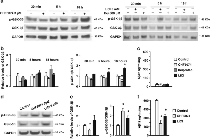
To verify whether the γ-secretase modulators might increase the inhibitory phosphorylation of GSK-3β inde-pendently of changes in Aβ42 production, we exposed primary cortical neurons from wild-type mice to 3 μM CHF5074 and 500 μM ibuprofen for diverse times, starting from 30 min to 18 h. Cortical cultures, which release hardly detectable amounts of Aβ42(Fig.4c), were
treated with the GSK-3β inhibitor LiCl at 5 mM concen-tration as a positive control. The concenconcen-trations of CHF5074 and ibuprofen were chosen on the basis of their activity in lowering Aβ42 generation in hAPP cells (Weggen et al.2001; Imbimbo et al.2007) and the cerebral level of CHF5074 (3±0.6μM) detected in mice chronically treated with the compound (Imbimbo et al.2009). Contrary to LiCl, CHF5074 as well as ibuprofen did not increase the p-GSK-3β/GSK-3β ratio at any time tested (Fig. 4a, b). Ibuprofen even decreased the p-GSK-3β/GSK-3β ratio at 18 h in cortical cultures.
When tested in H4-APPswe cells expressing hAPP with the Swedish mutation and releasing high amounts of Aβ42 (Fig. 4f), either CHF5074 or LiCl increased the inhibitory phosphorylation of GSK-3β as indicated by the elevated p-GSK-3β/GSK-3β ratio (Fig. 4d, e). Cell treatment with CHF5074 also reduced the level of total GSK-3β. The CHF5074 effect on GSK-3β correlated with the inhibition of Aβ42release in the culture medium after 18-h incubation (Fig. 4f). Taken together, these results suggest that the inhibitory phosphorylation of GSK-3β induced by the γ-secretase modulation may be an event secondary to the reduction of endogenous Aβ generation.
Tg control CHF5074 PHF-1 Tau46
a
c
d e f
g
i
l m
n
50 µm 50 µm Ibuprofenb
h
Fig. 2 Immunohistochemical detection of hyperphosphory-lated and total tau in cortical sections of Tg mice. Analysis was performed using anti-hyperphosphorylated (PHF-1) and total tau (Tau46) antibodies in slices from right brain hemi-spheres of Tg mice. Pictures show that positive inclusions (arrows) to PHF-1 (a, b, c and relative inset magnification d, e, f) were reduced in sections from animals treated with CHF5074 and ibuprofen. The Tau46 posi-tive neurons (g, h, i and relaposi-tive inset magnifications l, m, n) appeared markedly reduced in CHF5074-treated group, but not in the ibuprofen group. Bar=50 μmDiscussion
This study provides the first evidence that chronic treatment with γ-secretase modulators reduces the hyperphospho-rylation and pathological accumulation of native tau as well as the level of activated GSK-3β in the brain of APP Tg mice. We previously reported that chronic treatment of APPSLmice with CHF5074 reduces the Aβ burden without modifying the generation of APP carboxy-terminal frag-ments, reduces the number of plaque-associated microglia, and attenuates spatial memory deficit (Imbimbo et al.
2009). Similar effects on Aβ plaque accumulation and
microglial immunoreactivity were observed with ibuprofen (Lim et al. 2000; Yan et al. 2003; Heneka et al. 2005; Morihara et al. 2005), though in APPSL mice ibuprofen showed to be less effective than CHF5074 (Imbimbo et al.
2009). Together with Aβ deposition, accumulation of
intracellular hyperphosphorylated tau represents the neuro-pathological hallmark of AD (Selkoe2001). A large body of evidence indicates that Aβ peptides, the primary
constituents of neuritic plaques, may initiate the process of neurodegeneration in AD brains (Clippingdale et al.
2001; Casas et al. 2004; Oakley et al. 2006; Smith et al.
2006). Aβ deposits promote hyperphosphorylation,
accu-mulation, and aggregation of tau in a conformationally altered and argyrophilic form (Samura et al. 2006) in HEK293 cells overexpressing APP (Rank et al. 2002), in transgenic mice expressing double mutated hAPP form (Bellucci et al.2007), in double transgenic mice expressing mutant hAPP(swe) and presenilin-1 (Samura et al. 2006), and in triple transgenic mice expressing the hAPP(swe), presenilin-1 and tau (Oddo et al. 2003a, b). Song and colleagues (2008) observed that phosphorylation of tau increases in cortical neurons as early as 3 h of Aβ42 treatment, evoking an AD-like accumulation of phosphor-ylated tau. The abnormal phosphorylation of tau promotes its misfolding, decreases the degradation, and induces the self-assembly into tangles of paired helical filaments which alter the microtubule stability and normal neuronal functions (Iqbal et al. 2009). In addition, cytosolic abnormally hyperphosphorylated tau sequesters normal tau and two other neuronal microtubule-associated pro-teins (MAPs), MAP1A/MAP1B and MAP2, further contributing to microtubule disassembly and neuronal damage (Alonso et al. 2008; Iqbal et al. 2008). Neurofi-brillary degeneration appears to be required for the clinical expression of dementia asβ-amyloidosis in the absence of neurofibrillary degeneration is not associated with the development of cognitive symptoms (Alafuzoff et al.
1987; Arriagada et al.1992; Dickson et al.1992). In line with this evidence, tau reduction without changing Aβ plaque deposition prevents learning and memory deficits and protects mice against excitotoxicity (Roberson et al.
2007). On the other hand, in mice expressing a human tau variant under the control of an inducible promoter (tet-off), after the suppression of tau expression, memory function recovers and neuron number stabilizes although NFTs continue to form, suggesting that accumulation of hyperphosphorylated tau, and not its aggregation, is required for cognitive decline and behavioral impairment (Santacruz et al. 2005). Thus, although it is supposed to occur as a consequence of Aβ pathology (Wilcock et al.
2010), the tau hyperphosphorylation may be considered per se a useful therapeutic target for slowing AD progression (Churcher2006; Roberson et al.2007).
In this study, we investigated the effects produced by chronic administration of the γ-secretase modulators, CHF5074 and ibuprofen (Imbimbo et al.2009), on native tau. By checking tau phosphorylation, we found that ibuprofen treatment produced a significant reduction of phosphorylated tau recognized by both the PHF-1 and CP13 antibodies, which respectively identify the late (pSer396/404) and the early (pSer202) forms of
phosphory-a
b
Relative levels of GSK-3 ββ cont rol CH F507 4 0 0.25 0.50 0.75 1.00 1.25***
***
no n-Tg p-GSK-3β GAPDH GSK-3β p-GSK-3 β /total GSK-3 β ratio contr ol CH F507 4 0 0.25 0.50 0.75 1.00 no n-Tgc
**
Ibup rofen Ibup rofen CHF5074 control Tg mice 36 KDa 46 KDa 46 KDa non-Tg Ibuprofen Tg mice Tg mice _ _ _ _ _ _Fig. 3 Effects of CHF5074 and ibuprofen treatments on GSK-3β and phospho-GSK-3β levels. a Picture from a representative western blot analysis performed using anti-phospho-GSK-3β(pSer9
) and anti-GSK-3β antibodies in brain extracts from non-Tg mice or Tg mice treated with standard diet or diet with CHF5074 and ibuprofen. The amount of total GSK-3β did not change in Tg mice compared to non-Tg group, while it significantly reduced in mice treated with ibuprofen and CHF5074. b Data from densitometry analysis of GSK-3β immunoblots, expressed as relative levels to non-Tg value, are the ratio of GSK-3β to relative GAPDH. Blots were obtained by simultaneous exposure of membranes to anti-GSK-3β and anti-GAPDH antibodies. Bars are the means ± SEM of 9–10 animals per group; ***p<0.001 vs. Tg control value as well as vs. non-Tg value. c Ratio of p-GSK-3β to relative GSK-3β; **p<0.01 vs. Tg control value; Tg, but not the CHF5074 and ibuprofen groups, were significantly different from the non-Tg group
lated tau (Klein et al.2004; Liu et al.2006). This is in line with previous evidence showing that ibuprofen reduces hyperphosphorylated tau in 3xTg-AD mice expressing hAPP(swe), presenilin-1(M146V), and tau(P301L) (Mckee et al. 2008). The effect of CHF5074 was more evident on PHF-1- than on CP13-positive phospho-tau. In particular, CHF5074 and to a lesser extent ibuprofen limited the accumulation of tau protein, as suggested by the parallel decrease in total tau level. This evidence suggests that the γ-secretase modulators can reduce the accumulation of phosphorylated tau and not simply tau phosphorylation.
It is recognized that GSK-3β acts as a major kinase responsible for tau hyperphosphorylation and subsequent resistance to degradation (Iqbal et al. 2009) in AD
pathology (Gong et al. 2005). GSK-3β is constitutively
active and is a substrate for other kinases capable of phospho-regulating its activity through both inhibition and activation (Grimes and Jope 2001). In the case of deactivation, signaling through phosphoinositide 3-kinase (PI3K) and subsequent activation of the Ser-Thr kinase Akt inhibits GSK-3β activity via Ser9
phosphorylation. A body of evidence suggests that Aβ promotes tau phosphorylation through activation of GSK-3 (Takashima et al.1996,1998; Tomidokoro et al. 2001; Hoshi et al. 2003; Song et al.
2008). The GSK-3β activation mediated by its Ser9 dephos-phorylation (Wang et al. 2006; Song et al.2008) has been demonstrated after exogenous application of Aβ peptide as well as during intracellular accumulation of endogenous Aβ in cell-based models of AD. In line with previous
a
46 KDa 46 KDa 36 KDa GSK-3 GAPDH p-GSK-3 _ + + CHF5074 3 µM 30 min 5 h 18 h _ _ _ _ _ + _ _ _ GSK-3 p-GSK-3 LiCl 5 mM GAPDH 46 KDa 46 KDa 36 KDa 30 min 5 h 18 h _ _ _ Ibu 500 µM _ + _ + _ + _ + _ _ _ + _ + _ _ _ _ GSK-3 p-GSK-3 GAPDH Contro l CHF5 0743µ M LiCl 5 mMd
46 KDa 46 KDa 36 KDae
b
0 1 p-GSK-3 / GSK-3 0 1 2 3 p-GSK-3 /GSK-3*
*
0 1 2 3 Relative levels of GSK -3 Relative levels o f GSK -3 0 1 2 3 Control CHF5074 Ibuprofen LiCl Control CHF5074 LiCl 30 min 5 hours 18 hours2
*
0 150 300 450 600 A β 42 ng/ml/mg*
*
*
330 min 5 hours 18 hours
0 150 300 450 600 A β 42 ng/ml/mg
c
f
*
*
Fig. 4 Western blot analysis of GSK-3β phosphorylation in neuronal cultures exposed to 3μM CHF5074, 500 μM ibuprofen, or 5 mM LiCl. a Immunoblot, representative of three different experiments, shows the p-GSK-3β and GSK-3β levels in extracts of cortical neurons exposed to the various treatments for 30 min, 5 h, and 18 h. b Densitometry analysis of immunoblots, expressed as relative levels to control value, reveals no change of GSK-3β or p-GSK-3β/GSK-3β ratio in neurons exposed to CHF5074 or ibuprofen. Neurons treated with LiCl for 5 or 18 h displayed a significant increase in p-GSK-3β/ GSK-3β ratio (*p<0.05 vs. corresponding control value). c Aβ42 released in the medium of cortical neurons exposed for 18 h to 3μM
CHF5074, 500 μM ibuprofen, or 5 mM LiCl; *p<0.01 vs. corresponding control value. d Representative immunoblot of GSK-3β and p-GSK-GSK-3β in H4-APPswe cells exposed to CHF5074 and LiCl for 18 h. Similar results were obtained in two additional experiments. e Data from densitometry analysis, expressed as relative levels to control value, show the means of GSK-3β and p-GSK-3β/GSK-3β ratios of three separate experiments. Bars are the means ± SEM (*p< 0.05 vs. control value). f Aβ42 released in the medium of H4-APPswe cells exposed for 18 h to CHF5074 3μM or LiCl 5 mM; *p<0.01 vs. corresponding control value
evidence (Rockenstein et al.2007; Bitner et al. 2009), we found that the level of total GSK-3β was not modified in APPSLmice when compared to wild-type littermates, while its phosphorylated form significantly reduced, suggesting a sustained activation of the kinase in AD mice. This effect was more evident in our 12-month-old mice than in 3-month-old APPSL mice previously investigated by Rock-enstein et al. (2007). The chronic treatment with CHF5074 or ibuprofen inhibited the GSK-3β activity by increasing the pGSK-3β/GSK-3β ratio near to the non-Tg group and by reducing the total protein level. Likewise theγ-secretase modulators, caffeine was recently found to decrease active and total GSK-3β level, in addition to Aβ burden and cognitive impairment in AD mice (Arendash et al.2009). It remains to be ascertained whether the diminished levels of GSK-3β are associated with decreased protein synthesis or increased degradation. We checked the possibility that the γ-secretase modulators might indeed regulate the GSK-3β level and activation independently of their anti-amyloidogenic effect. However, this hypothesis was denied by experiments on primary cortical neurons from wild-type mice which showed that, contrary to the positive control LiCl, neither brief (30 min) nor long-time (18 h) applica-tions of CHF5074 and ibuprofen could decrease the level of GSK-3β or enhance its phosphorylation. Ibuprofen even reduced the p-GSK-3β/GSK-3β ratio at 18 h. It can be inferred that ibuprofen neuroprotective activity reported in pure cortical neurons (Iwata et al.2010) is associated with signaling pathway other than the GSK-3β inhibition as, possibly, the activation of the peroxisome proliferator-activated receptor-γ pathway (Dill et al. 2010). The capability of CHF5074 to reduce GSK-3β and increase the GSK-3β inhibitory phosphorylation was restored in H4swe cells overexpressing hAPP. Thus, while possible direct effects elicited in vivo by CHF5074 metabolites on GSK-3β cannot be excluded by our study, it is conceivable that reduction of kinase activity and tau phosphorylation is the resultant of decreased generation of endogenous Aβ overproduction.
The central role of Aβ in driving tau pathology has recently been underlined by studies showing that in APP mice expressing native or transgenic tau as well as in AD patients, the pathological accumulation of hyperphosphory-lated tau develops concomitantly with Aβ accumulation in hippocampal synaptic terminals, linking these intraneuronal pathological signs to the synaptic alteration and cognitive dysfunction of AD (Guo et al.2006; Takahashi et al.2008). Moreover, decreasing Aβ in APP Tg mice by immunother-apy significantly lowers tau pathology and reverses memory deficits (Wilcock et al.2010). In this context, the higher efficacy of CHF5074 compared to ibuprofen in reducing the cognitive deficits in APPSL mice may be associated with its higher efficacy in reducing Aβ
gener-ation (Imbimbo et al.2009) and tau dysregulation, besides its capability to conserve the COX-2-mediated memory retrieval (Murray and O’Connor 2003; Sharifzadeh et al.
2006). Consistent with this evidence, our data support Aβ
as therapeutic target for the disease-modifying treatment and propose the γ-secretase modulator, CHF5074, as a suitable tool to afford this approach by avoiding heavy collateral effects associated with the immunotherapy (von Bernhardi2010).
Acknowledgments A part of this study was presented at the 12th International Conference on Alzheimer Disease (ICAD 2009), Wien, 11–16 July 2009. The study was sponsored by Chiesi Farmaceutici, Parma, Italy, and by EX 60%.
References
Alafuzoff I, Iqbal K, Friden H, Adolfsson R, Winblad B (1987) Histopathological criteria for progressive dementia disorders: clinical–pathological correlation and classification by multivari-ate data analysis. Acta Neuropathol 74:209–225
Alonso AC, Li B, Grundke-Iqbal I, Iqbal K (2008) Mechanism of tau-induced neurodegeneration in Alzheimer disease and related tauopathies. Curr Alzheimer Res 5:375–384
Arendash GW, Mori T, Cao C, Mamcarz M, Runfeldt M, Dickson A, Rezai-Zadeh K, Tane J, Citron BA, Lin X, Echeverria V, Potter H (2009) Caffeine reverses cognitive impairment and decreases brain amyloid-beta levels in aged Alzheimer’s disease mice. J Alzheimers Dis 17:661–680
Arriagada PV, Growdon JH, Hedley-Whyte ET, Hyman BT (1992) Neurofibrillary tangles but not senile plaques parallel duration and severity of Alzheimer’s disease. Neurology 42:631–639 Ballatore C, Lee VM, Trojanowski JQ (2007) Tau-mediated
neuro-degeneration in Alzheimer’s disease and related disorders. Nat Rev Neurosci 8:663–672
Baum L, Hansen L, Masliah E, Saitoh T (1996) Glycogen synthase kinase 3 alteration in Alzheimer disease is related to neurofibril-lary tangle formation. Mol Chem Neuropathol 29:253–261 Bellucci A, Rosi MC, Grossi C, Fiorentini A, Luccarini I, Casamenti F
(2007) Abnormal processing of tau in the brain of aged TgCRND8 mice. Neurobiol Dis 27:328–338
Bitner RS, Nikkel AL, Markosyan S, Otte S, Puttfarcken P, Gopalakrishnan M (2009) Selective alpha7 nicotinic acetylcholine receptor activation regulates glycogen synthase kinase3beta and decreases tau phosphorylation in vivo. Brain Res 1265:65–74 Casas C, Sergeant N, Itier JM, Blanchard V, Wirths O, van der Kolk
N, Vingtdeux V, van de Steeg E, Ret G, Canton T, Drobecq H, Clark A, Bonici B, Delacourte A, Benavides J, Schmitz C, Tremp G, Bayer TA, Benoit P, Pradier L (2004) Massive CA1/2 neuronal loss with intraneuronal and N-terminal truncated Abeta42 accumulation in a novel Alzheimer transgenic model. Am J Pathol 165:1289–1300
Churcher I (2006) Tau therapeutic strategies for the treatment of Alzheimer’s disease. Curr Top Med Chem 6:579–595
Clippingdale AB, Wade JD, Barrow CJ (2001) The amyloid-β peptide and its role in Alzheimer’s disease. J Pept Sci 7:227–249 Dias-Santagata D, Fulga TA, Duttaroy A, Feany MB (2007) Oxidative
stress mediates tau-induced neurodegeneration in Drosophila. J Clin Invest 117:236–245
Dickson DW, Crystal HA, Mattiace LA, Masur DM, Blau AD, Davies P, Yen SH, Aronson MK (1992) Identification of normal and
pathological aging in prospectively studied nondemented elderly humans. Neurobiol Aging 13:179–189
Dill J, Patel AR, Yang XL, Bachoo R, Powell CM, Li S (2010) A molecular mechanism for ibuprofen-mediated RhoA inhibition in neurons. J Neurosci 30(3):963–972
Fulga TA, Elson-Schwab I, Khurana V, Steinhilb ML, Spires TL, Hyman BT, Feany MB (2007) Abnormal bundling and accumu-lation of F-actin mediates tau-induced neuronal degeneration in vivo. Nat Cell Biol 9:139–148
Gong CX, Liu F, Grundke-Iqbal I, Iqbal K (2005) Post-translational modifications of tau protein in Alzheimer’s disease. J Neural Transm 112:813–838
Grimes CA, Jope RS (2001) The multifaceted roles of glycogen synthase kinase 3beta in cellular signaling. Prog Neurobiol 65:391–426
Guo JP, Arai T, Miklossy J, McGeer PL (2006) Abeta and tau form soluble complexes that may promote self aggregation of both into the insoluble forms observed in Alzheimer’s disease. Proc Natl Acad Sci USA 103:1953–1958
Harr SD, Hollister RD, Hyman BT (1996) Glycogen synthase kinase 3 alpha and 3 beta do not colocalize with neurofibrillary tangles. Neurobiol Aging 17:343–348
Heneka MT, Sastre M, Dumitrescu-Ozimek L, Hanke A, Dewachter I, Kuiperi C, O’Banion K, Klockgether T, Van Leuven F, Landreth GE (2005) Acute treatment with the PPARg agonist pioglitazone and ibuprofen reduces glial inflammation and Aβ1-42 levels in APPV717I transgenic mice. Brain 128:1442–1453
Hoshi M, Sato M, Matsumoto S, Noguchi A, Yasutake K, Yoshida N, Sato K (2003) Spherical aggregates of beta-amyloid (amylosphe-roid) show high neurotoxicity and activate tau protein kinase I/ glycogen synthase kinase-3beta. Proc Natl Acad Sci USA 100:6370–6375
Hutter-Paier B, Huttunen HJ, Puglielli L, Eckman CB, Kim DY, Hofmeister A, Moir RD, Domnitz SB, Frosch MP, Windisch M, Kovacs DM (2004) The ACAT inhibitor CP-113,818 markedly reduces amyloid pathology in a mouse model of Alzheimer’s disease. Neuron 44:227–238
Imbimbo BP, Del Giudice E, Colavito D, D’Arrigo A, Dalle Carbonare M, Villetti G, Facchinetti F, Volta R, Pietrini V, Baroc MF, Serneels L, De Strooper B, Leon A (2007) 1-(3′,4′-Dichloro-2-fluoro[1,1′-biphenyl]-4-yl)-cyclopropanecarboxylic acid (CHF5074), a novel gamma-secretase modulator, reduces brain beta-amyloid pathology in a transgenic mouse model of Alzheimer’s disease without causing peripheral toxicity. J Pharmacol Exp Ther 323:822–830
Imbimbo BP, Hutter-Paier B, Villetti G, Facchinetti F, Cenacchi V, Volta R, Lanzillotta A, Pizzi M, Windisch M (2009) CHF5074, a novel gamma-secretase modulator, attenuates brain beta-amyloid pathology and learning deficit in a mouse model of Alzheimer’s disease. Br J Pharmacol 156:982–993
Imbimbo BP, Giardino L, Sivilia S, Giuliani A, Gusciglio M, Pietrini V, Del Giudice E, D’Arrigo A, Leon A, Villetti G, Calzà L (2010) CHF5074, a novel gamma-secretase modulator, restores hippocampal neurogenesis potential and reverses contextual memory deficit in a transgenic mouse model of Alzheimer’s disease. J Alzheimers Dis 20:159–173
Iqbal K, Alonso Adel C, Grundke-Iqbal I (2008) Cytosolic abnormally hyperphosphorylated tau but not paired helical filaments seques-ter normal MAPs and inhibit microtubule assembly. J Alzheimers Dis 14:365–370
Iqbal K, Liu F, Gong CX, Alonso Adel C, Grundke-Iqbal I (2009) Mechanisms of tau-induced neurodegeneration. Acta Neuro-pathol 118:53–69
Ishizawa T, Sahara N, Ishiguro K, Kersh J, McGowan E, Lewis J, Hutton M, Dickson DW, Yen SH (2003) Co-localization of glycogen synthase kinase-3 with neurofibrillary tangles and
granulovacuolar degeneration in transgenic mice. Am J Pathol 163:1057–1067
Iwata Y, Nicole O, Zurakowski D, Okamura T, Jonas RA (2010) J Thorac Cardiovasc Surg 139(2):489–493
Jackson GR, Wiedau-Pazos M, Sang TK, Wagle N, Brown CA, Massachi S, Geschwind DH (2002) Human wild-type tau interacts with wingless pathway components and produces neurofibrillary pathology in Drosophila. Neuron 34:509–519 Klein RL, Lin WL, Dickson DW, Lewis J, Hutton M, Duff K, Meyer
EM, King MA (2004) Rapid neurofibrillary tangle formation after localized gene transfer of mutated tau. Am J Pathol 164:347–353
Lee VM, Goedert M, Trojanowski JQ (2001) Neurodegenerative tauopathies. Annu Rev Neurosci 24:1121–1159
Lim GP, Yang F, Chu T, Chen P, Beech W, Teter B, Tran T, Ubeda O, Ashe KH, Frautschy SA, Cole GM (2000) Ibuprofen suppresses plaque pathology and inflammation in a mouse model for Alzheimer’s disease. J Neurosci 20:5709–5714
Liu F, Liang Z, Shi J, Yin D, El-Akkad E, Grundke-Iqbal I, Iqbal K, Gong CX (2006) PKA modulates GSK-3beta- and cdk5-catalyzed phosphorylation of tau in site- and kinase-specific manners. FEBS Lett 580:6269–6274
Lucas JJ, Hernandez F, Gomez-Ramos P, Moran MA, Hen R, Avila J (2001) Decreased nuclear beta-catenin, tau hyperphosphorylation and neurodegeneration in GSK-3beta conditional transgenic mice. EMBO J 20:27–33
McKee AC, Carreras I, Hossain L, Ryu H, Klein WL, Oddo S, LaFerla FM, Jenkins BG, Kowall NW, Dedeoglu A (2008) Ibuprofen reduces Abeta, hyperphosphorylated tau and memory deficits in Alzheimer mice. Brain Res 1207:225–236
Morihara T, Teter B, Yang F, Lim GP, Boudinot S, Boudinot FD, Boudinot FD, Frautschy SA, Cole GM (2005) Ibuprofen suppresses interleukin-1b induction of proamyloidogenic a1-antichymotrypsin to ameliorate beta-amyloid (Ab) pathology in Alzheimer’s models. Neuropsychopharmacology 30:1111–1120 Murray HJ, O’Connor JJ (2003) A role for COX-2 and p38 mitogen
activated protein kinase in long-term depression in the rat dentate gyrus in vitro. Neuropharmacology 44:374–380
Nishimura I, Yang Y, Lu B (2004) PAR-1 kinase plays an initiator role in a temporally ordered phosphorylation process that confers tau toxicity in Drosophila. Cell 116:671–682
Oakley H, Cole SL, Logan S, Maus E, Shao P, Craft J, Guillozet-Bongaarts A, Ohno M, Disterhoft J, Van Eldik L, Berry R, Vassar R (2006) Intraneuronal beta-amyloid aggregates, neurodegenera-tion, and neuron loss in transgenic mice with five familial Alzheimer’s disease mutations: potential factors in amyloid plaque formation. J Neurosci 26:10129–10140
Oddo S, Caccamo A, Kitazawa M, Tseng BP, LaFerla FM (2003a) Amyloid deposition precedes tangle formation in a triple transgenic model of Alzheimer’s disease. Neurobiol Aging 24:1063–1070
Oddo S, Caccamo A, Shepherd JD, Murphy MP, Golde TE, Kayed R, Metherate R, Mattson MP, Akbari Y, LaFerla FM (2003b) Triple-transgenic model of Alzheimer’s disease with plaques and tangles: intracellular Abeta and synaptic dysfunction. Neuron 39:409–421
Pei JJ, Tanaka T, Tung YC, Braak E, Iqbal K, Grundke-Iqbal I (1997) Distribution, levels, and activity of glycogen synthase kinase-3 in the Alzheimer disease brain. J Neuropathol Exp Neurol 56:70–78 Pei JJ, Braak E, Braak H, Grundke-Iqbal I, Iqbal K, Winblad B, Cowburn RF (1999) Distribution of active glycogen synthase kinase 3beta (GSK-3beta) in brains staged for Alzheimer disease neurofibrillary changes. J Neuropathol Exp Neurol 58:1010–1019
Rank KB, Pauley AM, Bhattacharya K, Wang Z, Evans DB, Fleck TJ, Johnston JA, Sharma SK (2002) Direct interaction of soluble human recombinant tau protein with Aβ1-42 results in tau
aggregation and hyperphosphorylation by tau protein kinase II. FEBS Lett 514:263–268
Roberson ED, Scearce-Levie K, Palop JJ, Yan F, Cheng IH, Wu T, Gerstein H, Yu GQ, Mucki L (2007) Reducing endogenous tau ameliorates amyloid beta-induced deficits in an Alzheimer’s disease mouse model. Science 316:750–754
Rockenstein E, Torrance M, Adame A, Mante M, Bar-on P, Rose JB, Crews L, Masliah E (2007) Neuroprotective effects of regulators of the glycogen synthase kinase-3beta signaling pathway in a transgenic model of Alzheimer’s disease are associated with reduced amyloid precursor protein phosphorylation. J Neurosci 27:1981–1991
Samura E, Shoji M, Kawarabayashi T, Sasaki A, Matsubara E, Murakami T, Wuhua X, Tamura S, Ikeda M, Ishiguro K, Saido TC, Westaway D, St George Hyslop P, Harigaya Y, Abe K (2006) Enhanced accumulation of tau in doubly transgenic mice expressing mutant βAPP and presenilin-1. Brain Res 1094:192–199
Santacruz K, Lewis J, Spires T, Paulson J, Kotilinek L, Ingelsson M, Guimaraes A, DeTure M, Ramsden M, McGowan E, Forster C, Yue M, Orne J, Janus C, Mariash A, Kuskowski M, Hyman B, Hutton M, Ashe KH (2005) Tau suppression in a neurodegen-erative mouse model improves memory function. Science 309:476–481
Sarnico I, Lanzillotta A, Boroni F, Benarese M, Alghisi M, Schwaninger M, Inta I, Battistin L, Spano PF, Pizzi M (2009) NF-kappaB p50/RelA and c-Rel-containing dimers: opposite regulators of neuron vulnerability to ischaemia. J Neurochem 108:475–485
Schilling S, Zeitschel U, Hoffmann T, Heiser U, Francke M, Kehlen A, Holzer M, Hutter-Paier B, Prokesch M, Windisch M, Jagla W, Schlenzig D, Lindner C, Rudolph T, Reuter G, Cynis H, Montag D, Demuth HU, Rossner S (2008) Glutaminyl cyclase inhibition attenuates pyroglutamate Abeta and Alzheimer’s disease-like pathology. Nat Med 14:1106–1111
Selkoe DJ (2001) Alzheimer’s disease results from the cerebral accumulation and cytotoxicity of amyloid beta-protein. J Alz-heimers Dis 3:75–80
Sharifzadeh M, Tavasoli M, Soodi M, Mohammadi-Eraghi S, Ghahremani MH, Roghani A (2006) A time course analysis of cyclooxygenase-2 suggests a role in spatial memory retrieval in rats. Neurosci Res 54:171–179
Shiurba RA, Ishiguro K, Takahashi M, Sato K, Spooner ET, Mercken M, Yoshida R, Wheelock TR, Yanagawa H, Imahori K, Nixon RA (1996) Immunocytochemistry of tau phosphoserine 413 and tau protein kinase I in Alzheimer pathology. Brain Res 737:119–132 Smith WW, Gorospe M, Kusiak JW (2006) Signaling mechanisms
underlying Abeta toxicity: potential therapeutic targets for Alz-heimer’s disease. CNS Neurol Disord Drug Targets 5:355–361
Song MS, Rauw G, Baker GB, Kar S (2008) Memantine protects rat cortical cultured neurons against beta-amyloid-induced toxicity by attenuating tau phosphorylation. Eur J Neurosci 28:1989–2002
Steinhilb ML, Dias-Santagata D, Fulga TA, Felch DL, Feany MB (2007) Tau phosphorylation sites work in concert to promote neurotoxicity in vivo. Mol Biol Cell 18:5060–5068
Takahashi RH, Capetillo-Zarate E, Lin MT, Milner TA, Gouras GK (2008) Co-occurrence of Alzheimer’s disease beta-amyloid and tau pathologies at synapses. Neurobiol Aging 31:1145–1152 Takashima A, Noguchi K, Michel G, Mercken M, Hoshi M,
Ishiguro K, Imahori K (1996) Exposure of rat hippocampal neurons to amyloid beta peptide (25–35) induces the inacti-vation of phosphatidyl inositol-3 kinase and the actiinacti-vation of tau protein kinase I/glycogen synthase kinase-3 beta. Neurosci Lett 203:33–36
Takashima A, Honda T, Yasutake K, Michel G, Murayama O, Murayama M, Ishiguro K, Yamaguchi H (1998) Activation of tau protein kinase I/glycogen synthase kinase-3beta by amyloid beta peptide (25–35) enhances phosphorylation of tau in hippocampal neurons. Neurosci Res 31:317–323
Tomidokoro Y, Ishiguro K, Harigaya Y, Matsubara E, Ikeda M, Park JM, Yasutake K, Kawarabayashi T, Okamoto K, Shoji M (2001) Aβ amyloidosis induces the initial stage of tau accumulation in APPswe mice. Neurosci Lett 299:169–172
von Bernhardi R (2010) Immunotherapy in Alzheimer’s disease: where do we stand? Where should we go? J Alzheimers Dis 19:405–421
Wang ZF, Li HL, Li XC, Zhang Q, Tian Q, Wang Q, Xu H, Wang JZ (2006) Effects of endogenous beta-amyloid overproduction on tau phosphorylation in cell culture. J Neurochem 98:1167–1175 Weaver CL, Espinoza M, Kress Y, Davies P (2000) Conformational
change as one of the earliest alterations of tau in Alzheimer’s disease. Neurobiol Aging 21:719–727
Weggen S, Eriksen JL, Das P, Sagi SA, Wang R, Pietrzik CU, Findlay KA, Smith TE, Murphy MP, Bulter T, Kang DE, Marquez-Sterling N, Golde TE, Koo EH (2001) A subset of NSAIDs lower amyloidogenic Abeta42 independently of cyclooxygenase activ-ity. Nature 414:212–216
Wilcock DM, Gharkholonarehe N, Van Nostrand WE, Davis J, Vitek MP, Colton CA (2010) Amyloid reduction by amyloid-beta vaccination also reduces mouse tau pathology and protects from neuron loss in two mouse models of Alzheimer’s disease. J Neurosci 30:1197–1198
Yan Q, Zhang J, Liu H, Babu-Khan S, Vassar R, Biere AL, Citron M, Landreth G (2003) Anti-inflammatory drug therapy alters b-amyloid processing and deposition in an animal model of Alzheimer’s disease. J Neurosci 23:7504–7509