Contents lists available at
ScienceDirect
Pharmacological
Research
j o u r n a l h o m e p a g e :
w w w . e l s e v i e r . c o m / l o c a t e / y p h r s
Review
Blocking
the
FGF/FGFR
system
as
a
“two-compartment”
antiangiogenic/antitumor
approach
in
cancer
therapy
Arianna
Giacomini,
Paola
Chiodelli,
Sara
Matarazzo,
Marco
Rusnati,
Marco
Presta,
Roberto
Ronca
∗
DepartmentofMolecularandTranslationalMedicine,UniversityofBrescia,25123Brescia,Italy
a
r
t
i
c
l
e
i
n
f
o
Articlehistory:
Received22January2016
Receivedinrevisedform15March2016 Accepted16March2016
Availableonline22March2016 Keywords: Angiogenesis Cancer Cancertherapy FGF FGFR Stroma Therapeutictarget
a
b
s
t
r
a
c
t
Fibroblastgrowthfactors(FGFs)areafamilyofpleiotropicfactorsproducedbystromalandparenchymal tumorcells.EventhoughFGFshavebeenfirstlycharacterizedasangiogenicfactors,theyexertautocrine andparacrinefunctionsnotonlyonendothelialcellsbutalsoontumorcellsandotherstromal com-ponents.Thus,theFGF/FGFreceptor(FGFR)pathwaymayrepresentakeyplayerintumorgrowthby regulatingthecomplexcross-talkbetweenstromalandtumorcompartments.
TheliganddependentorindependentactivationoftheFGF/FGFRsystembygeneupregulation, onco-genicmutationoramplificationoccursinavarietyofhumantumorsandisimplicatedinvariouskeysteps oftumorgrowthandprogression.Inaddition,FGF/FGFRactivationhasbeendescribedasamechanism oftumorescapeinresponsetoantiangiogenic/anti-VEGFtherapies.
Experimentalandclinicalevidencesprovideacompellingbiologicrationaleforthedevelopmentof anti-FGF/FGFRtargetingagentsincancertherapy.However,thedevelopmentofdrugsspecifically target-ingtheFGF/FGFRpathwayprovedtobedifficult,alsoduetothehighredundancyandpleiotropiceffects ofFGFandFGFRfamilymembers.Ontheotherhand,thepossibilitytodevelop“two-compartment” targetingagentsendowedwithbothantiangiogenicandantitumoractivitiesremainspromising.
Herewewillreviewthepreclinicalandclinicalapproachesandpotentialtherapeuticscurrently avail-abletoblocktheFGF/FGFRsysteminhumancancer.
©2016ElsevierLtd.Allrightsreserved.
Contents
1. TheFGF/FGFRsystem...173
1.1. Fibroblastgrowthfactors...173
1.2. Fibroblastgrowthfactorreceptors...173
2. TheFGF/FGFRsysteminendothelialcells ... 174
3. DeregulationoftheFGF/FGFRsystemincancercells...175
3.1. Activatingmutations ... 175
3.2. Geneoverexpressionandamplification...176
3.3. Chromosomaltranslocations...177
3.4. Aberrantautocrineandparacrineligandsignalling...177
4. TheroleoftheFGF/FGFRsystemintumor/stromacross-talk...177
5. InhibitionoftheFGF/FGFRsystem:therapeuticapproaches...178
5.1. InhibitionofFGF/FGFRsystemattheextracellularlevel...178
5.1.1. InhibitionoftheexpressionofFGF/FGFR/FGFRco-receptors...178
5.1.2. PreventingFGF/FGFR/co-receptorinteractions...179
5.2. InhibitionofsignaltransductiontriggeredbyFGFRactivation...180
∗ Correspondingauthorat:UniversityofBrescia,DepartmentofMolecularandTranslationalMedicine,vialeEuropa11,25123Brescia,Italy. E-mailaddress:[email protected](R.Ronca).
http://dx.doi.org/10.1016/j.phrs.2016.03.024
6. Concludingremarks...180
Conflictofinterest ... 181
Acknowledgments...181
References...181
1.
The
FGF/FGFR
system
1.1.
Fibroblast
growth
factors
Fibroblast
growth
factors
(FGFs)
are
secreted
proteins
that
act
as
paracrine,
autocrine
or
endocrine
factors.
The
FGF
family
encom-passes
22
members,
grouped
into
seven
subfamilies
on
the
basis
of
phylogenetic
analysis
and
sequence
homology
[1]
.
The
subfam-ilies
FGF1/2/5,
FGF3/4/6,
FGF7/10/22,
FGF8/17/18
and
FGF9/16/20
act
as
canonical
FGFs;
FGF11/12/13/14
are
intracellular
factors
act-ing
in
an
FGF
receptor
(FGFR)
independent
manner;
FGF19/21/23
subfamily
members
function
as
hormones
[2]
.
Canonical
FGFs
are
paracrine
factors
that
mediate
their
bio-logical
responses
by
binding
to
and
activating
tyrosine
kinase
(TK)
FGFRs.
The
interaction
with
heparin/heparan
sulfate
(HS)
proteoglycans
(HSPGs)
plays
a
pivotal
role
in
mediating
the
bio-logical
activity
of
FGFs,
leading
to
the
formation
of
signalling
FGF/FGFR/HSPG
ternary
complexes
[3]
.
Moreover,
HSPGs
sequester
FGF
molecules
near
the
site
of
action,
providing
a
reservoir
for
the
growth
factor
and
allowing
the
formation
of
extracellular
matrix
(ECM)-associated
FGF
gradients
[4]
.
Canonical
FGFs
mediate
a
plethora
of
functions
during
develop-ment.
They
are
involved
in
patterning
of
germ
cell
layers,
formation
of
body
axes,
induction
of
organogenesis
and
morphogenesis.
Moreover,
FGFs
display
homeostatic
functions
in
the
adult,
being
involved
in
tissue
repair
and
remodelling
processes.
Finally,
dereg-ulation
of
FGF
signalling
can
contribute
to
pathological
diseases,
including
cancer.
In
this
context,
several
alterations
affecting
the
FGF/FGFR
system
have
been
reported
in
tumors,
including
gain-or
loss-
of
function,
altered
gene
expression
or
changes
in
binding
specificity
[2]
.
Intracellular
FGFs
act
as
intracellular
signalling
molecules
in
a
FGFR-independent
manner;
they
play
a
major
role
in
neu-ronal
functions
at
postnatal
stages
by
interacting
with
intracellular
domains
of
voltage-gate
sodium
channels
and
with
the
neuronal
mitogen-activated
protein
kinase
scaffold
protein
islet-brain-2
[5]
.
Hormone-like
FGFs
exhibit
poor
affinity
for
HSPGs,
resulting
in
more
diffusive
properties
through
blood
circulation
[6]
.
These
FGFs
depend
on
Klotho
co-receptors
(see
below)
to
activate
intracellular
signalling
responses
[7]
.
FGF19
(orthologue
of
murine
FGF15)
acts
as
a
growth/differentiation
factor
in
the
heart
and
brain
at
embry-onic
stages
and
plays
a
crucial
role
in
regulating
hepatic
bile
acid
production
[8]
.
FGF21
is
a
metabolic
regulator
of
lipolysis
in
the
white
adipose
tissue
[9]
and
FGF23
acts
as
a
physiological
regulator
of
phosphate
and
active
vitamin-D
blood
levels
[10]
.
The
wide-ranging
biological
roles
of
FGFs,
the
variety
of
activated
signalling
pathways
and
the
complex
and
dynamic
expression
of
FGF
ligands
and
receptors
implies
that
the
FGF/FGFR
system
must
be
tightly
regulated.
1.2.
Fibroblast
growth
factor
receptors
In
mammals,
FGFRs
are
encoded
by
four
distinct
genes
(FGFR1-4).
FGFRs
consist
of
three
extracellular
immunoglobulin-like
(Ig)
domains
(D1-3),
a
single
transmembrane
helix
domain
and
an
intracellular
TK
domain
[11]
.
D2
and
D3
domains
are
responsible
for
FGF
binding.
FGFRs
show
diverse
specificities
for
FGF
ligands.
In
addition,
alternative
splicing
of
the
D3
domain
that
may
occur
in
FGFR1,
2
and
3,
but
not
in
FGFR4,
generates
“IIIb”
and
“IIIc”
isoforms
with
additional
ligand-binding
properties.
For
instance,
FGFR2IIIb
binds
FGF7
and
FGF10,
but
not
FGF2,
whereas
the
FGFR2IIIc
iso-form
binds
FGF2
and
FGF18,
but
not
FGF7
and
FGF10
(see
Ref.
[12]
for
further
details
about
the
ligand-binding
properties
of
the
dif-ferent
FGFRs).
Interestingly,
FGFR1-3IIIb
and
FGFR1-3IIIc
isoforms
often
display
differential
expression
in
epithelial
and
mesenchymal
tissue,
respectively
[13]
.
FGFRs
interact
with
HSPGs
via
the
D2
domain.
The
formation
of
a
2:2:2HSPG/FGF/FGFR
ternary
complex
[14]
causes
receptor
dimer-ization
with
conformational
shift
in
receptor
structure
that
leads
to
trans-phosphorylation
of
multiple
residues
in
the
intracellu-lar
TK
domain.
Receptor
phosphorylation
activates
multiple
signal
transduction
pathways
that
generate
distinct
cellular
responses.
As
summarized
in
Fig.
1
,
major
substrates
of
FGFR
TK
are
the
intracellular
specific
adaptor
protein
FGFR-substrate-2
(FRS2)
and
phospholipase-C
␥
(PLC
␥)
[15]
.
Activated
FRS2
allows
the
recruit-ment
of
the
adaptor
protein
GRB2
that
in
turn
recruits
SOS
or
GAB1
to
the
signal
complex.
The
recruitment
of
SOS
activates
RAS
and
the
downstream
RAF/mitogen-activated
protein
kinase
(MAPK)
pathway.
The
downstream
effect
of
this
pathway
is
mainly
cell
pro-liferation,
even
though
cell
differentiation
or
cell
cycle
arrest
can
also
be
induced
depending
on
the
different
cellular
context.
The
recruitment
of
GAB1
causes
the
PI3K–mediated
activation
of
the
AKT
antiapoptotic
pathway.
PLC
␥
leads
to
the
activation
of
protein
kinase
C
(PKC)
that
sustains
MAPK
and
AKT
pathways
and
plays
a
role
in
cell
migration.
Other
pathways
may
be
activated
in
different
cell
subtypes,
including
p38
MAPK,
JAK-STAT
and
RSK2
[16]
.
Several
extracellular
and
intracellular
mechanisms
have
been
described
able
to
regulate/attenuate
FGFR
signalling
at
differ-ent
levels.
FGFRs
are
internalized
upon
receptor
activation
[17]
,
inducing
receptor
degradation
or
recycling.
Relevant
to
this
point,
N-glycosylation
of
the
receptor
affects
the
assembly
of
the
FGF/FGFR1/HSPG
complex
[18]
and
internalization
of
FGFR2
[19]
.
At
intracellular
level,
MAPK
signalling
may
phosphorylate
threo-nine
residues
on
FRS2,
inhibiting
the
recruitment
of
GRB2
[20]
.
Sprouty
proteins
are
negative
modulators
that
compete
for
GRB2
binding
by
preventing
RAS
activation
or
directly
binding
RAF
and
disrupting
MAPK
signalling
[21]
.
In
addition,
FGFs
can
induce
the
activation
of
phosphatases,
including
SEF
and
MAPK-phosphatase
3
(MKP3).
SEF
interacts
directly
with
FGFRs,
thus
preventing
their
activation,
whereas
both
enzymes
can
dephosphorylate
and
inac-tivate
ERK
1/2[22]
.
Different
molecules
can
act
as
cell
surface
co-receptors
for
FGFs
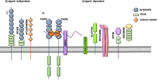
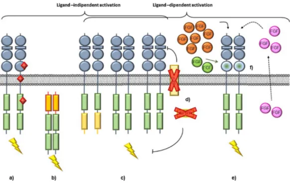
(
Fig.
2
).
As
already
mentioned,
HSPGs
are
required
for
a
produc-tive
FGF/FGFR
interaction
that
enables
FGFR
signalling
[23]
.
For
this
reason,
structural
modifications
of
the
HS
chains
deeply
affect
FGFR
signalling
and
can
be
responsible
for
its
fine-tuning.
As
an
excep-tion,
hormone-like
FGFs
have
reduced
affinity
for
HSPGs
and
their
activity
depends
on
the
presence
of
Klotho
proteins
as
co-receptors.
Cell
surface
-Klotho
and
␣-Klotho
are
co-factors
for
FGF19/21
and
FGF23,
respectively,
and
convert
FGFRs
into
high
affinity
recep-tors
for
endocrine
FGFs,
limiting
nonspecific/off-target
signalling.
The
cell
membrane
ganglioside
GM1
acts
as
a
FGF
co-receptor
by
interacting
with
FGF2
and
promoting
its
biological
activity
in
endothelial
cells
[24]
.
In
addition,
␣v3
integrin
promotes
FGF-mediated
endothelial
cell
proliferation,
motility,
and
FGFR1
recruitment
[25]
,
thus
contributing
to
the
cross-talk
between
FGFR
and
integrin
signalling
[26]
.
Neural
cell
adhesion
molecule
(N-CAM),
neuronal
cadherin
(N-cadherin)
and
L1
can
activate
FGFR1-2
Fig.1.FGFRsignallingpathways.
FGFsbindtoFGFRs,inducingreceptordimerizationandtransphosphorylationoftheirTKdomain.This,inturn,leadstothedockingofadaptorproteinsandconsequent activationofdownstreamsignallingpathways.ActivatedFGFRsubstrate2(FRS2)recruitsandactivatesRAS-RAF-MEK-ERK1/2andPI3K-AKTpathwaysinvolvedincell proliferationandantiapoptoticactivity,respectively.RecruitmentandphosphorylationofPLC␥inducesPKCactivationandintracellularCa++release,eventsthatregulate
cellmotility.ThenegativeregulatorsMKP3andSPRYcanmodulateFGFRsignallingwhereasSEFmayalsointerferewithligandbinding.
Fig.2.FGFRco-receptors.
(a)FGFsbindtoFGFRsandHSPGs,leadingtotheformationofaternarycomplexthatinducesreceptordimerizationandactivation.Moreover,variousligand-independent (b)andligand-dependent(c)cell-surfaceproteinsandglycolipidsmayactasFGFRco-receptors.
in
the
absence
of
canonical
FGFR
ligands
and
this
interaction
is
mediated
by
the
acid
box
motif
in
the
linker
region
between
the
D1
and
D2
Ig-like
loops
of
the
receptor
[27]
.
Therefore,
N-CAM
and
N-cadherin
act
as
a
nonconventional
FGFR1
ligand,
promote
receptor
stabilization
and
exert
a
peculiar
control
on
FGFR
intracel-lular
trafficking
[28]
.
Extracellular
matrix-associated
glycoprotein
anosmin-1
binds
FGFR1
in
a
HS-dependent
manner,
modulating
FGFR
signalling
during
development
[29]
.
Finally,
EphA4
has
been
identified
as
a
binding
partner
for
FGFRs.
The
interaction
occurs
between
the
juxtamembrane
region
of
FGFR
and
the
cytoplasmic
domain
of
EphA4
and
leads
to
enhanced
level
of
MAPK
signalling
and
stimulation
of
cell
proliferation
[30]
.
2.
The
FGF/FGFR
system
in
endothelial
cells
Angiogenesis
is
an
essential
process
for
tumor
growth
and
progression,
since
the
large-scale
growth
of
a
tumor
ultimately
requires
an
adequate
blood
supply
[31]
.
Indeed,
once
a
tumor
lesion
exceeds
a
few
millimeters
in
diameter,
hypoxia
and
nutrient
depri-vation
trigger
an
‘angiogenic
switch’
to
allow
the
tumor
to
progress
[32]
.
In
1980s,
the
purification
to
homogeneity
of
tumor
angiogenic
proteins
led
to
the
first
identification
of
the
two
heparin-binding
angiogenic
growth
factors
FGF1
and
FGF2
[33,34]
.
At
present,
FGF1
and
FGF2
still
represent
the
prototypical
and
best
studied
mem-bers
of
the
canonical
FGF
subfamily.
In
vivo
they
exert
a
potent
pro-angiogenic
effect
in
different
experimental
models,
including
the
chick
embryo
chorioallantoic
membrane
(CAM)
[35]
,
rab-bit/mouse
cornea
[36]
and
murine
subcutaneous
Matrigel
plug
[37]
assays.
Besides
FGF1
and
FGF2,
only
scattered
pieces
of
information
indicate
that
other
FGFs
show
clear
pro-angiogenic
properties
(like
FGF4
and
FGF8)
whereas
few
or
controversial
data
have
been
reported
for
the
remaining
members
of
the
FGF
family
(see
Ref.
[38]
for
an
extensive
review).
Interestingly,
the
apparent
redundancy
of
the
FGF
family
may
lead
to
complementary/compensatory
actions,
Fig.3. DeregulationoftheFGF/FGFRsystemincancer.
a)Activatingmutations(reddiamonds)canoccurintheextracellular,transmembraneorTKdomainofFGFR,leadingtoconstitutivedimerizationandactivationofthe receptor.b)Chromosomalrearrangementsmayleadtointragenictranslocationsthatresultintheexpressionoffusionproteinswhosedimerizationtriggersconstitutive activationoftheFGFRsignalling.c)FGFRoverexpressioncanbeinducedbygeneamplification,translocationoraberranttranscriptionalregulation.FGFRoverexpressioncan alsobeaccompaniedbyalteredC-terminalsplicing(lightyellowboxes)thatmayinterferewithreceptorinternalizationwithconsequentaccumulationofthereceptoratthe cellsurface.d)Negativeregulators(e.g.SEF)canbedownmodulated,thusincreasingFGFRsignalling.e)CancercellsmayinducestromalcellstooverexpressFGFs(orange), thusactivatingaparacrineloopofstimulation.Inaddition,FGFs(pink)areproducedbycancercellsandactinanautocrinefashion.f)Switchingbetweenalternativelyspliced receptorisoformsmayinduceimbalancedFGFRsignallingwithalteredspecificityforFGFligands(green).
making
difficult
the
identification
of
the
biological
significance
of
the
various
FGFs
by
the
gene
knockout
approach.
For
instance,
FGF2
knockout
and
FGF1/FGF2
double-knockout
mice
develop
normally
with
only
mild
phenotypic
defects
in
their
wound
healing
capacity
associated
with
FGF1/FGF2
deletion
[39]
.
Of
note,
human
umbilical
vascular
endothelial
(HUVE)
cells
express
several
canonical
FGFs
(including
FGF1,
FGF2,
FGF5,
FGF7,
FGF8,
FGF16,
FGF18)
and
two
FGF
homologous
factors
(FGF11
and
FGF12)
[40]
,
thus
suggesting
that
FGFs
may
also
exert
autocrine
functions
in
endothelium.
Even
though
a
comprehensive
study
is
still
missing,
endothe-lial
cells
express
different
members
of
the
FGFR
family,
FGFR1IIIc
being
the
most
represented
receptor
while
FGFR2-IIIc
and
FGFR3-IIIc
are
expressed
at
lower
levels
[40]
.
Experiments
performed
on
FGFR1
and
FGFR2
null
mice
in
both
endothelial
and
hematopoi-etic
cells
indicate
that
these
receptors
are
not
required
for
vascular
homeostasis
or
physiological
functions.
However,
FGFR
signalling
in
endothelial
cells
plays
a
pivotal
role
in
tissue
repair
and
neovas-cularization
following
injury,
pointing
to
endothelial
cell
FGFRs
as
a
target
for
the
therapy
of
diseases
characterized
by
an
aberrant
vascular
proliferation
[41]
.
The
formation
of
HSPG/FGF/FGFR
ternary
complexes
causes
receptor
dimerization
and
trans-phosphorylation
of
multiple
residues
in
the
intracellular
FGFR
TK
domain.
This
leads
to
the
activation
of
a
complex
“pro-angiogenic
phenotype”
in
endothelial
cells
that
recapitulates
several
aspects
of
the
in
vivo
angiogene-sis
process,
including
modulation
of
endothelial
cell
proliferation,
migration,
protease
production,
integrin
and
cadherin
receptor
expression,
and
intercellular
gap-junction
communication
(sum-marized
in
Ref.
[42]
).
For
instance,
FGF1,
FGF2,
FGF4,
FGF7
and
FGF8b
bind
and
activate
FGFR1
or
FGFR2,
stimulating
endothelial
cell
proliferation
[43]
.
In
addition,
FGFs
can
modulate
extracellular
matrix
degradation,
as
reported
for
the
capacity
of
FGF1
and
FGF2
to
induce
the
secretion
of
MMP1
and
MMP3
in
endothelial
cells
[44]
and
the
capacity
of
FGF2
to
stimulate
the
shedding
of
endothelial
membrane
vesicles
containing
MMP1,
MMP9
and
metalloprotease
inhibitors
TIMP-1
and
TIMP-2
[45]
.
Also,
various
studies
demon-strate
that
FGFs
promote
endothelial
cell
migration,
as
shown
by
the
ability
of
FGF1,
FGF2,
FGF7,
FGF16
and
FGF18
to
induce
a
chemo-tactic
response
in
endothelium
[43,46]
.
3.
Deregulation
of
the
FGF/FGFR
system
in
cancer
cells
An
aberrant
regulation
of
the
FGF/FGFR
system
may
occur
in
human
tumors,
leading
to
the
deregulated
activation
of
ligand-dependent
or
ligand-independent
FGFR
signalling.
As
summarized
in
Fig.
3
,
this
may
represent
the
consequence
of
activating
FGFR
mutations
that
occur
in
the
extracellular,
transmembrane
or
TK
domain
of
the
receptor;
chromosomal
rearrangements
that
result
in
the
expression
of
FGFR
signalling
fusion
proteins;
FGFR
over-expression
induced
by
gene
amplification,
translocation,
aberrant
transcriptional
regulation
or
down-modulation
of
negative
regula-tors;
FGF
overexpression
by
stromal
and/or
tumor
cell,
leading
to
the
activation
of
autocrine/paracrine
loops
of
stimulation.
Clearly,
while
FGFR
mutations
are
anticipated
to
impact
mainly
the
tumor
cell
behavior,
FGF
overexpression
by
tumor
cells
may
exert
both
autocrine
and
paracrine
effects,
thus
contributing
to
the
epithe-lial/stroma
cross-talk
that
occurs
in
the
tumor
microenvironment.
In
addition,
depending
on
the
molecular
mechanism
responsible
for
the
ligand-dependent
or
ligand-independent
deregulation
of
FGFR
signaling
in
a
given
neoplasm,
different
approaches
can
be
envis-aged
aimed
at
targeting
the
FGF/FGFR
system
at
the
extracellular
or
intracellular
level
(see
below).
3.1.
Activating
mutations
The
screening
from
210
different
human
cancers
of
1000
somatic
mutations
in
the
coding
exons
of
518
protein
kinase
genes
Table1
Chemotherapeutics,otherdrugsandnaturalproductsendowedwithantiangiogenicactivity.Theantiangiogenicactivityofthesecompoundshasbeendemonstratedtobe due,atleastinpart,totheircapacitytoinhibitFGFproductionand/orFGFRexpressionortointerferewiththeintracellularsignallingtriggeredbytheFGF/FGFRsystemin endothelialcells(seereferencesforfurtherdetails).
Chemotherapeutics Maintumortarget(s)(FDAapproved) Ref
6-methylmercaptopurine-riboside acutelymphaticleukemia [159]
topoisomerase-Iinhibitortopotecan smallcelllungcancer,metastaticovariancancer [160]
medroxyprogesterone-acetate endometrialcancer,breastcancerinpostmenopausalwomen [161]
Tamoxifen breastcancerinpostmenopausalwomen [162]
Thalidomide multiplemyeloma [163]
quinazoline-derived␣1-adrenoreceptorantagonistdoxazosin prostatecancer [164]
6-thioguanine acutemyelogenousleukemia [165]
Atiprimod relapsedacutelymphoblasticandmyeloidleukemias [166]
etoposide smallcelllungcancer,testicularcancer [167]
combinationoftegafuranduracil(UFT) advancedcolorectalcancer,variouscancera [168]
Otherdrugs Originaltherapeuticindication
Tranilast anti-allergicdrug [169]
Spironolactone heartfailure [170]
Zoledronicacid variousskeletalcomplications [171]
Cidofovir cytomegalovirusretinitisinAIDSpatients [172]
Indomethacin nonsteroidalanti-inflammatorydrugs [173]
Celecoxib [174]
Cerivastatin hypercholesterolemia [175]
Ticlopidine(derivatives) plateletantiaggregatingagent [176]
Triamcinoloneacetonide intraoculardisorders [177]
HyPE(secretoryphospholipase-A2inhibitor) asthma [178]
Naturalproducts Source
curcumin Curcumalonga [179]
epigallocatechin-3-gallate greentea [180]
Gleditsiasinensis fruitextract [181]
1,2,3,4,6-penta-O-galloyl-beta-d-glucose GallaRhois [182]
4-O-methylgallicacid dietarylegumeCanavaliagladiate [183]
resveratrol grapesandwine [184]
glyceollins soybean [185]
alliin garlic [186]
stilbeneglycosides Boswelliapapyriferai [187]
salvicine SalviaprionitisHance [188]
polymethoxyflavonoidnobiletin citrus [189]
aplidine marine-deriveddepsipeptide [190]
philinopsideA, seacucumberPentactaquadrangulari [191]
psammaplinA marinesponge [192]
carrageenan edibleseaweeds [193]
carotenoids marinealgae [194]
epoxydocosapentaenoicacids(EDPs) omega-3dietaryfattyacids(fishoil) [195]
1-O-alkylglycerols fishliveroils [196]
Neovastat(AE-941) cartilage [197]
aApprovedinUKandJapan.
highlighted
various
components
of
the
FGF
signalling
pathways
as
the
most
commonly
mutated
genes
in
the
subset
of
non-synonymous
mutations
[47]
.
For
instance,
∼50%
of
bladder
cancers
have
somatic
mutations
in
the
FGFR3-coding
sequence
[48]
,
more
than
half
of
the
mutations
occurring
at
a
single
position
(S249C)
in
the
extracellular
domain
of
the
receptor.
This
mutation
leads
to
the
formation
of
an
aberrant
intermolecular
cysteine
bridge
that
results
in
ligand-independent
constitutive
dimerization
and
activa-tion
of
the
receptor
[49]
.
FGFR3
mutations
have
also
been
identified
in
many
other
cancer
types,
including
cervical
cancers
[50]
,
mul-tiple
myeloma
(MM)
[51,52]
,
prostate
cancers
[53]
,
spermatocytic
seminomas
[54]
and
oral
squamous
carcinomas
[55]
.
Other
muta-tions
located
in
the
TK
domain
can
change
the
FGFR
conformation,
leading
to
a
constitutive
ligand-independent
receptor
activation,
as
observed
for
FGFR4
in
the
childhood
rhabdomyosarcoma
[56]
.
At
variance
with
FGFR
genes,
FGF
mutations
are
rare
in
human
cancers
and
their
impact
on
cancer
biology
is
unclear.
Indeed,
to
the
best
of
our
knowledge,
somatic
mutations
have
been
described
only
for
FGF9
in
colorectal
and
endometrial
cancers
[57]
.
They
are
predicted
to
result
in
loss-of-function
and
it
is
not
known
whether
these
mutations
participate
in
tumor
formation.
3.2.
Gene
overexpression
and
amplification
Elevated
FGFR
levels
can
be
observed
in
human
cancers
as
the
consequence
of
deregulated
gene
transcription
or
amplification.
Also
in
this
case,
this
will
lead
to
the
activation
of
FGFR
signalling
in
a
ligand-independent
manner.
At
variance
with
the
activation
of
FGFR3
by
somatic
mutations,
FGFR3
gene
amplification
has
been
rarely
described
in
cancer
[58]
.
In
contrast,
both
FGFR1
and
FGFR2
amplifications
are
more
commonly
found.
For
instance,
amplifica-tion
of
the
chromosomal
region
8p11–12,
the
genomic
location
of
FGFR1,
is
one
of
the
most
common
focal
amplifications
in
breast
cancer
[59]
.
It
occurs
in
approximately
10%
of
breast
tumors
and
predominantly
in
estrogen
receptor
(ER)-positive
cancers
[59]
.
Recent
studies
have
demonstrated
focal
FGFR1
amplification
in
non-small
cell
lung
carcinoma
cells
in
3%
of
lung
adenocarcinomas
and
21%
of
squamous
cell
carcinomas
[60,61]
.
FGFR1
amplifica-tions
have
been
observed
also
in
oral
squamous
carcinomas
[62]
and
are
found
at
a
low
incidence
in
ovarian
cancer
[63]
,
bladder
cancer
[58]
and
rhabodomyosarcoma
[64]
.
As
to
FGFR2,
approx-imately
10%
of
gastric
cancers
show
FGFR2
amplification,
which
is
associated
with
poor
prognosis
in
diffuse-type
cancers
[65]
.
In
addition,
FGFR2
amplification
occurs
in
approximately
2%
of
breast
cancers
and
breast
cancer
SUM52PE
and
MFM-223
cell
lines
Table2
NaturalFGF2-trapmolecules.
FGF2-trapmoleculea
TSP-1
fibstatin(fibronectinfragment) gangliosides PDGF ␣2-macroglobulin PTX3 heparin,freeHSPGs CXCL13 CXCL4
solubleformoftheextracellularportionofFGFR1
aSee[114,198]andreferencestherein.