Comparison of Different Fixation Techniques of the Long Head of the Biceps Tendon (LHBT) in Superior Capsule Reconstruction for Irreparable Posterosuperior Rotator
Abstract Background
Purpose The purpose of this study was to investigate the effect of using the long head of the biceps tendon (LHBT) for reconstruction of the superior capsule on shoulder kinematics along with different fixation constructs in a dynamic biomechanical model. The authors hypothesized that each of the three proposed fixation techniques would restore native joint kinematics; including glenohumeral superior translation, maximum abduction angle, cumulative deltoid force, and subacromial contact pressure.
Study Design Controlled laboratory study
Methods Eight fresh-frozen cadaveric shoulders (mean age: 53.4±14.2 years) were tested using a dynamic shoulder simulator. Each specimen underwent the following five conditions: (1) native, (2) irreparable posterosuperior rotator cuff tear (psRCT), (3) V-shaped reconstruction, (4) Box-shaped reconstruction, (5) Single-stranded reconstruction. Maximum abduction angle (MAA), glenohumeral superior translation (ghST), cumulative deltoid force (cDF) and subacromial contact pressure (sCP) were assessed in each tested condition.
Results Each of the three LHBT techniques for reconstruction of the superior capsule significantly increased MAA, while significantly decreasing ghST and cDF compared to the psRCT (P < .001, respectively). Additionally, the V-shaped and the Box-shaped technique significantly decreased sCP (p=.009 and p=.016, respectively) compared to the psRCT. The V-shaped technique further showed a significantly increased MAA (P < .001, respectively) and decreased cDF (p= .042; p=.039, respectively) when compared to the Box-shaped and Single-stranded techniques as well as a significantly decreased ghST (p=.027) when compared to the Box-shaped technique.
Conclusion Reconstruction of the superior capsule using the LHBT significantly improved shoulder kinematics when compared to a posterosuperior rotator cuff tear.
Clinical Relevance Using a biologically viable and locally available LHBT autograft is a cost-effective, potentially timesaving and technically feasible alternative for reconstruction of the superior capsule, which may result in favorable outcomes in irreparable posterosuperior rotator cuff tears due to biological advantages. Moreover, each of the three techniques restored native shoulder biomechanics, which may help improving shoulder function by preventing superior humeral head migration and the development of rotator cuff tear arthropathy in young patients with irreparable rotator cuff tears.
Key Terms Superior Capsular Reconstruction; SCR; Rotator Cuff Tear; Irreparable Rotator Cuff Tear; Long Head of the Biceps Tendon; Biomechanics
What is known about the subject Recent literature focuses on utilizing locally available and biological viable autografts, such as the long head of the biceps tendon (LHBT) for reconstruction of the superior capsule, with promising early resultsA LHBT autograft has biological advantages, such as local availability ad viability, and is easily accessible for harvest without donor side site morbidity when compared to an allograft.
What this study adds to existing knowledge Using the LHBT for reconstruction of the superior capsule may help to improve shoulder function by preventing superior humeral migration, thus delaying the development of rotator cuff tear arthropathy in young patients with irreparable RC tears and an intact LHBT.
Introduction
In the past decade, superior capsular reconstruction (SCR) has emerged as a potential surgical approach in young patients with irreparable posterosuperior rotator cuff tears (RCT) and absence of severe degenerative changes. 32 Originally proposed by Mihata et al, 38 continuous evolvement of arthroscopic techniques has led to the use of different materials for SCR, including allografts, autografts, xenografts or synthetic patches. 12, 16, 19, 28, 29, 32, 49 Subsequent preliminary clinical trials yielded promising satisfactory results, 7, 14, 35-39 however, optimal choice of graft type remains a matter of debate among shoulder surgeons due to donor side morbidity, time of surgery, cost-effectiveness, and graft failures with subsequent functional impairment. 9, 14, 17, 21, 31
Thus, recent literature focuses on utilizing locally available and biological viable autografts, such as the long head of the biceps tendon (LHBT) for reconstruction of the superior capsule, with promising early results. 6, 11, 16, 19, 45, 48 As the LHBT plays a dynamic role in contributing to glenohumeral stability and humeral head depression, especially in chronic rotator cuff tears, 23-25, 27, 46, 50 reconstruction of the superior capsular using a biologically viable LHBT autograft may be a feasible alternative resulting in more favorable outcomes compared to SCR autografts due to biological advantages. 5, 6, 33, 50 Additionally, the mechanical properties of the LHBT such as ultimate strength, ultimate strain and strain energy density have been reported to be similar or even higher compared to other tendons around the shoulder joint, indicating that the LHBT may have the ability to act as a humeral head depressor. 34 Thus, using a LHBT in these patients may be appealing, as it is locally available, free of additional costs, technically less demanding and timesaving when compared to conventional SCR.
So far, LHBT autograft techniques have only been described and tested in static biomechanical shoulder models 6, 16, 42, without a consensus on how to position the graft. The purpose of this dynamic biomechanical study was to investigate the effect of using the long
head of the biceps tendon (LHBT) for reconstruction of the superior capsule on shoulder kinematics along with different fixation techniques. The authors hypothesized that each of the three proposed fixation techniques of the LHBT for reconstruction of the superior capsule would restore native joint kinematics; including glenohumeral superior translation, maximum abduction angle, cumulative deltoid force, and subacromial peak contact pressure.
Materials and Methods
This study was reviewed via Human Research Determination Form by the institutional review board (IRB) of the University of Connecticut, and it was concluded that no IRB approval was required. Eight fresh-frozen, independent cadaveric shoulders specimens with a mean age of 53.4 ± 14.2 years (range: 20 – 64 years, eight males, five left shoulders) were obtained from Medcure Inc. (Portland, OR). All specimens underwent visual and radiographic inspection to detect and exclude those with any tears of the rotator cuff tendons or capsule, moderate to severe osteoarthritis, bony defects, or joint contractures. No specimens had to be excluded.
Specimen Preparation
Specimen preparation was performed according to a previously described method. 1, 15, 47 Specimens were thawed overnight at room temperature prior to dissection. Specimens were then dissected free of skin, subcutaneous tissue and muscles leaving the rotator cuff muscles and the coracoacromial ligament carefully preserved. At the deltoid tuberosity, the anterior, middle, and posterior portions of the deltoid tendon were detached from the muscle belly and preserved with anchor loops being sutured to the tendinous insertions using a locking running stitch (No. 2 FiberWire, Arthrex Inc., Naples, FL, USA), allowing for attachment of each of the three deltoid heads to an individual shoulder simulator actuator. 1, 15, 47 The rotator cuff muscles (supraspinatus (SSP), subscapularis (SSC), infraspinatus (ISP), and teres minor (TM)) were sharply released of the scapula and separated from the underlying capsule, while meticulously preventing disruption of the tissue. The ISP and TM were simulated as one unit as previously described. 1, 15, 22, 47
The individual rotator cuff tendons were sutured to pulley-straps using No. 2 FiberWire to avoid pull-through during load application. The scapular body was placed in a custom rectangular box with the medial border aligned perpendicular to the ground and the glenoid tilted 10° superiorly and bone cement was poured in to ensure proper fixation.1, 15, 20, 47, 53 A
steel rod was cemented into the distal humerus and loaded with 1.7 kg, 30 cm distal from the center of the humeral head, representing a constant moment arm for each tested shoulder.22, 53 The glenohumeral joint capsule was vented by opening the rotator interval, in order to prevent changes during testing.1, 15, 47
Testing Setup
For biomechanical testing, a standard dynamic shoulder model was utilized, adapted from previously validated cadaveric shoulder studies. 1, 13, 15, 20, 22, 40, 47, 53 (Figure 1) The shoulder simulator consisted of up to six linear screw-driven actuators (Bimba, Monee, IL, USA) connected to 444 N load cells (Futek, Irvine, CA, USA). A universal strain gauge signal conditioner (Futek Model CSG110) was linked to a panel mount display (Futek Model IMP 650), and a test and measurement software (Sensit V2.5.1.0, Futek, Irvine, CA, USA) was used for load cells data acquisition in real time. 1, 15, 47
The specimen was mounted to the simulator on a 6 degrees-of-freedom jig with the scapula in 10° of anteflexion, 10° superior tilt of the glenoid, resulting in a 110 ° angle between the scapular spine and vertical axis. 53 The anatomic lines of action of the three portions of the deltoid, SSC and ISP/TM unit were routed using custom 7 mm-diameter frictionless pulleys. The cable attached to the SSP tendon was aligned with a tilt of 10° to the horizontal. 53 The pulley for the anterior deltoid was placed over the tip of the coracoid process, approximately 5 mm anteriorly to the anterolateral corner of the acromion with the middle deltoid pulley routed over a point 5 mm posteriorly to the anterolateral corner of the acromion, whereas the posterior deltoid pulley was placed at the posterolateral edge of the acromion in line with the scapular spine in order to mimick the native force vectors. 1, 15, 47, 53
Prior to testing, four infrared cameras (Vero v1.3, Vicon Motion Capture Systems, Centennial, CO, USA) were mounted around the shoulder simulator to cover a 180° field of view. A stationary triad, consisting of 3 optical markers, was placed on the acromion with its center being in line with the pulley of the middle deltoid. A second moving triad was mounted to the humeral shaft with its longitudinal axis being in line with the center of the stationary triad placed on the acromion. In a displacement-controlled setting, computer software (SiNet Hub Programmer V1.29; Applied Motion Products, Inc., CA, USA) was utilized to generate custom motion profiles for the individual actuators of the SSP as well as the anterior, middle, and posterior deltoid separately for each specimen. 1, 15, 47 3-dimensional (3D) motion tracking (Vicon Nexus 2.8, Vicon Motion Capture Systems, Oxford, UK) and four infrared cameras (Vicon Vero v1.3) with a frame rate of 250 Hz and a position accuracy of 0.01 mm and 0.1 degrees, recorded each motion profile with the arm being abducted in neutral rotation from 0° to 60° in the scapular plane with the scapula fixed, corresponding to approximately 90° of total shoulder abduction. 1, 15, 47 For calculation of these custom motion profiles, the SSC and ISP/TM unit were loaded statically with a 1.36 kg hanging weight, allowing for a balanced abduction motion. 44 In order to generate reliable data of applied forces, each motion cycle was repeated three times. 1, 15, 47 Maintaining joint centering at the resting position was guaranteed by applying 10 N to the SSP as well as the anterior, middle, and posterior deltoid, respectively. 1, 15, 47, 51 Every testing cycle started with the specimen in its resting position (0° of abduction, neutral rotation). The motion profile was recorded to articulate the arm from 0° to 60° of glenohumeral abduction in the scapular plane and in neutral rotation. 13, 47 Individual tendon excursion and velocity was calculated to reach 60° of glenohumeral abduction as previously described, 13, 47 while all tendons reached the abduction angle simultaneously. Force in each muscle was specified to increase linearly. 47For each specimen, an individual motion profile was generated in the intact state (condition 1) and maintained throughout all further testing conditions. The optical markers were tracked and recorded by infrared cameras, allowing for
accurate evaluation of ROM using computer software (Vicon Nexus 2.8 and Vicon proCalc 1.2.1, Vicon Motion Capture Systems).
Testing Conditions
The specimens remained in the shoulder simulator throughout all testing and surgical repairs. To avoid performance bias, all surgeries were performed by the same surgeon. In total, every specimen underwent 5 different conditions with each specimen being its own control (Figure 2 and Figure 3). First, the specimen was tested in the (1) native state. Secondly, a (2) massive, irreparable posterosuperior rotator cuff tear was created by sharply dissecting the footprint of the supraspinatus and cranial part of the infraspinatus on the greater humeral tuberosity. Subsequently, the supraspinatus muscle belly was detached from the fossa supraspinata in order to create a massively retracted rotator cuff tear. Care was taken not do damage the glenoidal attachment of the LHBT.
In condition (3), reconstruction of the superior capsule using the V-shaped LHBT configuration was performed. The LHBT was tenotomised sharply underneath the bicipital groove, just above the musculotendinous junction distally. With the shoulder being placed in 30° of abduction in neutral rotation, the LHBT was fixed on the glenoidal side 2-cm posterior to the tuberculum supraglenoidale using a double-loaded 3-mm suture anchor (SutureTak, Arthrex Inc., FL,USA) For the lateral attachment of the graft, a double-loaded 3-mm suture anchor (SutureTak, Arthrex Inc., FL,USA) was placed mid onto the greater tuberosity. No posterior or anterior side-to-side suturing was performed.
For condition (4), the glenoidal attachment of the graft was detached by cutting the sutures at the glenoid and retrieving the LHBT, leaving any excess of the graft free. No damage to the graft was noted. For condition (5), the LHBT was released from the humeral and glenoidal attachment. No damage to the graft was noted. Two single-loaded 3-mm anchors were placed 5-mm lateral to the bicipital groove (anterior-medial anchor) and postero-lateral on the greater
tuberosity (postero-lateral anchor), respectively. Fixation of the graft was performed first antero-medial; then postero-lateral; and finalised by fixing the graft on the glenoidal side. For fixation, the shoulder was placed in 30° of abduction in neutral rotation.
Outcome parameters
Four parameters were directly measured in the cadaveric shoulders: (1) maximum glenohumeral abduction angle (degree), (2) glenohumeral superior translation (%), (3) subacromial peak contact pressure (MPa), and (4) cumulative deltoid force (N).1, 15, 47 Glenohumeral abduction angle and glenohumeral superior translation were measured using 3-dimensional (3D) motion tracking (Vicon Nexus 2.8, Vicon Motion Capture Systems, Oxford, UK) and four infrared cameras (Vicon Vero v1.3). Glenohumeral superior translation for conditions 2 – 5 was calculated by dividing each value by the value for the native, intact condition (1). 47 Subacromial peak contact pressure was measured between the coracoacromial arch (coracoacromial ligament and acromion) and the humerus throughout abduction by using a pressure-measuring system (saturation pressure, 0.56 MPa; pressure mapping sensor model 4205 Tekscan). 1, 39, 47 Deltoid force was recorded in real time throughout range of motion by loadcells (Futek) connected to the actuators. 1, 15, 47 Cumulative deltoid force was calculated as the summation of anterior, middle, and posterior deltoid forces. Every specimen underwent 3 trials for each measurement. 1, 15, 47
Statistical analysis
A power analysis was performed to determine detectable differences in the dependent variables given estimated standard deviations. 47 For the glenohumeral abduction angle, an error variance of 1° across all conditions with a correlation of 0.3 between measurements was
assumed. A sample size of 6 specimens will provide 80% power to detect a 1° difference in shoulder angle at an α level of .05.
Descriptive statistics including mean and standard deviation were calculated to characterize the specimens. Repeated measures analysis of variance was performed to examine differences in maximal glenohumeral abduction angle, glenohumeral superior translation, subacromial peak contact pressure, and cumulative deltoid force among the various testing conditions. There were 6 comparisons of interest. For simplicity, the adjusted P values were reported. Specifically, the unadjusted p-values were multiplied by 6. When significant, post-hoc paired t tests with a Bonferroni corrected alpha were performed to determine which pairwise comparisons were statistically significant. The P value for the omnibus analysis of variance was P < .001 for each outcome measure. Given that there was only 1 independent variable (condition, defined as intact shoulder, simulated irreparable rotator cuff tear, V-shaped reconstruction, Single-stranded reconstruction, Box-shaped reconstruction), an interaction term was not included as a second independent variable would have been required. The alpha level for all analyses was set at .05. All statistical analysis was performed using Stata 15.2 software (StataCorp 2017. Stata Statistical Software: Release 15).
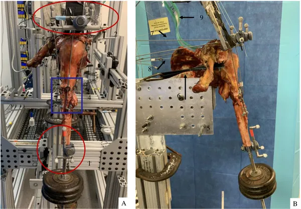
Figure 1. Figure demonstrating the dynamic shoulder simulator A. 1. a moving triad was mounted to the humeral shaft with its longitudinal axis being in line with the center of the 2. stationary triad placed on the acromion; 3. a steel rod was cemented into the distal humerus and loaded with 1.7 kg, 30 cm distal from the center of the humeral head, representing a constant moment arm for each tested shoulder; 4. at the deltoid tuberosity, the anterior, middle, and posterior portions of the deltoid tendon were preserved with anchor loops being sutured to the tendinous insertions using a locking running stitch, allowing for attachment of each of the three deltoid heads to an individual shoulder simulator actuator; B. 6. the subscapularis and infraspinatus/teres minor unit were loaded statically with a 1.36 kg hanging weight, allowing for a balanced abduction motion; 7. the anatomic lines of action of the three portions of the deltoid were routed using custom 7 mm-diameter frictionless pulleys; 8. the cable attached to the supraspinatus tendon was aligned with a tilt of 10° to the horizontal; 9. The Tecscan sensor was placed underneath the acromion and the free end was connected to the measuring device.
A 1 2 3 4 B 5 6 9 7 8
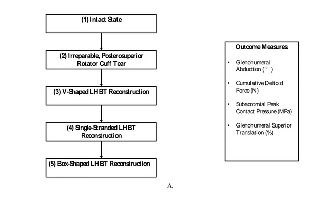
Figure 2. Flowchart displaying the A. five testing conditions and B. four outcome measures
(1) Intact State
(3) V-Shaped LHBT Reconstruction (2) Irreparable, Posterosuperior
Rotator Cuff Tear
(5) Box-Shaped LHBT Reconstruction (4) Single-Stranded LHBT Reconstruction Outcome Measures: • Glenohumeral Abduction ( ° ) • Cumulative Deltoid Force (N) • Subacromial Peak
Contact Pressure (MPa) • Glenohumeral Superior
Translation (%)
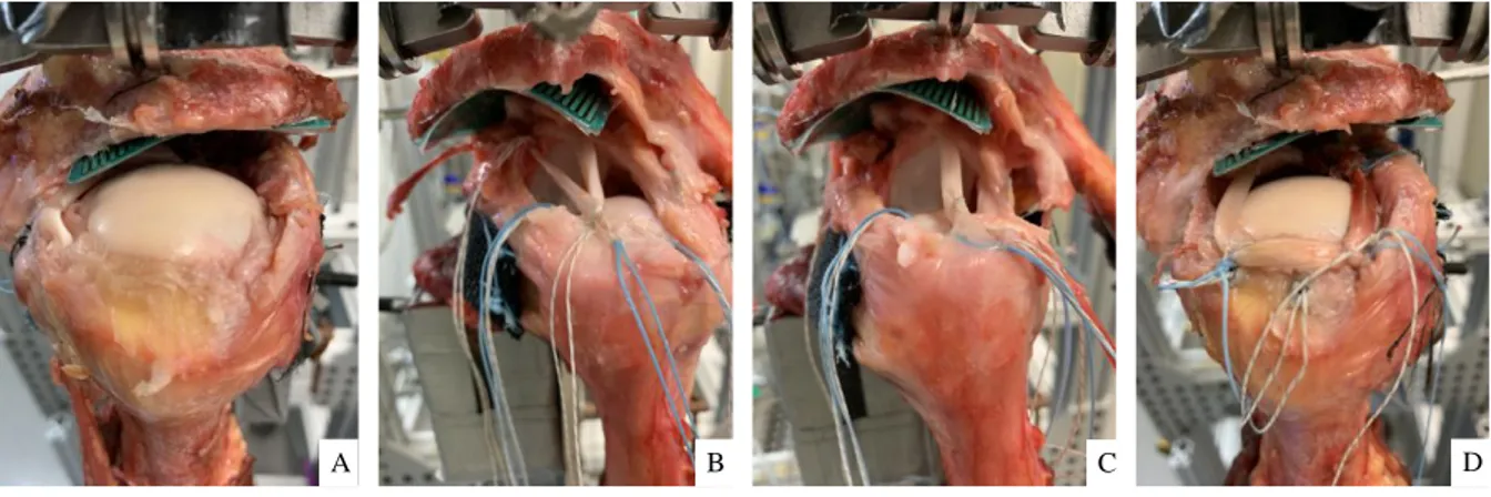
Figure 3. Figure demonstrating the A. created posterosuperior rotator cuff tear; B. the V-shaped reconstruction; C. the Single-stranded reconstruction ; D. and the Box-shaped reconstruction
Results
Irreparable Rotator Cuff Tear
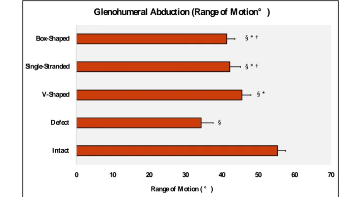
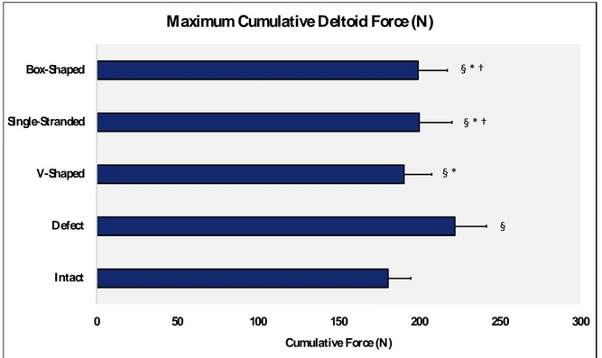
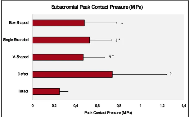
Intact shoulders achieved a mean maximum glenohumeral abduction of 55.3 °± 2.3°, requiring on average 180.7 N± 14.1 N of total deltoid force. Compared to the intact condition, a posterosuperior rotator cuff tear significantly decreased maximum abduction angle (62 % of intact; P < .001), and significantly increased glenohumeral superior translation (163 % of intact; P < .001), cumulative deltoid force (122 % of intact; P < .001) and subacromial peak contact pressure (296% of intact; P <.001).
Reconstruction of the Superior Capsule Using the LHBT
Each of the three reconstruction techniques significantly increased mean maximum glenohumeral abduction, and significantly decreased glenohumeral superior translation and cumulative deltoid force when compared to the posterosuperior rotator cuff tear state (P < .001, respectively).
There was no statistical difference in maximum glenohumeral abduction, glenohumeral superior translation, and cumulative deltoid force between the Box-shaped and Single-stranded technique. However, the V-shaped technique showed increased maximum glenohumeral abduction (P < .001, respectively) and decreased cumulative deltoid force (p= .042; p=.039, respectively) when compared to the Box-shaped and Single-stranded technique, respectively. No statistically significant difference was found when comparing glenohumeral superior translation and the V-shaped and the Single-stranded technique. Although, the V-shaped technique significantly decreased glenohumeral superior translation (p=.027) when compared to the Box-shaped technique. (Table 1, Table 2)
Peak Contact Pressure
The V-Shaped and the Box-shaped technique significantly decreased peak contact pressure (p=.009 and p=.016, respectively) compared to the defect state. When comparing peak contact pressure between the Single-stranded technique and the psRCT, there was no statistically significant increase. Additionally, there was no statistically significant difference in peak contact pressure when comparing the three techniques.
Intact Defect V-Shaped Single-Stranded Box-Shaped
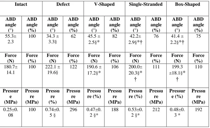
ABD angle (°) ABD angle (%) ABD angle (°) ABD angle (%) ABD angle (°) ABD angle (%) ABD angle (°) ABD angle (%) ABD angle (°) ABD angle (%) 55.3± 2.3 100 34.3 ± 3.3§ 62 45.5 ± 2.5§* 82 42.2± 2.9§*† 76 41.4 ± 2.2§*† 75 Force (N) Force (%) Force (N) Force (%) Force (N) Force (%) Force (N) Force (%) Force (N) Force (%) 180.7± 14.1 100 222.1 ± 19.6§ 122 190.6 ± 17.2§* 106 200.0± 20.3§* † 111 199.3 ±18.1§* † 110 Pressur e (MPa) Pressu re (MPa) Pressu re (%) Pressu re (MPa) Pressu re (%) Pressu re (MPa) Pressu re (%) Pressu re (MPa) Pressur e (%) Pressu re (MPa) 0.25±0. 08 100 0.74±0. 5 § 296 0.47±0. 2 §* 188 0.53±0. 2 §* 212 0.48±0. 3 * 192
Table 1. Table displaying the maximum glenohumeral abduction; the mean cumulative deltoid force; the mean subacromial peak contact pressure for each testing condition. Values are given as mean ± standard error; % glenohumeral abduction, % cumulative deltoid force and % subacromial contact pressure were calculated by dividing each value by the value for the native, intact condition. §: Significant difference compared with condition 1; *: Significant difference compared with condition 2, posterosuperior rotator cuff tear.
Intact Defect V-Shaped Single-Stranded Box-Shaped Glenohumeral Superior Translation (%) Glenohumeral Superior Translation (%) Glenohumeral Superior Translation (%) Glenohumeral Superior Translation (%) Glenohumeral Superior Translation (%) 100 163 § 120 § * 124 § * 130 § *†
Table 2. Table displaying the mean glenohumeral superior translation for each testing condition. Values are given as mean ± standard error. % glenohumeral superior translation was calculated by dividing each value by the value for condition 1, intact. §: Significant difference compared with condition 1; *: Significant difference compared with condition 2, posterosuperior rotator cuff tear.
Figure 4. Figure displaying glenohumeral abduction (range of motion °) across the testing conditions; §: Significant difference compared with condition 1; *: Significant difference compared with condition 2, posterosuperior rotator cuff tear.
0 10 20 30 40 50 60 70 Intact Defect V-Shaped Single-Stranded Box-Shaped Range of Motion ( ° )
Glenohumeral Abduction (Range of Motion° )
§ * § * †
§
Figure 5. Figure displaying maximum cumulative deltoid force (N) across the testing conditions; §: Significant difference compared with condition 1; *: Significant difference compared with condition 2, posterosuperior rotator cuff tear.
0 50 100 150 200 250 300 Intact Defect V-Shaped Single-Stranded Box-Shaped Cumulative Force (N)
Maximum Cumulative Deltoid Force (N)
§ * †
§ * †
§ *
Figure 6. Figure displaying subacromial peak contact pressure (MPa) across the testing conditions; §: Significant difference compared with condition 1; *: Significant difference compared with condition 2, posterosuperior rotator cuff tear.
0 0,2 0,4 0,6 0,8 1 1,2 1,4 Intact Defect V-Shaped Single-Stranded Box-Shaped
Peak Contact Pressure (MPa)
Subacromial Peak Contact Pressure (MPa)
*
§ *
§ *
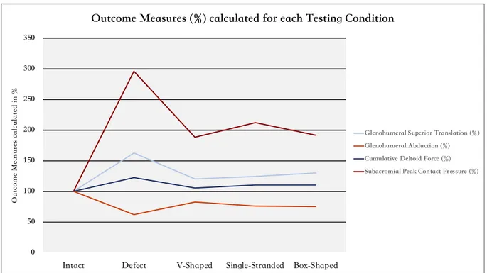
Figure 7. Figure displaying the changes (in %) of all outcomes measures across the testing conditions; % glenohumeral superior translation, % glenohumeral abduction, % cumulative deltoid force and % subacromial contact pressure were calculated by dividing each value by the value for the native, intact condition.
Discussion
The most important finding of this study was that reconstruction of the superior capsule using a V-shaped, Box-shaped or Single-stranded technique significantly increased mean maximum glenohumeral abduction, and significantly decreased glenohumeral superior translation, cumulative deltoid force and subacromial contact pressure when compared to the posterosuperior rotator cuff tear. Among the tested techniques, the V-shaped LHBT technique was shown to be superior to the Box-shaped and Single-stranded techniques. In consideration of the data gathered from this dynamic biomechanical investigation, using a locally available LHBT autograft may be a biological viable, technically feasible and potential cost- and timesaving alternative for reconstruction of the superior capsule, which may result in favorable outcomes.
When approaching massive, irreparable RCT in young and active patients without severe signs of osteoarthritis, choosing the optimal treatment remains a major challenge for shoulder surgeons. A plethora of surgical techniques have been described covering arthroscopic salvage techniques or more sophisticated open approaches such as tendon transfers, SCR or reverse total shoulder arthroplasty. However, a lack of clear evidence-based guidelines and high clinical failure rates emphasize the incertitude on how to best treat this challenging patient cohort. 41
Recent studies have focused on using locally available and biological viable autografts for reconstruction of the superior capsule. 5 When using the LHBT for reconstruction of the superior capsule, promising early results may be expected, 5, 10, 18 however, detailed biomechanical understanding and its effect on joint kinematics remains limited. 6, 11, 16, 19, 45, 48As to date, current data has only been gathered on small patient cohorts with limited follow-ups, or static shoulder simulators, there is no consensus on which technique may result in favorable shoulder function.
Reconstruction of the superior capsule using the LHBT in massive, irreparable posterosuperior RCT is based on the principles of SCR. First described by Mihata in 2012 using fascia lata, SCR has since then evaded the field of orthopaedic surgery with more than 18,000 procedures performed worldwide in 2018. Initial clinical results yielded promising outcomes 4, 8, 14, 35, 36, 38, 43, although clinical failure rates have been reported to vary between 4 -55 % of cases, highly dependent on technique and graft choice.8, 14, 26, 37, 43, 52, 54 The most common failures include graft tears on the glenoidal side and concomitant graft failures, which may be higher for allografts, when compared to autografts. 14, 26, 32, 38, 54 Thus, potential donor-site morbidity, graft reactions, minimal needed size of graft, high-learning curves, complex techniques and high-costs, raises concerns among orthopaedic surgeons when indicating patients for conventional SCR. Consequently, using the LHBT in reconstruction of the superior capsule may be a cost-effective, timesaving and technically feasible procedure, which does not destroy the path, even if reconstruction fails. Compared to conventional SCR, less anchors on the glenoid and humeral side are needed, which may be of clinical relevance, when converting patients with failed SCR to rTSA. To date, it remains unknown how failed SCR with considerable anchors at the glenoidal and humeral side will subsequently affect outcomes and complications in future interventions.30 Additionally, due to the biological viability of the proximal attachment, glenoidal and subsequently graft tears may be avoided.
Biomechanically, the V-shaped LHBT technique was superior compared to the Box- and Single-Stranded technique. By using a V-Shaped technique, the intra-articular biceps tendon, which is typically wide and flat, 2 may act as a “reverse trampoline” at lower abduction angles, whereas the box-technique may act as a “pillow” or a “spacer” underneath the acromion. When compared to the Single-stranded reconstruction, a V-shaped LHBT configuration may allow for a more dynamic humeral head depression, thus may improving shoulder function. Additionally, the thickness 34 and the flatting 50 of the intra-articular LHBT may also contribute to the “spacer” effect described by Singh et al. 49 Compared to commercially available allografts
(thickness 3.0 mm), the LHBT used in this study was notably thicker (4.0 – 6.0-mm), thus being comparable to the native rotator cuff as well as the 6.0- and 8.0-mm thickness recommended by Scheiderer et al and Mihata et al, to have the proposed spacer effect. 39, 47, 49
There are several limitations to this study. First, graft healing and remodeling are not considered in biomechanical studies, as only time-zero effects are examined. Further, the high age of the donors of the cadaveric shoulders does not always reflect clinical practice, as the soft tissue qualities may be different in comparison to younger patients, which are usually indicated for SCR. In addition, the latissimus dorsi or pectoralis major, known as substantial muscles of the shoulder influencing shoulder kinematics, were not taken into account in this study. To this, as it is necessary to securely mount the specimen to the shoulder simulator, any scapulothoracic motion was eliminated by fixing the scapula. Additionally, optimal fixation angle and graft tensioning have not been investigated in this setup, may inducing more biomechanical studies in the near future. Further, it remains unknown if changes in biceps kinematics may induce pain in patients with degenerative tendon changes or SLAP lesions, as the LHBT is a highly innervated structure and is considered as a shoulder pain generator. 3 Similarly, lesions around the tendon origin may occur. Finally, using the LHBT as reconstruction of the superior capsule may not be feasible in a certain number of patients as tenotomies may have been performed in previous surgeries. Thus, these patients may benefit from using a strong autograft/allograft in a similar fashion. Clinically, the length of the biceps tendon required for a Box-shaped reconstruction of the superior capsule may not always be realistic, as 13 - 15 cm of tendon are necessary for an adequate Box-shaped reconstruction.
Conclusion
Reconstruction of the superior capsule using the LHBT significantly improved shoulder kinematics when compared to a simulator posterosuperior rotator cuff tear.
Disclosures
The University of Connecticut Health Center / UConn Musculoskeletal Institute has received direct funding from the German speaking Society for Arthroscopy and Joint Surgery (AGA).
Funding
The University of Connecticut Health Center/UConn Musculoskeletal Institute has received direct funding and material support from Arthrex Inc. (Naples. Fl). The company had no influence on study design, data collection, or interpretation of the results or the final manuscript.
The University of Connecticut Health Center / UConn Musculoskeletal Institute has received direct funding from the German speaking Society for Arthroscopy and Joint Surgery (AGA).
No-one of the all mentioned authors has received personal financial support related to this study.
Conflict of interest
Authors Berthold DP, Muench LM, Obopilwe E, Scheiderer B. Cote MP and Dyrna F declare that they have no conflict of interest. Author Beitzel K is a consultant for Arthrex. Author Imhoff AB is a consultant for Arthrosurface, Arthrex, and mediBayreuth. Author Mazzocca AD is a consultant for Orthofix and Arthrex and receives research grants from Arthrex. Voss A is a consultant for DJO Global. Author Milano G is a consultant for Arthrex and receives research grants from Arthrex and FGPAuthor Milano G is a consultant for Arthrex and receives research grants from Arthrex and FGP.
Dr. Kriftler?
Ethical approval
Ethical approval was obtained via Human Research Determination Form to the institutional review board (IRB) of the University of Connecticut and it was documented that no IRB approval was required (de-identified specimen do not constitute human subjects research).
References
1. Adams CR, Comer B, Scheiderer B, et al. The Effect of Glenohumeral Fixation Angle on Deltoid Function During Superior Capsule Reconstruction: A Biomechanical Investigation. Arthroscopy. 2020;36(2):400-408.
2. Ahrens PM, Boileau P. The long head of biceps and associated tendinopathy. J Bone Joint Surg Br. 2007;89(8):1001-1009.
3. Alpantaki K, McLaughlin D, Karagogeos D, Hadjipavlou A, Kontakis G. Sympathetic and sensory neural elements in the tendon of the long head of the biceps. JBJS.
2005;87(7):1580-1583.
4. Altintas B, Higgins B, Anderson N, Millett PJ. Superior Capsule Reconstruction for the Treatment of Irreparable Rotator Cuff Tears. Oper Tech Orthop. 2018;28(4):226-231.
5. Barth J, Olmos MI, Swan J, Barthelemy R, Delsol P, Boutsiadis A. Superior Capsular Reconstruction With the Long Head of the Biceps Autograft Prevents Infraspinatus Retear in Massive Posterosuperior Retracted Rotator Cuff Tears. Am J Sports Med. 2020:0363546520912220.
6. Boutsiadis A, Chen S, Jiang C, Lenoir H, Delsol P, Barth J. Long head of the biceps as a suitable available local tissue autograft for superior capsular reconstruction:“The Chinese Way”. Arthrosc Tech. 2017;6(5):e1559-e1566.
7. Burkhart SS, Denard PJ, Adams CR, Brady PC, Hartzler RU. Arthroscopic Superior Capsular Reconstruction for Massive Irreparable Rotator Cuff Repair. Arthrosc Tech. 2016;5(6):e1407-e1418.
8. Burkhart SS, Hartzler RU. Superior Capsular Reconstruction Reverses Profound Pseudoparalysis in Patients With Irreparable Rotator Cuff Tears and Minimal or No Glenohumeral Arthritis. Arthroscopy. 2019;35(1):22-28.
9. Castagna A, Garofalo R, Maman E, Gray AC, Brooks EA. Comparative cost-effectiveness analysis of the subacromial spacer for irreparable and massive rotator cuff tears. Int Orthop. 2019;43(2):395-403.
10. Chillemi C, Mantovani M, Gigante A. Superior capsular reconstruction of the shoulder: the ABC (Arthroscopic Biceps Chillemi) technique. Eur J Orthop Surg Traumatol. 2018;28(6):1215-1223.
11. Cho NS, Yi JW, Rhee YG. Arthroscopic biceps augmentation for avoiding undue tension in repair of massive rotator cuff tears. Arthroscopy. 2009;25(2):183-191.
12. de Campos Azevedo CI, Ângelo ACLPG, Vinga S. Arthroscopic Superior Capsular Reconstruction With a Minimally Invasive Harvested Fascia Lata Autograft Produces Good Clinical Results. Orthop J Sports Med. 2018;6(11):2325967118808242.
13. Debski RE, McMahon PJ, Thompson WO, Woo SL, Warner JJ, Fu FH. A new dynamic testing apparatus to study glenohumeral joint motion. J Biomech. 1995;28(7):869-874.
14. Denard PJ, Brady PC, Adams CR, Tokish JM, Burkhart SS. Preliminary Results of Arthroscopic Superior Capsule Reconstruction with Dermal Allograft. Arthroscopy. 2018;34(1):93-99.
15. Dyrna F, Kumar NS, Obopilwe E, et al. Relationship Between Deltoid and Rotator Cuff Muscles During Dynamic Shoulder Abduction: A Biomechanical Study of Rotator Cuff Tear Progression. Am J Sports Med. 2018;46(8):1919-1926.
16. El-Shaar R, Soin S, Nicandri G, Maloney M, Voloshin I. Superior Capsular Reconstruction With a Long Head of the Biceps Tendon Autograft: A Cadaveric Study. Orthop J Sports Med. 2018;6(7):2325967118785365.
17. Ferguson DP, Lewington MR, Smith TD, Wong IH. Graft Utilization in the
Augmentation of Large-to-Massive Rotator Cuff Repairs: A Systematic Review. Am J Sports Med. 2016;44(11):2984-2992.
18. Guven O, Bezer M, Guven Z, Gokkus K, Tetik C. Surgical technique and functional results of irreparable cuff tears reconstructed with the long head of the biceps tendon. Bull Hosp Jt Dis Orthop Inst. 2001;60(1):13-17.
19. Han F, Kong CH, Hasan MY, Ramruttun AK, Kumar VP. Superior capsular
reconstruction for irreparable supraspinatus tendon tears using the long head of biceps: A biomechanical study on cadavers. Orthop Traumatol Surg Res 2019;105(2):257-263.
20. Henninger HB, Barg A, Anderson AE, Bachus KN, Tashjian RZ, Burks RT. Effect of deltoid tension and humeral version in reverse total shoulder arthroplasty: a
biomechanical study. J Shoulder Elbow Surg. 2012;21(4):483-490.
21. Hirahara AM, Adams CR. Arthroscopic superior capsular reconstruction for treatment of massive irreparable rotator cuff tears. Arthrosc Tech. 2015;4(6):e637-e641.
22. Hurschler C, Wülker N, Mendila M. The effect of negative intraarticular pressure and rotator cuff force on glenohumeral translation during simulated active elevation. Clin Biomech (Bristol, Avon). 2000;15(5):306-314.
23. Itoi E, Hsu H-C, Carmichael S, Morrey B, An K. Morphology of the torn rotator cuff. J Anat. 1995;186(Pt 2):429.
24. Itoi E, Kuechle DK, Newman SR, Morrey BF, An K. Stabilising function of the biceps in stable and unstable shoulders. J Bone Joint Surg Br. 1993;75(4):546-550.
25. Itoi E, Motzkin NE, Morrey BF, An K-N. Stabilizing function of the long head of the biceps in the hanging arm position. J Shoulder Elbow Surg. 1994;3(3):135-142.
26. Jordan RW, Sharma N, Daggett M, Saithna A. The role of Superior Capsule Reconstruction in the irreparable rotator cuff tear - A systematic review. Orthop Traumatol Surg Res. 2019:S1877-0568(1819)30292-30290.
27. Kelkar R, Wang VM, Flatow EL, et al. Glenohumeral mechanics: a study of articular geometry, contact, and kinematics. J Shoulder Elb Surg. 2001;10(1):73-84.
28. Kooistra B, Gurnani N, Weening A, van den Bekerom M, van Deurzen D. Low level of evidence for all treatment modalities for irreparable posterosuperior rotator cuff tears. Knee Surg Sports Traumatol Arthrosc. 2019.
29. Ladermann A, Collin P, Athwal GS, Scheibel M, Zumstein MA, Nourissat G. Current concepts in the primary management of irreparable posterosuperior rotator cuff tears without arthritis. EFORT Open Rev. 2018;3(5):200-209.
30. Leroux TS. Editorial Commentary: Superior Capsule Reconstruction With Dermal Allograft: Effective Marketing or the Real Deal? Arthroscopy 2018;34(1):102-104.
31. Lim S, AlRamadhan H, Kwak JM, Hong H, Jeon IH. Graft tears after arthroscopic superior capsule reconstruction (ASCR): pattern of failure and its correlation with clinical outcome. Arch Orthop Trauma Surg. 2019;139(2):231-239.
32. Makovicka JL, Chung AS, Patel KA, Deckey DG, Hassebrock JD, Tokish JM. Superior capsule reconstruction for irreparable rotator cuff tears: a systematic review of biomechanical and clinical outcomes by graft type. J Shoulder Elbow Surg. 2019.
33. McGarry MH, Nguyen ML, Quigley RJ, Hanypsiak B, Gupta R, Lee TQ. The effect of long and short head biceps loading on glenohumeral joint rotational range of motion and humeral head position. Knee Surg Sports Traumatol Arthrosc. 2016;24(6):1979-1987.
34. McGough R, Debski R, Taskiran E, Fu F, Woo SL. Mechanical properties of the long head of the biceps tendon. Knee Surg Sports Traumatol Arthrosc. 1996;3(4):226-229.
35. Mihata T, Lee TQ, Fukunishi K, et al. Return to Sports and Physical Work After Arthroscopic Superior Capsule Reconstruction Among Patients With Irreparable Rotator Cuff Tears. Am J Sports Med. 2018;46(5):1077-1083.
36. Mihata T, Lee TQ, Hasegawa A, et al. Five-Year Follow-up of Arthroscopic Superior Capsule Reconstruction for Irreparable Rotator Cuff Tears. J Bone Joint Surg Am. 2019.
37. Mihata T, Lee TQ, Hasegawa A, et al. Arthroscopic Superior Capsule Reconstruction Can Eliminate Pseudoparalysis in Patients With Irreparable Rotator Cuff Tears. Am J Sports Med. 2018;46(11):2707-2716.
38. Mihata T, Lee TQ, Watanabe C, et al. Clinical results of arthroscopic superior capsule reconstruction for irreparable rotator cuff tears. Arthroscopy. 2013;29(3):459-470.
39. Mihata T, McGarry MH, Kahn T, Goldberg I, Neo M, Lee TQ. Biomechanical Effect of Thickness and Tension of Fascia Lata Graft on Glenohumeral Stability for Superior Capsule Reconstruction in Irreparable Supraspinatus Tears. Arthroscopy.
2016;32(2)(3):418-426.
40. Mihata T, McGarry MH, Pirolo JM, Kinoshita M, Lee TQ. Superior capsule reconstruction to restore superior stability in irreparable rotator cuff tears: a biomechanical cadaveric study. Am J Sports Med. 2012;40(10):2248-2255.
41. Muench LN, Kia C, Williams AA, et al. High Clinical Failure Rate After Latissimus Dorsi Transfer for Revision Massive Rotator Cuff Tears. Arthroscopy 2020;36(1):88-94.
42. Park MC, Itami Y, Lin CC, et al. Anterior cable reconstruction using the proximal biceps tendon for large rotator cuff defects limits superior migration and subacromial contact without inhibiting range of motion: A biomechanical analysis. Arthroscopy 2018;34(9):2590-2600.
43. Pennington WT, Bartz BA, Pauli JM, Walker CE, Schmidt W. Arthroscopic Superior Capsular Reconstruction With Acellular Dermal Allograft for the Treatment of Massive Irreparable Rotator Cuff Tears: Short-Term Clinical Outcomes and the Radiographic Parameter of Superior Capsular Distance. Arthroscopy.
2018;34(6):1764-1773.
44. Poitras P, Kingwell SP, Ramadan O, Russell DL, Uhthoff HK, Lapner P. The effect of posterior capsular tightening on peak subacromial contact pressure during simulated active abduction in the scapular plane. J Shoulder Elbow Surg. 2010;19(3):406-413.
45. Rhee SM, Oh JH. Bridging Graft in Irreparable Massive Rotator Cuff Tears: Autogenic Biceps Graft versus Allogenic Dermal Patch Graft. Clin Orthop Surg. 2017;9(4):497-505.
46. Rodosky MW, Harner CD, Fu FH. The role of the long head of the biceps muscle and superior glenoid labrum in anterior stability of the shoulder. Am J Sports Med.
1994;22(1):121-130.
47. Scheiderer B, Kia C, Obopilwe E, et al. Biomechanical Effect of Superior Capsule Reconstruction Using a 3-mm and 6-mm Thick Acellular Dermal Allograft in a Dynamic Shoulder Model. Arthroscopy. 2020;36(2):355-364.
48. Sershon RA, Van Thiel GS, Lin EC, et al. Clinical outcomes of reverse total shoulder arthroplasty in patients aged younger than 60 years. J Shoulder Elbow Surg.
2014;23(3):395-400.
49. Singh S, Reeves J, Langohr GDG, Johnson JA, Athwal GS. The subacromial balloon spacer versus superior capsular reconstruction in the treatment of irreparable rotator cuff tears: A biomechanical assessment. Arthroscopy 2019;35(2):382-389.
50. Takahashi N, Sugaya H, Matsuki K, et al. Hypertrophy of the extra-articular tendon of the long head of biceps correlates with the location and size of a rotator cuff tear. Bone Joint J 2017;99(6):806-811.
51. Veeger HE, Van der Helm FC, Van der Woude LH, Pronk GM, Rozendal RH. Inertia and muscle contraction parameters for musculoskeletal modelling of the shoulder mechanism. J Biomech. 1991;24(7):615-629.
52. Woodmass JM, Wagner ER, Borque KA, Chang MJ, Welp KM, Warner JJP. Superior capsule reconstruction using dermal allograft: early outcomes and survival. J Shoulder Elbow Surg. 2019;28(6s):S100-s109.
53. Wuelker N, Wirth CJ, Plitz W, Roetman B. A dynamic shoulder model: reliability testing and muscle force study. J Biomech. 1995;28(5):489-499.
54. Zastrow RK, London DA, Parsons BO, Cagle PJ. Superior Capsule Reconstruction for Irreparable Rotator Cuff Tears: A Systematic Review. Arthroscopy. 2019;35(8):2525-2534.e2521.