Scuola di Ingegneria dei Sistemi
Corso di Laurea in
Ingegneria Biomedica
A NEW SYSTEM TO MONITOR CHANGES OF LUNG MECHANICS
DURING RESUSCITATION OF NEWBORNS BY FOT
Relatore: Prof. Dellacà Raffaele Correlatore: Ing. Veneroni Chiara
Ing. Zannin Emanuela
Tesi di Laurea di: Alberto MOTTA matr. 766020
A Marco ed Alice. Che questo mio grande traguardo,
i
Table of Contents
SUMMARY ... I SOMMARIO ... VI INTRODUCTION ...4 1 - THEORETICAL BASES ...71.1 Physiology and Mechanics of Breathing...8
1.1.1 The Respiratory System ...8
1.1.2 Definition of Lung Volumes and Pressures ...9
1.1.3 Lung Ventilation ...11
1.1.4 Mechanical Behavior of the Respiratory System ...12
1.1.5 Oscillatory Mechanics and FOT ...19
1.2 Newborns Peculiarities and Lung’s Recruitment ...24
1.2.1 Fetal and Postnatal Lung Development ...24
1.2.2 Preterm Birth ...27
1.2.3 Recruitment Maneuvers ...28
1.2.4 Sustained Lung Inflation (SLI) ...30
1.3 Mechanical Ventilators ...32
1.3.1 The Neonatal Intensive Care Unit (NICU)...32
1.3.2 Classification of Ventilators ...34
1.3.3 Physical Characteristics ...37
1.3.4 Components of Breath Delivery ...46
1.3.5 Basic Modes of Ventilation ...50
2 – MATERIALS AND METHODS ...56
2.1 Hardware and Firmware ...57
2.1.1 Fabian’s Acutronic jsc ...57
2.1.2 Anemometer ...62
2.1.3 Samsung Galaxy Tab2 P5110 ...63
2.1.4 USB To Serial Converter ...64
2.1.5 OTG USB Host Converter ...67
2.2 Software ...67
ii
2.2.4 Android ...72
3 - LUNG MECHANICS MEASUREMENT SYSTEM ...80
3.1 Setup overview...81
3.2 Fabian HFOV Firmware Changes ...82
3.2.1 Threshold Limitation and Filtrage ...82
3.2.2 Firmware Data Communication ...84
3.3 Android Application ...85
3.3.1 Code Architecture ...88
3.3.2 The Least Mean Squares Method and the Calculation of Respiratory Impedance ...92
3.4 Pressure-Flow shift compensation ...95
Flow and Pressure Correction – HFO mode ...95
Flow and Pressure Correction – CPAP mode ...98
4 - IN VITRO MEASURES ... 101
4.1 Lung Models ... 102
4.2 Setup Validation ... 104
4.2.1 Z Computation... 105
4.2.2 FOT Measurement During SLI ... 109
4.2.3 Data Communication and Impedance Computation Test ... 110
5 – IN VIVO MEASUREMENTS ... 113
5.1 Set-up ... 114
5.2 Protocol... 115
5.3 Study Population ... 116
5.4 Data Processing ... 116
5.5 Results and Discussion ... 117
5.5.1 Z Comparison ... 117
5.5.2 Lung Mechanics During SLI ... 117
CONCLUSIONS and FUTURE DEVELOPMENTS ... 122
Further Developments ... 124
REFERENCES ... 125
APPENDIX ... 129
APPENDIX A Acronyms ... 129
iii B.3 DemoActivity.java ... 134 B.4 Model.java ... 139 B.5 CPU.java ... 141 B.6 SaveData.java ... 144 RINGRAZIAMENTI ... 146
I
SUMMARY
At birth, lung ventilation becomes the first step for respiration and it is fundamental to provide the correct amount of oxygen to the tissues.
Birth represents a critical event associated with dramatic changes in the lung function: the fetal lung fluid which maintains the lungs in a distended state during fetal life needs to be absorbed, the lungs have to be filled with air, adequate gas-exchanging surface area has to be established, the pulmonary blood flow has to greatly increase and the surfactant system has to ensure the lungs remain expanded by decreasing the alveolar surface tension.
For the onset of ventilation the respiratory muscles have to provide an adequate force to overcome the elastic recoil of lung and chest wall, the viscous friction of air flowing and the forces caused by the visco-elastic behaviour of the lung tissues and the inertial forces. The infant respiratory system has several physiological and anatomical differences with respect to the adults' one. These differences result to bring more difficulties to overcome a respiratory stress.
Moreover, preterm infants are compounded with an incomplete lung structure.
When, for any reason, the respiratory muscles cannot provide the necessary pressure for respiratory flow, the lung ventilation must be guaranteed externally, by artificial ventilators. Although mechanical ventilation is indispensable for the survival of little patients, excessive tidal volumes and inadequate lung recruitment may contribute to mortality by causing ventilator-induced lung injury – such as VILI.
Nowadays, recruiting techniques are investigated in order to assist directly newborn at birth helping them to remove the fetal fluid from lung and establish an adequate volume recruitment, as recruitment of alveoli could bring to avoid ventilation and thus the eventually derived injury, or at least to reduce the ventilator induced lung injury. A lot of techniques has been proposed to recruit the lung. Between them, SLI –Sustained Lung Inflation, a technique which insufflates air at high pressure peaks for few seconds - is gaining popularity.
However as an excessive stress applied to the lung tissue at birth could damage the lung, monitoring respiratory mechanic during this manoeuvre, could help to define an optimal
II
approach to maximize the final FRC and to minimize the stress applied to pulmonary tissue.
Forced oscillation technique – FOT – is a non-invasive, versatile method to assess respiratory mechanics even without patient’s cooperation. Therefore it is very useful to study infants who are not capable to direct collaboration. FOT has been proved to be sensitive to alveolar recruitment in an animal model and applicable to the study of lung mechanics in ventilated preterm infants. During forced oscillations, a small amplitude sinusoidal pressure stimulus is applied to the airway opening. The mechanical response of the respiratory system is studied thanks to the total respiratory input impedance (ZIN). ZIN, or ZRS, is a complex number which can be expressed as real part, called resistance (RRS), and imaginary part, called reactance (XRS). Particularly, XRS measured at 5Hz is very sensible to changes in the mechanics of lung periphery and provides accurate information about lung volume recruitment and de-recruitment. A FOT setup usually includes a loud speaker which imposes a sinusoidal wave above the ventilator waveform but FOT can be easily implemented in ventilators. However up to now there are no commercial ventilator s that implement this technique and the traditional FOT set up is not suitable for application in a critical situation as during SLI at birth.
The purpose of this Thesis work is to develop and validate a FOT setup suitable to evaluate lung changes during SLI resuscitations of newborns. The developed setup exploits a newborn ventilator produced by Acutronic: Fabian HFO. Fabian High Frequency Oscillations module of ventilation has been used as generator of oscillations, as FOT requires. In collaboration with Acutronic, few changes have been implemented in the ventilators’ firmware including a communication protocol to send flow and pressure data through serial port and small changes in data elaboration. In order to avoid excessive ventilators’ firmware changes, we acquired the flow and pressure data from the ventilator with an android tablet and developed an application to permit the clinician to have online computation of R and X. The tablet is a very common device, with low cost, which has an open source program language and guarantees high portability.
The developed android application calculates the respiratory impedance ZRS on line, divided in its real and imaginary components. Thus the two recorded measures – flow and pressure – and the two others derived measures, are stored in a file in order to be observed
III
also later and sub-sampled to be plotted on line. This setup has the advantages of being user friendly AND compact, as it involves only a tablet and a ventilator. Therefore it can be used in emergency cases, such as in NICU – newborns intensive care units – or during very critical moments such as birth.
In vitro tests were performed to validate the developed setup. After that, the set up was
tested in vivo. Respiratory impedance was measured in three newborns in the neonatal intensive care unit of Mangiagalli e Regina Elena Hospital in Milan. Infants were studied at baseline with the developed set up and with an already validated, but cumbersom, setup. Using the developed setup lung mechanics were studied during the application of SLI.The aim of this study was to evaluate the feasibility of monitoring changes in respiratory mechanics induced by SLI in preterm newborns with the developed set up.
The above tracted topics are mapped in the 5 major chapters of my Thesis.
The first chapter deals with the fundamental of the respiratory system and with the differences between adults and newborns’ one. FOT is presented as a non invasive method to monitor respiratory mechanics.
After these physiological hints, the SLI recruitment manoeuvre and the functioning principles of mechanical ventilator are explained.
In the second chapter instead the hardware and basis of android language programming are described. The Fabian HFOV ventilator has been studied and different ways of implementing FOT have been considered. To minimally change the ventilator firmware, we decided to use the HFO module to generate FOT pressure stimulus as it permits to have a mean average pressure, with oscillations superimposed.
A tablet has been chosen for data elaboration. An android tablet seems to be a good choice because of its low cost, its high portability and because of the possibility to quite easily create a suited application – thanks to open source codes. Hints of object oriented programming language are presented, because android programming language needs these theoretical bases.
IV
In the third chapter, the developed setup is presented. Acutronic Fabian firmware has been modified in order to deliver the flow and the pressure data through serial communication, in a precise bounded data packet.
Moreover the original flow data elaboration procedure in the ventilator was modified to permit the observation of small flow changes around zero. As a time shift between flow and pressure data has been found, a correction was computed to correct for it.
Secondly, this chapter deals with the description of the android application. It records data delivered by the ventilator, calculates the components of respiratory impedance - after compensate for the time-shift between flow and pressure, plots and saves them.
Chapter number four deals with the in vitro tests. Bottles and glass capillaries have been mixed to constitute three different lung models, with different values of Resistance and Reactance and used to test the developed set up.
As to measure ZRS, is necessary to have perfect data synchronization. The correction found in the previous chapter is now tested on different lung model to verify that the delay between flow and pressure is not dependent on the mechanical properties of the used test lung. The validation consists in the comparison of values obtained with the developed setup and with another setup, considered as gold standard. Error between these two measurement in the all the test lungs is lower than 10%, compliant with European FOT guidelines:
Resistance Error % 4.37 ± 3.99 cmH2O*s/l, Reactance Error % 5.42 ± 4.21 cmH2O*s/l. Other tests were performed. It has been tested the possibility of applying FOT during SLI, whether lost of data can occur and the accuracy of Z online computation.
The fifth Chapter deals with in vivo measurements. To test whether the developed system is completely reliable, it was tested even in critical and no ideal conditions. Thus three newborns at Clinica Mangiagalli di Milano, were studied. They have been chosen by clinicians; mean gestational age 31.33 ± 2.3 weeks, mean age after birth: 18 ± 10.3 days and mean weight at birth: 1195 ± 169 g – all females.
The comparison between the values obtained with the “Ventilator and Tablet” setup and a gold standard one was performed. The results permit to proceed in the application of SLI manoeuvre.
V
These preliminary results highlight the difference of lung mechanics response to SLI maneuver between infants. This suggest that monitoring lung mechanics could help in discerning between lungs that improves or not to SLI and adapt pressure and time of the SLI maneuver according to infants’ needs.
In conclusion:
- the set-up allows the measurement of respiratory system impedance during SLI in newborns;
- the set-up is easy to manage by the clinicians during the manoeuvre; - the set-up and procedure is well tolerated by infants;
- the set-up is suitable to be used in the clinical environment.
The criticality that emerged from these first attempts to monitor lung impedance changes during SLI is the interface with the infant particularly in terms of leaks, which should be avoided as much as possible to get reliable measurements.
Further developments would deal with the generation of oscillations by the control of the PEEP valve, and not using HFOV module.
Another development, could occur on Android application. It could automatically chose stable tracts during the inflation and give a feedback of only reliable values.
Further studies can address the choice of the stimulus frequency. Low frequencies are more sensitive to lung peripheral but the SNR – Signal to Noise Ratio - is low because of interference with the frequency components of the breathing signal. In adults 5Hz represents the best compromised between sensitivity to lung periphery and SNR, however as infants have higher breathing frequencies and higher resonant frequency, an higher stimulus frequency could be a best choice.
VI
SOMMARIO
Alla nascita, la ventilazione polmonare è il primo passo per la respirazione ed è fondamentale che sia fornito il giusto quantitativo di ossigeno ai tessuti. La nascita rappresenta un evento critico associato a drammatici cambiamenti nella funzionalità polmonare: il liquido polmonare fetale che mantiene i polmoni in uno stato disteso durante la gestazione deve essere assorbito, i polmoni devono essere ventilati con aria, deve essere mantenuto un adeguato scambio di gas rispetto alla superficie respiratoria. Il flusso sanguigno polmonare aumenta notevolmente ed il tensioattivo deve garantire che i polmoni rimangano pervi, diminuendo la tensione superficiale alveolare.
I muscoli respiratori per permettere la ventilazione, devono fornire una forza sufficiente a superare il ritorno elastico di polmone e parete toracica, l'attrito viscoso dell’aria che fluisce e le forze di opposizione, causate dal comportamento viscoelastico dei tessuti polmonari e dall’inerzia.
La conoscenza completa dello sviluppo polmonare nei bambini e la comprensione delle peculiarità del polmone del neonato, sono i prerequisiti per la scelta di trattamento corretto per i disturbi respiratori infantili.
Il sistema respiratorio del bambino ha grandi differenze fisiologiche e anatomiche rispetto a quello adulto. Queste differenze sembrano aumentare le difficoltà nel superare uno stress respiratorio. Inoltre, la condizione dei neonati pretermine è nuovamente peggiorata, a causa dell’incompleta struttura polmonare.
Quando, per qualsiasi motivo, i muscoli respiratori non possono fornire la pressione necessaria per il normale flusso respiratorio, la ventilazione polmonare deve essere garantita esternamente, da ventilatori artificiali. Nonostante la ventilazione meccanica sia indispensabile per la sopravvivenza dei piccoli pazienti, eccessivi volumi tidali ed eccessive pressioni sottoposte a livello polmonare possono contribuire alla mortalità provocando un danno polmonare indotto da ventilatore, altrimenti detto VILI.
Oggi, tecniche di reclutamento sono molto studiate per assistere il neonato direttamente alla nascita, aiutandolo a rimuovere il liquido fetale dal polmone e stabilendo un reclutamento adeguato. Il reclutamento alveolare potrebbe portare ad evitare la
VII
ventilazione artificiale e quindi il danno polmonare stesso. Molte tecniche sono state proposte per reclutare il polmone. Tra esse, la SLI (Sustained Lung Inflation o sostenuta inflazione polmonare) una tecnica che fornisce alta pressione per qualche secondo, sta guadagnando popolarità.
SLI si è dimostrata essere una tecnica efficace di reclutamento, tuttavia è fondamentale la conoscenza della meccanica respiratoria durante questa manovra, per definire un approccio ottimale per massimizzare la capacità funzionale residua (CFR) finale e minimizzare la sollecitazione applicata al tessuto polmonare.
La tecnica delle oscillazioni forzate (FOT) è una metodologia non invasiva e versatile per la valutazione della meccanica respiratoria, anche senza la collaborazione del paziente. Per questo è molto utile per studiare i bambini che, per natura, non sono in grado di offrire collaborazione. FOT è stato dimostrato essere sensibile al reclutamento alveolare in un modello animale e applicabile allo studio della meccanica polmonare in neonati pretermine ventilati. Durante le oscillazioni forzate, un piccolo stimolo in pressione, di ampiezza sinusoidale viene applicata l'apertura delle vie aeree. La risposta meccanica del sistema respiratorio è studiata grazie alla impedenza totale di ingresso respiratoria (ZIN). ZIN, o ZRS da impedenza respiratoria, è un numero complesso che può essere espresso come parte reale, chiamata resistenza respiratoria (RRS), e parte immaginaria, detta reattanza (XRS). In particolare, XRS misurata a 5Hz è molto sensibile ai cambiamenti della meccanica della periferia polmonare e fornisce informazioni precise sul reclutamento del volume polmonare e de-reclutamento. Una configurazione FOT solitamente comprende un altoparlante che impone un'onda sinusoidale sopra la forma d'onda del ventilatore. Nonostante le oscillazioni forzate possano essere facilmente implementate in un ventilatore, tuttavia fino ad oggi non ci sono ventilatori commerciali con questa tecnica ed il set-up tradizionale (quello con implementazione esterna) non è certamente adatto per uno studio in una situazione critica come durante l’applicazione della SLI alla nascita.
Lo scopo di questo elaborato di tesi è, come indica il titolo, quello di creare e validare un nuovo sistema capace di investigare tramite FOT, i cambiamenti che avvengono a livello di meccanica polmonare, durante la rianimazione dei neonati. Il setup sviluppato sfrutta un ventilatore neonatale fornito da Acutronic’s: il Fabian’s HFO. Il modulo di alta frequenza del Fabian, HFOV è stato utilizzato come generatore di oscillazioni sinusoidali, come
VIII
necessita la tecnica FOT. In accordo con la ditta Acutronic, alcune modifiche al firmware sono state fatte, incluso anche il trasferimento dei dati di flusso e pressione, tramite un preciso protocollo di comunicazione seriale. Altri cambiamenti software sono stati fatti. Per permettere ai care-giver di visualizzare on line la resistenza e la reattanza R ed X, senza cambiare eccessivamente il firmware del ventilatore, si è pensato ad un tablet android . Il tablet è un dispositivo estremamente utilizzato al giorno d’oggi, per l’estrema portabilità, il basso costo e per l’apprezzata filosofia del codice open source. Proprio grazie a queste caratteristiche, è stato possibile creare un’applicazione android ad hoc. L’applicazione registra i dati di pressione e flusso trasmessi dal ventilatore. Inoltre, calcola l’impedenza respiratoria ZRS, scomposta nelle sue componenti reali ed immaginarie, R ed
X. Le due misure registrate e queste ultime due derivate, sono plottate online, seppur sotto campionate, e salvate in un file per poter essere osservate successivamente. Il set-up creato ha il vantaggio di essere facile da usare e di essere compatto in quanto comporta solo un tablet ed un ventilatore. Pertanto può essere utilizzato in casi di emergenza, ad esempio nelle terapie intensive neonatali- NICU - o durante momenti critici come la nascita.
Test in vitro sono stati condotti per verificare la configurazione sviluppata. Dopo di che, il set-up è stato utilizzato in vivo. L’impedenza respiratoria è stata misurata in tre neonati in terapia intensiva neonatale della Mangiagalli e Regina Elena di Milano. I neonati sono stati studiati a livello basale con due setup: quello creato, ed uno già convalidato, tuttavia ingombrante e non utilizzabile nei momenti critici dei quali si è già parlato. Successivamente, la meccanica polmonare è stata studiata durante l'applicazione del SLI, solamente con il set-up sviluppato in questa tesi. Lo scopo di questo studio era di valutare la fattibilità del monitoraggio dei parametri di meccanica respiratoria durante la manovra SLI, in bambini nati pre termine.
Ciò di cui sopra, è stato mappato in questo mio elaborato di tesi, in 5 principali capitoli.
Il primo capitolo comprende la teoria inerente il sistema respiratorio completo e sviluppato (quello adulto), e le principali differenze tra questo e quello dei neonati. FOT è presentata come una tecnica non invasiva per monitorare le meccanica respiratoria. Dopo questi cenni teorici, sono spiegate la manovra di reclutamento SLI ed i principi di funzionamento dei ventilatori meccanici.
IX
Il secondo capitolo invece tratta i principali strumenti utilizzati. Sono quindi descritti i componenti hardware ed il software del ventilatore Fabian. Il ventilatore è stato osservato ed è stata vagliata ogni possibilità di modifica, per creare il sistema di monitoraggio desiderato. Sono state trovate due possibilità per implementare le oscillazioni forzate FOT, che permetterebbero lo studio della meccanica polmonare dei soggetti: implementazione esterna al ventilatore, o interna ad esso.
Per la necessità di salvare spazio, specialmente in situazioni di emergenza, e per il desiderio di creare un singolo e compatto sistema, è stata scelta la strada dell’implementazione interna al ventilatore. Per cambiare il meno possibile il firmware del ventilatore, si è scelto di utilizzarlo nella modalità HFOV. Tale modalità di ventilazione con oscillazioni ad alta frequenza permette di fornire al paziente, un valore medio di pressione, al quale sono sovraimposte oscillazioni sinusoidali, con frequenza impostabile. Secondariamente, si è scelto un tablet per gestire i dati. Il tablet android sembra essere un’ottima scelta per il relativamente basso costo d’acquisto, l’alta portabilità e la possibilità di creare un’app apposita abbastanza facilmente, grazie ai codici open source reperibili in rete. Dunque, sono anche fatti accenni al linguaggio di programmazione ad oggetti, alla base della programmazione android.
Dopo aver preso le suddette decisioni, si è quindi creato il set-up. Nel terzo capitolo sono quindi descritti tutti i passaggi necessari per l’ottenimento del set-up clinico sviluppato. Il firmware del ventilatore di proprietà Acutronic è stato modificato in modo da inviare i dati di flusso e pressione percepiti prossimalmente al paziente, secondo un preciso pacchetto di dati e tramite protocollo seriale. Grazie all’applicazione android appositamente creata, il tablet controlla la ricezione dei dati e, in tempo reale, calcola le componenti reali ed immaginarie dell’impedenza respiratoria ZRS. Il calcolo è possibile grazie all’abbassamento della soglia reimpostata sulla ricezione dei dati da parte del ventilatore. Tale soglia serve per eliminare eventuale rumore nei dati, tuttavia inficia la ricezione delle oscillazioni forzate necessarie per la FOT, la quale necessita di un’alta sensitività. È stata fondamentale anche la comprensione dello slittamento nel tempo dei dati di pressione e flusso, i quali quindi non risultavano perfettamente sincronizzati. Dopo aver compreso l’entità dell’errore, questo è stato compensato. In ultimo, il capitolo parla dell’applicazione
X
android. Il tablet registra i dati inviati dal ventilatore, calcola le grandezze derivate di interesse, le plotta e le salva. Inoltre, viene applicata la compensazione che corregge lo slittamento nel tempo.
Il capitolo numero 4 tratta i test eseguiti in vitro. Alcune bottiglie e capillari in vetro, di grandezze note, costituiscono dei modelli polmonari (test lung). Ne sono stati creati 3, con diversi valori di resistenza e reattanza. Per trovare l’impedenza respiratoria ZRS, è necessario che ci sia perfetta sincronia tra i dati di flusso, e quelli di pressione relativi allo stesso istante di campionamento. La compensazione di questo sfasamento nel tempo è stata trovata nel capitolo precedente. In questo quarto capitolo invece, la si è testata, grazie ai due rimanenti test polmonari. La validazione consiste nel comparare i valori ottenuti con il set-up clinico, creato per questa tesi, con quelli ottenuti dal set-up di riferimento, considerate come gold standard. La media degli errori in percentuale, tra le due misure è sotto il 10%, pertanto accettabile secondo le linee guida europee per la FOT:
Errore % Resistenza 4.37 ± 3.99 cmH2O*s/l, Error % Reattanze 5.42 ± 4.21 cmH2O*s/l. Sono stati eseguiti anche altri test per verificare l’applicabilità della FOT durante la manovra SLI, e per verificare le performance del tablet. Più precisamente, per vedere se il tablet perde qualche dato, rispetto al gold standard, e se i dati sono processati nella maniera corretta, anche se i calcoli vengono eseguiti run time e non successivamente.
Il quinto capitolo riguarda la validazione clinica ed i risultati. Per verificare che il set-up sviluppato sia del tutto affidabile, sarebbe infatti necessario testarlo in condizioni critiche e, certamente, non ideali. Quindi dei neonati sono stati osservati alla clinica Mangiagalli di Milano. I pazienti sono stati scelti dal personale medico: età gestazionale media 31.33 ± 2.3 settimane, età dopo la nascità: 18 ± 10.3 giorni e peso medio alla nascita: 1195 ± 169 g. Il confronto tra i valori ottenuti nelle due modalità di acquisizione ormai note, è stato fatto sui tratti basali, ovvero senza insufflazione di alte pressioni, né l’applicazione di particolare manovre. Si è quindi potuti procedere con lo studio durante l’applicazione della manovra SLI.
In questo caso, la manovra SLI è stata applicata unicamente con il set-up clinico. Si son considerati diversi momenti temporali ed i risultati sono elencati e commentati direttamente nello stesso quinto capitolo. Su 3 neonati studiati, la SLI si è dimostrata non
XI
essere sempre soddisfacente. Ciò dipende, probabilmente, dalle caratteristiche polmonari del singolo individuo. È quindi possibile verificare, tramite il set-up creato, quali pazienti necessitino o meno di reclutamento, ed in quale entità.
In conclusione, il setup sviluppato è:
- permette misure accurate di resistenza e reattanza durante la manovre SLI, applicata su neonati pretermine
- il set-up non è ingombrante, ma compatto - il set-up è user friendly
- il set-up e la procedura sono ben tollerati dai pazienti
Il sistema creato può essere utilizzato per eseguire successivi studi, i quali scopi saranno la valutazione dei benefici clinici portati dalla scelta dei parametri di reclutamento polmonare.
Nel futuro si auspica una modifica del set-up attuale, se non altro per vedere quale configurazione si rivela essere la migliore; la generazione delle oscillazioni forzate, necessarie per la FOT, potrebbe essere gestita quindi da un controllo sulla valvola PEEP e non più tramite il modulo HFO.
Un ulteriore sviluppo, potrebbe riguardare il software android. Questo, automaticamente potrebbe riconoscere i tratti più stabili di resistenza e pressione, evitando gli spike dovuti ad eccessive perdite o ad eccessiva opposizione respiratoria da parte del bambino. Il riconoscimento del tratto più stabile di fine espirazione dovrebbe avvenire non solo tramite l’osservazione del flusso respiratorio, ma anche del volume tidalico espiratorio.
Prossimi studi, in ultimo, potrebbero essere indirizzati sulla scelta della frequenza di stimolazione ottimale. Basse frequenze sono ottimali per l’analisi delle zone periferiche polmonari, tuttavia il SNR – rapporto segnale rumore è basso a causa delle interferenze con le componenti del segnale respiratorio. Negli adulti 5 Hz è il compromesso ideale, mentre nei neonati che hanno frequenze respiratorie nettamente maggiori ed una più alta frequenza di risonanza, la scelta di una frequenza di stimolo maggiore potrebbe essere l’ideale.
4
INTRODUCTION
The respiratory system conducts and exchanges gas with the pulmonary circulation. Adult respiration is very different from newborns’ one, because when infants are born, the respiratory system is not completely developed yet – particularly when birth occurs preterm [1, 2]. These differences result in more a higher susceptibility to respiratory stress. Birth represents a critical event associated with dramatic changes in lung function: the fetal lung fluid which maintains the lungs in a distended state during fetal life needs to be absorbed, the lungs have to be filled with air, adequate gas-exchanging surface area has to be established, the pulmonary blood flow has to greatly increase and the surfactant system has to ensure the lungs remain expanded by decreasing the alveolar surface tension [3]. All these features make infants and especially the preterm ones very exposed to respiratory system pathologies [4]. Ten to fifteen percent of all newborn babies require care in NICUs, specialized Intensive Care Units for Neonates. For the onset of ventilation the respiratory muscles have to provide an adequate force to overcome the elastic recoil of lungs and chest wall, the viscous friction of air flowing and the forces caused by the viscoelastic behaviour of the lung tissues and the inertial forces.
When, for any reason, the respiratory muscles cannot provide the necessary pressure for respiratory flow, ventilation must be guaranteed externally, by artificial ventilators. Assisted ventilation aims at re-expanding atelectatic lung units and at preventing the alveoli from collapsing, resulting in increased gas-exchange surface and in improved ventilation-perfusion matching. Although mechanical ventilation is indispensable for the survival of little patients, excessive tidal volumes and inadequate lung recruitment may contribute to mortality by causing ventilator-induced lung injury (VILI).
Nowadays, recruiting strategies are investigated in order to assist newborns right at birth helping them to remove the fetal fluid from the lung and establish an adequate volume recruitment, as alveolar recruitment could prevent mechanical ventilation and thus the secondary derived injury, or at least to reduce the ventilator induced lung injury. A lot of techniques have been proposed to recruit the lung. One of them, the Sustained Lung Inflation (SLI), a technique which insufflates air at high pressure peaks for few seconds, is gaining popularity [5, 6]. SLI has been proved to be an effective way of recruiting, anyway
5
the knowledge of respiratory mechanics during this manoeuvre is fundamental in order to define an optimal approach to maximize the final functional residual capacity (FRC) and to minimize the stress applied to pulmonary tissue [6, 7].
SLI has been proved to be more effective than Intermittent Mandatory Ventilation (IMV) for term newborns, according to the FRC with asphyxia. Moreover, recent studies tested the good scores achieved even in pre-term newborns. This recruitment aims at achieving, after the inflation, to a natural respiration [6].
The need of a monitoring system, able to observe these changes in lung mechanics, is strong. In the ‘50s DuBois et al. proposed a method to calculate the respiratory impedance, due to the elastic recoil of lung and chest wall, the viscous friction of air flowing and the forces caused by the viscoelastic behavior of the lung tissues and the inertial forces. This methods, called Forced Oscillation Technique (FOT), permits to obtain the input impedance of the respiratory system, a complex number, which accounts for gas compression, airways and tissue impedance. In particular, the imaginary part of the impedance represents the compliance and the inertance of the overall respiratory system and is called reactance, XRS, while the real part represents the resistance of the overall respiratory system, RRS. FOT is performed by measuring respiratory system’s response to a forcing signal applied externally [8, 9, 10]. It is a simple and non-invasive technique and it does not require patient’s collaboration and therefore it is very useful to study infants who are not capable to direct collaboration. Particularly, it has been recently shown that XRS measured at 5Hz – at least in adults - is very sensible to changes in the mechanics of lung periphery and provides accurate information about lung volume recruitment and derecruitment. The above mentioned Forced Oscillation Technique could be useful to measure newborns’ mechanical properties without imposing a direct collaboration [11].
It should be very useful to have a unique device to measure and stimulate the subjects, in order to reduce set-up in NICUs and delivery rooms, where there are already a lot of instruments and space needs to be saved. In this way it should be possible to ventilate subjects, observing even mechanical parameters such as the respiratory impedance.
The main purpose of this work is thus to develop a setup which is able to monitor changes in lung mechanics, during the application of SLI resuscitating manoeuvre, by FOT. The
6
setup has been developed with forced oscillations implemented by the ventilator. An android tablet works with the ventilator, and processes data delivered by it in order to calculate resistance and reactance values, which are saved and on line displayed too.
The developed system has been tested both in vitro e in vivo.
Introduction References
1. Horsfild, K., G. Dart, D. E. Olson, and G. Cumming. Models of the human bronchial tree. J Appl Physiol. 1971;31:207-17.
2. Merkus PJ, ten Have-Opbroek AA, Quanjer PH. Human lung growth: A review. Pediatr Pulmonol. 1996;21(6):26-36.
3. Guyton AC HJ. Il sistema respiratorio. In: Fisiologia Medica. ; 2006.
4. Garvey J. Infant respiratory distress syndrome. Am J of Nursing. 1975;75(4):614. 5. Dellaca RL, Andersson OM, Zannin E, Kostic P, Pompilio PP, Hedenstierna G et al.: Lung recruitment assessed by total respiratory system input reactance. Intensive Care Med 2009.
6. Harling E, Beresford MW, Yoxall CW. Does sustained lung inflation at resuscitation reduce lung injury in the preterm infant?. Arch Dis Child. 2005;90(5):406-10.
7. te Pas AB, siew M, Yagi N et al. Establishing functional residual capacity at birth: The effect of sustained inflation and positive end-expiratory pressur in rabbit lung model.. Pediatric Research. 2009;65(5):597-41.
8. Oostveen E, MacLeod D, Lorino H, Farre R, Hantos Z, Desager K, Marchal F, ERS. The forced oscillation technique in clinical practice: methodology, recommendations and future developments. Eur Respir J. 2003;22(6):1026-41.
9. Dellaca RL, Veneroni C, Vendettuoli V, Zannin E, Matassa P, Pedotti A, Colnaghi M, Mosca F In press, Relationship between respiratory impedance and positive end-expiratory pressure in mechanically ventilated neonates Intensive Care Med
10. Gauthier R, Beyaert C, Feillet F, Peslin R, Monin P, Marchal F. Respiratory oscillation mechanics in infants with bronchiolitis during mechanical ventilation. Pediatr Pulmonol. 1998;25(1):18-31.
11. Weber K, Courtney SE, Pyon KH, Chang GY, Pandit PB, Habib RH. Detecting lung overdistention in newborns treated with high-frequency oscillatory ventilation. J Appl Physiol. 2000;89(1):364-72.
7
1 - THEORETICAL BASES
This chapter deals with the fundamental of respiratory system, the characteristics of the preterm newborns’ respiratory system, the recruitment maneuvers and the mechanical ventilator functioning principles.
Lung ventilation is the first step for respiration and it is fundamental to provide the correct amount of oxygen to the tissues. Infant respiratory system has several physiological and anatomical differences with respect to the adults' one. Moreover, preterm infants are compounded with an incomplete lung structure. When, for any reason, the respiratory muscles cannot provide the necessary pressure for respiratory flow, the lung ventilation must be guaranteed externally, by artificial ventilators. Anyway excessive tidal volumes and inadequate lung recruitment may contribute to mortality by causing Ventilator Induced Lung Injury – VILI. The recruitment of alveoli is necessary to reduce lung injury. SLI – Sustained Lung Inflation - is a recruiting technique which insufflates air at high pressure peaks for few seconds that is entering the clinical practice especially for recruiting lung immediately after birth. The knowledge of what happens to lung tissues during these moments, will help to define an optimal approach to maximize the final FRC and to minimize the stress applied to pulmonary tissue. Up to now there are no methods to monitor this maneuvers, however the forced oscillation technique (FOT) is a non-invasive and versatile technique to assess respiratory mechanic, without patient’s cooperation, which can be implemented in ventilator and can be used for this purpose.
8
1.1 Physiology and Mechanics of Breathing
1.1.1 The Respiratory System
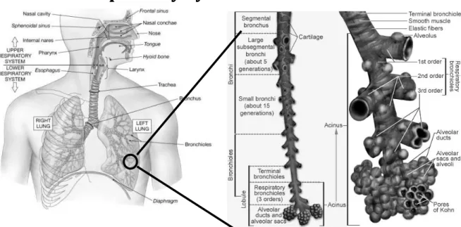
Figure 1. 1 Global view of respiratory system (left) and zoomed view of bronchial tract (right)
The developed respiratory system is the human system which conducts and exchanges gas with the pulmonary circulation. It is composed by different anatomical parts. From the external body, moving towards the internal one, there are mouth and nose, upper and lower airways.[1] The former airways consist of sinuses, vocal cords and larynx; the latter instead of trachea, bronchi, bronchioles. [2, 3]
The trachea divides into two bronchi, nominated “right“ and “left”. They can be furthermore divided in bronchioles. Although gas exchanges starts in the terminal zone of bronchioles, alveoli are the active part of exchange of gas. Since there are no alveoli in this initial part of airways (conducting airways), it is defined anatomic dead space (about 0.15 l). The “respiratory zone” is so composed of bronchioles, which have occasional alveoli on their walls, and of alveolar ducts which are completely filled with alveoli. The alveolus is the air-blood interface and allows gas exchange by diffusion. [4] There are about 300 million alveoli, about 0,3 mm in diameter each. Anyway, the respiratory system is not only composed of the lung but also by the chest wall. The pleural liquid, a thin liquid film at sub-atmospheric pressure, keeps lungs and ribcage close to each other. The interaction between these two structures determines the lung volume which plays a fundamental role in gas exchange and in work of breathing.
9
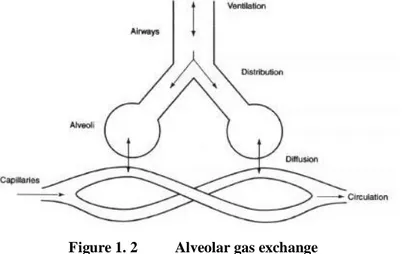
Figure 1. 2 Alveolar gas exchange
1.1.2 Definition of Lung Volumes and Pressures
A simple method for studying pulmonary ventilation is to record the volume movement of air into and out of the lungs: the spirometry. It is possible to define lung volumes under different conditions – as showed in Figure 1. 3: [4,5]
Figure 1. 3 Definition of lung volumes
Tidal Volume (VT) is the volume of a spontaneous breath (about 500 ml).
Inspiratory Reserve Volume (IRV) is the maximum volume inspirable from an end-tidal inspiratory level.
Expiratory Reserve Volume (ERV) is the maximum volume that can be expired from the end-expiratory level.
Residual Volume (RV) is the volume of gas in the lungs and airways after as much gas as possible has been exhaled (about 1.2 l).
10
Total Lung Capacity (TLC) is the volume of gas in the lungs and airways after as much gas as possible has been inhaled (about 6 l).
Vital Capacity (VC) is the volume of the deepest breath.
Functional Residual Capacity (FRC) is the volume of gas in the lungs and airways either at the end of spontaneous expiration or at the resting volume of the respiratory system.
Inspiratory Capacity (IC) is the maximum volume that can be inspired from the end-expiratory level.
To best compare lung volumes among subjects, generally these variables are corrected expressing them in percent of VC. This method is very efficient also because in normal subjects of equal sex, habits and age these values may reflect noticeable differences. Changes in lung volume are the result of the air flow in and out the respiratory system. Convective flow occurs as a result of a difference in pressure between two points. The respiratory system is able to establish a difference in pressure between open air and alveolar space by dimensional variations of thoracic cavity, generated thanks to the contraction and the relaxation of the chest muscles. Thus different following pressure can be defined:
External Pressure or Pressure at the Body Surface (PBS)
Pressure at the Opening Airways (PAO) is the pressure measured at the mouth.
Alveolar Pressure (PALV): the one registered within the alveoli.
Pleural Pressure (PPL) refers to the pressure within the pleural liquid that in normal condition is sub-atmospheric.
Transpulmonary Pressure (PTR): it is defined as the difference between the alveolar pressure and the pleural pressure. It is a measure of elastic forces that tend to collapse the lung at any level of lung expansion.
Wall Pressure (PW) results from the difference between the pleural pressure and the external pressure.
Respiratory System Pressure (PRS) is the pressure to which the respiratory system is exposed. It is calculated as the sum of the pressure drop due to airways (PAW, the difference between PAO and PALV), the transpulmonary pressure and the wall pressure. PRS can be divided in a resistive component (PRES), due to flow and to
11
dissipation properties of the system, and in two elastic components, referred to lung (L-PEL) and to chest wall (CW-PEL), due to volume and conservative properties of the system. [6]
1.1.3 Lung Ventilation
The lung ventilation is performed let flowing air into and out of the lungs, in alternation.
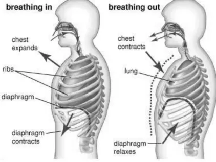
Figure 1. 4 Mechanic of inspiratory and expiratory phases
Inspiration
One of the two above mentioned phases, is the inspiration. Air flows in the lung when atmospheric pressure overcomes the pressure in the alveoli. Pressure can be generated both by the diaphragm and the intercostal muscles. When diaphragm contracts, abdominal pressure is increased and this generates forces able to elevate and enlarge the power part of the ribcage. If the action effectively transmits to the upper part, lung volu me changes and inspiration can continues.
This transmission of actions depends on the compliance of the rib cage: the less it is compliant, the more the risk to have paradoxical breathing. External intercostal muscles helps the rotation of the ribcage forward and upward. Moreover, it pushes the sternum increasing the antero-posterior and cross-section diameter of the thoracic cage and producing negative pressure in the intrapleural space. As before said, the increasing of the chest volume makes the lungs expanded because of the cohesive forces between the
12
visceral pleura and the parietal one; the lungs and their inner pressure decrease letting the atmospheric air enter alveoli. [4, 6]
Expiration
The second phase is usually passive, at least in quiet breathing. It deals as the inverse of the physical phenomena which provoke the inspiration. It is driven by the elastic recoil of the chest and of the lungs which were stretched during inspiration. It depends on surface tension, elastic properties of the tissues and bony elements of the rib-cage too. Active expiration instead is determined by the muscles of the abdominal wall and by the internal intercostals. When muscles of the abdominal wall contract, the abdominal pressure raises pushing the diaphragm upward. The thoracic volume decreases. [4, 6]
1.1.4 Mechanical Behavior of the Respiratory System
The mechanical behavior of the respiratory system can be explained using an electrical equivalent model - Figure 1. 5. It concerns four elements: airways, lung, chest wall and respiratory muscles. As usual, electric charge is the volume and thus the derivate, the electric current represents the flow of air; electric potential stands on the other hand for the pressure. The respiratory muscles are represented as voltage generator because they are able to change pleural pressure (PPL) through the chest wall (CW). Because of compression and expansion of gas, a capacitor is inserted between the airways (AW) and the lung (L) – the pressure between airway and lung is the alveolar pressure, PALV. The equivalent describes the behavior of lungs which neither suddenly expand during expiration nor decrease in volume when inspiration starts. Capacitor manages the usual exponential increment and decrement.[5] PBS is the pressure at body surface, while AO is the acronym for airway open. The gradient is PRS, respiratory pressure.
13
Static Behavior
Volume-Pressure Curve of the Lung
The lung is an elastic structure which would collapse expelling all its air through trachea if there were no force that keeps the air inflated. Lung tissue has fibers of elastin and collagen in it.
Pressure-Volume curves represent the behavior of lung or of the total respiratory system in static or dynamic condition. The static P-V curve is assessed by plotting the volume of the lung versus the driving pressure: the slope of this curve is known as compliance and describes the distensibility of the lung. Static P V curve can be observed in Figure 1. 6: two borders are defined as HIP and LIP as explained in the caption.
Figure 1. 6 Static P V curve. High and low pressure values are decided in the range of HIP –high inflection point- and LIP – low infection point
The steeper the slope, the grater the compliance and then the distensibility of the lung. Considering the whole breathing, the curve is hysteretic, namely it has two different P-V relations for expiration and inspiration [7], as depicted in Figure 1. 7:
14
Figure 1. 7 Hysteric dynamic P-V curve. A tract corresponds to inspiration while B to expiration phase. There are three zones where alveoli behave in different ways.
The curve deserves to be analyzed at least in 3 zones: the central and the two external regions. The lower end of the curve has a low compliance. This region is below critical opening pressure so pressure is required to open terminal airways and alveoli. At the centre of the curve the compliance increases resulting in a large change in volume for a small change in pressure. Normal tidal breathing usually occurs here because efficiency is maximal. At the upper end of the curve the compliance decreases once more. At high expanding pressures, in fact, the lung is overinflated and opposes to further volume increases. [8]
Lung’s major elastic component is probably due to the geometrical disposition of these fibers. [3, 6] Furthermore, the compressibility of the gas and the surface tension at the alveoli cause elastic behavior too. However the surface tension at the air-alveolus interface that allows mechanical equilibrium is really lower than in the presence of air-water interface. In fact it is highly dependent on the presence of the surfactant which reduces surface tension at the alveolar air-liquid interface. Surfactant is a complex lipoprotein formed by type II alveolar cells that lower the surface tension and consequently reduce the work of inspiratory muscles. This material is adsorbed at alveolar interface and its effect depends on alveolar size: the smaller the alveolus’ radius the more concentrated is the surfactant and this results in a further reduction of the tension. [7, 9] Surfactant helps to keep the alveoli dry. In fact, the surface tension tends to collapse alveoli and to adsorb fluid into the alveolar space from the capillaries. The surface tension of alveoli reduces the hydrostatic pressure outside the capillaries by reducing surface forces; surfactant prevents the transudation of fluid into the interstitium. Anyway, all these components concur in the definition of elastic nature and a curve testifies a scientific and proportional correlation between the driving pressure and the change in volume.
An important factor in the pressure-volume behavior of the lung is, as said, the surface tension of the liquid film covering the alveoli. It is a contractive tendency of the surface of a liquid that allows it to resist an external force. To be more explicit, surface tension arises because the attractive forces between adjacent molecules of the liquid are stronger than those between the liquid and gas and thus, it keeps occupying the smallest possible area. It
15
is revealed, for example, in the ability of some insects to run on the water surface. This property is caused by cohesion of similar molecules, and it is responsible of many of the behaviors of liquids. The tendency to occupy the smallest possible area, brings to make a sphere-like form. The pressure within a liquid sphere should be influenced both by the surface tension forces offered by the liquid and by the size of the sphere. Indeed, Laplace, found that pressure within a sphere is directly related to the surface tension of the liquid and inversely related to the radius of the sphere, or
[Eq. 1. 1]
where P is the pressure within the sphere, is the surface tension of the liquid , and R is the radius of the sphere – make reference to Figure 1. 8. As equal, thinking the alveolus as a spherical cavity
Figure 1. 8 Spherical alveolus
with a thin wall covered by a liquid film, the total pressure across the alveolar wall (P) is the sum of the pressure gradient at the air liquid-interface (PL-PEXT) and the pressure between the liquid film and the alveolar wall (PALV-PL):
[Eq. 1. 2]
where γ is the surface tension and R the radius of the sphere ([Eq. 1. 1] previously written) This relationship shows that the pressure (P) needed to stabilize the alveolus is directly proportional to the surface tension (γ) at air-liquid interface and inversely proportional to the radius (R). So the larger the radius of the alveoli, the less pressure is needed to hold them open, the smaller the radius, the more pressure is required to hold the airways open. [6]
16
Volume-Pressure Curve of the Entire Respiratory System
It has been spoken about lungs and their tendency to collapse; the main force which opposes the collapse is the one provided by the rib cage. The curve of the global mechanical answer of the respiratory system corresponds so to the sum of the curve of lung and chest wall measured separately, using the principle of superposition of effects. The chest wall curve alone, has negative pressure at low volumes so as at FRC and becomes positive when the volume is increased to about 75% of the vital capacity (VC). In the lung curve alone, pressure is always positive and the lung tends to collapse because of its elastic properties. At volumes above FRC, the total pressure of lung and rib cage together is positive and at smaller volumes the pressure becomes sub-atmospheric. At zero pressure, the lung is at its minimal volume, which is below residual volume (RV), the minimal volume reached by the chest wall only. [7, 10]
P-V curves have great importance in deciding the optimal parameters to recruit lungs, as it will be discussed in the next paragraph.
Dynamic Behavior
During respiration the respiratory muscles have to provide an adequate force to overcome the elastic recoil of lung and chest wall, the viscous friction of air flowing through the airways, the forces caused by the viscoelastic behavior of the lung tissues and the inertial forces. In expressing this, it is not used an electrical model, but a mechanical one is preferred. The balance of forces is described by equation [Eq. 1. 3,which is known as the equation of motion of the respiratory system.
[Eq. 1. 3]
In this equation PAO is the pressure at the airway opening, while V, V˙ and V˙˙ are the volume of the lung above end-expiratory volume and its time derivatives, flow and acceleration. PMUS is the pressure generated by the respiratory muscles. Other fundamental parameters are compliance C, resistance R and inertance I. The parameters relates pressure to Volume, flow and acceleration respectively.
17
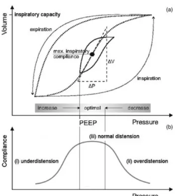
Figure 1. 9 Dynamic P V curve (upper) and related compliance (lower). In the optimal zone, the compliance assumes the highest and smoothest value.
Compliance measures the elastic recoil of the lung and chest wall. It depends primarily on lung’s size in a direct proportional way. [4, 6]
[Eq. 1. 4]
Elastance is a very similar way to refer compliance; it is exactly the inverse of C. Static and dynamic compliance can be distinguished. Static compliance is obtained considering the slope of the static P-V curve while dynamic compliance is related to the dynamic P-V curve. Dynamic pressure-volume loops show the pressure-volume relationship during inspiration and expiration (make reference to Figure 1. 9).
At end inspiration flow is zero, but pressure is lower than that expected from the static curve. The difference between static and dynamic compliance is due to resistance heterogeneities in the alveolar units of the same generation. If the airway diameter is smaller, flow is reduced and volume changes are slower. As frequency increases, if alveoli have not enough time to empty, the total change in lung volume decreases and therefore also compliance decreases. [6]
18
Resistance
Resistance represents the mechanical opposition to flow because of frictional forces. The resistance of the respiratory system is the sum of viscous resistance and airways resistance. Viscous resistance is the resistance generated within the lung tissue and chest wall during inflation and deflation. [6] Airway resistance is defined as the pressure gradient needed to move gas though the airways at a constant flow rate:
[Eq. 1. 5]
R not only depends on length and inside diameter of the conducting airways, but also on viscosity and density of the gas, on flow rate and on the kind of flow, which can be laminar or turbulent. Reynolds’ number is used to characterize these regimes of flow. Flow through straight tubes starts to become turbulent when Reynolds’ number exceeds approximately 1500 and is generally considered to be fully turbulent when Reynolds’ number is above 4500. [11] It has the following expression:
[Eq. 1. 6]
where v is fluid velocity, ρ gas density, ID is the internal diameter of the tube and μ is the viscosity of the gas. When both the flow regimes are presents the relationship between pressure and flow is expressed by Rohrer's equation:
[Eq. 1. 7]
where K1 and K2 are called the Rohrer constants. When flow is fully turbulent, the pressure difference is proportional to the square of the gas, only the second part of equation has to be considered and the resistance coincides whit K2; while when flow is fully laminar only the linear part of equation has to be considered and the resistance coincides whit K1.
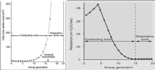
The resistance of the respiratory system (RRS) represents the sum of airways, lung tissue and chest wall resistance and it is rarely linear so the Rohrer's equation is to be considered. Thus the human lung can be modeled as a multiple tube system whose resistance depends on the total cross-sectional area of all the tubes. (Figure 1. 10) Because resistance increases with the fourth power as the airway is narrowed, even small airway constrictions can cause significant increases in resistance to flow.
19
Figure 1. 10 Total cross sectional area (left) and resistance (right) values, as function of air way generation
Inertance
Inertance of the respiratory system is the analogue of the electric inductance and it is a measurement of the tendency of the respiratory system to resist changes in flow. At frequencies normally reached, during spontaneous and mechanical ventilation, inertance is usually negligible and in general ignored. The pressure caused by the inertance is in the opposite direction to that generated by the elastance. Therefore, inertial forces tend to balance the impedance to the flow provided by the stiffness of the respiratory system (make reference to [Eq. 1. 13] in the next paragraph). [6]
1.1.5 Oscillatory Mechanics and FOT
Oscillatory mechanic of the respiratory system is the study of the mechanical properties of the lung and airways as derived by the response of the system to small pressure stimuli generated externally and applied during normal breathing. The relationship between pressure and flow is non linear in the respiratory system but we can consider its linearization around a work point in order to analyze the behavior of the system using tools for studying linear systems. In fact in this way it is possible to define the transfer function between two variables, one chosen as input and the other one as output and to use it to evaluate the mechanical properties of the respiratory system. When considering pressure (P) as output and flow (V˙) as input we obtain the respiratory impedance ZRS, or input
20 [Eq. 1. 8]
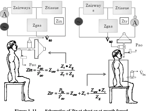
The respiratory system can be modeled as a generalized two-port system in which one port is the airways opening and the other one the body surface. For a two port system like that two different mechanical impedances can be defined.
Figure 1. 11 Schematics of Ztr at chest or at mouth forced.
The input impedance of the respiratory system (ZIN) is then obtained as the spectral relationship between pressure and flow (defined positive if entering the port) both measured at the airways opening when the stimulus is imposed to the same side, while the transfer impedance (ZTR) is obtained when the oscillations are imposed and pressure and flow are measured at different sites of the respiratory system. The expression for ZIN and ZTR are the following:
[Eq. 1. 9] [Eq. 1. 10]
where PAO and V˙AO are the pressure and flow measured at the airways opening respectively, while PBS and VBS are the pressure and flow measured the body surface,
21
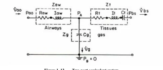
respectively. Since ZIN and these two variants of ZTR are affected differently by the parallel elements of the respiratory system, such as alveolar gas compressibility and upper air way wall movements, they can be selected or combined to obtain more reliable estimates of the airway and tissue impedance. If the measured system is linear, the two expressions above for ZTR must be equivalent [56]. The internal connection between the two ports can be modeled using the T-network, proposed by DuBois et al. 41] (Figure 1. 12)
Figure 1. 12 Two-port equivalent system
It includes airways impedance (ZAW), tissue impedance (ZT) and gas compression (ZG). ZAW is composed of the correspondent resistance and reactance, capacitor element (CG) accounts for gas compressibility and the tissue properties are described by the relative resistance, compliance and reactance. If alveolar gas compressibility is small (CG ≈0 and
•VG≈0), then this model reduces to the series R-I-C model, where RRS=RAW+RT, IRS=IAW+IT and CRS=CT. The electrical analogue can be simplified as it is possible to express the system as a one-port system:
22
The total impedance of this simplified model of the respiratory system can be calculated as following, summing partial pressure drops:
[Eq. 1. 11] Eq. 1. 12]
considering that PBS is the atmospheric pressure results:
[Eq. 1. 13]
The imaginary part of the impedance represents the compliance and the inertance of the overall respiratory system and is called reactance, XRS(ω), while the real part represents the resistance of the overall respiratory system, RRS(ω). Another way to consider respiratory impedance, is to divide its magnitude and its phase, as showed in [Eq. 1. 8]:
Where |ZRS| is the magnitude of respiratory impedance and ΦRS represents its phase:
[Eq. 1. 14]
At low frequencies, because 1/ωCRS is greater than ωIRS, the system behaves like a compliant one, the impedance magnitude decrease with frequency, ΦRS is negative (pressure lags flow) and the imaginary part is negative. At higher frequencies, because ωIRS is larger than 1/ωCRS, the effect of inertance is predominant, the impedance magnitude increases with frequencies, ΦRS is positive and the imaginary part is positive. At an intermediate frequency called resonant frequency (f0), the effects of compliance and inertance cancel each other so that |ZRS|=RRS and ΦRS = 0 (pressure in phase with flow). FOT or Forced Oscillations Technique makes reference to this theory to gain respiratory impedance and it has been exploited in the experimental set-up.
Forced Oscillation Technique (FOT) is a simple and minimally invasive method, useful to study the mechanical properties of the respiratory system. This observation is performed by measuring respiratory system’s response to a forcing signal applied externally. The mechanical response is generally defined as impedance, as said before. To best measure Z,
23
not Pressure’s sources different from the externally applied, have to be recorded. To do this, the signal’s frequency needs to be higher than the physiologic one (f > 4Hz). This so called “high-frequency FOT” permits the patient to breathe spontaneously and so it can be performed both in healthy and ill subjects.
FOT superimposes small pressure oscillation (about 1-5 cmH2O) at the respiratory system while recording pressure and flow signals. This oscillatory pressure induces an oscillatory flow whose amplitude and phase is related to the mechanical impedance of the patient’s respiratory system. As the respiratory system is not linear, the use of small oscillations permit to focus on a work point and to consider that as a linearization. Respiratory system impedance (ZRS) is defined as the complex ratio between the applied pressure (P) and the resulting volumetric flow rate (V˙) at the frequencies contained in the forcing signal. Therefore this complex function of frequency depicts the linear (or better, the linearized) behavior of the system in response to pressure oscillations and characterizes its mechanical response. ZRS can be factorized in its real and imaginary part, respectively called respiratory Resistance and Reactance. RRS represents the dissipative mechanical properties of the system, while the latter is related to the energy storage capacity and it is determined both by elastic and inertive properties. [Eq. 1. 13]
The technique was studied in ‘50s by Dubois [13] but only several years later it was greatly considered because of its technological difficulties to be applied. It gained interest because of patients who does not have to collaborate, and so it revealed a very useful technique for infants and very sick people unable to help doctors in measurement assessment.[14]
In recent years XRS at 5Hz has been proved to be sensitive to alveolar recruitment and to permit the identification of the lowest PEEP that maintains lung volume recruitment minimizing lung mechanical stress in an animal model of ALI [15,16].
Moreover it has been shown that FOT is applicable in ventilated newborns and can provide useful information for tailoring the ventilator settings according to the patho-physiological characteristics of the patient. [17]
24
1.2 Newborns Peculiarities and Lung’s
Recruitment
1.2.1 Fetal and Postnatal Lung Development
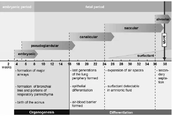
Newborns’ respiratory system is very different from adult’s one. It offers a much greater physical difficult to breathe, i.e. it has a total respiratory impedance greater than adults’ respiratory system. Thus, diseases that affect the small airways and cause large changes in peripheral resistance may be clinically silent in an adult, but can cause significant problems in infants (e.g., bronchiolitis). [19, 20] When birth occurs, some anatomical systems are not completely mature; the respiratory system is one of them. The development of the lung covers a period that begins with the appearance of lung bud in embryos and ends in the first infancy. [4, 20] The prenatal development of the lung can be divided in five periods described in synthesis in Figure 1. 14:
Figure 1. 14 Embryonic and fetal lung development
Most respiratory airways appear between the 16th week of gestation and birth while alveolar development starts relatively late in the 28th week of intrauterine life.
The development is not completed at 37th week of gestation, in fact the alveoli, as they exist in the adult, do not begin to appear until about 8 weeks of age. Most of them (85%)