U
NIVERSITÀ DELLA
C
ALABRIA
Dipartimento di M
ECCANICA
D
OTTORATO DIR
ICERCA IN INGEGNERIAM
ECCANICACICLO XXIII(2007-2010)
S.S.D. ING-IND/14 Mechanical Design and Machine Building
Biomechanics of the
Human Eye Sclera
A Dissertation Submitted in Partial Fulfilment of the Requirements for the Doctoral Research Degree in Mechanical Engineering
Doctoral Research Director Supervisors
Prof. Sergio RIZZUTI
Dr. Eng. Luigi BRUNO
Eng. J. Crawford DOWNS, PhD
Candidate
Abstract
The biomechanics of the optic nerve head and peripapillary sclera have been hypothesized to play a central role in the individual susceptibility to glaucoma, which is the second leading cause of blindness worldwide.
The aim of this work is to provide a mechanical testing and experimental data analysis procedure to characterize mechanical properties of human sclera.
The overall goal of performing scleral tissue mechanical testing is to elucidate the role of ocular biomechanics in the development of this disease by constructing eye-specific finite element models of the posterior pole.
In this work it has been developed and validated a new methodology for computing sub-micrometer scale IOP-induced deformation obtained during pressurization testing of human scleral shells.
Genetic Algorithm-based procedure and a microstructure-based numerical fitting method have been adopted to assess structural material properties of the testes eyes.
Preliminaries results show the range of nonlinear, anisotropic mechanical properties of posterior sclera from normal eyes of human donors.
Acknowledgments
I would like to thank you some colleagues and friends that helped me during my studies on ocular tissue biomechanics. I want to thank you Amedeo Lucente who introduced me in the study of the Glaucoma disease. He has been an invaluable source of motivations on ideas. I want to give a special thanks to Juan Reynaud who daily supported me in all the lab issues during the experimental testing period. I am profoundly thankful to Crawford Downs for every single help I received in the daily work spent at Devers Eye Institute and for providing me all the training I received about ocular tissue biomechanics. I thank you Ian Sigal and Rafael Grytz for being an inexhaustible source of ideas, motivations, discussions, and human support. I will be always thankful to Andrea Poggialini and Luigi Bruno for taking care of my technical formation. They have been much more than the mentors of my carrier.
1
TABLE OF CONTENTS
INTRODUCTION
3
The Importance of the Biomechanics of Human Sclera 3
The Existing Scleral Testing and Data Analysis Method 4
Description of the Activities Performed by the Candidate During the 3 Years of the
Doctoral Research Course 5
Introduction to the Chapters of this Dissertation 7
CHAPTER 1
9
Glaucoma and Ocular Tissues Biomechanics 9
1.1. Introduction 9
1.2. The ONH as a Biomechanical Structure 10
1.3. Association Between Normal Aging and the Vulnerability to IOP Related Injury 14
1.4. The Affects of Normal Aging on ONH Biomechanics 16
1.5. Changes in Laminar Material Properties and Morphology with Age 17
1.6. Changes in Biomechanical Behavior with Age 18
CHAPTER 2
20
Speckle interferometry - Theoretical Introduction 20
2.1. The Speckle Phenomenon 20
2.2. Interferometers for Measuring Displacements 24
2.3. Single-Light and Single-Image Absolute interferometer 25
2.4. Double Illumination and Dual Image Differential Interferometer 27
2.5. The Phenomenon of Speckle Decorrelation 28
2.6. Determination of the Phase 30
CHAPTER 3
33
Investigation Methods 33
3.1. Introduction 33
3.2. Preliminary Considerations 33
3.3. Material Constitutive Model 36
3.4. The Inflation Test 39
3.5. Inflation Test Set-up 41
3.6. Geometric Properties Reconstruction 46
2
Interferometric Data Analysis 50
4.1. The Recorded Data 50
4.2. The Problem of the Wrapped Phase 52
4.3. Reconstruction of the Displacement Field 56
4.4. B-spline Based Fitting Method 60
4.5. Numerical Analysis - Introduction 64
4.6. Numerical Analysis - FEM Model Implementation 65
4.7. Numerical Analysis - Material Properties 67
4.8. Numerical Analysis - Genetic Algorithm Optimization 70
4.9. Numerical Analysis - Genetic Algorithm Based Procedure 73
CHAPTER 5
78
Experimental Results 78
5.1. Human Eyes Data Fitting 78
5.2. Preliminary Outcomes from Human Eyes Data Fitting – Testing Procedure 80 5.3. Preliminary Outcomes From Human Eyes Data Fitting – Numerical Results 83 5.4. Preliminary Outcomes From Human Eyes Data Fitting – Comments 87
CHAPTER 6
88
A New Approach of Speckle interferometric Data Analysis for 3D Objects 88
6.1. Introduction 88
6.2. Fitting and Analysis Method 90
6.3. Comments on the Adopted Procedure 102
CHAPTER 7
103
3D Interferometric Data Analysis and Microscale-based Numerical Fitting
Method – A New Approach 103
7.1. Inverse Numerical Estimation of Eye-Specific intrinsic Material Properties and
Local Collagen Architecture 103
CHAPTER 8
108
Comments and Conclusions 108
8.1. Overview of the Mechanical Testing and Analysis Procedure 108
8.2. Comments on the Presented Preliminary Results 109
8.3. Final Conclusions 111
3
Introduction
The Importance of the Biomechanics of Human Sclera
The sclera is the most extensive load bearing tissue of the eye. Its major function is to provide structural support to the other two most extended tissues of the eye, the choroid and the retina. The retina is the tissue containing the optical receptors of the eye, while the choroid is meant to provide the nutritional blood supply to the retina.
The spherical shape of the eye is essential to allow the rays of light coming into the eye to be in focus on the retina. The sphericity of the eye is achieved by maintaining positive pressure the liquid inside the eye (i.e. humor vitreous and aqueous). This pressurized liquid creates a hydrostatic load on the ocular coats that is commonly referred to as intraocular pressure (IOP). Mechanically, the IOP inside the eye creates a hydraulic load, and the main function of the sclera is to provide structural stiffness to carry this mechanical load. The sclera also provides structural support to the choroid and the retina.
IOP is usually indirectly measured by applanation tonometers and its normal value is in the range of 10 - 21 mmHg (millimeters of Mercury). A pathological increase of IOP is associated to an increased risk in developing an optic neuropathy called glaucoma.
Glaucoma is the second leading cause of blindness in the world after cataracts, although cataracts can be treated surgically to reverse blindness. Loss of vision due to glaucoma is irreversible.
The mechanisms of damage to the retinal ganglion cell axons in glaucoma is not well understood, but it is hypothesized that elevated IOP affects the biomechanics of the tissues of the eye. In particular, glaucomatous damage takes place at the optic nerve head (ONH), a structure through which the axons that transport the visual information exit the eye on their path to the
4
brain. It is thought that the ONH is particularly susceptible to the effects of IOP, as it is generally less mechanically resistant to increased pressure loads. The sclera plays the major role in the global mechanical behavior of the eye, as it is the principal load bearing tissue of the globe. The sclera also surrounds the ONH, and therefore plays an important role in the biomechanics of the ONH.
In order to build an understanding of the role of ocular biomechanics in glaucoma, one must possess a detailed characterization of the mechanical properties of the sclera. The aim of this work is to develop a testing and analysis procedure for the mechanical characterization of the scleral tissue from human donor eyes.
The Existing Scleral Testing and Data Analysis Method
The mechanical testing procedure that served as the basis of this work is based on a procedure previously developed by Michael Girard in studies of scleral biomechanics in the monkey model of glaucoma (Girard M. J., 2008). The existing procedure was based on the inflation test of posterior sclera shells clamped and imaged in a customized apparatus. The deformations experienced by the pressurized scleral shell were recorded by a commercial laser speckle interferometer (Q100, Dantec Dynamics A/S, Denmark), and the deformation field was calculated by the commercial software package that accompanied the interferometer. The computed experimental displacement fields were then used as the target displacement for a genetic algorithm-based inverse finite element procedure that fitted a set of mechanical parameters (defining the material model assumed to describe the mechanics of the sclera). The mechanical parameter set was varied until the finite element model displacement predictions closely matched the experimentally measured displacement field.
5 Eyemesh, a semi-automated MatlabTM routine written by Michael Girard, was used to create an individual specific FE model of the tested eye. Eyemesh requires the following data inputs:
A point cloud obtained from a 3D mechanical digitizer that was used to represent the 3D shape of the scleral outer surface.
Scleral thickness measurements at 20 discrete locations collected by using an ultrasound transducer.
The 3D displacements of the scleral outer surface calculated by the commercial software package that accompanied the interferometer. These input data required a great deal of manual pre-processing, and were fraught with error as described in the next section.
Description of the Activities Performed by the Candidate During the 3 Years of the Doctoral Research Course
The goal the presented work was to revise, modify, and extend the previous work done by Michal Girard in mechanical testing of monkey sclera shells in order to perform automated, error corrected, mechanical testing of human scleral shells, and allow subsequent calculation of accurate mechanical properties.
A customized routine has been written in MathematicaTM to reconstruct the experimentally recorded displacement fields from the raw interferometer output, thereby bypassing the erroneous processing in the commercial interferometry software. All the optical errors inherent in the testing set-up were taken into account. This approach eliminated substantial errors in the displacement field calculation previously disregarded by the commercial software package.
The corrected displacement fields computed by the new custom routine were then used as the target values of a material property fitting procedure to estimate the material properties of the sclera. The derived custom routine
6
eliminated the errors in the experimentally measured displacement maps, which otherwise would cascade into wrong and missleading fitted scleral material properties.
In particular, portions of the genetic algorithm procedure were rewritten in
Fortran 77 and Fortran 90to perform a different numerical formulation of the eye-specific FE models used to estimate the sclera’s mechanical response to the load produced by IOP.
The Eyemesh routine was modified and extended such that all the tasks necessary to create the individual specific FE model of the eye are completely automated. This automation brought some important improvements:
A vast reduction in processing time.
An objective and repeatable pre-processing of the experimental data. The addition of graphic-based control of the experimental data
pre-processing.
Previously, the 3D displacement fields were calculated by the commercial software that accompanied the interferometer. This software has been entirely replaced by new custom routine written in MathematicaTM that reconstructs the experimental displacement fields at any IOP level directly from the raw data recorded by the interferometer. The key features of the now fully automated processing program are the following:
Direct management of the raw data recorded by the interferometer and of the scleral thickness and scleral surface geometry data.
Computation of the absolute phase change maps of the recorded speckle field (a function of the recorded deformation fields) by performing a custom unwrapping procedure.
Elimination of the errors associated with the change of the laser wavelength and the angle of the laser beams induced by speckle
7
recording through an optical window in a saline-filled chamber (refraction changes).
Providing a mathematical representation of the geometry of the scleral shell, and of the experimentally recorded 3D displacement fields. The new analysis procedure is now performed by this completely automated, graphics-assisted program. The new program completely replaced the previously described Eyemesh routine, except for the creation of the nodal coordinates of the scleral shell finite element mesh.
Introduction to the Chapters of this Dissertation
The present dissertation is split in several chapters each one describing a step of the work done in the 3 years Doctoral Research Course attended by the candidate. The main topic of each chapter is summarized in the following list:
Chapter 1: In this chapter, the importance of sclera biomechanics and how it potentially relates to the mechanical damage seen in Glaucoma is
discussed.
Chapter 2: In this chapter, an overview on speckle methodology is
presented. The speckle methodology presents the basis of the interferometric technique used to record the scleral surface displacement fields during
mechanical pressure testing.
Chapter 3: This chapter gives a detailed description of the experimental set-up used to perform the mechanical inflation test and a description of the testing instrumentation. Furthermore, the mathematical formulation used for the constitutive material description and the inverse FE models are presented.
Chapter 4: In this chapter, the procedure to analyze the experimental data from mechanical inflation test is described. This includes the management
8
and computation of the speckle interferometric data. After a brief introduction to the genetic algorithm-based optimization technique, it is here introduced the genetic algorithm used in this work, and how material properties are computed adopting this procedure is presented.
Chapter 5: Preliminary results obtained by the genetic algorithm-based fitting procedure and achieved by the analysis performed on four human eyes are reported in this chapter. Results shown in this section have been obtained by the genetic algorithm-based procedure described in Chapter 4, and the main outcomes of the procedure are analyzed and discussed.
Chapter 6: In this chapter, the new computational method that was developed to analyze the recorded speckle interferometric data is introduced and described. The optical set-up used in the scleral inflation experiments did not satisfy the simplifying assumptions typically employed in classic holography (and used by the commercial software accompanying the interferometer); the developed procedure accounts for all the optical features considering the real testing conditions of the mechanical testing procedure and of the experimental set-up are described.
Chapter 7: A microstructure-based numerical fitting method developed by Rafael Grytz is presented. This fitting method is used to estimate the mechanical properties of the human sclera form the corrected displacement measurments. It allows to assess some important features of the biological structure of the scleral tissue. Some preliminary results obtained with this new approach are reported.
Chapter 8: This chapter includes the discussion of the preliminary results obtained from the genetic algorithm-based procedure (Chapter 4) and from the microstructure-based numerical fitting method (Chapter 7).
9
Chapter 1
Glaucoma and Ocular Tissues Biomechanics
1.1. Introduction
The principal aim of this work is to provide a mechanical testing and analysis procedure to assess the mechanical properties of human eye sclera. Mechanical testing has been performed on about 40 pairs of human eyes and 4 pairs of monkey eyes with advanced experimental glaucoma.
The mechanical testing of the scleral shells from the human donor eyes has been carried out to fulfill the aims of the following grants from the U.S. National Institute of Health:
NIH R01-EY018926, Age-related Changes in Optic Nerve Head
Structure and Biomechanics.
Principal investigators: J. Crawford Downs and Christopher A. Girkin. NIH R01-EY019333 Racial Variations in Optic Nerve Head Structure
and Biomechanics.
Principal investigators: J. Crawford Downs and Christopher A. Girkin. Tested eyes from human cadaver have been collected from young normal donors and old normal donors of European descent, as well as donors of African descent.
The overall goals of these projects are to evaluate the influence of ocular biomechanics on the increased age- and race-related susceptibility to glaucoma. In particular, the presented work focuses on the differences in scleral stiffness due to aging and racial variations, and how scleral mechanical properties affect the biomechanics of ONH in the human eye.
10
The ONH biomechanics are understood here as the interactions between IOP and the connective tissues of the ONH and peripapillary sclera, or in particular, the structural stiffness these tissues (the combination of tissue architecture and material properties). Our overarching hypothesis is that the ONH biomechanics are inextricably linked, either primarily or secondarily, to IOP-related and pathophysiologic changes in the blood flow, nutrient diffusion and cellular activity within those tissues. The immediate goals of focused in this research grants are to characterize age- and race-related differences in ONH biomechanics and elucidate their effects on ONH susceptibility to IOP insult. The long-term goal of the research proposal is to develop clinical diagnostics and interventions designed to manage each important biomechanical risk factor in the development and progression of glaucoma. Sclera biomechanics is thought to be important in the development and progression of glaucomatous optic neuropathy. This is described in the
Age-related Changes in Optic Nerve Head Structure and Biomechanics grant
proposal submission made by the Principal investigator of this research proposal J. Crawford Downs, which is included below. This grant proposal provides a brief, detailed review of the scientific considerations on the topic that lead to the formulation of the hypothesis that changes in sclera mechanical properties (considered dependent to aging and ethnicity) are likely to increase the vulnerability of the ONH connective and neural tissues to IOP-related axonal injury in glaucoma.
1.2. The ONH as a Biomechanical Structure
The lamina cribrosa (Figure 1) provides structural and functional support to the retinal ganglion cell (RGC) axons as they pass from the relatively high-pressure environment within the eye to a lower high-pressure region in the retrobulbar cerebrospinal space (Zeimer, 1995). To protect the RGCs in this unique anatomic region, the lamina in higher primates has developed into a complex structure composed of a three-dimensional network of flexible
11
beams of connective tissue, nourished by a capillary bed primarily arising from the short posterior ciliary arteries penetrating the immediate peripapillary sclera (Burgoyne, Downs, Bellezza, & Hart, 2004) (Burgoyne, Downs, Bellezza, Suh, & Hart, 2005) (Quigley & Addicks, 1981) (Jeffery, Evans, Albon, Duance, Neal, & Dawidek, 1995). This scleral and intra-laminar vasculature is unique in that it is encased in load-bearing connective tissue, either within the scleral wall adjacent to the lamina cribrosa, or within architecture of the laminar beams themselves (Hayreh, 1986).
The anatomy of the lamina cribrosa and peripapillary sclera merits several considerations regarding the etiology of glaucomatous cupping and implies that the classic “mechanical” and “vascular” mechanisms of glaucomatous injury (Fechtner & Weinreb, 1994) are inseparably intertwined (Burgoyne, Downs, Bellezza, Suh, & Hart, 2005) (Figure 2 and Figure 3). For example, prior to structural damage, purely IOP-related stress could detrimentally affect the blood supply to the laminar segments of the axons by direct deformation of the capillary-containing connective tissue structures or due to changes in the extracellular matrix that may limit the diffusion of nutrients to RGC axons in the laminar region. Reciprocally, primary insufficiency in vascular supply to the laminar region could induce connective tissue changes that would serve to weaken the laminar beams, making them more prone to failure under similar levels of IOP-related mechanical stress.
To incorporate these concepts within a global hypothesis, a biomechanical model of glaucomatous optic neuropathy has been proposed (Burgoyne, Downs, Bellezza, Suh, & Hart, 2005). This model proposes that IOP-related stress and strain play an essential and causative role in the pathophysiology of the changes seen in all of the tissue types within the ONH and in its blood supply. These not only include the load-bearing connective tissues within the lamina cribrosa, the scleral canal wall, and the peripapillary sclera, but also the cellular components of these tissues, including astrocytes, glial cells, endothelial cells, and pericytes, along with their basement membranes and the RGC axons.
12
Figure 1. Anatomy of the human eye. Lamina cribrosa is highlighted in the boxed region at the
posterior pole. Figure provided by the National Eye institute and National institutes of Health, http://www.nei.nih.gov/photo/.
Figure 2. The optic nerve head (ONH) is influenced by IOP-related stress and strain. (A) The ONH is
made up of prelaminar, laminar and retrolaminar regions. (B) Within the clinically visible surface of the normal ONH (also referred to as the optic disc), central retinal vessels enter the eye and retinal ganglion cell (RGC) axons appear pink/orange due to their capillaries. (C) the blood supply for the laminar region (LC) is from the posterior ciliary arteries (PCA). The primary site of RGC axon insult in glaucoma is within the lamina cribrosa, which is schematically depicted with axon bundles (brown) in (D). The laminar connective tissues, isolated by trypsin digest in a scanning electron micrograph in (E), provide structural support to the ONH, which is the weak spot in an otherwise strong pressure vessel. The laminar beams, drawn in (F) with stippled extracellular matrix (ECM), central capillary (red) and surrounding astrocytes (yellow with basement membranes in black), also contain the capillary network and much of the cellular structure that support the axons in the laminar zone. The clinical manifestation of IOP-induced damage to the ONH is most commonly “deep cupping” (G), but in some eyes cupping can be shallower accompanied by pallor (H). Z-H, circle of Zinn-Haller; PCA, posterior ciliary arteries; NFL, nerve fiber layer; PLC, prelaminar region; LC, lamina cribrosa; RLC, retrolaminar region; ON, optic nerve; CRA, central retinal artery. (A) Reprinted with permission from Doug Anderson ; (C) reprinted with permission from Cioffi GA; (D) Reprinted with permission from Quigley HA; (E) Reprinted with permission from Quigley HA; (F) Reprinted with permission from Morrison JC.
13
Figure 3. IOP-related stress and strain influence both the ONH connective tissues (CT) and the
volume flow of blood primarily, and the delivery of nutrients secondarily, through chronic alterations in CT stiffness and diffusion properties (blue and red). Other primary insults to the ONH CT (yellow) can damage the ONH CT and/or axons. Once damaged, ONH CT can become more or less rigid depending upon lamina cribrosa astrocyte and glial response. If weakened, ONH CT deform in a predictable manner. The clinical ONH appearance of cupping can be divided into “laminar” and “prelaminar” components. Prelaminar cupping follows loss of prelaminar neural tissues without laminar involvement (white arrows, lower right). Deep or laminar cupping follows ONH CT damage and deformation. Laminar cupping is important because it establishes that IOP is actively involved (either 1°or 2°). Laminar cupping may be accompanied by a prelaminar component (small black arrows, lower left).
14
Regardless of the primary insult in glaucomatous injury, IOP-related stress and strain in the laminar connective tissues are key elements in this model and are dependent on the 3D ONH architecture and material properties of these connective tissues (Zeimer R. , 1995) (Burgoyne, Downs, Bellezza, Suh, & Hart, 2005) (Burgoyne & Morrison, 2001) (Quigley H, 1995) (Bellezza, Rintalan, Thompson, Downs, Hart, & Burgoyne, 2003) (Downs, Suh, Thomas, Bellezza, Hart, & Burgoyne, 2005) (Quigley & Addicks, 1981) (Sigal, Flanagan, & Ethier, 2005) (Zeimer & Ogura, 1989).
1.3. Association Between Normal Aging and the Vulnerability to IOP Related Injury
The results of the several randomized prospective trials have identified the risk factors associated with the development or progression of Glaucoma (Table 1) (AGIS Investigators, 2002) (EGPS Investigators, 2007) (Leskea, Heijl, Hyman, Bengtsson, & Komaroff, 2003). Across several studies, IOP, age, central corneal thickness, increased optic disc cupping, and African ancestry are independently associated with glaucomatous progression. It is important to note that all of these risk factors have a biologically plausible association with either the level of IOP, the severity of disease (visual field severity), or biomechanical properties of the ONH (age, (Albon, Purslow, Karwatowski, & Easty, 2000), African ancestry (Girkin, McGwin, & Xie, 2005), corneal thickness (Lesk, Hafez, & Descovich, 2006), increased cupping). Importantly, age is the only risk factor other than IOP that is independently associated with the onset and progression of glaucoma across all of the major prospective clinical trials conducted over the past twenty years.
15
In addition to data from prospective trials in glaucoma and ocular hypertension, every population-based survey conducted to date has demonstrated a strong relationship between the prevalence of glaucoma with advancing age (Rudnicka, Mt-Isa, Owen, Cook, & Ashby, 2006), despite almost all studies showing no changes in IOP (Leske, Connell, Wu, Hyman, & Schachat, 1997) (Weih, Mukesh, McCarty, & Taylor, 2001). Furthermore, while normal tension glaucoma is not uncommon within elderly populations (Levene, 1980) (Chumbley & Brubaker, 1976), it is not seen in children or young adults other than in a few isolated case reports (Geijssen, 1991). These findings indicate that the aging ONH becomes increasingly vulnerable to glaucomatous injury at similar levels of IOP.
Developing techniques to quantify the biomechanical behavior of the ONH will allow for the determination of how the affects of aging are mediated in the causality of glaucomatous injury. This will also form the groundwork for continued studies investigating the effects of individual variation in ONH structure and biomechanics on the vulnerability to IOP-related injury as seen in at–risk groups such as individuals of African ancestry.
16
1.4. The Affects of Normal Aging on ONH Biomechanics
As with any solid structure, the degree of local deformation (strain) experienced by the ONH under a given level of stress is dependent upon its 3D architecture and material properties (Bellezza, Hart, & Burgoyne, 2000) (Downs, Suh, Thomas, Bellezza, Hart, & Burgoyne, 2005) (Sigal, Flanagan, & Ethier, 2005). Thus, variation in ONH anatomy and material properties that occur with aging may account for the increased susceptibility to IOP-induced injury (Figure 3and Figure 4).
Previous researches have shown that even at physiologic level of IOP substantial stress and strain may occur within the lamina and peripapillary sclera that may lead to alteration in the ONH architecture (Bellezza, Hart, & Burgoyne, 2000) with subsequent increase in the vulnerability to mechanical failure.
Figure 4. The complex relationships between normal aging, IOP, mechanical stress and strain, and
ONH susceptibility. Susceptibility 1: for a given ONH, IOP generates low or high levels of mechanical stress and strain depending upon the 3D architecture and mechanical properties of the ONH connective tissues. Some ONHs will have relatively low mechanical stress at high IOP (d), while others will have high mechanical stress at low IOP (e). Susceptibility 2: Whether a given level of IOP-related stress and strain is physiologic or pathophysiologic depends upon the individual ONH’s resistance to IOP-related connective tissue strain, blood flow reduction, and cellular activation, all of which are affected by aging. Strong connective tissues, a robust blood supply and stable astrocytes and glia should increase the chance of an individual undergoing normal ONH aging and withstanding the deleterious effects of IOP, even if that IOP is higher than normal.
17
It has also been shown that the material properties of the sclera are altered early in glaucoma in the monkey model (Downs, Suh, Thomas, Bellezza, Hart, & Burgoyne, 2005), which indicates that the cellular response to connective tissue strain can quickly alter the mechanical behavior of the tissue.
These findings may have important clinical ramifications in that for a given magnitude of IOP insult, the aged ONH ought to demonstrate shallower cupping than a young ONH, as seen in the senile sclerotic variant of glaucoma (Broadway, Nicolela, & Drance, 1999) (Nicolela MT, 1996). Interestingly, preliminary data from an analysis of imaging results from patients participating in a longitudinal glaucoma study seem to indicate that older patients tend to have shallower degrees of cupping than young patients when controlled for disease severity, race, pre-treatment IOP, and current IOP.
1.5. Changes in Laminar Material Properties and Morphology with Age
A variety of age-related changes in the ONH connective tissue may affect ONH compliance (Morrison, LHernault, Jerdan, & Quigley, 1989) (Albon, et al., 2007) (Albon J. , Karwatowski, Avery, Easty, & Duance, 1995) (Bailey, Paul, & Knott, 1998) (Hernandez MR, 1989). In the young adult, the laminar beams are composed of a central core of elastin fibers and fibrillar collagen surrounded by basement membrane components (collagen IV, and laminin). During aging, immunohistochemistry has demonstrated increases in collagens I, III, IV, and V, along with thickening of the elastic fibers within the laminar beams. In addition, quantitative studies revealed an increase in the total collagen and advanced glycation end products, with reduction in the proportion of collagen III, total sulphated glycosaminoglycans, and lipid concentration (Quigley, Brown, Morrison, & Drance, 1990) (Albon, et al., 2007) (Albon J. , Karwatowski, Easty, Sims, & Duance, 2000) (Brown, Vural,
18
Johnson, & Trinkaus-Randall, 1994) (Hernandez MR, 1989). In all, these changes should result in a stiffening of the connective tissues of the ONH (Zeimer & Ogura, 1989) (Albon, Purslow, Karwatowski, & Easty, 2000). Age-related increases in the thickness of the laminar beams have been reported, with associated reductions in the proportional area of laminar pores [63,66]. Since changes in morphology will affect the biomechanical behavior of any load bearing structure, these age-related morphologic variations certainly must exert some effect on the biomechanical behavior of the ONH.
1.6. Changes in Biomechanical Behavior with Age
Several in-vivo techniques have been developed to study the compliance of the ONH. In cadaver eyes, ONH compliance has been quantified using scanning laser tomography (Burgoyne, Mercante, & Thompson, 2002) (Yan, Coloma, Metheetrairut, Trope, Heathcote, & Ethier, 1994), Doppler velocimetry (Zeimer, Wilensky, Goldberg, & Solin, 1981), and radiographic evaluation of platinum wires embedded within the lamina (Zeimer & Ogura, 1989). Albon and colleagues demonstrated that there is an overall increase in structural stiffness of the lamina cribrosa with age, with greater deformation of the lamina seen in younger eyes at any given IOP (Albon, Purslow, Karwatowski, & Easty, 2000). While these results confirm the increased stiffness of the lamina with age, they are limited to imaging only the surface of the lamina and peripapillary sclera.
Sigal and colleagues determined that variations in scleral thickness, radius of the eye, laminar stiffness, and scleral thickness have the greatest influence on the biomechanical response of the ONH using finite element modeling based on axisymmetric idealized geometries (Sigal, Flanagan, Tertinegg, & Ethier, 2004). This research demonstrates that variation in the material properties of the sclera and lamina are important determinates of the stress/strain relationship within the ONH. However, these idealized models lack sufficient anatomic detail to define the biomechanical changes within the
19
microarchitecture of the lamina, which is the primary site of axonal injury. In contrast to prior work, this proposal will combine methods that will define both the material properties and microarchitecture of the ONH necessary to develop biomechanical models based on individual human eyes.
20
Chapter 2
Speckle interferometry - Theoretical Introduction
2.1. The Speckle Phenomenon
When a surface - with a roughness greater than or equal to the wavelength of the illuminating light - is illuminated with a coherent light beam at a certain distance from the surface, it can be observed in a field of light characterized by a myriad of light and dark spots randomly distributed in the space. This speckle phenomenon is created by the scattering of light waves when reflected by the rough surface. Theoretically, this phenomenon can occur not only when the illuminating light is in the visible spectrum, but it can be also generated by other oscillatory phenomena of different wavelengths such as radio waves or X-ray.
The formation of these structures was observed the first time by researchers working in the field of holography. The speckle was thus initially considered as a noise effect inevitably superimposed on the information encoded in the recorded holographic fringes. The speckle signal was later investigated as a means of measurement. The statistical distribution of light intensity, amplitude, and phase of the electromagnetic field generated by the speckle effect have been obtained theoretically (Goodman, 1975).
A spatial random distribution of a speckle field is shown Figure 5, in which the three-dimensional surfaces are equal light intensity regions.
The size of the single speckle, denoted by , is a very important parameter. It can be evaluated using the following formula:
21
where is the wavelength of the light source, z and D are defined in terms of how the speckle field is observed.
If the speckle is directly observed (without any optical means) it is termed
objective speckle and in this case z is the distance between the observation
point and the observed surface, while D is the diameter of the illuminated area (assumed to be circular and uniformly illuminated).
If the surface is observed through a lens, it is termed subjective speckle; in this case, z is the distance between the plane of the lens and the plane of focus, while D is the diameter of the smallest lens in the optical set-up used.
Figure 5.Three-dimensional reconstruction of the speckle observed at far distance (left) and short distance (right).
For this reason, the speckle size changes as a function of the observation distance z.
The transverse size of the speckle also changes as a function of the optical set-up. In this case, the size of the single speckle is
22
It can be seen in Figure 5 that the surfaces of the speckle are quite spherical and the structure changes rapidly when viewed from afar (left side), but the speckles tend to stretch and grow when viewed closely (right side), and the structure tends to remain the same.
This last condition is necessary for conducting experimental measurements using the speckle phenomenon. In fact, this speckle field created by illuminating a rough surface can be directly used for applications such as metrology.
Speckle effect can be regarded as a sort of “fingerprint” of a surface, and it can be used to detect any changes in the surface as rigid motions or local deformations.
Some techniques based on digital correlation or aperiodic decorrelation of speckle fields have been adopted by many researchers. Digital correlation (Oliver & Pharr, 1992) (De Fazio, Syngellakis, & Fugiuele, 2001) is a technique that can be regarded as the modern version of the speckle photography (Beghini M, 1999 ). Speckle photography consists of using a photographic plate to record the same speckle field at two different configurations, i.e. before and after the observed surface is deformed. To measure deformations by exploiting the speckle phenomenon, it is necessary that interference occur between the speckle field generated by the beam illuminating the observed surface and another light field consistent with the first (created by a beam generally called reference beam).
The basic principle is shown schematically in Figure 6, wherein two coherent sources interfere with each other. If one source undergoes a shift at a generic point in space, it produces a cyclical variation of the light intensity whose frequency depends on the wavelength of illuminating light used.
23
Figure 6. Schematic representation of an interferometer.
So, if two stationary waves with same frequency interfere, the resulting light intensity ( ) depends only on the phase relationship existing between the two waves. Using a complex notation, the resulting light intensity equation for a generic stationary wave can be written as follow:
(eq. 3)
where A represents the waves amplitude and the phase value. By summing the two interfering waves and calculating the resulting intensity, it can be shown that:
(eq.4)
The evolution of the resulting intensity is shown in Figure 7, where it can be seen that the change in light intensity depends on the phase difference. Hence, the cyclical variation in light intensity can be traced back to the dealignment between the two sources that created the phase difference. Speckle interferometry, as well as holographic interferometry, have the advantage of being able to operate on surfaces that don’t need to be observed along their normal direction because the light is diffused by the
24
object and theoretically, every point in the space has a light intensity that is function of the diffused light made by all the points on the surface.
This feature imputes a great advantage to speckle interferometry in that it is able to measure both out-of-plane displacement components (as in classical interferometry) and in-plane components (such as in moiré interferometry) of displacement.
Figure 7. Resulting light intensity as a function of the phase shift
Alternate and Mean values are plotted in the ordinate coordinate axis.
2.2. Interferometers for Measuring Displacements
As shown by the multitude of scientific publications (Doval, 2000) (Sirohi R. S., 2002) and specialized texts (Erf, 1978) (Sirohi, Speckle metrology, 1993) there are different types of interferometers that have been proposed by many researchers working in speckle interferometry. One way to classify interferometers has been proposed in (Bruno & Poggialini, 2000), where the classification is made on the basis of which displacement component the device is sensitive to, and on how the illumination and observation directions are set up.
Another feature that distinguishes an interferometer is the number of beams used for illuminating the object. By using this classification method
25
interferometers are classified as single or double illumination, depending on the number of light sources, and single or double observation, depending on how many images of the object observed are focused on the CCD of the observation camera.
The interferometer used in this work can be classified as a Single-Light and
Single-Image Absolute interferometer.
2.3. Single-Light and Single-Image Absolute interferometer
This kind of interferometer is the simplest set-up, it is sensible to only a single generally oriented displacement component. By considering in the Figure 8 a generic point P(x,y,z) belonging to the surface under examination, it is possible to define a direction of illumination by joining the light source point S and the generic point P. The direction of observation can be defined by joining the point P and the observation point O (which normally coincides with the optical center of the lens). The direction of illumination and observation are uniquely identified by two unit vectors
k
iandk
o, and the angle between the viewing directions and lighting is 2.
ki ko d O S 2 P(x,y,z) k
26
Indicating with = { } the shift affecting the point P and neglecting (in this instance) the variations of the unit vectors and , it can be written the well known formula of the holographic interferometry (Vest, 1979)(still applicable in speckle interferometry):
=
(eq. 5)
where is the wavelength of the used light and is the phase shift that arises due to the displacement . The vector
=
is called sensitivity vector, its module is(
and it is oriented along the line bisecting the illumination and the observation directions.Therefore, by using an interferometer of this type is theoretically possible to measure a displacement vector oriented along any direction in the space. By choosing a suitable point of view and three linearly independent lighting directions, it is possible to reconstruct three independent displacement components describing the surface deformation.
Of course, the determination of the phase change must be made by using a reference beam interfering with the light diffused by the object and constant in direction throughout the deformation of the object.
It has to be considered that the observed objects are never perfectly diffusing and most of the light is sent back, more or less, in the direction of illumination. This implies that to have good contrasted light intensity distributions in the space the sensitivity vector has to lay close to the illuminating direction
27
2.4. Double Illumination and Dual Image Differential Interferometer
In the case of double illumination and dual image differential interferometer the phase relationship is obtained differentiating the phase relationship of the single image interferometer.
The optical set-up of this interferometer is shown in Figure 9, and the relation between the change in phase with the measurements is the follows:
(eq. 6) ki1 ko d S1 P(x,y,z) d+ d P(x+ x,y+ y,z) Measurement plane k ki2 S2
28
This type of interferometer is mainly sensitive to the in-plane components, so it is suitable to measure the first derivatives of displacements in the plane. This type of interferometer (shearing interferometer or shearometer) is not widely used due to the low visibility of the fringes (Tyrer & Petzing, 1997). An interesting application of this interferometer has been recently adopted to analyze human corneas shell mechanical properties (Knox Cartwright, Tyrer, & Marshall, 2010).
A more effective method to measure the first derivatives of the in-plan displacements is to use the shearometer by recording two measures in order to have a sensitivity vector symmetrical to the normal (Steinchen, Kupfer, Mäckel l, & Vössing, 1999) (Fan, Wang, & Tan, 1997).
In this way, by using a simple sum and difference operation on the double measurement of the phase detected at any point, it is possible to separate the in-plan and out-of-plane deformation components.
2.5. The Phenomenon of Speckle Decorrelation
Using speckle interferometry as a measuring device for deformations introduces some carefulness to be taken into account. The most relevant problem that has to be avoided is the speckle decorrelation.
This phenomenon occurs when there is a change in the structure of the speckle field. In speckle interferometry it is assumed that while a point of a surface is undergoing a deformation each pixel of the camera keeps intercepting the same portion of the speckle field created by the surface. The light intensity varies cyclically according to the diagram of Figure 7 and the cyclical variation has to be compared with the light intensity spatial distribution of the undeformed body using a simple subtraction operation. The decorrelation occurs when a portion of the speckle field intercepted by a pixel changes. In this case the light intensity of the deformed condition cannot be compared with the reference condition. of course, this phenomenon does not occur instantaneously but it occurs gradually.
29
There are several models to explain the decorrelation, and the simplest geometric model is shown schematically in Figure 10 (Bruno, Pagnotta, & Poggialini, 2000) where speckle decorrelation is due to a rigid rotation of the surface. The objective speckle field moves in front of the lens changing the structure of the speckle observed by a single pixel. The decorrelation of the field is higher the most the lens is far and the most is great the rotation .
Figure 10. Schematic model of the decorrelation effect due to a rotation of the observed surface.
Without addressing particular details to the problem of decorrelation, for which aim there are many articles in the literature (Huntley, 1997) (Lehmann, 1997) (Joenathan, Haible, & Tiziani, 1999), it can be assessed that in the most of the cases decorrelation effects cannot be compensate. In particular, it is never possible to recover the decorrelation due to oxidation or surface plasticity, nor for decorrelation induced by particular cases of large deformations.
Instead, in theory at least, decorrelation can be compensated when made by effects of rigid motions of the observed body. Speckle decorrelation compensation technique can be adopted in some cases to properly reconstruct the speckle field (Molimard, Cordero, & Vautrin, 2008) (Hung, Wang, & Hovanesian, 1997).
30
2.6. Determination of the Phase
Figure 7 shows a sinusoidal variation in light intensity made by two interfering sources and how light intensity can be characterized by a mean value and an alternate value commonly named average and modulation intensity.
Figure 11 it is shown a vector-based diagram that plots on the horizontal axis the measured light intensity represented as the projection on this axis of the vector sum of the average intensity (always parallel to the horizontal axis) and the modulation (whose contribution depends on the value of the phase).
Figure 11. Light intensity as a function of the phase value .
A useful notation to represent the light intensity as a function of the phase value is the complex notation:
(
(eq. 7)
Each pixel of the image can be considered as a standalone speckle interferometer. The light intensity detected for each pixel contains three bits
31
of information: the phase value ( ), the average intensity ( ) and modulation ( ). Therefore, to determine these three variables, it has to be performed three independent measurements of the light intensity. To obtain these three characteristics of the light intensity it is necessary to introduce a known variations of phase so that it can be defined an invertible system of equations. It is also possible to make a larger number of measurements obtaining a system with a redundant number of equations. In this way the unknowns will be obtained through a method of minimization that would allow a reduction of measurement errors.
A known phase shifts i can be introduced by changing the length of the
optical path in one of the two interfering beams.
If a phase shift of i is introduced, the detected intensity light would be:
(eq. 8) The easiest way to impose these phase shifts (though not always the cheapest) is to shift a mirror with piezoelectric actuators, which are devices capable of controlling with high stability and repeatability movements of the order of the light wavelength.
For N measurements it can be obtained a redundant system of equations:
(eq. 9)
32
With the coefficient matrix, which depends only on the introduced phase shifts, and
I
the vector of known values obtained from the measured intensity. The real part, the complex part, and the modulation of the intensity light can be derived using the following equations:Defining and
I
(eq. 10)
Light intensity components can be obtained as follow:
I
(eq. 11)Phase and modulation can be now obtained by:
=
(eq. 12)
(eq. 13)
A so defined phase have a value included in the interval [- , [.
33
Chapter 3
Investigation Methods
3.1. Introduction
In this chapter, the constitutive model used in this work to model the scleral material response and its physiologic motivation will be presented. In addition, the choice of an inflation test as the mechanical testing procedure will be discussed. The experimental set-up used to perform the inflation test on the sclera shells will be described.
3.2. Preliminary Considerations
Soft tissues often show having a little compressibility when hydrostatically compressed. A parameter used to express the elastic compressibility of a material is the bulk modulus (
B
), that when expressed as a function of the elastic modulus (E
) and of the coefficient of Poisson ( ) takes the form:)
2
-3(1
E
=
B
(eq. 14) B can be also expressed as a function of the volume (V
) change of the tissue:V / V)
(
/
P
-=
B
(eq. 15) An incompressible material or a barely compressible material doesn’t change in volume when uniformly compressed by a pressure load. These materials are usually considered as having an infinite value of the bulk modulus. Because of that, Poisson ratio value for incompressible material is usually approximated to about 0.5.34
(eq. 16)
Biological soft tissues have a high water content, and human sclera consists of about 70% water (Nicoli, et al., 2009). This high water content makes the sclera barely compressible when hydrostatically loaded.
The high density of collagen fibers in the sclera is responsible of the non-linear mechanical behavior. When the tissue is unloaded, collagen fibers crimp or buckle. When mechanically loaded, the induced deformation of the tissue straightens and stretches the collagen fibers constituting the sclera. Straightening the collagen fiber crimp results in a nonlinear stiffening of the tissue (Grytz & Meschke 2009). Because of the inhomogeneous alignment of these collagen fibers, the sclera is characterized by an anisotropic (or orientation dependent) constitutive response as has been reported for porcine sclera (Girard M. J., Downs, Burgoyne, & Suh, 2008), monkey sclera (Girard M. J., Downs, Bottlang, Burgoyne, & F. Suh, 2009), and in human sclera by using a bi-axial testing procedure (Eilaghi, Flanagan, Tertinegg, Simmons, Brodland, & Ethier, 2010).
Incompressibility, non-linearity and anisotropy make the mechanical characterization of this tissue difficult to achieve.
The first studies on mechanical characterization of scleral tissue used simplified material models and low-resolution techniques for fitting experimental data obtained by traction test (Friberg & Lace, 1988) and inflation test (Woo, Kobayashi, Schlegel, & Lawrence, 1972).
While these first attempts to find a load-deformation relationship are still valid references for a qualitatively estimation of the constitutive response of human sclera, more detailed and extensive analysis are needed to quantitatively estimate the anisotropic non-linear sclera mechanical properties.
A common approach of mechanical characterization of soft tissues is based on techniques of reverse engineering. This procedure consists of finding a
35
set of material properties best fitting experimental data directly measurable on the investigated material.
Reverse engineering technique applied on soft tissue is a new branch of scientific research. For this reason, material models implemented ad hoc for describing mechanical behavior of soft tissues are in progress of development. While material models based on physical parameters at the micro- and meso-scales have recently been developed (Grytz & Meschke, Constitutive modeling of crimped collagen fibrils in soft tissues, 2009), engineers originally approached biological tissues characterization by using phenomenological material models.
The tendency of biological tissue to allow large deformations when mechanically loaded, their incompressible behavior, and the anisotropy caused by the inhomogeneous distribution of its collagen fibers, make the scleral constitutive response similar to a fiber reinforced rubber- like material. Available rubber-like material constitutive models account for finite deformations, non linearity, as well as for anisotropic fiber-architecture.
Moreover, rubber-like material constitutive models are often formulated to account for incompressibility.
Due to their capability to fit complex mechanical behaviors, rubber-like constitutive models are considered as suitable to be used in the mechanical characterization of biological soft tissues by mean of reverse engineering procedure.
36
3.3. Material Constitutive Model
Constitutive model of rubbers are predominantly formulated by defining a
strain energy density function whose derivative with respect to the strain
describes the stress tensor.
Strain energy function (W) is a scalar valued function describing the amount of elastic energy stored in the material. It doesn’t account for the loading condition history of the material, neither for thermal loads.
For isotropic homogenous materials the strain energy function is commonly written as a function of the principal stretches ( or of the deformation invariants ( ).
(eq. 17) As mentioned, biological tissues are constituted by collagen fibers whose rate of alignment depends on the nature of the tissue. For example, tendons have a high rate of collagen fibers alignment making this tissue strongly anisotropic; at the opposite, collagen fibers in skin are randomly distributed, this confers an isotropic mechanical behavior to this soft tissue (Fratzl, 2008). Collagen fibers distribution stands in the middle of these two extreme cases. Fibers orientation rate widely changes through the whole sclera tissue. Sclera exhibits both regions with high and very low rate of fibers’ alignment. Histological analysis on human sclera gave a rough mapping of collagen fibers distribution as shown Figure 12 (Kokott, 1934).
37
Figure 12. Fiber distribution obtained by histological section (Kokott, 1934); fiber orientation widely
changes in the different regions of the sclera.
Modern techniques such Small Angle Laser Scattering applied on rats (Girard, Dahlmann, Vijay, Khaw, & Ethier, 2010) and other optical techniques based on laser scattering (Yan, McPheeters, Johnson, Utzinger, & Vande Geest, 2010) confirmed this not homogeneous distribution of the collagen fibers distribution through the whole sclera.
38
While elasticity is a reasonable hypothesis to be considered, linearity and isotropic behavior are clearly not suitable for describing such a complex structure present in the sclera.
The sclera can be macroscopically considered as constituted by two major constituents:
1. A ground substance matrix which contains the elastin fibers, proteoglycans, fibroblasts, tissue fluid and all other tissue constituents except collagen.
2. Collagen fibers.
Since the structural stiffness of the ground substance is much lower than the stiffness of the collagen fibers, its anisotropic contribution to the constitutive response can be disregarded. Therefore, the ground substance matrix is assumed to deform isotropically.
With these physiologically-based assumptions, the sclera can be thought as a fiber-reinforced hyperelastic material.
To account for anisotropy, strain energy function (
W
) can be reformulated decoupling an isotropic (W
isotr) plus a transverse isotropic (W
transv.isotr) energetic contribution (Weiss, Maker, & Govindjee, 1996).isotr transv
isotr
W
W
W
. (eq. 18)This formulation can be definitively adopted to built a material constitutive model describing sclera mechanics.
Deformation of the ground substance matrix can be accounted in the isotropic part of the strain energy function, while collagen stretching can be considered affecting the transverse isotropic energetic contribution part of the function.
Use of a transverse isotropic model is justified by a mechanical interpretation of a physiological feature of the scleral tissue.
Collagen fiber direction is assumed to be stress orientation dependent, as it has been observed in bones remodeling (Wolff, 1892) and in cardiac tissue (Rubbens, Mol, Boerboom, Bank, & Baaijens, 2009).
39
While a physiological explanation of this collagen fibers’ attitude is not clearly revealed, it is experimentally observed that collagen fiber orientation changes as a function of the stress/strain surrounding conditions.
Eye bulb can be considered as a thin walled recipient under pressure, so stress field can be considered approximately constant through eye’s thickness (Timoshenko, 1940). By considering the mentioned stress-dependent orientation feature of collagen fibers and relating it to an approximately constant stress field through the thickness of the sclera, we assume that collagen fibers architecture doesn’t significantly change through the sclera thickness.
In this condition the anisotropic behavior of this tissue is represented by a transverse isotropic formulation.
3.4. The Inflation Test
Adopting a material constitutive model depending on the principal stretch components to describe the mechanical behavior of this soft tissue arises important complexities in the mechanical testing procedure.
A purely traction test carried on strips of sclera tissue would create a mono-directional strain deformation field. In that case the only not null component of the stress tensor would be the one oriented along the traction direction. While perfectly suitable to characterize linear elastic response of typical engineering materials such as steel or other rigid metals, traction tests seem not be a suitable testing procedure components of . A multi-axial mechanical test has to be performed to assess a multi-parameter of a hyperelastic constitutive model (Kao & Razgunas, 1986) such as the one used in this work. Furthermore, anisotropic characteristics cannot be assessed by a single uniaxial test.
The eye can be understood as an nearly spherical recipient subjected to intra-ocular pressure produced by the internal liquid constituting the humor vitreous. The nearly spherical geometry makes the eye a self-equilibrating hyperstatic structure. In this condition the stress tensor does not only
40
dependent on the loading condition, but it is also dependent on the eye geometry.
Because of these mechanical features, an experimental testing set-up that preserves the natural geometrical configuration of the tissue might result in a more realistic mechanical characterization of the tissue properties compared to uniaxial or biaxial testing of tissue strips.
The nonlinear mechanical response of the slera makes its material properties dependent on the absolute magnitude of the tissue deformation. Clamping effects, stress concentration effects, and geometrical distortions caused by the loading procedure may alterate the experimental measurements and should, therefore, be minimized.
Bi-axial tests are widely used in hyperelastic material characterization since multi-directional stress condition may be investigated. Bi-axial testing is commonly performed on thin soft materials, such as rubber-based membranes.
In case of an in-plane stress loading condition, several simplifying assumptions can be incorporated on the tensor describing the stress condition in the material. For this reason, biaxial and uniaxial tests need to minimize undesired bending momentum forces that would introduce out-of-plane stress components in the stress tensor violating the typically applied assumptions.
While bi-axial test can be considered as a 2-dimensional testing procedure, inflation tests are performed on 3-dimensional shaped specimens.
Hyperelastic material models are usually calibrated and validated by FEM modeling of the testing procedure. Therefore, it is desirable to keep the loading condition during the experiment close to the idealized one considered in the FEM model.
Thin rubber-like membranes are useful materials to validate experimental procedures such as inflation tests as they satisfy the equi-biaxal stress
41
conditions in the center of the inflated membrane, thereby achieving a uniform stress distribution similar to the idealized condition considered in the FEM formulation.
The physiological spherical shape of the eye can be closely maintained during an inflation experiment. Accordingly, experimental data obtained from inflation tests can be considered as a suitable basis for inverse engineering techniques, which try to elucidate realistic biomechanical properties of scleral shells.
3.5. Inflation Test Set-up
In this section the customized charging apparatus used to perform the inflation tests on the scleral tissue samples is presented. The testing set-up used in this work was modified from a previous set-up used on monkey scleral tissue samples (Girard M. J., Downs, Bottlang, Burgoyne, & F. Suh, 2009). The customized testing set-up is illustrated in Figure 13.
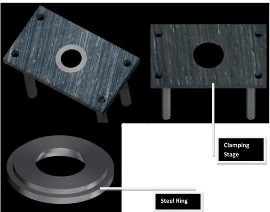
A circular hole was machined in the clamping stage to accept stainless steel clamping rings of different sizes. Clamping is performed by elevating the vertical stage and by squeezing the tissue close to the equatorial region of the posterior pole of the scleral shell between the border of the steel ring and the plastic ring shown in Figure 14. This clamping condition eliminates any rigid movements of the sample during the mechanical test and ensures the sealing condition that is always verified after clamping the eye.
42
Figure 13. Charging apparatus for mechanical inflation test.
Figure 14. Clamping stage after and before the steel ring is mounted. Clamping Stage
43
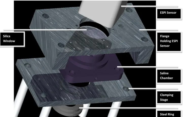
A sealed chamber filled of saline solution (Phosphate Buffered Saline, or PBS) is mounted on the steel plate. The saline chamber (Figure 15) maintains tissue hydration during the entire inflation test.
Two fluid lines allow for inflow and outflow of saline solution inside the hemisphere of the eye mounted on the pressurization apparatus. The pressure inside the eye is controlled by a digital manometer connected in parallel to the outflow line.
Pressure increases are obtained by raising a bottle of saline solution via a motor-controlled vertical stage. An automated system controls the pressure step increase by integrating information from the digital manometer with the position of the saline bottle. The phase variations of the speckle fields caused by the deformation of the outer scleral
surface induced by the pressure increase is recorded by a commercial Electronic Speckle Pattern interferometer (Q100, Dantec Dynamics A/S, Denmark).
The interferometer consists of a unit containing four lasers symmetrically positioned around the CCD (Coupled Charge Device) of the unit, connected to a central controller.
Part of the final set-up is shown in Figure 16.
Figure 15. Saline chamber mounted on the
44
All the figures describing the mechanical testing set-up have been made in Pro-Engineer™. During the data collection period the following parts were redesign from its original form: The Design of new components have been designed with the mentioned CAD program.
For all the tested eyes internal pressure has been raised from a baseline of 5
mmHg up to 45 mmHg. The baseline of 0 mmHg couldn’t be used since the
posterior hemisphere doesn’t keep its shape at zero pressure. A 5 mmHg initial internal pressure was just enough to pre-stretch the eye into its natural shape while keeping the pre-existing stress low.
Each scleral shell was subjected to IOP preconditioning, consisting of 20 IOP cycles from 5 mmHg to a maximum of 30 mmHg at a rate of 5 mmHg per second, and then allowed to recover for 15 minutes.
Figure 16. Part of the experimental set-up for the mechanical testing.
ESPI Sensor Flange Holding ESPI Sensor Saline Chamber Clamping Stage Steel Ring Silica Window