Maxillomandibular Advancement in Obstructive
Sleep Apnea Syndrome: A Surgical Model to
Investigate Reverse Face Lift
Francesco Arcuri, MD, Matteo Brucoli, MD, Rodolfo Benech, MD,
Mariangela Giarda, MD, and Arnaldo Benech, MD, PhD
Background: The aging process is characterized by multiple signs affecting the upper, the middle, and the lower third of the face; coventionally, face-lift procedures and structural fat grafting are performed to create a younger face. During the life, craniofacial skeleton atrophies, leading to a reduction of the facial height and depth, while increasing the facial width. Maxillomandibular ad-vancement (MMA) by orthognathic surgery restores the lost space dimension, projecting the cheeks, the mouth, and the nose. The aim of this study was to analyze the morphologic change of the face af-ter MMA in patients with obstructive sleep apnea syndrome, fo-cusing on the previously mentioned stigmata of the middle and the lower third of the aging face.
Methods: The records of 16 patients who underwent MMA for obstructive sleep apnea syndrome between January 2005 and December 2008 in the Unit of Maxillofacial Surgery at the Novara Major Hospital were included in this study. We explained to the patients the stigmata of a standard aging face, and we asked them to evaluate each sign affecting the middle and the lower third of their preoperative condition. One positive point was given for the pres-ence of each sign reported by the patients. At 2 years after surgery, we asked the patients to evaluate the previously mentioned aging signs of their postoperative face. Again, 1 positive point was given for the presence of each sign reported by the patients.
Results: Although we did not perform statistical evaluation, 13 patients showed a degree of rejuvenation after MMA (the score of the postoperative face is less than the score of the preoperative face). Three patients reported no postoperative change; none re-ported a more aging face, with a successful ‘‘reverse face-lift’’ oc-curred in 81% of our cases.
Conclusions: Simultaneous maxillary and mandibular advance-ments change the skeletal framework of the face, improving soft-tissue support and resulting in rejuvenation of the middle and the lower third of the face. This condition is demonstrated by the results
of our study in that all patients appeared postoperatively more youth-ful from a self-evaluation.
Key Words: Maxillomandibular advancement, obstructive sleep apnea syndrome, reverse face-lift, facial rejuvenation
(J Craniofac Surg 2011;22: 2148Y2152)
T
he aging face is characterized by multiple signs affecting the upper third (forehead furrows, glabellar frown lines, brow pto-sis, excess of upper eyelid skin, pseudoherniation of the intraorbital fat pad, and evidence of the tear through deformity), the middle third (development of the nasojugal fold and accentuation of the para-buccal fat pad), and the lower third (formation of the facial jowls, evidence of the labiomental fold, and accentuation of the submental fat pad).1Y5According to the classic belief that, during the life, the force of gravity stretches and attracts the cervicofacial teguments down, face-lift procedures replace the tissue up, both conventionally3,6and endoscopically.7,8
In 1998, Coleman9 changed the surgical approach to the aging face by pioneering the concept of the volumetric face-lift.10,11 The face reaches its volumetric peak between 20 and 30 years of age, and thereafter, it slowly atrophies, collapsing toward the geometric center; the soft tissues become flaccid and lose internal pressure. Structural fat grafting projects the atrophic facial soft tissue and recreates the lost young tension.
Recently, in relation to the aesthetic results of the orthognathic procedures performed on older patients by experienced maxillofacial surgeons,12Y14a new concept started to arise in craniomaxillofacial
surgery: ‘‘reverse face-lift.’’
During the life, craniofacial skeleton atrophies, leading to a reduction of the facial height and depth, while increasing the facial width; maxillary and mandibular bone reabsorption leads to a loss of support of the lips and the nose.15Y19 Maxillomandibular ad-vancement (MMA) by orthognathic surgery restores the lost space dimension, projecting the cheeks, the mouth, and the nose.
The aim of this study was to analyze the morphologic change of the face after MMA in patients with obstructive sleep apnea syndrome (OSAS), focusing on the previously mentioned stigmata of the middle and the lower third of the aging face.
MATERIALS AND METHODS
The records of 16 patients who underwent MMA for OSAS between January 2005 and December 2008 in the Unit of Maxillo-facial Surgery at the Novara Major Hospital were included in this study. The study group comprised 13 men (81.2%) and 3 women (18.8%), with a mean age of 49.6 years (range, 34Y64 years). The From the Department of Maxillo-Facial Surgery, Azienda Ospedaliera
Maggiore della Carita`, University of Piemonte Orientale ‘‘Amedeo Avogadro,’’ Novara, Italy.
Received May 16, 2011.
Accepted for publication July 7, 2011.
Address correspondence and reprint requests to Dr. Francesco Arcuri, SCDU di Chirurgia Maxillo-Facciale, Ospedale Maggiore della Carita`, Corso Mazzini 18, 28100 Novara, Italy; E-mail: [email protected] The authors report no conflicts of interest.
Copyright* 2011 by Mutaz B. Habal, MD ISSN: 1049-2275
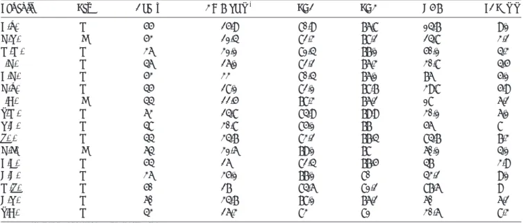
mean body mass index (BMI) was 28.7 kg/m2(range, 21.4Y35.1 kg/ m2). The mean presurgical cephalometric horizontal measurement for to the maxilla (SNA) was 81.5 degrees (range, 77.1Y85.1); the same preoperative analysis related to the mandible (SNB) was 78 degrees (range, 76.1Y81.2). The mean preoperative respiratory disturbance index (RDI) was 47.1 events per hour (range, 14.7Y87.6). The mean preoperative posterior airway space (PAS) was 6.2 mm (range, 3.2Y9.1). No patients had concurrent orthodontic therapy (Table 1).
We explained to the patients the stigmata of a standard ag-ing face, and we asked them to evaluate each sign affectag-ing the middle and the lower third of their preoperative condition. One pos-itive point was given for the presence of each sign reported by the patients. A hypothetical medium score of 7 means a preoperative aging face affected by all the anatomic signs (Table 2).
All of the patients had the surgical procedures executed by the same surgeon (A.B.); maxillary and mandibular advancements were obtained by Le Fort I osteotomy and bilateral sagittal split TABLE 1. Preoperative Physical Profile
Patients Sex Age, y BMI, kg/m2 SNA SNB RDI PAS, mm
B.V. M 55 25.9 80.9 76.8 14.7 9.1 G.R. F 53 21.4 82.3 78.2 24.8 3.2 M.M. M 36 31.1 81.4 77.1 50.1 4.3 P.G. M 46 26.1 82.2 76.3 30.8 4.5 B.G. M 53 33 80.4 76.1 76 5.1 G.V. M 45 28.1 82.1 78.7 39.8 5.9 P.A. F 44 22.5 78.3 76.2 18 6.2 T.M. M 63 24.8 84.9 79.9 30.1 6.1 R.C. M 48 30.8 85.1 77 56 8 S.P. M 44 34.7 83.2 77.4 84.7 7.3 G.F. F 64 31.6 79.1 78 60.1 4.1 B.E. M 54 26 82.4 77.5 47 3.9 C.C. M 36 35.1 77.1 80 43.2 9.1 M.S. M 50 27 84.6 81.2 87.6 9 C.R. M 60 34.7 78.1 76.2 60 6.2 T.A. M 43 26.3 83 81 30.6 8.3
RDI indicates Respiratory Disturbance Index.
TABLE 2. Preoperative Signs of the Aging Face
Patients
Nasojugal Fold Labiomental Fold Facial Jowl
Submental Fat Pad Total
Right Left Right Left Right Left
B.V. & & & & & & & 7
G.R. & & & & & 5
M.M. & & & & 4
P.G. & & & & & 5
B.G. & & & & & & 6
G.V. & & & & & 5
P.A. & & & & & & 6
T.M. & & & & & & & 7
R.C. & & & & & 5
S.P. & & & & 3
G.F. & & & & & & & 7
B.E. & & & & 4
C.C. & & & 3
M.S. & & & & 4
C.R. & & & & & & & 7
T.A. & & & 3
osteotomy, respectively; no patients had bone grafting. Concomi-tant to the bimaxillary surgery, 5 patients underwent septoplasty/ turbinoplasty; 1 patient sustained uvulopalatopharyngoplasty, and 3 patients underwent simultaneously septoplasty/turbinoplasty and uvulopalatopharyngoplasty.
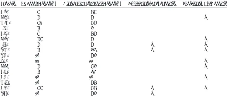
Additional ancillary procedures included (1) piriform rim re-contouring, (2) anterior nasal spine modeling, (3) alar base cinch suture, and (4) V-Y closure. No patients underwent genioplasty. The osteosynthesis was performed using plates and monocortical/ bicortical screws, as dictated by the magnitude of the advancements and the anatomic variations (Table 3).
RESULTS
At 2 years after surgery, after a follow-up scheduled every 4 months, we performed clinical evaluation, radiographic exami-nation, and cephalometric analysis. The mean postoperative cepha-lometric horizontal measurement for to the maxilla (SNA) was 85.8 (range, 81.6Y90.3); the same postoperative analysis related to the mandible (SNB) was 82.2 (range, 80.1Y86.1). The mean postoper-ative RDI was 7 (range, 0Y15.3). The mean postoperative PAS was 13.2 (range, 8.4Y16; Table 4).
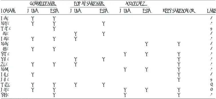
We asked the patients to evaluate the previously mentioned aging signs of their postoperative face. Again, 1 positive point was given for the presence of each sign reported by the patients (Table 5). Although we did not perform statistical evaluation, 13 pa-tients showed a degree of rejuvenation after MMA (the score of the postoperative face is less than the score of the preoperative face). Three patients reported no postoperative change; none reported a more aging face, with a successful reverse face-lift occurred in 81% of our cases (Figs. 1 and 2).
DISCUSSION
Maxillomandibular advancement has been successfully per-formed to treat patients affected by OSAS for several years. Bimax-illary manipulation expands the skeletal framework and stretches the oropharyngeal soft tissues; this procedure leads to a larger PAS, de-creasing the collapsibility of the airway. Maxillomandibular
advance-ment needs large surgical moveadvance-ments, generally on the order of 10 mm, to achieve a significant airway improvement.20,21
The position of the facial soft tissue after bimaxillary surgery is influenced by several factors: (1) preoperative profile (sex, age, and BMI), (2) concurrently orthodontic therapy, (3) direction and magnitude of the maxillary movements, (4) additional surgical pro-cedures, and (5) postoperative edema.22Y26
Orthognathic surgical procedures lead to postoperative edema that is expected to resolve within 6 months after surgery. In our retrospective study, the measures are obtained at 2 years after sur-gery, making extremely unlikely that postoperative edema modifies the position of the facial soft tissue. No patients had orthodontic TABLE 3. Surgical Procedures
Patients Le Fort I Osteotomy Bilateral Sagittal Split Osteotomy Uvulopalatopharyngoplasty Septoplasty/Turbinoplasty
B.V. 8 7.8 G.R. 9 9 X M.M. 8.1 8.9 P.G. 7 5 B.G. 8 7.9 G.V. 9.8 9 X P.A. 9 9 X X T.M. 7 5.6 X X R.C. 10 9.5 S.P. 11 11 X G.F. 9 8.5 X B.E. 7 6.3 C.C. 10 10 X M.S. 10 9.7 C.R. 8.8 8.7 X X T.A. 10 9.5 X
The cephalometric measure of the surgical advancement is calculated as the difference (in millimeters) between the postoperative and the preoperative (A) McNamara (maxilla) and (B) McNamara (mandible).
TABLE 4. Postoperative Profile
Patients SNA SNB RDI PAS
B.V. 84.6 80.2 3.5 15 G.R. 85.1 83 5.6 12 M.M. 83.1 80.1 10.1 11.1 P.G. 87.4 80.1 1.6 8.4 B.G. 83.2 80.2 15.3 10.5 G.V. 90.3 82.1 3.5 15.5 P.A. 84 81.1 2.9 16 T.M. 88.4 80.7 3.3 9.3 R.C. 88.6 82.3 8.6 12.1 S.P. 86.6 81.3 13.9 12.5 G.F. 86 85 8.1 16 B.E. 83.2 81.3 12.4 15.5 C.C. 81.6 83.6 11.3 15 M.S. 87.2 83.9 6.5 13 C.R. 85.2 84.6 0 14.3 T.A. 88.2 86.1 6.3 15.1
therapy during treatment; therefore, dental movements play no role in the postoperative changes of the soft tissue.
The direction and the magnitude of the maxillary movements play the greatest role in changing the postoperative position of the soft tissue. In our study, all jaw movements were virtually horizontal advancements; no genioplasty was performed, then analysis of the soft tissue was studied for maxillary and mandibular advancements. Additional surgical procedures influence the final position of the soft tissue. Several surgical techniques, inherited from our clinical experience by treating orthognathic cases, were used in this study to modify the facial soft tissue; V-Y closure of the upper lip, alar base cinch suture, anterior nasal spine recontouring, piriform-plasty, and septoplasty were virtually executed in all patients, trying to minimize unaesthetic nasolabial changes.
The surgical profile of a patient with OSAS is different from an orthognathic case: the former is a middle-aged man, who is obese and with normal dentofacial relations, requiring advancement on the order of 10 mm; and the latter is a young woman with normal weight, with a specific dentofacial deformity, needing maxillary movements on the order of 5 mm (Figs. 1 and 2).22,27,28
The effect of bimaxillary manipulation on the facial soft tis-sue for dentofacial deformities has long been studied23Y26; to the
best of our knowledge, the resultant facial changes of patients treated by MMA for OSAS has not been adequately investigated, and the concept of reverse face-lift has never been mentioned in the scien-tific literature.
Simultaneous maxillary and mandibular advancements change the skeletal framework of the face, improving soft-tissue support and resulting in rejuvenation of the middle and the lower third of the face. This condition is demonstrated by the results of our study in that all patients appeared postoperatively more youthful from a self-evaluation.
Conversely, MMA more than 10 mm can lead to an unaes-thetic prominence of the face, affecting the final aesunaes-thetic outcome; nevertheless, our study was made to evaluate only the patient’s per-ception of the postoperative facial rejuvenation.
Preoperative analysis of facial proportions with cephalometric measures, as performed with standard orthognathic cases, is nec-essary before doing MMA for OSAS. Eventual unaesthetic facial changes must be preoperatively discussed with the patient, and the necessity of clockwise/counterclockwise rotation of the occlusal plane needs to be assessed to obtain a satisfactory result for aes-thetics and functionality.
Based on our clinical experience by treating orthognathic cases, patients with a high risk for unfavorable facial changes include young patients, with normal weight, having a preoperative maxilloman-dibular protrusion. This risk is increased in patients with thin facial soft tissue, which does not mask the large maxillary advancements. TABLE 5. Postoperative Face
Patients
Nasojugal Fold Labiomental Fold Facial Jowl
Submental Fat Pad Total
Right Left Right Left Right Left
B.V. & & 2
G.R. & & & 3
M.M. & 1
P.G. & & 2
B.G. & & & 3
G.V. & & 2
P.A. & & 2
T.M. & & & 3
R.C. & & & 3
S.P. & & & & 3
G.F. & & & 3
B.E. & & 2
C.C. & 1
M.S. & & & & 4
C.R. & & & & & 5
T.A. & & & 3
& indicates presence.
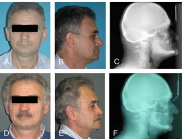
FIGURE 1. Female patient’s preoperative and postoperative frontal views (A, D), profiles (B, E), and teleradiographic images (C, F), respectively.
Finally, the result of postoperative facial aesthetics is depen-dent on the perception of the patient, which is likely to be signifi-cantly influenced by the outcomes of the OSAS treatment and the patient’s presurgical satisfaction with its own appearance.
CONCLUSIONS
The physiopathologic basis of the aging face is actually not completely understood; nevertheless, 3 factors contribute to the de-velopment of the previously mentioned processes: (1) soft-tissue lax-ity, (2) soft-tissue atrophy, and (3) skeletal reabsorption.
Conventional face-lift procedures are related to the first is-sue; structural fat grafting solves the second problem. Theoretically, MMA can be a very powerful tool to mask the physiological bone atrophy.
We think that reverse face-lift by bimaxillary advancements is a surgical procedure that, potentially associated to concomitant conventional face-lift technique and structural fat grafting, can be indicated for a selected group of middle-aged patients, very moti-vated to an extreme rejuvenation; further clinical studies, eventually associated to more sophisticated preoperative and postoperative soft-tissue analysis, should be necessary to support our speculation.
REFERENCES
1. Zimbler MS, Kokoska MS, Thomas JR. Anatomy and pathophysiology of facial aging. Facial Plast Surg Clin North Am 2001;9:179Y187 2. O¨ zdemir R, Kilinc H, U¨nlu¨ E, et al. Anatomicohistologic study
of the retaining ligaments of the face and use in face lift: retaining ligament correction and SMAS placation. Plast Reconstr Surg 2002;110:1134Y1147
3. Sherris DA, Larrabee WF Jr. Anatomic considerations in rhytidectomy. Facial Plast Surg 1996;12:215Y222
4. Gosain AK, Klein MH, Sudhakar PV, et al. A volumetric analysis of soft-tissue changes in the aging midface using high-resolution MRI: implications for facial rejuvenation. Plast Reconstr Surg
2005;115:1143Y1152
5. Owsley JQ. Elevation of the malar fat pad superficial to the orbicularis oculi muscle for correction of prominent nasolabial folds.
Clin Plast Surg 1995;22:279Y293
6. Tessier P. Subperiosteal face-lift. Ann Chir Plast Esthet 1989;34:193Y197 7. Isse NG. The endoscopic approach to forehead and brow lifting.
Aesthet Surg J 1998;18:462, 464
8. Ramirez OM. The central oval of the face: tridimensional endoscopic rejuvenation. Facial Plast Surg 2000;16:283Y298
9. Coleman SR. Structural fat grafting. Aesthet Surg J 1998;18:386, 388 10. Trepsat F. Volumetric face lifting. Plast Reconstr Surg 2001;108:
1358Y1370
11. Ellenbogen R, Youn A, Yamini D, et al. The volumetric face lift. Aesthet Surg J 2004;24:514Y522
12. Arnett GW, Gunson MJ. Esthetic treatment planning for orthognathic surgery. J Clin Orthod 2010;44:196Y200
13. Hwang SJ, Haers PE, Seifert B, et al. Non-surgical risk factors for condylar resorption after orthognathic surgery. J Craniomaxillofac Surg 2004;32:103Y111
14. Reyneke JP, Bryant RS, Suuronen R, et al. Postoperative skeletal stability following clockwise and counter-clockwise rotation of the maxillomandibular complex compared to conventional orthognathic treatment. Br J Oral Maxillofac Surg 2007;45:56Y64
15. Bartlett SP, Grossman R, Whitaker LA. Age-related changes of the craniofacial skeleton: an anthropomorphic and histologic analysis. Plast Reconstr Surg 1992;90:592Y600
16. Pessa JE, Zadoo VP, Mutimer KL, et al. Relative maxillary retrusion as a natural consequence of aging: combining skeletal and soft-tissue changes into an integrated model of midfacial aging. Plast Reconstr Surg 1998;102:205Y212
17. Pessa JE, Zadoo VP, Yuan C, et al. Concertina effect and facial aging: nonlinear aspects of youthfulness and skeletal remodeling and why perhaps infants have jowls. Plast Reconstr Surg 1999;103:635Y644 18. Richard MJ, Morris C, Deen BF, et al. Analysis of the anatomic changes
of the aging facial skeleton using computer-assisted tomography. Ophthal Plast Reconstr Surg 2009;25:382Y386
19. Farkas LG, Eiben OG, Sivkov S, et al. Anthropometric measurements of the facial framework in adulthood: age-related changes in eight age categories in 600 healthy white North Americans of European ancestry from 16 to 90 years of age. J Craniofac Surg
2004;15:288Y289
20. Riley RW, Powell NB, Guilleminault C. Obstructive sleep apnea syndrome: a review of 306 consecutively treated surgical patients. Otolaryngol Head Neck Surg 1993;108:117
21. Waite PD, Shetar SM. Maxillomandibular advancement: a cure for obstructive sleep apnea. Oral Maxillofac Surg Clin North Am 1995;7:327
22. Joss CU, Joss-Vassalli IM, Berge´ SJ, et al. Soft tissue profile changes after bilateral sagittal split osteotomy for mandibular setback: a systematic review. J Oral Maxillofac Surg 2010;68:2792Y2801 23. Jensen AC, Sinclair PM, Wolford LM. Soft tissue changes associated
with double jaw surgery. Am J Orthod Dentofacial Orthop 1992;101:266
24. Kolokitha OE, Athanasiou AE, Tuncay OC. Validity of computerized predictions of dentoskeletal and soft tissue profile changes after mandibular setback and maxillary impaction osteotomies. Int J Adult Orthod Orthognath Surg 1996;11:137
25. Enacar A, Taner T, Toroglu S. Analysis of soft tissue profile changes associated with mandibular setback and double jaw surgeries. Int J Adult Orthod Orthognath Surg 1999;14:27
26. Lin SS, Kerr WJ. Soft and hard tissue changes in class III patients treated by bimaxillary surgery. Eur J Orthod 1998;20:25
27. Demetriades N, Chang DJ, Laskarides C, et al. Effects of mandibular retropositioning, with or without maxillary advancement, on the oro-naso-pharyngeal airway and development of sleep-related breathing disorders. J Oral Maxillofac Surg 2010;68:2431Y2436 28. Abramson Z, Susarla S, August M, et al. Three-dimensional computed
tomographic analysis of airway anatomy in patients with obstructive sleep apnea. J Oral Maxillofac Surg 2010;68:354Y362
FIGURE 2. Male patient’s preoperative and postoperative frontal views (A, D), profiles (B, E), and preoperative (F) and postoperative (C) teleradiographic images.