UNIVERSITY OF URBINO CARLO BO
Department of Biomolecular Sciences
Ph.D. Course in Life Sciences, Health and Biotechnologies
XXXI cycle
Effect of exercise in Breast Cancer and
its association with tumor characteristics,
risk factors for recurrence and lifestyle
SSD: BIO/10
Supervisor:
Ph.D. student:
Prof. Luciana Vallorani
Dr. Valentina Natalucci
Co-Advisor:
Prof. Marianna Capecci
A
BSTRACT
Triple-negative breast cancer (TNBC) does not express estrogen receptors, progesterone receptors, or human epidermal growth factor receptor 2, and it is characterized by its aggressive nature, lack of targeted therapies and early peak of recurrence. Recent observational research suggests that inactivity is associated with an increased risk of breast cancer (BC) including the Triple Negative subtype. Little is known about the potential driving molecular events for each subtype of BC and the mechanisms through which exercise yields protective effects against disease progression and recurrence risk. The present thesis focuses on the protective effects of exercise against TNBC through the induction of biochemical metabolic system modifications able to reduce the cell proliferation of BC.
Organization of the thesis and experimental approaches
The first chapter provides possible explanations of causative mechanisms for cancer control examining recent findings regarding the basic biological effects of exercise on modulation of the mTOR pathway in TNBC and the benefits induced by various exercise and training protocols.
The second chapter of the thesis examines how circulating serum factors released after exercise (High-Intensity Endurance Cycling (HIEC) Test), before (pre) and after (post) a training protocol, could reduce TNBC cell line proliferation, ex vivo.
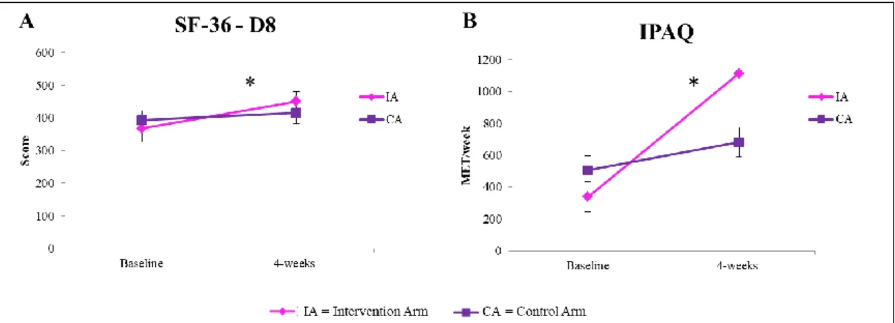
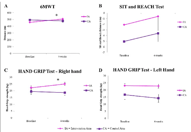
The third chapter evaluates the effect of structured and supervised physical activity (PA) intervention on breast cancer survivors (BCS) - “Lifestyle program”.
We showed that PA counseling and supervised exercise training improve functional exercise capacity and could motivate subjects suffering from BC to adopt a healthier lifestyle based on the maintenance of physical activity levels (PAL) recommended in this population. Women enrolled in the “Lifestyle program” took part in the Dragon Boat Race Day event, representing the first team of women in pink from the Marche Region.
INTRODUCTION ... 1
Breast Cancer ... 2
MOLECULAR AND SYSTEMIC EFFECTS LINKING EXERCISE TO CANCER PREVENTION AND CANCER CONTROL ...3
PHYSICAL ACTIVITY COUNSELING IN BREAST CANCER SURVIVORS ...4
PROTOCOL FOR EXERCISE PRESCRIPTION AMONG BREAST CANCER SURVIVORS ...4
HIGH INTENSITY INTERVAL TRAINING (HIIT): NEW EVIDENCE IN BCS ...5
AIMS OF THE THESIS ... 8
CHAPTER 1 ... 10
New Insights into the Role of Exercise in Inhibiting mTOR Signaling in Triple-Negative Breast Cancer ...11
INTRODUCTION ...14
MTORSIGNALING ...15
EVIDENCE OF MTORMODULATION BY EXERCISE IN TNBC ...20
ENERGY INTAKE IN TNBC AND MTORMODULATION ...31
EXERCISE PRESCRIPTION IN BCSURVIVORS ...33
BENEFITS OF EXERCISE PRE- AND POSTDIAGNOSES...38
CONCLUSIONS ...40
REFERENCES ...42
CHAPTER 2 ... 54
Effects of exercise on triple-negative breast cancer cell proliferation ...55
INTRODUCTION ...58
MATERIALS AND METHODS ...59
RESULTS AND DISCUSSION ...64
CONCLUSIONS ...68
REFERENCES ...69
CHAPTER 3 ... 71
Promotion and prescription of exercise in breast cancer survivors to improve quality of life and health outcomes ...72
BACKGROUND ...75
THE STUDY DESIGN ...76
RESULTS...87
DISCUSSION ...92
REFERENCES ...95
CONCLUSIONS ... 98 REFERENCES ... 103
O
RIGINAL
P
APERS
This Thesis is based on the following original research articles, which will be referred to by their Roman numerals.
I. Barbieri E, Falcieri E, De Santi M, Natalucci V, Vallorani L, Agostini D, Annibalini G, Stefani L, Szychlinska MA and Musumeci G. 2018. Editorial The “Journal of Functional Morphology and Kinesiology” Journal Club Series: Highlights on Recent Papers in Physical Activity and Sedentary Behavior. ISSN 2411-5142. doi:10.3390/jfmk3020023
II. Agostini D^, Natalucci D^, Baldelli G^, De Santi M, Donati Zeppa S, Vallorani L, Annibalini G, Lucertini F, Federici A, Izzo R, Stocchi V and Barbieri E. New Insights into the Role of Exercise in Inhibiting mTOR Signaling in Triple-Negative Breast Cancer – Oxidative Medicine and Cellular Longevity, 2018 Sep 30; 2018:5896786. doi: 10.1155/2018/5896786.
^Authors contributed equally to this work.
III. Natalucci V. et al. Effects of exercise on triple-negative breast cancer cell proliferation.
In preparation.
IV. Natalucci V. et al. Promotion and prescription of exercise in breast cancer survivors to improve quality of life and health outcomes.
1
2
Breast Cancer
Today breast cancer (BC) is the most common invasive tumor in women worldwide and is the most common cancer in all age groups, albeit at different rates (41% in young women vs. 22% in older women). In Italy, there were an estimated 50,500 new cases in 2017 and the average 5-year survival rate (87%) in our country is higher than the European average (82%) [1]. Studies have shown that BC is the result of an intricate interaction among genetic, epigenetic and environmental factors. Among the most significant factors influencing the risk of BC onset and relapse are aging, gender, family history, inherited factors (mutations in the BRCA1 and BRCA2 genes), menstrual and reproductive history, dense breasts, race and ethnicity, radiation exposure, birth control pills, combined post-menopausal hormone therapy (PHT), and diethylstilbestrol exposure (DES).
In recent years a growing body of evidence points to the important effect that healthy lifestyle choices (modifiable factors) such as not smoking, adopting a healthy diet and doing regular PA could have on the onset and clinical course of BC.
Growing amounts of data indicate a relationship between PA, biological makers of BC aggressiveness (e.g., levels of insulin, estrogen and IGF-1, and inflammatory status) and lower recurrence risk.
BC is a heterogeneous disease and differs greatly among patients (inter- intra-tumor heterogeneity) [2]. Although in Western countries the incidence of BC is increasing, mortality is steadily decreasing mainly due to prevention strategies, including regular mammography screenings for early detection of BC and primary prevention based on healthy lifestyle choices (not smoking, healthy diet and regular PA).
Several epidemiological studies suggest that physically active women have a significantly lower risk (a reduction of about 25%) of developing BC than sedentary women [3]. Exercise could also reduce the side effects of cancer treatments such as chemotherapy, radiotherapy and hormone therapy and have a positive effect on psychological wellbeing since woman undergoing cancer therapy are more at risk of developing depressive and cognitive disturbances [4-6]. Additional positive effects of PA include a reduction in body fat, improvement in immune system regulators, alterations in free-radical generation and an improvement in the ability of cancer patients and survivors to deal with fatigue, lymphedema and bone metastasis [7]. Another important consideration is that, after
3
diagnosis, most BC patients and survivors reduce PA level (by 11%) [8], and sedentary behaviors are strongly associated with overweight and obesity, both recognized risk factors for BC. Indeed, adipose tissue contributes to the establishment of a systemic low‐ grade inflammatory state characterized by activation and infiltration of pro‐inflammatory immune cells and a deregulated production of pro‐inflammatory cytokines [9,10,11].
Molecular and Systemic Effects Linking Exercise to Cancer Prevention and Cancer Control*
It has been 20 years since skeletal muscle was recognized as an endocrine organ, and skeletal muscle contraction and myokine release have received attention as exercise-induced health benefits. Physical activity (PA) has been shown to reduce cancer incidence, inhibit tumor growth, and improve clinical outcomes following a diagnosis of a primary disease [12], yet daily energy expenditure from PA is decreasing at an alarming rate in Western society [13]. In particular, inactivity is associated with an increased risk of breast cancer (BC). Pernille Hojman and collaborators from Bente K. Pedersen’s research team describe the latest molecular understanding of the systemic effect of exercise on cancer progression control. They defined a direct effect on tumor-environment factors linked to whole-body exercise remodeling, mitigation of cancer-related adverse events, and amelioration of anti-cancer treatment efficacy. Several preclinical studies on the effect of exercise on cancer outcomes point to a reduction in the rate of tumor growth [14,15]. However, an important finding of these studies is that exercise, per se, is not capable of directly eliminating already established tumors. For example, in response to exercise-conditioned serum treatment, BC cell growth is inhibited by 10–15% compared to tumor growth observed in the control setting, but there is not a complete eradication of cancer cells [16,17]. Recent investigations have stressed the role of circulating growth factors, prohormones, and sexual hormones, which are able to stimulate cancer cell proliferation through the activation of their respective receptors [18,19]. In particular, the phosphoinositide 3-kinase-AKT-mammalian target of rapamycin (PI3K-AKT-mTOR) pathway is a frequently hyperactivated pathway in cancer and is important for tumor cell growth and survival. Many studies have shown a down-regulation of PI3K-AKT-mTOR signaling after exercise; however, to date, none have established the exercise-dependent mechanisms underlying this association. Exercise
4
inhibits mTOR functions in both AMPK-independent and -dependent manners. AMPK is also induced in tumors during exercise [20,21], probably as a transient condition. It is known that intra-tumoral metabolism is regulated, but how this affects tumor growth and metastatic rate during exercise is not currently understood. Chronic exercise induces physiological adaptation across homeostatic control circuits, such as oxidative metabolism, mitochondrial biogenesis in several tissues, angiogenesis, immune regulation, and metabolism. Moreover, it diminishes BMI and blood concentrations of estrogens, and insulin, decreases insulin resistance, and strengthens anti-inflammatory pathways against tumor cells. Hence, exercise has an increasingly protective tumorigenic effect [22,23]. These findings have wide-ranging implications for society and may lead to improve the way cancer is managed.
Physical Activity Counseling in Breast Cancer Survivors*
The overall prevalence of women living with a diagnosis of breast cancer (BC) is increasing in industrialized countries. Survivors who maintain a healthy weight and stay physically active (PA) have a better response to treatment and better survival outcomes. Thus, it is necessary to identify an appropriate promotion and prescription of regular PA for BC survivors to improve their prognosis, response to therapy, and quality of life. In this issue, Fong AJ and colleagues [24] examine factors affecting exercise counseling by clinicians and discuss future strategies to address oncology clinicians’ perceptions of PA counseling and barriers to such counseling in BC survivors. Focus group discussions were transcribed verbatim and analyzed using inductive thematic analysis. In order to facilitate PA counseling, healthcare facilities, and clinicians need to better support and promote patient-managed PA, making it available on multiple platforms and providing better referral pathways to exercise professionals and physiologists.
Protocol for Exercise Prescription among Breast Cancer Survivors*
Recent systematic reviews and meta-analyses tend to aggregate exercise programs into general categories and rarely investigate the specific features of these programs, which may make them more or less effective. What physical activity (PA) should be prescribed for breast cancer (BC) survivors? Not all types of exercise carry the same benefits.
5
Specificity is a core principle of exercise training to promote the desired adaptations, and the specificity of adaptation is underpinned by the acronym FITT, which stands for frequency, intensity, time and type. The FITT principle outlines the key components of an effective exercise program Overall recommendations for cancer survivors are consistent with the American Cancer Society, who recommends that cancer survivors engage in a minimum of 150 weekly minutes of moderate-intensity exercise, and it is important to note that the American College of Sports Medicine [25] has established FITT guidelines that are easy to follow for exercise prescription that everyone should know. Frequency refers to how often exercise is performed, intensity refers to the intensity of the exercise undertaken, time refers to the amount of time spent exercising, and type refers to the kind of exercise that is performed. However, adherence to any exercise program is the most difficult thing to achieve, and currently there is a growing number of initiatives designed to increase the effectiveness of long-term PA in BC patients in the most comfortable environment: at home. The aim of the study carried out by Mascherini G. and collaborators was to verify the long-term effectiveness of a home-based program for active lifestyle change in overweight BC survivors. They enrolled premenopausal women, whose PA levels, baseline aerobic capacity, flexibility, strength, and anthropometric measurements were assessed. These parameters were assessed six times during one year of unsupervised exercise. After being prescribed an individual exercise program, a significant reduction in BMI and skinfold sum was observed, as well as maintenance of muscle cell mass. Assessments also showed an increase in lower limb muscle fitness and a reduction in diastolic blood pressure after a six-minute walking test. Exercise is highly recommended for cancer patients, and the prescription of a home-based model of unsupervised exercise described by Mascherini G. et al. seems to ensure ideal compliance, favoring long-term therapeutic efficacy [26]. This intervention strategy could target all patients affected by BC and might motivate these subjects to adopt a healthier lifestyle based on regular PA, as well as proper nutrition.
High Intensity Interval Training (HIIT): new evidence in BCS
In recent years, it has been suggested that a reassessment of the exercise prescription guidelines for BCS is needed. Generally, the beneficial effects induced by exercise are commonly attributed to two types of adaptation, long-term (months/years), such as
6
improvement in cardiorespiratory fitness, strength and body composition [27,28,29] and short-term effects (hours/days) due to stimulation of muscle contraction. In particular, the marked systemic response to a single bout of exercise reduces the vitality of BC [30]. The available exercise guidelines for BCS emphasize the importance of participating in moderate aerobic and resistance exercise and recommend flexibility building exercises or combined exercises (aerobic and resistance exercise) [31]. However, research on the effects of different types of exercise on physical fitness and various biomarkers in BCS is ongoing, and in the past year, a great deal of evidence has emerged regarding a novel strategy of training for BCS: High-Intensity Interval Training (HIIT). HIIT is a cardio session consisting of short sets of intense exertion, and it represents a valid alternative to traditional endurance-based exercise training for preventing cardiovascular disease (CVD) in BCS [32]. In particular, the principle of HIIT is based upon high intensity aerobic exercise training bouts that are separated by periods of lower intensity that allow for recovery. The target intensity during HIIT is between 85-95% of peak heart rate (HRpeak) for four minutes, while during the two-to-three-minute lower intensity recovery
periods, the target intensity is between 60-70%.
Only a few studies have been conducted on HIIT training in BCS where it was shown that HIIT can be performed by female cancer survivors without adverse health effects, and it may be an effective way to improve certain aspects of health, including protection against chronic diseases, especially CDV, in this population.
The beneficial effects of HIIT consist of improved quality of life, reduced cancer-related fatigue [33] and reduces the adverse cardiotoxic effects due to treatments in different stage of disease. An ongoing pilot study is examining the HIIT effects on endothelial function and maximal oxygen consumption (VO2max) [34].
In the HIIT protocol, the exercise training intensity is relative to the individual’s peak oxygen uptake (VO2peak) and HRpeak and should be measured for each individual before
starting a HIIT program to control the exercise training intensity properly. Furthermore, it has been shown that VO2max and other cardiovascular efficiency measures, such as endothelial function, improve both with HIIT exercise and with higher volumes of moderate continuous intensity exercise [35]. Both types of exercise enhance the patient’s quality of life (QoL), reduce fatigue, and improve energy expenditure and body composition. However, one of the added benefits of HIIT workouts is the reduced time
7
this protocol requires compared to other exercise programs and the fact that there are currently no contraindications for its use by BCS.
*A part of the introduction was taken by the Editorial The “Journal of Functional
Morphology and Kinesiology” Journal Club Series: Highlights on Recent Papers in Physical Activity and Sedentary Behavior. Barbieri E, Falcieri E, De Santi M, Natalucci V, Vallorani L, Agostini D, Annibalini G, Stefani L, Szychlinska MA and Musumeci G. 2018.
8
9
The first aim of this thesis was to provide new evidence of the effects of exercise on breast cancer prevention, control and healthy benefits.
The primary outcome of this thesis was to examine if systemic responses to acute exercise and training could modulate breast cancer cell proliferation and/or intracellular pathways, ex vivo. Particular attention will be focused on the anti-proliferative effect of exercise serum on Triple-Negative Breast Cancer cells.
The secondary outcome was an ameliorating effect induced from the introduction of targeted exercise counseling and a structured physical activity intervention on functional exercise capacities and lifestyle in breast cancer survivors.
10
11
Review Article
New Insights into the Role of Exercise in
Inhibiting mTOR Signaling in Triple-Negative
Breast Cancer
Deborah Agostini1, Valentina Natalucci1, Giulia Baldelli1, Mauro De Santi1, Sabrina Donati Zeppa1, Luciana Vallorani1, Giosuè Annibalini1,
Francesco Lucertini1, Ario Federici1, Riccardo Izzo1, Vilberto Stocchi1, and Elena Barbieri1,2
1
Department of Biomolecular Sciences, University of Urbino Carlo Bo, Urbino, Italy
2
Interuniversity Institute of Myology (IIM), University of Urbino Carlo Bo, 61029 Urbino, PU, Italy.
Published in Oxidative Medicine and Cellular Longevity. Volume 2018, Article ID 5896786; doi: 10.1155/2018/5896786.
12
New Insights into the Role of Exercise in Inhibiting mTOR Signaling in
Triple-Negative Breast Cancer
Deborah Agostini1§, Valentina Natalucci1§ , Giulia Baldelli1§ , Mauro De Santi1, Sabrina Donati Zeppa1, Luciana Vallorani1, Giosuè Annibalini1,
Francesco Lucertini1, Ario Federici1, Riccardo Izzo1, Vilberto Stocchi1, and Elena Barbieri1,2*
1
Department of Biomolecular Sciences, University of Urbino Carlo Bo, Urbino, Italy
2
Interuniversity Institute of Myology (IIM), University of Urbino Carlo Bo, 61029 Urbino, PU, Italy.
*Author to whom correspondence should be addressed. Department of Biomolecular Sciences, University of Urbino Carlo Bo, Via A Saffi 2, Urbino (PU) 61029 Italy. Tel: +39 0722-303417; Fax: +39 0722-303401; e-mail: [email protected]
§
Equally contributed to this work.
Abstract
Triple-negative breast cancer (TNBC) does not express estrogen receptor, progesterone receptor and human epidermal growth factor receptor 2, and is characterized by its aggressive nature, lack of targets for targeted therapies and early peak of recurrence. Due to these specific characteristics, chemotherapy does not usually yield substantial improvements and new target therapies and alternative strategies are needed. The beneficial responses of TNBC survivors to regular exercise, including a reduction in the rate of tumour growth, are becoming increasingly apparent. Physiological adaptations to exercise occur in skeletal muscle but have an impact on the entire body through systemic control of energy homeostasis and metabolism, which in turn influence the TNBC tumour microenvironment. Gaining insights into the causal mechanisms of the therapeutic cancer control properties of regular exercise is important to improve the prescription and implementation of exercise and training in TNBC survivors. Here we provide new evidence of the effects of exercise on TNBC prevention, control and outcomes, based on the inhibition of the phosphatidylinositol-3-kinase (PI3K)/protein kinase B (PKB also
13
known as Akt)/mammalian target of rapamycin (mTOR) (PI3K-Akt-mTOR) signaling. These findings have wide-ranging clinical implications for cancer treatment, including recurrence and case management.
Keywords
14
1. Introduction
Breast cancer (BC) is one of the most common carcinomas and one of the main causes of cancer-related death worldwide [1]. Among the various subtypes, triple-negative BC (TNBC) accounts for approximately 20% of BC cases. The absence of estrogen and progesterone receptors and human epidermal receptor 2 (HER2) in malignant cells reduces treatment options and increases the risk of recurrence and death, especially in the first 3–5 years of follow-up after surgery [2]. Thus, TNBC exhibits a more aggressive clinical course than non-TNBC. Most TNBC cases are diagnosed in women under the age of 60, and in 20% of diagnosed cases, there is a mutation of the germinal BC (BRCA) gene [3–7]. In patients with metastatic TNBC, there are currently no available targeted therapies and chemotherapy is the only possible treatment option. In addition to the biological-molecular aspects associated with prognosis and BC development, a growing body of evidence highlights the impact of lifestyle on disease-related outcomes. Unhealthy lifestyles with low levels of physical activity (PA) result in overweight and obesity, which appear to have a negative impact on BC [8], increasing the risk of recurrence and death in all subtypes, including TNBC [9]. Conversely, proper diet, weight loss, and increased PA lead to more favourable outcomes in the short and long term [10, 11]. The mechanisms underlying the effects of exercise on breast carcinogenesis are not clear, but experimental evidence suggests that PA induces phosphatidylinositol-3-kinase (PI3K)/protein kinase B (PKB also known as Akt)/mammalian target of rapamycin (mTOR) (PI3K-Akt-mTOR) signaling inhibition and slows TNBC tumor cell growth [12–14]. Physiological adaptations to exercise occur primarily in skeletal muscle, but the effects of exercise and training also impact other tissues through systemic control of energy homeostasis and metabolism, thus influencing the TNBC tumor microenvironment and mTOR inhibition [15].
Given the scope of this review, we summarise recent discoveries related to the underlying biology of exercise-induced modulation of the mTOR pathway in TNBC, examining the benefits induced by different exercise and training protocols.
We also consider how exercise affects the level of microRNAs (miRNAs) linked to the mTOR pathway involved in TNBC initiation and progression [16, 17], and how nutrients can influence mTOR signaling.
15
Finally, we discuss how exercise induces beneficial adaptations and why it should be prescribed as a coadjuvant “medicine,” which has the potential to improve TNBC outcomes.
2. mTOR Signaling
2.1. mTOR Pathway and mTOR Activation in BC
mTOR is a serine-threonine kinase that interacts with several proteins to form two distinct complexes, mTORC1 and mTORC2, which show different sensitivities to rapamycin [18]. mTORC1 is acutely sensitive to rapamycin and responds to growth factors, stress, amino acids, and energy, promoting protein translation and synthesis, cell growth, mass, division, and survival. mTORC1 comprises mTOR, the regulatory associated protein of mTOR (Raptor), the G-protein β-subunit-like protein (GβL), also known as mLST8, DEP domain-containing mTOR-interacting protein (Deptor), proline-rich Akt substrate of 40 kDa (PRAS40), and Tti1/Tel2 complex. mTORC2 is insensitive to acute rapamycin treatment and contains mTOR, the rapamycin-insensitive companion of mTOR (Rictor), the mammalian stress-activated map kinase-interacting protein 1 (mSIN1), GβL, Deptor, protein observed with Rictor-1/2 (Protor 1/2), and Tti1/Tel2. Raptor and PRAS40 are unique to mTORC1, while Rictor, mSIN1, and Protor 1/2 are unique to mTORC2 [18].
The various components of mTORC1, which is the most widely studied complex, have several regulatory effects: Raptor, Tti1, and Tel2 are positive regulators, whereas PRAS40 and Deptor are negative regulators [19]. Several factors regulating mTORC1 activation converge in the tubular sclerosis complex (TSC), consisting of hamartin (TSC1), tuberin (TSC2), and TBC1 domain family member 7 (TBC1D7) [20]; the complex works via the Ras homolog enriched in brain (Rheb) GTPase, negatively regulating mTORC1 [21].
An upstream regulator of TSC is the PI3K/Akt pathway activated by growth factors such as insulin-like growth factor 1 (IGF-1) and insulin. PI3K phosphorylates phosphatidylinositol (3,4)-bis-phosphate (PIP2) lipid to phosphatidylinositol (3,4,5)-tris-phosphate (PIP3), which recruits phosphoinositide-dependent kinase-1 (PDK1) and Akt. Akt phosphorylates TSC2 and PRAS40 inactivating them and inducing, in turn,
16
mTORC1 activation [22]. TSC2 can also be phosphorylated and inactivated by the activated Ras/extracellular signal-regulated kinase (ERK)/mitogen-activated protein kinase (MAPK) signaling pathway [19].
Another critical regulator of mTORC1 is the adenosine monophosphate-activated protein kinase (AMPK), which is activated when cellular energy level is low. AMP linking to AMPK allows its phosphorylation (while ATP availability prevents it) triggering repression of consuming processes, also inhibiting mTOR, and enhancing energy-producing processes. AMPK phosphorylates TSC2 in different sites than Akt, activating rather than inactivating TSC2, and phosphorylates Raptor, thus achieving mTORC1 repression [23].
mTORC1 activation requires sufficient amino acid levels, though it is not clear how these levels are sensed. Amino acid regulation requires the formation of a Rag GTPase complex, which binds Raptor, in order to translocate mTORC1 to the lysosome allowing its association with Rheb, and thus its activation [24].
The activation of mTORC1 leads to several downstream effects, including protein synthesis promotion. Raptor binds to the eukaryotic translation initiation factor 4E- (eIF4E-) binding protein 1 (4E-BP1) and the ribosomal protein S6 kinase beta-1 (S6K1), recruiting them to the mTORC1 complex and allowing their phosphorylation [25, 26]. Hyperphosphorylation of 4E-BP1 by mTOR prevents the association of 4E-BP1 and eIF4E, allowing eIF4E to bind eIF4G to begin translation. Phosphorylation of S6Ks, including several S6K1 isoforms and S6K2, by mTOR promotes their activation and thus the phosphorylation of their targets involved in mRNA translation. S6K1 is also involved in negative feedback on mTORC1 and mTORC2 [27].
The mTORC1 complex and AMPK also regulate the autophagic process, a cellular mechanism through which cells eliminate damaged components associated with a wide range of diseases, including cancer. After glucose deprivation, AMPK associates with, and directly phosphorylates, the serine/threonine Unc-51-like autophagy activating kinase (ULK1), an upstream component of the autophagy mechanism. By contrast, when nutrients are plentiful, mTORC1 phosphorylates ULK1, preventing its association with and activation by AMPK, inhibiting autophagy [28].
Aberrant activation of the PI3K/Akt/mTOR pathway is often found in human cancers and promotes cell proliferation [29]. Activation has been shown in the lung, head, and neck and breast, gynaecologic, colorectal, and prostate cancers and glioblastoma multiforme
17
[30] and also in B-lineage acute lymphoblastic leukemia [31]. PI3Ks are pivotal molecules in this pathway and possess eight isoforms grouped into class I, class II, and class III. Class I PI3Ks (PI3Kα, β, γ, and δ), stimulated by Tyr kinases, G protein-coupled receptors, and Ras, are currently the focus of research in drug development. Mutation of the PIK3CA gene, which encodes the catalytic subunit α (p110α), one of the class I PI3K isoforms, is found in several cancers [32]. The signaling and biological roles of class II and III PI3Ks are not clear, and they have not been implicated in oncogenesis [32].
In TNBC, the activation of the PI3K/Akt/mTOR pathway is induced by an overexpression of upstream regulators (i.e., growth hormone receptors), mutations of the PIK3CA gene, and by decreased activity of the phosphatase and tensin homolog (PTEN) and of the proline-rich inositol polyphosphatase, which are downregulators of PI3K [33– 35]. By contrast, activation of downstream effectors of PI3K (e.g., Akt and mTOR) and activation of downstream effectors of parallel pathways (MAPK and Ras) are rare events in TNBC [36]. Furthermore, other oncogenic pathways (i.e., FGFR, cMET, and RAF) regulated by P53 inactivation converge to activate the PI3K pathway [37].
Due to the frequent activation of the PI3K/Akt/mTOR pathway in human cancers, more than 50 inhibiting drugs are in development, and several clinical trials are ongoing [38]. The first established therapeutic anticancer agents targeting this pathway are everolimus and temsirolimus, which abrogate mTOR signaling, and have been approved by the U.S. Food and Drug Administration. Based on the results obtained with everolimus in pancreatic neuroendocrine tumors [39], and temsirolimus for advanced renal cell cancer [40], these agents are now approved for treatment of these diseases.
Therapies targeting other pathway members have been described. Monotherapy using pan-class I PI3K, which inhibits all class I PI3K isoforms, has effects at dose-limiting toxicity, leading to prolonged disease stabilization in some patients with advanced solid tumors (especially the lung) during phase I clinical trials [41]. Isoform-specific PI3K inhibitors have also been tested and have shown an antitumor activity in tumors such as p110δ- (isoform δ-) driven hematologic malignancies [42] or PIK3CA-mutant HR-positive BC [43]. Akt inhibitors and mTORC1/2 inhibitors aimed to suppress not only mTORC1, but also the feedback activation of Akt by mTORC2 [44], are currently being investigated in clinical studies [45]. The use of PI3K/Akt/mTOR pathway inhibitors is often associated with MAPK inhibitors, growth factor receptor inhibitors, and endocrine
18
therapy. Furthermore, they might sensitize tumors to chemotherapy synergistically inducing apoptosis, as showed in sarcomas [46].
These promising strategies are now under investigation for the treatment of several tumors, including nonsmall cell lung cancer [47], colorectal cancer [48], nonmedullary thyroid carcinoma [49], and B-lineage acute lymphoblastic leukemia [31]. Although these strategies have been shown to be effective, there is great variability in the duration and quality of their benefits and the long-term side effects for patients. Thus, the identification of protein and/or genetic biomarkers to recognize subjects that will benefit the most from these therapeutic strategies is essential [50]. In TNBC, the development of PI3K/Akt/mTOR-targeted therapies, taking into account the inhibitors of this pathway alone or in combination with other strategies, will provide new tools to control disease progression and improve outcomes [51]. In a recent phase 2 clinical trial, the efficacy of ipatasertib (an Akt inhibitor) in association with paclitaxel (an antineoplastic agent used in TNBC treatment) was shown [52].
2.2. MicroRNAs and mTOR Signaling in BC
Several studies highlight the role of circulating microRNAs (miRNAs), in different tumors, including BC and the TNBC subtype [16]. In particular, recent evidence has shown that miR10a is downregulated in triple-negative BC cells [53]. Furthermore, overexpression of miR-10a decreases the proliferation and migration of TNBC cell lines via PI3K/Akt/mTOR signaling and through the mitochondrial apoptotic pathway [53]. Recently, Phua et al. [54] demonstrated that miR-184 is also downregulated in TNBC patients and that miR-184 overexpression in TNBC cells leads to a reduced expression of mTOR. The decreased cancer cell proliferation, due to mTOR reduction, has been confirmed in vivo: mice injected with mir-184-transfected MDA-MB-231 cells showed a delayed primary tumor formation and reduced metastatic burden. Emerging evidence points to epigenetic silencing by hypermethylation as a possible mechanism through which these tumor suppressor/growth inhibitor miRNAs are downregulated in TNBC [55]. In metastatic breast tumors, miR-184 has been found to be hypermethylated compared to the methylation status of miR-184 in normal breast tissue, suggesting a selective pressure in silencing this miRNA during the metastatic process [54].
Upregulation of miR-21 was detected in TNBC tissues and in MDA-MB-468 cells by Fang et al. [56]. Inhibition of this miRNA resulted in decreased proliferation, viability,
19
and invasiveness of TNBC cells and enhanced apoptosis. Experiments to identify miR-21 targets have shown that PTEN is downregulated, suggesting an activation of mTOR and the oncogenic properties of miR-21 in TNBC, with increased proliferation and invasion by TNBC cells. Another miRNA that has been found to be upregulated in TNBC tissues in comparison to non-TNBC or adjacent tissues is miR-146a. Indeed, it has been reported to be significantly related to tumor size and histological stage: patients with elevated miR-146a expression have lower survival rates and worse prognoses than low-expression individuals [57]. In addition, miR-146a has been shown to bind the 3-UTR region of BRCA1, inhibiting its expression; the BRCA1 protein is absent or present at very low levels in about one-third of sporadic BCs [58]. Evidence suggests that downregulation of BRCA1 expression leads to Akt/mTOR oncogenic pathway activation [59]. Hence, strategies that could modify the deregulated status of these miRNAs in TNBC could have a pivotal role in inhibiting the Akt/mTOR pathway and could affect TNBC initiation and progression. It is not yet known how these miRNAs might be modulated by exercise and whether they can be associated positively or negatively with TNBC progression, for which there are no reliable prognostic factors.
2.3. Autophagy and mTOR Signaling
Autophagy is the cellular mechanism responsible for the degradation of cytoplasmic components. It is through this mechanism that cells maintain cellular homeostasis by eliminating damaged proteins and organelles and by providing substrates for energy generation and biosynthesis under stress conditions. The mTOR complex is a major negative regulator of autophagy. It suppresses autophagy in response to nutrients, growth factors, and hormone availability, promoting protein synthesis, cell division, and metabolism. The mTOR signaling pathway is frequently activated in tumor cells, resulting in the activation of its growth-promoting functions and the inhibition of autophagy [60]. In cancer, the cytoprotective role of autophagy could prevent tumorigenic transformation by inhibiting chronic tissue damage. By contrast, once cancer occurs, cancerous cells could utilize autophagy to enhance fitness and survive in the hostile tumor microenvironment, providing energy via substrate degradation. Autophagy could therefore be tumor suppressive (for example, via elimination of damaged cellular components), as well as tumor promoting in established cancers [61]. In addition, autophagy has recently been shown to play a role in necroptosis, and, together with
20
apoptosis, autophagy also regulates other death pathways, including immunogenic cell death, entosis, and pyroptosis [62]. It has been demonstrated that suppression of autophagy in epidermal growth factor receptor- (EGFR-) driven nonsmall cell lung adenocarcinoma xenografts promotes cell proliferation, tumor growth, and dedifferentiation, as well as resistance to EGFR tyrosine kinase inhibitor therapy [63]. Moreover, autophagy suppresses early oncogenesis in lung adenocarcinoma through effects on regulatory T cells [64], and autophagy genes are often required for the cytotoxic effects of chemotherapy [65]. In view of the complex- and context-dependent role of autophagy in cancer progression and response to therapy, it could be hypothesized that the inhibition of the mTOR pathway and the consequent induction of autophagy may be useful in certain cancers through autophagy-dependent antitumor immunity, autophagy-dependent cytotoxic effects, or other tumor-suppressor effects [66]. In addition to its effects on skeletal muscle, exercise has also been found to induce autophagy in the liver, pancreas, adipose tissue, and cerebral cortex in transgenic mouse models [67, 68]. Whether exercise-induced stress activates autophagy in healthy cells (or cells primed for malignant transformation), or cancer cells themselves, and whether such effects inhibit or potentiate tumorigenesis, is not known and needs further investigation [15].
3. Evidence of mTOR Modulation by Exercise in TNBC
3.1. mTOR and Exercise
PA reduces mortality for all diseases, including tumors [69], reducing the incidence of primary development and ameliorating the prognosis [15]. Hence, it should be prescribed like a medication indicating the correct typology, dose, and timing, i.e., the type, intensity, duration, and frequency of exercise as described in Exercise Prescription in BC Survivors. Physiological adaptations to exercise occur not only in skeletal muscle but also systemically in other metabolically active tissues involved in the exercise response (such as the bone, heart, adipose, endothelium tissue, and brain) profoundly altering the systemic milieu, in turn influencing the tumor microenvironment and cancer hallmarks [15]. In order to understand the effect of PA on mTOR and BC, muscular, systemic, and microenvironment effects should be considered.
21
3.1.1. Aerobic Exercise and Muscular Effects
In skeletal muscle, aerobic exercise activates several adaptive pathways, including protein kinases, transcription, and coregulatory factors that, by gene expression modification, increase mitochondrial biogenesis and stimulate metabolic reprogramming [70]. Exercise induces a depletion of nutrients, energetic substrates, and nicotinamide adenine dinucleotide (NAD)H that elevate the ratios of AMP : ATP and NAD+ : NADH, directly activating AMPK and other metabolic sensors, including NAD-dependent protein deacetylase sirtuin 1 (SIRT1) and kinases, such as ERK1/2, p38 MAPK, and Jun N-terminal kinase (JNK) [71]. These energy sensors trigger the transcriptional regulator peroxisome proliferator-activated receptor-γ coactivator 1α (PGC1α), which regulates the expression of mitochondrial biogenesis, increase the expression of mitochondrial transcription factor A (TFAM), which, once transferred to the mitochondria, controls transcription of mitochondrial DNA [71]. Moreover, aerobic exercise, through PGC1α phosphorylation, influences other transcription factors, including peroxisome proliferator-activated receptor-γ (PPARγ), an important regulator of fatty acid oxidation and estrogen-related receptor-α (ERRα) and ERRγ, which directly regulate mitochondrial energy metabolism by oxidative phosphorylation, fatty acid oxidation, and the tricarboxylic acid (TCA) cycle [72,73]. In this regard, the reactive oxygen species (ROS) and reactive nitrogen species produced by exercise also directly or indirectly regulate contraction-induced mitochondrial biogenesis [74] and skeletal muscle metabolic reprogramming via AMPK and PGC-1α [75]. AMPK-mediated cell survival requires inhibition of mTOR. Therefore, AMPK and mTOR play antagonistic roles in cells and inhibition of mTOR is essential for AMPK-mediated metabolic homeostasis [76].
3.1.2. Resistance Exercise and Muscular Effects
In skeletal muscle, resistance exercise causes an increase in muscle size and strength via mTOR activation. In canonical growth factor signaling, mTOR is activated by PI3K/Akt, through IGF-1 and insulin signaling, but a considerable body of evidence suggests that mTORC1 is also likely activated by a growth factor-independent movement of proteins to and from the lysosome, via resistance exercise-induced phosphorylation of TSC2 [77]. Cellular trafficking of mTOR and its association with positive regulators that occur in
22
human skeletal muscle leading to protein synthesis after resistance exercise, in fed condition, were recently confirmed by Song and colleagues [78].
3.1.3. Systemic and Microenvironment Effects of Exercise
Exercise stimulates the release of molecular signals such as muscle-derived regulatory RNAs, metabolites, and myokines with autocrine, paracrine effect on energetic substrate oxidation, hypertrophy, angiogenesis, inflammation, and regulation of the extracellular matrix. To better evaluate the systemic response to PA, a distinction must be drawn between long term (training) and acute exercise. Training induces a reduction of basal concentration of circulatory sex hormones and lowers adiposity, both recognized risk factors [79], while acute exercise causes a sharp increase in circulating hormones, cytokines, and immune cells [80–82]. Both the systemic adaptations to training and the strong response to acute exercise support plausible mechanisms that inhibit carcinogenesis by suppressing the activation of mTOR signaling network. Hence, exercise may improve BC outcomes [14] (Figure 1). Moreover, both long-term training and a single bout of exercise control energy availability and induce a hormetic response that accounts for the physiological cellular stress adaptation [83,84].
23
Figure 1: In this figure, we consider potential mechanisms regulated by physical activity and
caloric restriction in inhibiting the mTOR pathway. Both refer to energy availability inhibiting carcinogenesis by suppressing the activation of the mTOR signaling network in this subtype of mammary carcinoma. The mTOR inhibition is mediated through the effects of vigorous PA or long-term exercise on systemic response such as concentrations of the circulating growth factors and hormones (i.e., IGF-1 and insulin) that regulate the mTOR network. The network is controlled through the PI3K/Akt signaling pathway, the glycaemia and glutamine levels, inducing apoptosis and reversing malignancy-associated metabolic programming. Moreover, the control of energy availability by both exercise and CR induces a mitohormetic response that accounts for a physiological cellular stress adaptation through AMPK activation inducing mTOR inhibition. In this context, exercise should be considered in terms of its four components: frequency, intensity, time, and type; however, dose-dependent effects of each component on cancer protection via mTOR inhibition have not yet been clarified. Most data indicate that vigorous PA, either long-term or in adulthood, may reduce a woman’s risk of mammarian cancer, especially TNBC relapse. The inhibition of the mTOR complex and its cell growth-promoting functions leads to a reduction of cell proliferation, control of cancer progression, and consequent autophagy induction probably involved in tumorigenesis prevention. Thus, we hypothesized that the exercise-induced inhibition of the mTOR pathway may be useful in the control of cancer progression, including TNBC. PA: physical activity; CR: caloric restriction; CHOs: carbohydrates; mTOR: mammalian
24 target of rapamycin; IGF-1: insulin-like growth factor 1; IGF-1R: insulin-like growth factor receptor 1; IR: insulin receptor; IGFBPs: insulin-like growth factor binding proteins; PI3K: phosphatidylinositol-3-kinase; AMPK: adenosine monophosphate-activated protein kinase; TNBC: triple-negative breast cancer. FITT-VP principle, which reflects the frequency (F), intensity (I), time (T), and type (T) of exercise, and its volume (V) and progression (P) over time, in an individualized exercise training program.
Hormesis is a process whereby exposure to a low dose of a potential stress favours adaptive changes in the cell that enables it to better tolerate subsequent stress [85,86]. This type of stress is often related to reactive oxygen species (ROS) originating from the mitochondrial respiratory chain [87]. The accumulation of transient low doses of ROS through exercise influences signaling from the mitochondrial compartment to the cell [88]. Remarkably, this coordinated response to mild mitochondrial stress appears to induce mitochondrial metabolism, increase stress resistance, stimulate various long-lasting cytoprotective pathways, and favour the establishment of an oxidant-resistant phenotype, hence preventing oxidative damage and chronic diseases. Accordingly, low levels of ROS elicit positive effects on physiological cellular and systemic responses and ultimately increase lifespan [83,88–93]. The hormetic nature of exercise, which produces low levels of ROS, emerges as a key feature for cancer control. Indeed, in the tumor microenvironment, the activation of exercise-induced hormesis of the AMPK-p38-PGC1-α axis supports oxidative metabolism maintaining the cellular ATP pool and conserving cellular energy and viability during the metabolic stress condition: AMPK regulates metabolism and energy homeostasis [94,95]. Exercise-induced mitochondrial biogenesis improves mitochondrial function in addition to the upregulation of antioxidant defenses that function as back regulators of intracellular ROS levels, and leads to improved redox homeostasis [96,97] as well as significantly improved insulin sensitivity. By contrast, high levels of ROS cause functional oxidative damage to proteins, lipids, nucleic acids, and cell components, induce a significant increase in intracellular Ca2+, and promote signaling cascades for apoptosis or autophagy via NF-κB or forkhead box sub group O (FoxO) pathways. High ROS levels are therefore reputed to act as etiological, or at least exacerbating factors in chronic/aging-related diseases.
The typical hormetic response modulated by exercise involves kinases, deacetylases, and transcription factors; many of which have also been shown to be involved in the
25
carcinogenic process [86]. The most studied are sirtuins (SIRT), which are histone deacetylases, and the FoxO family of transcription factors. The pathways in which NF-kappaB and the Nrf-2/ARE are components are also involved in hormetic responses and implicated in carcinogenesis and are modulated by exercise [86].
FoxO transcription factors play a critical role in cell cycle control and cellular stress responses. FoxOs are known to be regulated by the insulin signaling pathway; however, recently, the research group of Burnet demonstrated that AMPK phosphorylates 6 specific residues on FoxO and opposes the phosphorylation of other FoxO sites by Akt [98]. Phosphorylation of FoxO by AMPK affects the conformation of the protein in such a way that sirtuin-mediated deacetylation is also modified [99]. The dependence of sirtuins on nicotinamide adenine dinucleotide (NAD(+)) links their activity to cellular metabolic status. Emerging evidence indicates that deacetylation of FoxO by SIRT1 favours expression of cell survival/stress resistance and the downregulation of proapoptotic genes [85,100,101]. Sirtuins therefore protect against cancer development as they regulate the cellular stress responses and ensure that damaged DNA is not propagated and that mutations do not accumulate [99]. However, how FoxO activation is influenced by exercise remains unclear. In addition, cytokines such as those that we and others have found to be regulated by exercise and training [14,102–104] have been reported to have direct and indirect effects on cellular stress responses modulated by acetylation/deacetylation reactions, and these effects can be further modified by cortical steroids, which exercise dramatically induces [105].
Similarly, various chemical mimetics of PA and caloric restriction (CR) such as AICAR, PPARδ agonist, resveratrol, and metformin can trigger a beneficial response by activation of key regulators of stress tolerance at the level of transcription, posttranscriptional modifications, and regulation of energy metabolism [92,106]. Cross talk between major CR hormesis-induced pathways, especially AMPK/PPAR and antioxidant systems, IGF-1, and homeostatic energy balance, reveals the correlation between CR and exercise mimetics [107].
Likewise, depending on the exercise, the level/persistence could induce an adaptive response that might turn the same process from “physiologic” into “pathologic,” as in the case of inflammation. Careful titration of ROS levels within specific tumor microenvironments may lie at the crossroads between the prevention, protection, and/or initiation and progression of disease, in particular, as regards the induction of
26
mitochondrial functionality, cellular homeostasis, and more generally, cellular metabolic health.
Considering the type of exercise, both aerobic and resistance training increase glucose uptake in skeletal muscle via insulin-independent mechanisms, with a subsequent decrease in circulating levels of insulin, IGF-1, and glucose [108]. In a model of mammary carcinogenesis, PA caused a delay in carcinogenesis with a concomitant activation of AMPK and reduction in Akt and mTOR activation and reduction in insulin and IGF-1 in circulation [12]. Reduction of insulin levels is an important aspect given that hyperinsulinemia and insulin resistance are commonly observed in obesity with adipokine alterations, conditions associated with increased risk of BC and poor prognosis [8]. Insulin resistance is a condition in which the target tissues of insulin such as skeletal muscle, adipose tissue, and liver show a reduction in their response to physiological concentrations of the insulin hormone. As a consequence, the pancreatic β-cells produce more of the hormone to compensate for the defective response of target tissues, thus leading to hyperinsulinemia. BC cells express high levels of the insulin receptor (IR), and increased circulating insulin is associated with BC recurrence and death [109]. In contrast, PA has a fundamental role in reducing muscle insulin resistance and normalizing circulating insulin levels. Regular exercise in both healthy and oncological conditions ameliorates glycemic control including glycated hemoglobin (HbA1c) and insulin sensitivity in a “dose”-dependent manner according to duration and intensity [110,111]. Skeletal muscle in virtue of its mass and high rate of insulin and exercise-stimulated glucose transport, represents the most important tissue in glucose uptake. Exercise per se increases trafficking of glucose transporter 4 (GLUT4) to the plasma membrane through insulin-independent mechanisms [112]. Under normal physiological conditions, in skeletal muscle, insulin actions are mediated by the IR-catalyzed phosphorylation of the IR substrates 1 and 2 (IRS1 and IRS2). The tyrosine-phosphorylated IRS proteins then interact with and activate PI3K, a critical player in insulin signaling, particularly with regard to glucose homeostasis. Activation of PI3K generates PIP3 that induces membrane translocation of the serine/threonine kinase Akt. PIP3 activation of PDK1 and the Rictor/mTOR complex 2 leads to phosphorylation and subsequent activation of Akt [113]. Akt phosphorylates TBC1D4 (also known as Akt substrate of 160 kDa, AS160) and TBC1D1 promoting the translocation of GLUT4 vesicles from intracellular compartments to membrane for glucose uptake [114].
27
Although recent findings help to better understand the effect of exercise on glycemic control, the specific exercise-induced signaling mechanisms leading to the acute and long-term adaptations favouring enhanced glycemic control are less clear [112,115]. Endurance and, to a lesser extent, resistance exercise represent a significant metabolic stress, activating AMPK and thus inhibiting mTOR also in nonmuscular tissue such as liver, fat, and tumor tissues. In order to better evaluate the impact of exercise on mTOR in the BC microenvironment, not only AMPK, but also other circulating factors, should be considered. IGF-1, as well as insulin, activates the MAPK pathway and the PI3K pathway, which are both involved in cancer development and progression. The importance of IGF-1 axis in the development and progression of BC has been clearly shown [116]. The overexpression of IGF-1R in BC has been reported and related to poorer survival rates [117].
The IGF signaling system is composed by IGF-1 and IGF-2, insulin-like growth factor binding proteins (IGFBPs), a family of binding proteins regulating IGF half-lives and available in circulation and extracellular fluids, IGF receptors, and insulin receptors. Furthermore, we recently evaluated the complexity of the IGF-1 gene [118] and the biological activity of IGF-1 isoforms in BC cell lines [119] showing that the IGF-1 isoforms induced cell proliferation via IGF1R phosphorylation. Some studies have reported conflicting results regarding the regulation of IGF-1. Such studies report an increase, no difference or a decrease in circulating IGF-1 levels associated with PA [120– 123]. These results are not surprising because the IGF-1 levels are influenced by several clinical factors such as gender, age, body mass index (BMI), sex steroid concentrations, nutrition, stress, level of PA, and intervening illness. Thus, exercise prescription should take into consideration most of these variables.
Another process through which exercise might regulate tumor metabolism is the autophagic machinery [15], as described in Autophagy and mTOR Signaling.
It is clear that exercise can ameliorate the BC microenvironment and can be very important in reducing BC risk and tumor burden when canonical radiochemotherapies or chemical mTOR inhibitors are not working, as in TNBC. Exercise workouts for these subjects will be explained in Exercise Prescription in BC Survivors. Ex vivo experimental data, using TNBC cell lines stimulated with sera collected before and after a single aerobic exercise bout (pre- or postexercise serum/a), are described in Experimental Evidence of mTOR Inhibition.
28
3.2. Experimental Evidence of mTOR Inhibition
As regards the mechanisms involved in the exercise-induced reduction of TNBC risk and tumorigenesis, few data are available. Ex vivo experiments, working with TNBC cells stimulated with sera collected before and after a single aerobic exercise bout (pre- or postexercise serum/a), are a good starting point to understand how exercise could affect the progression and recrudescence of TNBC. The research group of Dethlefsen has demonstrated that incubation of MCF-7 estrogen-responsive BC cells and MDA-MB-231 TNBC cells treated with postexercise serum, from both healthy volunteers [124] and operated cancer patients [14,124], resulted in a reduction of BC cell viability in comparison with BC cells incubated with preexercise sera. In particular, it has been demonstrated that MCF-7 and MDA-MB-231 stimulation with sera leads to a viability reduction of 11% in MCF-7 cells and 9% in MDA-MB-231 cells in the case of supplementation with postexercise serum from operated cancer patients receiving adjuvant chemotherapy compared to preexercise serum [124]. Furthermore, the viability of both BC cell lines supplemented with sera from healthy women was also significantly reduced by the exercise-conditioned sera, resulting in a 10% and 19% reduction in MCF-7 viability and a 14% and 13% reduction in MDA-MB-231 viability by 1 h and 2 h postexercise sera, respectively. The reduced viability of MDA-MB-231 supplemented with 5% of healthy women 2-hour postexercise serum has also been confirmed by a pilot study that we performed working with culture medium with a physiological concentration of glucose (80mg/dl), resulting in a statistically significant reduction in cell proliferation of about 10% compared to cells supplemented with preexercise human serum [103]. Promising data on the tumorigenic potential of cancer cells in mice are also available. As reported by Dethlefsen et al. in 2017 [124], different outcomes in incidence and growth of tumors were detected inoculating NMRI-Foxn1nu mice with MCF-7 or MDA-MB-231 BC cells preincubated for 48 hours with pre or postexercise sera from healthy volunteers. In particular, only 45% of the mice inoculated with MCF-7 supplemented with postexercise human serum formed tumors compared with 90% of mice inoculated with MCF-7 preincubated with at rest sera, and the volume of tumors was reduced by 76%. Moreover, tumor incidence in mice inoculated with MDA-MB-231 cells preincubated with postexercise sera tended to be lower than it was in mice inoculated with MDA-MB-231 cells preincubated with rest sera, but no difference in tumor volume was observed between the two groups. These results show that exercise-stimulated changes suppress
29
BC cell proliferation and reduce the tumorigenic potential of BC cells, also in the case of TNBC cells. Another important aspect to be considered is the fact that PA has been reported to lead to an increased level of the catecholamines epinephrine (EPI) and norepinephrine (NE) [82]; this result has also been confirmed in BC survivors two hours after a single exercise session [124]. Moreover, by blocking the β-adrenergic signaling pathway in BC cells, the effects of postexercise sera in BC cell viability is completely blunted, indicating the crucial role of catecholamines in inhibiting BC cells viability and tumor growth [124]. Their role in exercise-induced effects on BC cell viability has also been confirmed by MCF-7 and MDA-MB-231 treatment with different doses of EPI and NE, resulting in a dose-dependent growth inhibitory effect in both BC cell lines. Catecholamines have been shown to induce a dose-dependent phosphorylation of yes-associated protein (YAP) in MDA-MB-231 cells [125]; YAP is the main downstream target of the mammalian Hippo pathway and, when phosphorylated, it is retained in the cytoplasm. Hippo pathway is a tumor suppressor signaling cascade that regulates cell growth, and it has been shown to be a dysregulated pathway in several types of cancers, including BC, in which there is an activation of YAP oncoproteins and transcriptional coactivators with the PDZ-binding motif (TAZ) associated with tumor formation, growth and progression, metastasis, and drug resistance [126]. Dethlefsen et al. showed that the Hippo pathway is regulated by exercise-conditioned sera: incubation of BC cells with postexercise sera led to a time-dependent phosphorylation of YAP in MCF-7 BC cells and to a decreased expression of YAP target genes, due to phosphorylated-YAP cytoplasmic retention, in both MCF-7 BC cells and MDA-MB-231 TNBC cells [124]. Studies performed by Tumaneng et al. demonstrated that Hippo pathway is related to the mTOR signaling cascade: YAP mediates the effects of the Hippo pathway regulating target genes, including the miR-29; this miRNA family has been proven to inhibit PTEN, an upstream activator of mTOR [127]. In summary, the Hippo pathway can be activated by exercise through the production of the catecholamines EPI and NE and can inhibit BC cell growth through the action of YAP and miR-29, inactivating the mTOR pathway. As mentioned above, several miRNAs have been found to be deregulated in TNBC cells and patients; evidence suggests that different types of exercise can regulate these miRNAs in different ways. One of these miRNAs is miR-21, which has been found to be upregulated in TNBC patients; it has an oncogene activity and plays a crucial role in tumor cell proliferation and invasion, repressing PTEN [128].
30
Nielsen et al. [129] showed how miR-21 level significantly decreased 3–5 days after endurance training (60 min of cycle ergometer exercise at 65% of Pmax, 5 times a week
for 12 weeks), at rest. However, levels of miR-21 were also found to be upregulated immediately after a single exhausting cycling exercise at a low heart rate, just as it was after a training period of 90 days [130]. Discrepancies between data obtained by these two studies could be explained by the different types of exercise considered, as confirmed by Wardle et al. [131].
The microRNA precursor miR-146a has also been found to be an upregulated miRNA in TNBC tissues, and its level is related to tumor size and survival rate. Nielsen et al. [129] showed that miR-146a levels significantly decreased immediately after a single session of pedaling exercise performed at 65% of the maximal power output. In this case, depending on the different exercise considered, miR-146 levels can be dysregulated: after a single exhausting cycling exercise at a low heart rate, it has been found to be upregulated [130]. Variations in miR-146 levels when comparing strength or endurance exercise groups to controls were observed; levels increase in the endurance group, while they decrease in the strength group [131]. The downregulation of miR-146a after strength exercise was also confirmed by a study that involved a single strength exercise session performed at 70% of one-repetition maximum [132] in which the miR-146a level was found to have decreased 3 days after exercise.
In short, a subset of circulating miRNAs, including miR-21 and miR-146a, are associated with the whole-body adaptive response to differential forms of exercise and training. These miRNAs have been found to be upregulated in TNBC patients and related to the repression of PTEN or BRCA1 with consequent mTOR pathway activation. Hence, their downregulation with specific types of exercises could be a very promising approach to control TNBC initiation and progression.
31
4. Energy Intake in TNBC and mTOR Modulation
mTORC1 is a key regulator of cell growth and proliferation, and at the same time, it is also at the centre of nutrient regulation and utilization. In this regard, a large number of studies have demonstrated the role of excessive energy intake on cancer development, and by contrast, the protective effects of CR [133]. While the antitumorigenic effects of CR are well established, the mechanism behind this relationship is not completely clear, though it is believed that the tumor suppressive effects are mediated, as they are for exercise, by enhanced apoptosis, modulation of systemic signals such as IGF-1, insulin, metabolic, and inflammatory pathways, as well as by reduced angiogenesis [134]. Specifically, a large quantity of data points to the role of mTOR activation in cancer development through protein-induced IGF-1 signaling and to the beneficial effects of caloric and protein restriction not only on aging-associated diseases such as cancer but also on life span [135,136] (Figure 1).
CR increases the level of the circulating adiponectin, which can exert anticancer effects through mechanisms that include an increase in insulin sensitivity, a decrease in insulin/IGF-1 and mTOR signaling via AMPK activation as well as a reduction in the proinflammatory cytokine expression via inhibition of the nuclear factor κ-light-chain-enhancer of activated B-cells (NF-κB) [136,137].
AMPK, as mentioned above, is an important mediator in the maintenance of cellular energy homeostasis, and recently, it has gained attention for its possible role as a metabolic tumor suppressor and in cancer prevention and control. Since AMPK phosphorylation is regulated by energy availability (AMP : ATP ratio), AMPK activators, such as metformin, CR, and aerobic exercise, reduce the incidence of cancer.
Leptin is a peptide hormone produced by white adipose tissue. It affects several tissues and acts on the hypothalamus to regulate appetite and energy expenditure. It also impacts carcinogenesis, angiogenesis, immune responses, cytokine production, and other biological processes [138,139].
Intermittent CR is associated with the suppression of murine mammary tumor incidence and a decrease in the leptin-to-adiponectin ratio [139]. This ratio, when elevated, is related to metabolic syndrome and some cancers [140,141]. In TNBC metastases, CR decreases proliferation, increases apoptosis, and downregulates the IGF1-1R pathway, coadiuvating canonical therapies [142]. Taken together, these findings show that dietary
32
interventions can ameliorate the systemic milieu and tumor microenvironment. Chronic CR is not suitable for cancer patients at risk for weight loss, cachexia, and immunosuppression, but it can be substituted with intermittent CR, fasting-mimicking diets, low carbohydrate/ketogenic diets, or CR mimetic drugs.
Fasting and low carbohydrate diets have been shown to reduce side effects and to enhance the effectiveness of chemotherapy and radiation therapy in animal models, and there is a great deal of interest in the potential clinical value of these interventions.
Protein consumption has different effects on cancer mortality, which vary according to age, with an increased risk in middle age and a reduction in the elderly [143].
Protein restriction (PC) for the middle-aged followed by moderate protein intake in elderly subjects may increase longevity and health span since protein restriction is sufficient to reduce growth hormone receptor (GHR)-IGF1 activity and can reduce cancer incidence in model organisms regardless of energy intake [144].
Moreover, L-type amino acid transporter 1 (LAT1), which transports large quantities of neutral amino acids, was found highly expressed in human BC tissues. The upregulation of LAT1 plays an important role in BC progression because more amino acids are required for protein synthesis and cellular proliferation [145].
The activation of the mTOR/S6K1 signaling pathway depends on the availability of amino acids (AA), particularly branched chain AA, such as leucine, and also glucose [106]. Growth factor signals, which usually activate mTORC1 signaling, have little or no impact in the absence of AA.
Leucine deprivation causes an upregulation of insulin-like growth factor binding protein 1 through transcriptional activation and mRNA stabilization, probably decreasing the effects of IGF1 and thus lowering cell proliferation [146].
However, in most BC cell lines with constitutively activated Akt/mTOR signaling, leucine restriction is not efficient in inhibiting mTOR signaling since it is associated with activation of survival molecule Akt, making leucine deprivation an undesirable approach for BC therapy [146].
Glutamine is another AA involved in the regulation of the mTOR pathway inducing the uptake of leucine [147]. Tumor cells are more sensitive to amino acid deprivation than normal cells; thus, glutamine restriction and/or transporter inhibition decrease mTOR activity [147].