Oleoylethanolamide in the gut-brain axis
A Dissertation in Fulfillment of the Requirements for the Degree of Doctor of Philosophy in Pharmacology and Toxicology
Candidate: Justyna Barbara Koczwara
Department of Physiology and Pharmacology “Vittorio Erspamer” Sapienza University of Rome
Director of the Ph.D Program: Prof.ssa Maura Palmery
Thesis Advisor: Prof.ssa Silvana Gaetani
Committee: Prof.ssa Paola Casolini
Prof.ssa Grazia Graziani
Prof.ssa Luigia Trabace
To all our students,
thank you.
Try to realize it’s all within yourself, no one else can make a change. (“Within you without you”, The Beatles)
Now, here, you see, it takes all the running you can do to keep you in the same place. If you want to get somewhere else, you must run at least twice as fast! (“Alice through the looking glass”, Lewis Carroll)
For certain, you have to be lost to find a place that can’t be found, elseways everyone would know where it was. (“Pirates of the Caribbean: at world’s end”, Captain Barbossa)
Chapter 1: General Introduction
1.1. Obesity………pag.1 1.2. The gut-brain axis……….pag.4 1.2.1. The vagus nerve………pag.6 1.2.2. Mediators of energy balance……….pag.12 1.2.3. Central circuits involved in the homeostatic control of feeding behavior………pag.19 1.3. Gut microbiota……….pag.25 1.4. Changes induced by the prolongated exposure to a high fat diet
1.4.1. Dysbiosis………..pag.30
1.4.2. Alterations of gene expression………..pag.34 1.5. Oleoylethanolamide………pag.37 1.5.1. Biosynthesis, degradation, and distribution………....pag.37 1.5.2. Receptors……….pag.39 1.5.3. OEA’s effects on feeding, gene expression,
and lipid metabolism……….…..pag.40 1.5.4. OEA and the CNS………...pag.42 1.6. Aim of the study………..pag.45 1.7. References………..pag.48
Chapter 2: Role of vagal afferents on the neurochemical effects of oleoylethanolamide
Abstract………pag.76 2.1 Introduction………pag.77 2.2 Materials and methods………pag.80 2.2.1 Animals……….pag.80 2.2.2 Drugs and treatments……….pag.80 2.2.3 Intraperitoneal catheter implantation………pag.81 2.2.4 Subdiaphragmatic Vagal Deafferentation (SDA) surgery.pag.81 2.2.5 Immunohistochemistry………...pag.82 2.2.6 Image analyses………...pag.85 2.2.7 Statistical analysis………..pag.86 2.3 Results………...pag.87
2.3.1 OEA induces the expression of Fos and DBH in the AP and in the NST of both sham and SDA rats………….………...pag.87 2.3.2 SDA may not prevent OEA-induced increase of Fos expression in DBH+ neurons in the NST and AP………...pag.91 2.3.3 SDA does not prevent OEA-induced increase of Fos
expression in OXY+ neurons in the PVN……….pag.92 2.3.4 Effects of SDA on OEA-induced Fos expression in the
hypothalamus………..pag.94 2.4 Discussion………..………...pag.96 2.5 References………..pag.101
Chapter 3: Role of oleoylethanolamide in regulating gene expression in both brain and peripheral organs
Abstract………..pag.105 3.1 Introduction……….…pag.106 3.2 Materials and methods………..pag.109 3.2.1 Animals………...pag.109 3.2.2 Drugs and treatments………..pag.109 3.2.3 Behavioral experiment and organ harvesting…………..pag.109 3.2.4 RT-qPCR analyses………..pag.110 3.2.5 Statistical analyses………...pag.110 3.3 Results……….pag.111 3.3.1 Behavioral results……….pag.111 3.3.2 Effects on gene expression in different brain areas……pag.111 3.3.3 Effects on gene expression in different peripheral
organs……….pag.114 3.4 Discussion………...pag.118 3.5 References………..pag.121
Chapter 4: Role of oleoylethanolamide in the metabolic changes induced by a prolonged exposure to a high fat diet
Abstract………..pag.125 4.1 Introduction……….pag.126 4.2 Materials and methods………..pag.128
4.2.1 Animals and diets……….pag.128 4.2.2 Drugs and treatments………..pag.129 4.2.3 Chronic treatment……….pag.129 4.2.4 Terminal experiment……….pag.131 4.2.5 Quantification of total bacteria: qPCR………...pag.131 4.2.6 Pyrosequencing of the barcode rRNA 16s gene……….pag.132 4.2.7 PCR: analysis of gene expression in the hypothalamus and
brainstem………pag.133 4.2.8 Statistical analyses………...pag.134 4.3 Results……….pag.135 4.3.1 OEA decreases energy intake in all diet groups………..pag.135 4.3.2 The OEA-induced weight loss is observed in all diet groups,
and is linked to changes in the metabolism………..pag.137 4.3.3 Microbiota analysis: total amount of bacteria and
beta-diversity……….pag.141 4.3.4 Microbiota analysis: relative abundance of the main
phyla, classes and orders………pag.143 4.3.5 Effects of oleoylethanolamide on gene expression in the
brainstem and hypothalamus………..pag.146 4.4 Discussion………...pag.149 4.5 References………..pag.154
Chapter 5: General conclusions
5.1 OEA in clinical practice……….pag.157
5.2 References……….pag.162
1
Chapter 1:
General introduction
1.1 Obesity
According to the World Health Organization (WHO), obesity is the world’s most widespread chronic pathological conditions, to the point that it has been defined as a global epidemic1. In fact, in the past years, overweight and obesity have reached epidemic proportions, with 1,9 billion overweight adults, 600 million obese adults and over 100 milion obese children2. Obesity is a complex condition that leads to the impairment of the quality of life, and acts as a risk factor for the development of other diseases, such as cardiovascular diseases, diabetes, and hypertension3,4.
Obesity is characterized by the excessive accumulation of body fat in various districts of the body. The most used parameter to define the severity of obesity is the body mass index (BMI), obtained by dividing the weight of the person expressed in kilograms by the squared height expressed in meters. The value obtained will fall in a category defining a pathological condition, as follows:
- BMI < 16,5: severely underweight - 16,5 < BMI > 18,4: underweight - 18,5 < BMI > 24,9: normal weight - 25 < BMI > 29,9: overweight
- 30 < BMI > 34,9: obese I class (moderate obesity) - 35 < BMI > 39,8: obese II class (severe obesity) - BMI > 40: obese III class (very severely obese)
However, even though it is the most used index to describe body mass and obesity, BMI does not take into account other factors that influence weight, such as gender, age, and the percentage of lean/fat mass. Therefore, this index is usually accompained by the measurment of abdominal circumference, since the accumulation of fat in the visceral area is correlated to cardiovascular and metabolic disorders5.
Many factors contribute to the development of obesity, such as the uncontrolled consumption of foods rich in fats and sugars, sedentary lifestyle,
2 and genetic background6. Hence, obesity is considered a multifactorial pathological condition, that can be linked to a chronic disruption of energy balance, defined as the ratio between the energy assumed through food consumption and the energy expenditure (basal metabolism, body temperature maintenance, physical activity). Therefore, when energy intake exceeds the energy expenditure, the excessive energy can be stored as fat, laying the basis for the development of obesity7,8. Energy balance is controlled by multiple physiological mechanisms, that involve a plethora of signals that, from the periphery, communicate with the brain, and viceversa9. Many organs partake in this intricated interplay, such as adipose tissue (that acts as storage), liver (the center for lipid and glucose metabolism)10, and central nervous system (CNS), that acts as an integration center for all the signals conveyed from the periphery, that will result in a behavioral response11 (Fig. 1.1). Therefore, when these mechanisms are altered they may result in the onset of a pathological condition, for instance anorexia (with a low intake of energy) or obesity (with an excessive intake of energy)12,13.
During the past few years, an increasing number of evidence demonstrated that obesity is not only the result of disrupted physiological patterns, but also environmental, social, and behavioral factors play a crucial role in the regulation of energy balance and fat accumulation14. For instance, the availability of calorie-dense foods, such as snacks, the reduction of physical activity due to a sedentary lifestyle, together with unhealthy eating habits, are all considered as pivotal factors for the development of obesity15. To date, an improvement of lifestyle, the introduction of healthier eating habits, and an increase of physical activity are the most effective ways to prevent the onset of obesity and eating-related disorders16. However, since the European Association for the Study of Obesity (EASO) estimated that 3,3 billion people will be overweight by 203014, novel pharmacological approaches that would allow to regulate the metabolic alterations associated to this pathology are needed, in particular to control overfeeding. Therefore, in the past years, research has focused on investigating the mechanisms involved in the control of feeding and energy balance, that has led to the discovery of a variety of signaling pathways17,18, that are organized in a complex network of
3 heterogeneous molecules19–24, among which N-acylethanolamines (NAEs)25–28 and N-acylphosphatidylethanolamides29 (NAPEs) have gained a great deal of attention.
Fig 1.1:The pathways by which gut hormones regulate energy homeostasis. Schematic
representation of the main pathways of the gut-brain axis. (Kevin G. Murphy & Stephen R. Bloom, Gut hormones and the regulation of energy homeostasis, Nature volume 444, pages 854–859)
1.2 The gut-brain axis
The consumption of a quantity of calories that is sufficient to satisfy the energetic requirements of the body is of extreme importance. In fact, organisms that are able to regulate the intake of food based on the necessities are evolutionarly selected30. The reduction of the caloric intake or the increase of the energetic consumption lead to the activation of orexiant signals, that stimulate food intake and body weight gain. On the other hand, fat accumulation and the increase of body weight induce the release of anorexiant mediators, that decrease the caloric intake and the exploitation of fat storage31.
4 The gastrointestinal (GI) tract is innervated by the autonomic nervous system (ANS), that is divided in the parasympathetic and sympathetic divisions. The main nerves of the former are the vagus and the pelvic nerves, that exert and inhibitory tone on the GI tract; the main nerves of latter are the splanchnic nerves that have an exitatory effect on the digestive system.
Then, a third component of the ANS was added, composed of the net of neurons located in the myenteric plexus32, called enteric nervous system (ENS)33. In general, the parasympathetic efferent nervous fibers of the gut-brain axis, in particular vagal and pelvic efferents, are the main ways through which the activity of the ENS is regulated by the CNS during the digestive phase, whereas the sympathetic (splanchnic) efferent neurons are involved in the regulation of nociception and stress response34.
The afferent fibers of the gut-brain axis are mainly represented by vagal afferents and spinal nerves, for the parasympathetic and sympathetic components, respectively. They convey to the CNS the stimuli produced in the intestine, that can be mechanical (distention and contraction) or chemical, such as nutrients in the intestinal lumen, hormonal stimuli, neurotransmitters, neuromodulators, citokynes and other mediators of inflammation35.
The ingestion of food exerts the release of hormones and peptidic mediators, the modulation of the gastrointestinal motility and of bilio-pancreatic secretions36.
Incretins, such as the glucagone-like peptide-1 (GLP-1), and other hormones, like the peptide YY (PYY) and oxyntomodulin (OXM), secreted in the small intestine, inhibit the cephalic phase of digestion through vagal stimulation37,38. Moreover, other intestinal hormones, such as colecystokinin (CCK) and the gastric inhibitory peptide (GIP) inhibit gastric motility both by relaxing the fundus of the stomach and stimulating the contraction of the pylorus. These actions slow down gastric emptiyng, increase the duration of the digestion and of satiety, that, altogether, reduce caloric intake36,39.
Since the vagus nerve innervates the majority of the GI tract, it plays a pivotal role in the regulation of the energy intake, hunger and satiety40. In fact, the pharmacological or surgical lesion of the vagus reduces the amount of food consumed in a meal and increases meal duration38,41. Moreover, it has
5 been demonstrated that a low frequency stimulation of the vagus induces a reduction of food intake38. However, the manipulation of the vagus nerve is paricularly difficult due to the anatomic connections between vagal, sympathetic and ENS fibers38.
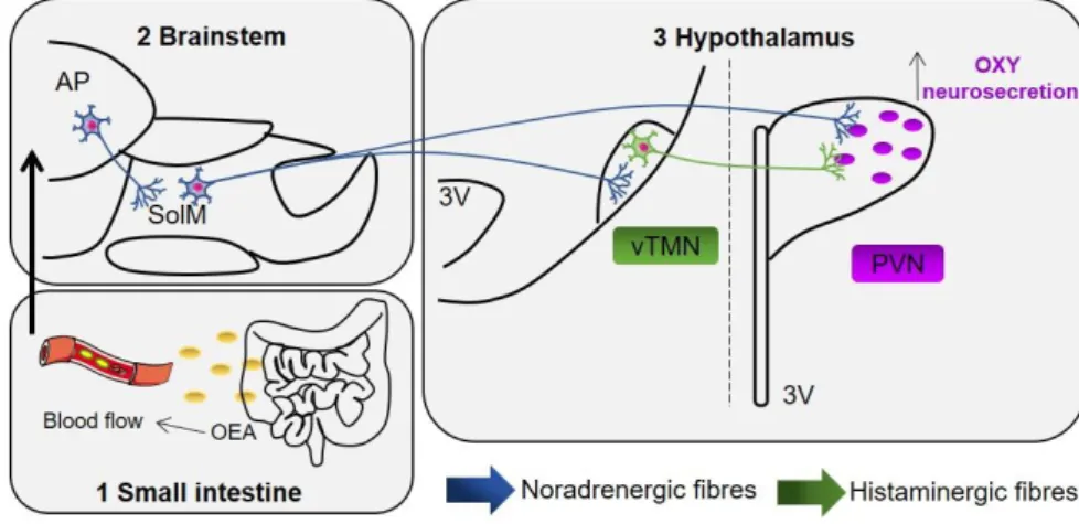
Circulating nutrients, that reflect the levels of nutrients in the periphery, are detected by the area postrema (AP), a circumventricular organ that lacks a functional blood-brain barrier (BBB) located on the floor of the fourth ventricle42, that, in turn, activates other nuclei in the brainstem41. In response to these stimuli, the brainstem, thanks to the activity of vagal efferent fibers, plays a crucial role in the control of the ENS, thus modulating many functions of the GI tract, and in the activation of neural circuits in the hypothalamus to reduce food intake. In particular, vagal afferents convey informations from the periphery to the nucleus of the solitary tract (NST)43, that, in turn, projects to hypothalamic nuclei43,44. The hypothalamus comunicates with vagal efferents, whose cell bodies are located in the dorsal motor nucleus of the vagus (DMV), through which it can slow gastric emptiyng32,45. Altogether, AP, NST and DMV form the dorsal vagal complex (DVC).
In the hypothalamus, a variety of molecules and receptors involved in the control of appetite are produced, in particular endocannabinoids (ECs)46, neuropeptide Y (NPY), pro-opiomelanocortin (POMC), alpha melanocyte-stimulating hormone (α-MSH), Agouti-related peptide (AgRP), cocaine- and amphetamine-regulated transcript (CART), CCK, and GLP-136,47.
The endocannabinoid system (ECS) plays a crucial role in the regulation of energy homeostasis and feeding behavior. The receptors for ECs are in fact largely expressed in the CNS, in particular in the hypothalamus and brainstem, and in the periphery, in organs that are important for the metabolism, such as liver, pancreas, muscle and adipose tissue48,49. Moreover, ECS is involved in many physiological aspects, such as the modulation of appetite and reward, lipid storage, energy consumption and insulin homeostasis50–52.
6
1.2.1 The vagus nerve
The vagus nerve is the tenth cranial nerve and innervates mainly the thoracic and abdominal cavities. Vagal fibers are not only composed of nerves but contains also glial and dendritic cells belonging to the immune system53. Moreover, paraganglia are often in close contact with its branches54,55 (Fig 1.2).
Fig. 1.2: Anatomy of the vagus nerve. Representation of the main branches of the vagus
nerve.
Cervical and thoracic vagus
The right and left branches of the cervical portion of the vagus nerve are both made of afferent and efferent fibers, that leave the skull from the jugular
7 foramen. Outside of the skull, the jugular (proximal) and nodose (distal) ganglions are located, where the cell bodies of sensitive neurons can be found. At this level, the vagus runs along the carotid artery and branches at the jugular ganglion into the auricular and meningeal branches, that provide sensorial innervation to the skin of the external acoustic meatus and to the dura of the posterior cranial fossa, respectively. Then, the pharyngeal branch departs at the nodose ganglion, and from its caudal part the superior laryngeal nerve branches, and runs under the carotid artery to the larynx, providing also smaller branches that innervate the caudal part of the pharynx and the esophagus. A cardiac cervical brach departs either from the cervical portion of the vagus or from the laryngeal nerve. This branch is called aortic56 and contains a large number of afferent fibers receiveng stimuli from the baroceptors in the aortic arch. The laryngeal nerve departs at the level of the subclavian artery in the right part of the body, and at the level of the aortic arch in the left, then running along and innervating the trachea and the exophagus. Then, the fibres that innervate bronchi, lungs, and heart branch in the superior mediastinum.
In particular, the afferent fibers that depart from the jugular ganglion produce neuroactvie peptides, such as substance P or Calcitonin Gene Related Peptide (CGRP), whereas those that depart from the nodose ganglion do not produce these molecules57,58.
Moreover, abdominal and thoracic branches contain afferent or efferent fibers that decussate from one side to the other54,59,60. In the rat, there are few lines of retrotracing evidences about decussating efferent branches, however, possibly 20% of afferent branches may decussate in the thoracic cavity61.
Abdominal vagus
The anterior (or ventral) portion, along with the left cervial vagus, branches in the common hepatic, gastric and coeliac portions. In the rat, there are about 11000 nervous fibers in each subdiaphragmatic branch, of which about 8000 are afferent and 3000 efferent. Moreover, less than 1% of all the fibers is myelinated. The common hepatic branch is important for the communication between the immune and nervous systems and for the thermoregulation, and
8 contains about 3000 fibres, of which 2200 are afferent, 200 efferent and 600 non-vagal62. The terminations of vagal afferents end in the connective tissue around the intrahepatic triads, extrahepatic bile ducts, portal vein and paraganglions63. Other afferents run along the common hepatic branch and, to a lesser extent, along the periarterial plexus of the common hepatic artery, around the portal vein63. Bile ducts are more innervated compared to portal vein: here, vagal afferents are tightly connected to the main branches of bile ducts and are found also in the walls of intra- and extra- biliary ducts63. This branch is called hepatic, but it innervates also the pylorus, the pancreas and the proximal duodenum64,65. Once the hepatic branch reaches the hepatic proper artery, it moves towards the hepatic common and gastroduodenal arteries, and then it divides in two branches, one following the right gastric artery (to the stomach), an the other following the dorsal and ventral duodeno-pancreatic arteries (to the proximal duodenum and pacreas). The anterior gastric branch innervates the ventral part of the stomach and, through small fibers inside the circular muscle of the pylorus, resches the duodenum.
The dorsal (posterior) part of the gastric branch, on the other side, enters the dorsal part of the stomach, near the cardias, running along the left gastric artery, and innervates the proximal duodenum through trans-pyloric fibers. In the stomach, vagal nerves are in both layers of smooth muscle, in the myenteric plexus and in the lamina propria. The fibers located in the longitudinal and circular layers have been described as long axon bundles parallel to muscles and connected by short branches64,65.
The most common vagal afferent ending is the intraganglionic laminar ending (IGLE), mainly linked to the neurons of the myenteric plexus66. This type of neuron is also observed in the whole intestine, both small and large, where vagal afferents penetrate through the myenteric plexus to the circular smooth muscle and the submucosa, to form a net of axon branches in the lamina propria. Thus, it has been hypothesized may function as distention receptor67,68. Then, the ventral celiac branch, after leaving the ventral portion of the esophagus, reaches the dorsla celiac branch near the left gastric artery. Both these branches go down the celiac artery to reach the celiac ganglion,
9 and then the small and large intestine running along the superior mesenteric artery.
Role of the vagus nerve in the homeostatic control of energy
balance
The intestine constantly sends signals about the quantity and quality of the nutrients to the CNS, that is able to process a variety of inputs and to transform them in information of various nature, that will determine a behavioral response. These processes can engage either the somatic or the autonomous nervous systems, the former being mainly controlled by the prefrontal cortex (PFC), hippocampus (Hippo) and nucleus accumbens (NAcc) and being influenced by the emotional cues linked to feeding; the latter, on the other hand, is more involved in the homeostatic control of energy balance. In particular, the parasympathetic nervous system, composed of afferent (sensitive) fibres that convey signals to the CNS, and efferent (motor) fibres, that convey signals from the CNS to the parasympathetic ganglions near target organs, receive stimuli from the brainstem, and in particular from the DVC and pars reticolata10,69. This bidirectional pathway, or vago-vagal reflex, is the major extrinsic neural way involved in the control of pancreatic and GI tract functions70. The main stimuli received by vagal afferents are:
Mechanic stimuli. The GI tract is rich in mechanoreceptors, in particular located in the mucosa, sensitive to friction71, and on the outer muscle layer, sentitive to distention, such as IGLE fibres72.
Chemicals and nutrients. In the rat, the majority of vagal fibers that innervate the duodenum respond, apart from mechanic stimuli, to chemical stimuli, such as pH and osmolality70. Moreover, these fibres respond to peptides acting in the intestine, such as CCK73,74, GLP-175, serotonin (5-HT)76, somatostatin75 and interleukin-β (IL-β)77.
Other stimuli. Vagal afferents are also sensitive to temperature, osmotic pressure74 and pain. For example, a prolonged gastric distention activates vagal afferents and induces Fos in the NST78.
Vagal afferent teminals are mainly found, in the CNS, in the NST, AP, and, to a lesser extent, in the DMV and in the trigeminus. The terminations of
10 the cardiac and pulmonar vagal afferents are located in the lateral subnuclei of the NST, whereas the terminations of the alimentary canal are found in the medial subnuclei of the NST79–81. Moreover, many of vagal afferents of the jugular ganglion produce peptides such as CGRP and substance P82,83, whereas glutammate has been found in cardiac afferents. In the same way, it has been demonstrated that pharmacological blockade of NMDA receptors is able to abrogate gustative inputs in the NST84. In fact, many studies suggest that ionotropic glutammate receptors, including NMDA, in the NST play a crucial role in the control of eating. For example, the blockade of NMDA receptors with a microinjection of dizocilpine, a non-competitive antagonist of this receptor, leads to a delay of the onset of satiety and to an increase of food consumption85. Moreover, the electric stimulation of the NST results in a current (either eccitatory or inhibitory) in the DMV, suggesting a connection between these two areas86. Moreover, pharmacological and histological evidences showed the presence of glutammate receptors in the DMV87, where the cell bodies of the neurons that innervate the GI tract are located88,89. In this area, two distinct populations of preganglionic neurons modulate the GI functions: on one hand cholinergic neurons, located in the rostral DMV and increase gastric motility90; on the other, non-adrenergic non-cholinergic (NANC) fibres, located in the caudal DMV and that decrease GI motility90,91. Therefore, the DMV is characterized by two parallel neuronal pathways that modulate gastric motility: an exitatory cholinergic pathway that increases gastric motility, and an inhibitory NANC pathway that slows gastric functions. Intraganglionic vagal afferent terminals are located in the capsule of connective tissue of the ganglions of the myenteric plexus, between the longitudinal and circular layers of smooth muscle32, and, therefore, respond to passive distention and active contractions of the muscles72. This type of vagal afferent has been found in the esophagus and along the GI tract92,93.
Intramuscolar bundles are almost exclusively located in the longitudinal and circular layers of the stomach smooth muscle64, where the presence of food is detected also by vagal afferents found in the gastric mucosa, sensitive to mechanical stimuli94, whereas the quantity of ingested food is detected by vagal afferents located in the outer muscle layer, sensitive
11 to distention95. Moreover, vagal afferents of the gastric mucosa can respond to hormones released locally, such as leptin and ghrelin96. In fact, vagal afferents express leptin receptors97,98. In addition, the appetite-stimulating effects of ghrelin are abolished by total subdiaphragmatic vagotomy (TVX) or the treatment with capsaicin in rats99. Hence, since vagal neurons located in the nodose ganglia express ghrelin receptor99,100, it maybe hypothesized that ghrelin, like leptin, acts through the activation of vagal afferents.
Several studies suggest that the inhibitory effects on feeding mediated by CCK101 involve vagal afferents, that express the CCK receptor A (CCKA)102,103. In the periphery, these neuronal terminals are located in the walls of the GI tract, both in the mucosa and in the muscolar layer104. Moreover, it has been demonstrated that the inhibition of gastric emptying is mediated by CCKA-expressing vagal afferents105. In addition, it has been observed that a total or selective vagotomy abolished or decreased, respectively, the satiety effect of CCK106. Furthermore, another important study demonstrated that the treatment with capsaicin, that destroys unmyelinated fibres (including unmyelinated vagal fibres), dampened the satiety effect of CCK107.
Food intake, and, in particular, the intake of carbohydrates, fats and proteins, induces the release of GLP-1, that seems to be induced indirectly by the stimulation of the nerves of the apical portion of the intestine, or directly by the contact with the lower portion of the intestine108,109. Since GLP-1 modulates pancreatic secrestions, it plays a crucial role in glucidic homeostasis110,111. The peripheral administration of GLP-1 induces satiety in rats and humans112,113. This effect may be due to the paracrine stimulation of the gastric mucosa by vagal afferents: in fact, GLP-1 receptor is expressed in the nodose ganglion, and, moreover, in the CNS, in the AP114,115, NST and lateral parabrachial nucleus (LPB)116.
1.2.2 Mediators of energy balance
Peripheral signals
The GI tract releases more that 20 different regulators and hormones, that are involved in the regulation of many physiological processes37. The release of
12 hormones such as PYY, GLP-1 and OXM is triggered by gastric distention and by the interaction between nutrients and the intestinal walls117,118, and, once released, intestinal hormones act on target organs, such as endocrine glands, smooth muscle and peripheral nervous system (PNS)39,119. It is well known that hormones and intestinal peptides play a crucial role in the modulation of hunger and satiety120: many studies support that signals like CCK, PYY, GLP-1 and OXM, on one side, reduce food intake by reducing the levels of orexigenic signals while increasing anorexigenic signals in the hypothalamus121,122, and, on the other, by triggering negative feedback mechanisms on the intestinal transit, that contribute to enhance the feeling of satiety during the intermeal interval123, hence aiding the regulation of the after meal GI motility110,118.
Ghrelin is a peptide hormone produced by the stomach and released
into the bloodstream, initially discovered for the affinity for the growth hormone secretagogue receptor (GHS-R). It is a peptidide chain of 28 aminoacids and undergoes one post-translational modification that involves the addition of one molecule of ottanoic acid on the serine 3119,124, necessary for the binding to the GHS-R and to cross the BBB125. Ghrelin is defined as the “hunger hormone”125, and its circulating levels increase during fasting and decrease after a meal. Moreover, the administration of ghrelin in the CNS produces an increase of food intake and the release of the growth hormone (GH) in rats, whereas its peripheral administration reduces the use of fat from adipose storage126. Several studies show that the disruption of the ghrelin-mediated signaling induce several alterations in the control of energy homeostasis127,128. The pharmacological inhibition of the GHS-R was thought to be a valid strategy for the treatment of obesity. In fact, GHS-R antagonists are able to decrease food intake in fasted animals127, and that the vaccination against ghrelin induces weight loss129. However, even though promising results have been obtained from animal models, an irreversible approach, such as vaccination, is not considered safe for human treatment124. Ghrelin agonists, on the other hand, may be used for the treatment of anorexia in oncologic patients, that experience appetite loss130, in patiens with dialysis-induced malnutrition130, or to improve gastric emptying in diabetic patients with
13 gastroparesis131. Moreover, it has been shown that the gene encoding for ghrelin encodes for another peptide, called obestatin. preliminary studies demonstrate that central administration of obestatin reduce food intake, while the periheral administration reduce body weight132. These effects seem to be exerted by the activation of the GPR39, an orphan receptor. However, further studies did not confirm the previous results, suggesting that obestatin does not bind GPR39 and does not control feeding behavior133,134.
PYY is a peptidic hormone related to the neuropeptide Y (NPY). Both
these peptides have a structural feature characteristic of the PP proteins and exert their action by binding to Y receptors. There are two endogenous isoforms of PYY, based on the presence of the N-terminal, PYY1-36 e PYY3-36, the latter having hagher affinity to Y2 receptor (Y2R). In fact, the effects of the PYY3-36 are dampened by the administration of a Y2R antagonist135 and abolished in Y2R-KO mice121. Preliminary studies demonstrated that the peripheral administration of PYY3-36 reduces foof intake in rodents and humans121. However, these preliminary findings were controversial, since many laboratories could not reproduce them136. Then, further studies demonstrated that the effects of the peripheral administration of PYY3-36 on feeding were dampened by stressful conditions, such as the lack of handling or the introduction to a new environment137,138. Many subsequent studies then confirmed that the acute administration of PYY3-36 reduces food intake113,139. The fact that the biological effects of PYY3-36 are dampened by the pretreatment with a Y2R antagonist135 and abolished in Y2R-KO mice136 further confirm that this peptide acts through the Y2R. Furthermore, an altered control of energy homeostasis is observed in PYY-KP mice, thus validating the role played by this peptide in its regulation140,141.
The role of the CCK on the esocrine pancreas and gallbladder became clear when, in 1973, it was the first intestinal hormone proven to be involved in the control of feeding behavior142. CCK is released during the aftermeal period and exerts its effect through the activation of the CCK receptor 1 (CCK1), expressed on vagal fibres. The pretreatment with antagonists of this receptor increase food consumption in rodent and humans124,143, and Otsuka Long-Evans Tokushima rats, KO for this receptor, are obese and
14 hyperphagic143. However, continuous infusions of CCK failed to reduce food intake, and, although the intermittent administrations reduce short-term food consumption, this effect is dampened by the compensatory increase of food intake during the intra-administration period144. Moreover, some studies suggest that physiological concentration of CCK do not activate vagal circuits, suggesting that the action of this peptide on FI is mainly due to its paracrin action more than to an endocrine one145.
The pancreatic polypetide (PP) is synthesized and released by the endocrine pancreatic parenchyma and has a high affinity for Y4 and Y5 receptors. As for PYY3-36, PP levels increase after a meal, and decrease during fasting. Acute PP administration reduce food intake in mice and humans146,147, while chronic administrations reduce body weight in ob/ob mice148. It has been hypothesized that the anorexiant effect of PP is due to the delay of gastric emptying147,149.
Amylin is a 37 aminoacids-long peptide, belonging to the calcitonin
peptides family, that is released, as insulin, by pancreatic β-cells after the ingestion of food. Although its main function seems to be linked to glucose homeostasis, it reduces food intake after peripheral administration150. In fact, the administration of Pramlintide, a synthetic analogous of amylin, reduces body weight in patients with type 1 and 2 diabetes151,152. The anorexiant effects of amylin may be due to the modulation of the serotonergic, histaminergic and dopaminergic systems, as long as the inhibition of NPY release153. Moreover, many studies reported that increased circulating levels of amylin are observed in obese patients154.
GIP is a polypeptidic chain composed of 42 aminoacids, synthesized
and released by the K cells of the duodenum after food consumption. There are no clear studies linking this peptide to the regulation of food intake, although mice with the genetic deletion of the gene encoding for its receptor are resistant to the diet-induced obesity (DIO)155. Therefore, further studies are required to investigate the physiological mechanism involved in the mechanism of action of GIP, that may involve adipocytes and neural circuits in the CNS156.
15
GLP-1 is co-secreted with PYY by the L cells of the intestinal mucosa
upon the ingestion of food. It is produced by the cleavage of a precursor, the preproglucagone, that, after enzymatic cleavage, produces several biologically active peptides, such as glucagone, GLP-1 and -2 and OXM. GLP-1 has two different active forms, GLP-17-37, found in the periphery, and GLP-17-36 amide, found in the CNS38. GLP-17-36 amide- positive neurons have been found throughout the CNS, in particular in the paraventricular nucleus (PVN), in the dorsomedial nucleus (DMN), in the DVC, in the hypophysis and in the thalamus157. Moreover, GLP-1 is a potent incretine: in fact, its release after food consumption stimulates the release of insulin38,124. Furthermore, central administration of GLP-1 drastically reduces food intake in rodents, whereas peripheral administration reduces appetite in both rodents and humans158. Exendin-4 is an agonist of the GLP-1R, while its cleaved form, exendin9-39, is an antagonist of the same receptor. It has been demonstrated that the acute central administration of exendin9-39 increase food intake, and its chronic administration increases body weight124,158. Although it seems that endogenous GLP-1 is involved in physiological processes, such as the regulation of feeding, in a mouse model knock-out for the GLP-1R feeding and body weight are not altered158. Moreover, a phase III clinical trial demonstrated that exendin-4 ameliorates glucose homeostasis in patients with type 2 diabetes, and that it induces a reduction of food intake159. Along with exendin-4, the GLP-1R agonist exenatide has already been approved for the treatment of type 2 diabetes in co-treatment with metformin.
GLP-2 shares the same synthesis pathway as GLP-1, glucagone and
OXM. High concentrations of this peptide have been found in the brain, and its central administration reduces food intake160. However, in the periphery, GLP-2 is involved in the regulation of gastric motility, digestion and absorption of nutrients, and does not seem to influence appetite161.
OXM is a 37 aminoacids-long plypeptidic chain produced by the
cleavage of preproglucagone. Like GLP-1, is released upon the ingestion of food, and, when administered centrally, reduces food intake162. Although many studies suggest tha OXM exerts its biological action through the activation of the GLP-1R, supported by the fact that its anorectic effect is
16 abolished in GLP-1R KO mice, it seems that these effects do not perfectly match those exerted by GLP-1R38,124. In fact, it has been demonstrated that the administration of exendin9-39 in the arcuate nucleus (Arc) abrogates the OXM-induced anorectc effects, but not thos of GLP-1162. Moreover, chronic administration of OXM, either peripheral or central, induce a body weght reduction greater than that of pair-feeding animals, suggesting that OXM may act by increasing energy expenditure162.
The role played by glucagone in the control of glucose homeostasis is well defined. It is produced by α-cells of Langerhans islets in the pancreas and increases glucose levels in response to a hypoglycemic state. Moreover, glucagone improves the stress response by increasing energy expenditure163. The administration of glucagone reduces food intake, probably through the modulation of the vagal tone and of the gastric emptying164. Furthermore, recent findings demonstrated that the co-administration of antagonists for glucagone receptore and GLP-1R ameliorates insulin sensitivity and DIO165.
Insulin, produced by the β-cells of the pancreas, is released after the
ingestion of food and induces its very well-known hypoglycemic effects166. Moreover, it acts on the CNS as a satiety factor: in fact, the central administration of insulin dose-dependently reduces food intake and body weight gain in rodents and baboons167,168. Insulin is carried in the CNS by a receptor-mediated transport169. Furthermore, insulin receptors are expressed throughout the brain, in particular in the Arc, the DMN and PVN170.
Neuroactive mediators
Neuropeptides are small proteic molecules, released from the cells of the nervous system in response to a stimulus, able to control the comunication between neurons by binding secific receptors. Hypothalamic peptides may be classified based on the effect they exert on feeding behavior. On the orexigenic side, NPY is produced by neurons in the Arc that project to other hypothalamic nuclei171. Although NPY can exert several different effects on feeding behavior172, the most known is in the increase of appetite upon central administration173. The synthesis of NPY occurs in Arc and its release in the PVN, and both the processes are negatively regulated by leptin and insulin, and positively regulated by ghrelin173. Five receptors for NPY have been
17 described: Y1, Y2, Y3, Y4, Y5 and Y6; among them, Y5 receptor is the most involved in the modulation of NPY effects on feeding174,175.
Another orexigenic peptide is the AgRP, a 132 aminoacid chain exclusively expressed in the Arc. It has been demonstrated that, upon central administration, either in the PVN or DMN, AgRP increases food consumption176.
Hypocretins 1 and 2, also known as orexins A and B, are neuropeptides produced in the lateral hypothalamus (LH) and linked to the stimulation of appetite by binding their receptors, OX1R and OX2R. OX1R is mainly expressed in the ventromedial hypothalamus (VMH), whereas OX2R in the PVN177. Moreover, orexin-secreting neurons are located in the LH and DMH, and project to other hypothlamic nuclei, in particular to the Arc178,179, and input on NPY secreting neurons expressing OX1R178.
Apart from orexigenic signals, there are a lot of anorexigenic peptides acting in the hypothalamus: CART is a neuropeptide synthesized in the DMH, PVN, LH and Arc180; melanocortins, in addition, are bioactive peptides that derive from the pro-opiomelanocortins (POMC) after a tissue-specific post-translational cleavage181. The gene encoding for POMC is expressed in several tissues, such as hypothalamic neurons, the adenohypophysis and the pars intermedia182. Moreover, the intermedial lobe of the hypophysis produces
α-MSH, an anorexiant peptide that binds to melanocortin receptors 3 and 4
(MC3R and MC4R), expressed by brain areas known to be involved in the control of feeding behavior and in telencephalic structures, such as the cortex182.
The corticotropin-releasing hormone (CRH) is a 41 aminoacids-long hormone and regulates the secretion of the adrenocorticotropic hormone (ACTH) from the hypophysis. CRH is highly expressed in the PVN and, when centrally administered, inhibits food intake and reduces body weight in rats171. On the other hand, the periheral administration of CRH in humans increases energy expenditure and fatty acid oxidation (FAO)171. Moreover, the infusion of leptin increases the expression of CRH, whereas the pretreatment with a CRH receptor antagonist attenuates the leptin-induced reduction of food intake and body weight171.
18 Among neurotransmitters, histamine is a hypophagic agent synthesized from the decarboxylation of histidine, exerted by the histidine-decarboxylase (HDC)183. In fact, the central infusion of α-fluoromethylhistidine, an inhibitor of HDC that, therefore, leads to a decrese of central histamine, is able to disrupt feeding and hydration patterns, along with deambulation184. Moreover, it has been demonstrated that histaminergic system is involved in the anticipatory phase of eating: in fact, the specific activation of the E3 subdivision of the ventral tuberomammillary nucleus (vTMN)185 is rats with restricted access to food. However, several lines of evidence suggest that histamine is also involved in the consummatory phase of feeding behavior, during which a rapid transitory increase of hypothalamic histamine levels is observed186.
Histamine binds to 4 GPCR receptors: H1R, H2R, H3R, H4R187,188.
H1R, coupled with Gq, is expressed in the brain, bronchial epithelium, cardiovascular system, liver and cells of the immune system189. In the CNS is expressed in the VMH, that is likely the site where histamine exterts its appetite-suppressing effects189.
H2R, coupled with Gs, is expressed in the gastric mucosa, in the muscular layer of arteries, in the cells of the immune system and in the brain189.
H3R, coupled with Gi/o, is highly expressed in the CNS, where it acts as presynaptic autoreceptor, thus inhibiting the release of histamine190. Moreover, it acts as heteroreceptor, modulating the release of other neuronsmitters, such as acetycholine, dopamine, noradrenaline, and serotonin.
Lastly, H4R has a primary role in the inflammatory response189.
Several studies have demontrated that the intra-hypothalamic administration of histamine, where the activation of H1R induces satiety, increases mRNA expression levels of the uncoupling protein 1 (Ucp1), marker of energy expenditure, in the brown adipose tissue (BAT), and, moreover, increases the electrophysiological activity of the sympathetic nerves around that area191,192. Furthermore, it is likely that histamine released in the periphery is involved in metabolic and homeostatic processes linked to food consumption193.
19
1.2.3 Central circuits involved in the homeostatic control of feeding
behavior
Feeding and metabolism are regulated by complex systems in the CNS194. The main area of the CNS involved in the control of food intake is the hypothalamus, that is costantly informed by signals secreted in the periphery about the nutritional and energetic statuses of the body43. These signals are then integrated and conveyed to other brain areas. Satiety signals are generated in the GI tract during meal consumption and modulate feeding through the release of peptides that reach the NST via the vagal afferent system195 (Fig. 1.3).
Among all the systems of the gut-brain axis that interplay in the regulation of feeding behavior, an important role is played by the ECS. In the CNS, this system plays a role in the motivation for the research and consumption of food, through the activation of mesolimbic pathways that regulate reward196. N-arachidonoylethanolamide (or anadamide, AEA) and 2-arachidonoylglycerol (2-AG) are the most studied ECs197 and have a hyperphagic effect by acting on CB1 receptors in the PVN198 and in the lateral hypothalamic area (LHA), or by influencing the action of hormones like ghrelin197. The role of this system has gained a great deal of attention, starting from the development of synthetic compounds acting on CB1 receptor, the rimonabant, as a potential target for the treatment of obesity50
20
Fig. 1.3: Crosstalk between circuits modulating feeding behavior. Lori M. Zeltser, Feeding
circuit development and early-life influences on future feeding behavior, Nature Reviews
Neuroscience volume 19, pages 302–316.
Brainstem
The brainstem is one of the most important areas for the homeostatic control of feeding behavior, since it acts as an integration center between sensitive inputs and motor outputs. the NST is located in the most caudal portion of the brainstem, and represents the first relay station of vagal afferents, that convey the informations about the quantity of food consumed from the periphery199. In particular, it is in contact with the preganglionic vagal neurons, in order to regulate nutrient adsorption in the alimentary canal79, and sympathetic preganglionic nerves, in order to modulate energy expenditure10. The NST is then connected with other brain areas involved in the homeostatic control of feeding behavior: in particular, it has direct inputs towards the hypothalamus, and contacts indrectly telencephalon and the cerebral cortex10. Moreover, it is known that part of the NST projections that contact the hypothalamic neurons are the A2 noradrenergic fibres200, and many studies have demonstrated that the ablation of these fibres dampens the effects of some satiety factors, such as CCK199.
The NST is composed by different subnuclei that specifically respond to stimuli received10. For instance, the gustative fibres of the tongue and the posterior oropharynx input in the rostral part of the NST79, whereas the
21 afferents from below the diaphragm project to the caudal part of this nucleus69. Neurons of the DMV respond mainly to gastric distention201, while the neurons below the AP respond mainly to duodenal signals, whereas the neurons located in the medial part of the NST respond both to gastric and duodenal signals201. Notably, in this nucleus, there are neurons that express POMC97, melanocortin202 and leptin97 receptors.
The AP, in close contact with the NST, is another important nucleus involved in the homeostatic control of feeding behavior, and expresses receptors for amylin, GLP-1 and ghrelin. Moreover, it is able to convey informations about gastric emptying42,203. Studies conducted in rats that underwent the surgical lesion of the AP highlighted the involvement of this area in the regulation of the signals sent from the periphery204,205. The AP is a circumventricular organ outside the BBB, that lacks tight junctions and is rich in fenestrated capillaries206. Thanks to these features, some peptides and other signaling molecules may have direct access to the AP wich is one of the main integration stations in the CNS that conveys numerous physiological signals from the bloodstream206. Moreover, many studies demonstrated that AP projects extensively to other nuclei of the brainstem and to the hypothalamus207.
In particular, it projects to the NST and LPB, known integrative stations of the brainstem207,208. Moreover, neurons of the AP project direclty to the dorsal and medial subnuclei of the NST207. Furthermore, the AP projects to a lesser extent to the nucleus ambiguous, to the DMV, the dorsal parts of the tegmental nucleus and to the spinal tract of the trigeminus207.
The afferent neurons pojecting to the AP come from functionally distinct parts of the brainstem and of the hypothalamus. Apart from the bidirectional connections to and from the NST and LPB, the majority of the hypothalamic connections comes from the PVN and the DMH207,209. Moreover, it has been demonstrated that the AP receives direct inputs from vagal afferents207.
It has been observed that the AP contains the receptors for a variety of hormones that regulate feeding behavior, such as CCK, intestinal vasoactive peptide, NPY, vasopressin (AVP), substance P and insulin210,211.
22 Another important brain structure involved in the homeostatic control of feeding behavior is the LPB, located in the pons. Its anatomical position, between the medulla and the prosencephalon, allows it to integrate the signals coming from both ways, hence helping to organize behavioral responses212. Moreover, the LPB integrates a variety of sensitive inputs, associated to food consumption or to stress212,213. In particular, the LPB receives sensitive informations mainly from the NST214, and projects to the PVN and Arc10.
Hypothalamus
The mammalian hypothalamus consists in over 40 nuclei in histologically distinct areas215. The medial part of the hypothalamus is mainly composed of large nuclei that receive bloodborne stimuli and pass them down onto other hypothalamic nuclei, controllig endocrine responses. Then, the lateral portion is a large area that bridges the hypothalamic nuclei with extrahypothalamic brain areas, such as the cortex and the limbic system215.
Since its lesion induces hyperphagia and body weight gain, the VMH has been identified as the satiety center in the hypothalamus23. On the other hand, the LH has been identified as the “hunger center” of the hypothalamus, since its lesions induce weight loss216.
Among hypothalamic nuclei, the PVN interplays with the endocrine and autonomic nervous systems and is involved in many behavioral responses. Moreover, the PVN is able to comunicate with the preganglionic neurons that innervate the pancreas, and with the parasympathetic and sympathetic nervous systems217. PVN neurons project towards di- and telencephalic structures218 and to other hypothalamic areas, such as DMH, VMH and Arc218,219. In general, the PVN can be divided in magnocellular and parvocellular. Magnocellular neurons contain oxytocin (OXY) and AVP, that are released in the posterior lobe of the pituitary gland220. Many medial parvocellular neurons contain CRH and tireotropin-releasing hormone, that are released in the ME. The dorsal, ventral and lateral parvocellular neurons, on the other hand, project to the periacqueduttal grey, the LPB and the NST, and to both sympathetic and parasymathetic preganglionic neuronal populations220,221. The better-known endocrine signals produced by the PVN
23 are those of the magnocellular projections, that reach the posterior part of the pituitary gland releasing OXY and AVP, and those of the parvocellular projections, that release CRH and TRH. The non-endocrine signals are those coming from the dorsal raphe, LPB, DMV and NST222. Therefore, the PVN seems to be in the best position to integrate internal stimuli, such as the availability of nutrients in the GI tract and adipostatic signals, to thus organize endocrine and autonomic responses10.
The supraoptic nucleus (SON) lies behind the optic chiasma, and is composed, as the PVN, of magnocellular neurons10. These neurons contact the posterior part of the hypophysis, through which the SON contributes to the endocrine control of the organism. The major inputs to the SON come from the branstem, in particular from the NST223.
The Arc is a hypothalamic region in close contact with the ME, a circumventricolar organ rich in fenestrated capillaries that lacks a functional BBB224. It contains two major neuronal populations that control appetite and satiety. The former is composed of orexigenic neurons, and express NPY and AgRP225, while the latter contains anorexigenic neurons, that contain α-MSH (derived from POMC), and CART10,225.
The TMN is a group of large cells located in the tuberal caudal and mammillary rostral areas and form the median hypothalamic area. In the medial part of the vTMN226 a complex network of histaminergic neurons is found, that project to several brain areas193,227. These neurons are involved in many physiological responses, such as circadian rythm, emotions, learning and memory228,229.
24
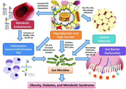
1.3 Gut microbiota
Microbes are everywhere, and it is known that microbial populations are resident, in humans, on the skin, in the oral cavity, in the urogenital tract and in the gastro-intestinal (GI) tract. In all these areas, the microbial populations partake in the physiological control of the homeostasis by establishing a symbiotic relationship with the host230. Human microbiome is involved in many mechanisms that allow the maintenance of the well-being of the host, like metabolic and immunological processes231. These lines of evidence, therefore, led to hypothesize the existence of the “olobiome”, represented by the symbiotic interaction between the microbiome and the host232. On the other hand, this interaction may play a role in the onset of pathological conditions, and, therefore, in the past 20 years many studies were conducted to demonstrate how feeding behavior can alter the gut microbiome233. In fact, it has been shown that different host’s characteristics, like genetic background, gender, age, and immunological profile, play a pivotal role in shaping the gut microbiome234, as long as environmental and behavioral factors, such as pharmacological therapies, surgeries, physical activity or stressful conditions235. In newborns, the microbiota is spread during delivery and is, then, influenced by a variety of factors, such as the type of delivery (natural or c-section) or breastfeeding236. For example, the microbial flora of naturally delivered babies is characterized by the presence of Lactobacillus, Prevotella, and Atopobium, whereas in babies delivered by c-section the flora is rich in Staphylococci, as for the mother’s skin236. Moreover, in the recent years, new lines of evidence show that the intrauterine environment is not sterile, but colonized by Enterococcus fecalis, Staphylococcus epidermis, e Escherichia coli237. The newborn’s flora, rich in aerobic bacteria, undergoes changes during the post-natal period towards a flora composed mainly by anaerobic bacteria238. This first colonization happens together with the activation of the hypothalamus-pituitary-adrenal axis (HPA axis), that has a strong impact on the ENS231. Moreover, it has been shown that the metabolites produced by the enteric flora may induce the release, by the enteroendocrine cells of the GI tract, of mediators involved in the control of feeding, lipid storage, and energy homeostasis7,239. Among these mediators,
25 short-chain fatty acids (SCFAs) activate the GPR41 receptor expressed by the enteroendocrine cells240, involved in the gut microbiota-mediated control of adiposity and inflammatory processes241.
Then, the population of the gut microbiome becomes more stable during adult life242, with a last change during the elderly life: in fact, aging is associated to physiological changes tha influese the composition of the gut microbiome231. The microbiome is composed by over a 100 billion of microorganisms, the majority belonging to the reign of Bacteria243. Moreover, the entire genome of the commensal flora contains over 3 million genes, 100 time more than the human genome243. Therefore, the variety of protein products yields a pool of metabolic and biochemical functions that have a high impact on the host’s physiological processes244. Both the host and the gut microbiota produce a variety of small molecules during the catabolism of food and xenobiotics, that play a crucial role in the communication between the cells of the host and of the microbiome. Moreover, the microbiota composition changes in different portions of the GI tract236,245, and an interplay between all the different populations along the GI tract has been observed246. The exchange of low molecular weight metabolites, like peptides and small proteins, underlies this type of chemical communication, together with pathway mediated by the immune system230.
The human body eliminates up to 100 mg of volatile phenols per day, in particolar 4-cresol and phenol, either as glucuronate conjugate or solfates247. In mammals, the production of these chemical species is due to the activity of different microbial populations: for example, cresol is produced by the species Clostridium, Bifidobacterium, and Bacteroides fragilis230, whereas E. coli has been associated with the production of phenol230. Moreover, an alteration of 4-cresol metabolites in urine has been associated with several pathophysiological conditions, that span from weight loss to inflammatory bowel diseases230. Moreover, these conditions have been associated to a variation of gut microbiota composition, in particular to a decrease of the populations of Lactobacillus and Bacteroidetes in inflammatory bowel diseases248, and to an unbalance between the populations of Firmicutes and Bacteroidetes in weight loss249. The enzymatic
26 activities of gut microbiota products can directly act on the fermentation of carbohydrates and on the metabolism of bile acids. Indigestible carbohydrates, the so-called functional fibers, are an important energy source for many members of the gut microbiota populations, such as Bacteroides thetaiotaomicron and Bacteroides ovatus250. In fact, the fermentation of these fibers leads to the production of SCFAs, such as acetate, propionate, butyrate and lactate249. In particular, butyrate is a crucial substrate for cell metabolism of colon epithelium. In fact, it has been observed that germ-free mice show a severe energetic deficiency251, characterized by an increased activity of the AMP kinase, that is involved in the monitoring of the energetic state of the cell252. Moreover, the hepatic metabolism of germ-free animals is different than that of colonized ones, and this difference is probably due to a higher presence of SCFAs in the liver: it has been shown that the liver uptakes acetate and propionate and uses them as substrates for lipo- and gluconeogenesis. Furthermore, SCFAs promote the differentiation and the proliferation of colon epithelial cells, probably through the modulation of genetic expression due to the butyrate-induced inhibition of the histone deacetylase (HDAC)253. Moreover, SCFAs are able to modulate gene expression by activating two different GPCR, GPR41 and GPR43, that partake in different pathways based on the cell type they are expressed in254. For example, the activation of GPR43, in neutrophiles, has an anti-inflammatory effect255, whereas, in intestinal L cells, it induces the release of GLP-1256. By activating GPR41, the gut microbiome induces the release of PYY257, and the genetic deletion of this receptor prevents the accumulation of fat. Although many studies demonstrate the interplay between GPR41/43, enteroendocrine cells activity, and metabolites produced by the gut microbiota, many lines of evidence suggest that other biologically active compounds, hence acting on different receptors, can induce the release of GLP-1/2 and PYY258, for example bioactive lipids such as oleoylethanolamide (OEA) or 2-oleoyl-glycerol (2-OG), that bind the GPR119 receptor259. In fact, a very recent finding demonstrates the beneficial effects of OEA on the gut microbiota260.
27 However, to date, little is known about the role that each bacterial species has in the production of bioactive metabolites. It has been observed that Akkermansia muciniphila, that breaks down mucine, produces propionate and butyrate, that bind the GPR43 receptor261. Moreover, a recent study reported that obese or insulin-resistant mice show a decrease of the abundance of this species, and that the daily administration of A. muciniphila for four weeks reverts this phenotype262. Moreover, a direct correlation between the abundance of this bacterial species and the secretory activity of L cells has been found263.
The gut microbiome regulates lipid and glucidic metabolisms, acting both on the liver and on the production of bile acids. Colonized mice display higher levels of triglycerides stored in the liver, and an increase in the synthesis of very low-density lipoproteins (VLDL)264, that are involved in the transport of triglycerides from the liver to other tissues. Furthermore, an increased production of triglycerides in colonized mice is associated to a reduction of the expression of the angiopoietin-like protein 4 (ANGPTL4) in the small intestine251,265, that is a potent inhibitor of the lipoproteic lipase (LPL), a mediator of the cellular uptake of triglycerides. It has been demonstrated that germ-free mice knockout for the gene encoding for ANGPTL4 show an increase of fat mass and of the body weight, suggesting a role of both gut microbiota and ANGPTL4 in regulating adiposity251,265.
Choline is an important constituent of plasma membranes, that can be either introduced with the diet (mainly through the consumption of eggs and meat) or endogenously synthesized266. It has a major role in lipid metabolism and in the synthesis of VLDL in the liver, and its inadequate supply is associated with an alteration of the gut microbiota and with hepatic steatosis, both in mice267 and in humans268. In particular, low levels of Gamma-proteobacteria combined with high levels of Erysipelotrichi in human fecal microbiota are linked to hepatic steatosis268. The enzymatic activities of the gut microbiota and of the host interplay in the transformation of the choline into toxic metabolites, such as trimethylamine, that is further converted into trimethylamine-N-oxide in the liver269,270, thus reducing choline availability, that may be the cause of the onset of the non-alcoholic fatty liver disease (NAFLD)
28 in mice270. Moreover, an increase in trimethylamine-N-oxide is linked to a higher cardiovascular disease risk271.
Primary bile acids, such as cholic and chenodeoxycholic acids, are synthesized in liver from cholesterol, and partake in the solubilization of lipids, dietary fats and liposoluble vitamins, to aid their intestinal adsorption, and the gut microbiota is able to metabolize these compounds into secondary bile acids. In fact, higher levels of primary bile acids, and a lower variability in secondary bile acids, has been observed in germ-free rodents272. Moreover, bile acids bind nuclear receptors273, as the farnesoid X receptor (FXR)274, involved in the regulation and the synthesis of bile acids, and the G protein-coupled bile acid receptor 1 (or TGR5). Both these receptor are involved in the regulation of glucose metabolism in mice, negatively in the case of FXR, and positively in that of TGR5275. FXR is activated by primary bile acids, whereas TGR5 binds secondary bile acids, like deoxycholic (derived from cholic acid) and lithocholic (derived from chenodeoxycholic acid) acids. The signal induced by the activation of TGR5 in L enteroendocrine cells leads to an increase of GLP-1 secretion, improving glucose tolerance in obese mice275. Moreover, in the BAT and in the muscle, the activation of this receptor increases energy expenditure and protects from the onset of a DIO phenotype276. Commensal flora may contribute to the pathophysiological mechanisms underlying the onset of type 2 diabetes and obesity, by controlling lipid and glucidic metabolism through the regulation of the production of bile acids. Therefore, alterations in the capability of the commensal microflora of processing cholesterol, choline, and dietary lipids may contribute to the development of cardiovascular diseases230,261.