International Journal of
Immunopathology and Pharmacology 2016, Vol. 29(2) 267 –273 © The Author(s) 2015 Reprints and permissions:
sagepub.co.uk/journalsPermissions.nav DOI: 10.1177/0394632015590949 iji.sagepub.com
Introduction
Obstructive sleep apnea syndrome (OSAS) is the most common respiratory disorder of sleep, char-acterized by repetitive episodes of partial or com-plete upper airway collapse or narrowing during sleep, followed by phasic oxygen desaturation.1,2
The apnea or hypopnea must last longer than 10 s, with a frequency above five per hour and often occurs in patients with chronic snoring.3
The decrease in partial pressure of oxygen and the hypercapnia are associated with an increased risk of cardiovascular and cerebral disease, stroke, hypertension, diabetes, and hypothyroidism.3,4
Evaluation of an oral appliance in patients
with mild to moderate obstructive sleep
apnea syndrome intolerant to continuous
positive airway pressure use: Preliminary
results
S Cantore,1 A Ballini,1 D Farronato,2 G Malcangi,3 G Dipalma,3 F Assandri,4 U Garagiola,4 F Inchingolo,3 D De Vito1 and N Cirulli1
Abstract
Obstructive sleep apnea syndrome (OSAS) is a phenomenon of repeated, episodic reduction, or cessation of airflow (hypopnea/apnea) as a result of upper airways obstruction. First-line treatment in younger children is adenotonsillectomy, although other available treatment options in middle-aged adults include continuous positive airways pressure (CPAP) and airway adjuncts. Oral appliances (OA) are a viable treatment alternative in patients with OSAS.
The objective of this study was to assess, in a 1-year follow-up study, an OA in OSAS patients. The participants were subjected to polysomnographic examination with a validated device (MicroMESAM). Eight participants were fitted with a Thornton Adjustable Positioner (TAP). The participants were asked to wear the test appliance for 7 nights, and in case of compliance, for 6 months. The selected patients record their usage of the appliance and any adverse effects in a treatment journal. The research focused on the following outcomes: sleep apnea (i.e. reduction in the apnea/hypopnea index) and the effect of oral appliances on daytime function.
In conclusion, the results suggest that OA have a definite role in the treatment of snoring and sleep apnea.
Keywords
apnea-hypopnea index, continuous positive airway pressure, obstructive sleep apnea syndrome, oral appliance, Thornton Adjustable Positioner
Date Received 6 February 2015; accepted 20 May 2015
1 Department of Basic Medical Science, Neurosciences and Sense
Organs, University of Bari Aldo Moro, Bari, Italy
2 Department of Morphologic and Surgical Sciences, Insubria University,
Varese, Italy
3 Department of Interdisciplinary Medicine, University of Bari Aldo
Moro, Bari, Italy
4 Department of Biomedical, Surgical and Dental Sciences, Unit of
Orthodontics and Paediatric Dentistry, University of Milan, Milan, Italy All authors contributed equally.
Corresponding author:
Andrea Ballini, Department of Basic Medical Science, Neurosciences and Sense Organs, University of Bari Aldo Moro, P.zza Giulio Cesare, 11, 70124 Bari, Italy.
Email: [email protected]
Sleep fragmentation, induces a series of distur-bances in the waking state, such as morning head-aches, daytime sleepiness, anxiety, irritability, depression, fatigue, retrograde amnesia, personal-ity change, as well as reduced performance in cog-nitive function, impaired concentration and reduced efficiency at work, relationship problems of couples, increasing the risk of road and home injuries with impaired quality of life.1–5
This clinical condition is now a major social health problem, whereas the percentage of undiag-nosed OSAS remains very high with progression to dangerous complications.5
There are three degrees of sleep apnea in relation to the Apnea Hypopnea Index (AHI) – number of apneas and hypopneas per hour of sleep registration: Mild (with AHI below 15 events per h); Moderate (with AHI in the range of 15–30 events per h); and Severe (with AHI greater than 30 events per h).6
From a neurological point of view, we recognize three types of apnea: central; peripheral (or obstruc-tive); and mixed.6
Treatment of sleep-disordered breathing can be divided into four general categories. These include: weight loss;7 lifestyle modification (alcohol, smoke); surgical treatment;8–10 ventilatory treat-ment with continuous positive airway pressure (CPAP);11,12 and oral appliances (OA).13–17
The American Sleep Disorders Association rec-ommends the use of oral devices in patients with mild to moderate OSAS and simple snoring or, alternatively, in patients with moderate to severe OSAS, who cannot tolerate or refuse CPAP.16,17
OA are dental devices that improve OSA by maintaining the patency of the posterior pharynx.
These devices are typically fit by a qualified den-tist and maintain pharyngeal patency by advancing the mandible forward (mandibular repositioning appliances) or maintaining the tongue in an anterior position (tongue-retaining devices) or both.
OA, such as mandibular repositioning devices (MRD), have been found to be less effective at improving oxygen saturation and reducing AHI when compared to continuous positive airway pressure (CPAP) therapy.18,19
OA are typically indicated for patients with mild to moderate OSA and for patients with severe OSA who are intolerant or choose not to use CPAP therapy
More recent data demonstrate that a trial of this approach to therapy may also be reasonable for
patients with more severe disease (AHI >30 events per h) and for those with a higher body mass index (BMI).20,21
The purpose of this study was to evaluate the effectiveness in the short and long term of a new orthodontic device for the treatment of patients with mild to moderate OSAS and in patients who, for various reasons, did not want or could not use CPAP. Materials and methods
Instrumental evaluation of OSAS and inclusion criteria
We considered all patients, aged younger than 65 years, enrolled in our research unit for the study of dental sleep disorders, at Centri Odontoiatrici Specialistici in Bari, Italy, from January 2013 to January 2014 who had been diagnosed with OSAS.
The dentists at the Center deliver custom-made OA for patients who have tried CPAP for at least 3 months and who show poor compliance, defined as average use of <5 h per night.
Exclusion criteria were: patients aged older than 65 years; bruxism; dental prosthesis; temporoman-dibular joint disorders; and inadequate tooth sup-port to retain the OA.
This work was conducted according to the rec-ommendations of the Declaration of Helsinki, and informed consent was procured from all the patients before starting the treatment.
Among all 21 potential patients, those suitable for the study were eight: three women and five men; average BMI, 28; age range, 55–63 years (average age, 58 years).
This sample was again subjected to polysomno-graphic examination with a validated device, MicroMESAM®22 (MAP, München), to confirm the diagnosis of OSAS and to assess the AHI. MicroMESAM® generated flow-time curves cor-respond well with pneumotachograph generated curves, producing automated AHIs that are highly sensitive in detecting sleep-disordered breathing. The sensitivity and specificity of AHI are 100% and 87.5%, respectively.22
All patients underwent a systematic dental exam-ination by a dentist, which put an indication to the use of OA, based also on the craniofacial features assessment. The Epworth Sleepiness Scale (ESS) was used to evaluate daytime sleepiness.14,16
Our study aimed to apply a medical device Thornton Adjustable Positioner (TAP®) type-1.19,20
The bite was taken with a mandibular advancement of 2–3 mm and vertical rise of 4–5 mm. The OA was supposed to be fitted only during the night. Demarcation of the cephalometric points and collection of the assessed measurements
Each patient was subjected to a pre- and post-treat-ment lateral cephalometric radiographs, taken with the teeth in occlusion and following a standardized procedure, which showed short or normal jaw and pharyngeal air space (PAS) less than 8 mm.
The cephalometric examination was carried out with a standardization of the magnification factor, and each cephalogram was double hand-traced by two different investigators who were unaware of the clinical results.
All eight radiographs were scanned by a single operator, a radiology technician, using the Epson Expression 1680 scanner. They were scanned with a 150 dpi resolution and processed in the OrisCEPH-RX3® for Windows (Elite Computer Italia). Using the same program, the cephalometric points were located in the eight images, using the mouse, by the same operator, generating in this way the cephalometric and radiographic analysis.
Furthermore, the last investigator compared all the cephalometric and radiographic data (hand-traced and software assisted), to verify possible errors.
The following angles were measured: SNA (angle between the sella, nasion, and subspinale point A), SNB (angle between the sella, nasion, and supramental point B), ANB (angle between the maxilla and the mandible), PNS (posterior nasal spine), Ba (Basion), PAS (the narrowest distance between the base of the tongue and the posterior pharyngeal wall – a measurement used to detect pharyngeal obstruction), MP-H (distance from the mandibular plane to the most anterior point of the hyoid bone), Co-A (the amount of maxillary advancement), and Co-Pog (the amount of man-dibular advancement), facial height, S (Sella) – Go (Gonion) – Na (Nasion) – Gn (Gnathion) – Me (Menton), soft palate length, base tongue position compared to back pharyngeal wall, sagittal and vertical position of hyoid bone, measured with the distance hyoid bone–jaw plane.
Moreover were measured the following angles: Ba-SNA angle (used to describe skull base, meas-ured on Na-S-Ba angle), and BaS-PNS angle
(describes horizontal position of hard and soft pal-ate, that often causes air way obstruction, meas-ured from Ba-S-SNP angle).
For the airway space percentage we measured the following variables: distance from PNS and the closer point to adenoids on Ba-PNS line (AD1-PNS); distance from PNS and the closer point to adenoids on the line passer by PNS and perpendicu-lar to the straight line; passer-by S and Ba (AD2-PNS); distance from the closer point to adenoids and a point 5 mm over the cross on the vertical to the pterygopalatine fossa (PTV:AD); thickness of adenoids on the line from pterygo-maxilla point to the middle S -Ba (T1); and thickness of adenoids on the line from pterygo-maxilla point to Ba (T2).
The analysis used to evaluate the nasopharyn-geal airway space was described by Schulhof.23 This analysis combines four cephalometric meas-urements used in the analysis of the nasopharyn-geal region forming a system of four factors for assessing the nasopharyngeal airspace.
The first factor described by Handelman and Osborne,24 corresponds to the percentage of airway occupied by adenoid tissue in the nasopharynx area.
The second factor was described by Linder-Aronson and Henrikson25 and it is represented by the distance from the point AD1 to the posterior nasal spine (D-AD1:PNS).
The third factor, also described by Linder-Aronson and Henrikson,25 represents the linear dis-tance from the point AD2 to the posterior nasal spine (D-AD2:PNS).
The fourth factor described by Schulhof23 is rep-resented by the linear distance from point AD to a point of pterygoid vertical line 5 mm above the posterior nasal spine (D-PTV:AD).
For all the values were calculated the mean and standard deviation.
Laboratory procedures
Maxillary and mandibular impressions of each par-ticipant were made with irreversible hydrocolloid (Jeltrate Plus; Dentsply), and poured in Type III dental stone (Microstone; WhipMix). Interocclusal records were made with poly(vinyl siloxane) (PVS) impression material (Regisil PB; Dentsply) at 60% of maximum protrusion using a George GaugeTM.
Stone casts and interocclusal records were sent to ORTOVIT S.R.L. (Gorle, Italy) for fabrication of No. 8 TAP oral appliances.
Clinical procedures
During the delivery appointment, the treatment appliances were fitted and the participants received usage and homecare instructions. The appliances were initially set at the protrusive position corre-sponding to that of the interocclusal record.
Participants were instructed to wear the OA for 7 nights and to increase the amount of mandibular protrusion by adjusting the screw 0.25 mm per night as far as comfort permitted.
A treatment journal consisting in questionnaire on the ‘state of health SF36’, quality of life, and daytime sleepiness (ESS) during treatment to assess patient compliance,14,16 before and after treatment, considering the side effects, associated with the use of this device, was given to each par-ticipant to record the time of insertion and the time of removal of the OA, as well as any side effects experienced.
After having worn the test appliance for 7 nights, and in case of compliance, for 6 months, the par-ticipants returned the treatment appliance and the treatment journal.
Results
Treatment was considered successful when the AHI was <5 or showed a substantial reduction, defined as a reduction in the index of at least 50% from the baseline value, to a value <20 in a patient without subjective OSAS symptoms, while under-going therapy (no excessive daytime sleepiness or fewer than two subjective OSAS symptoms). Compliant patients
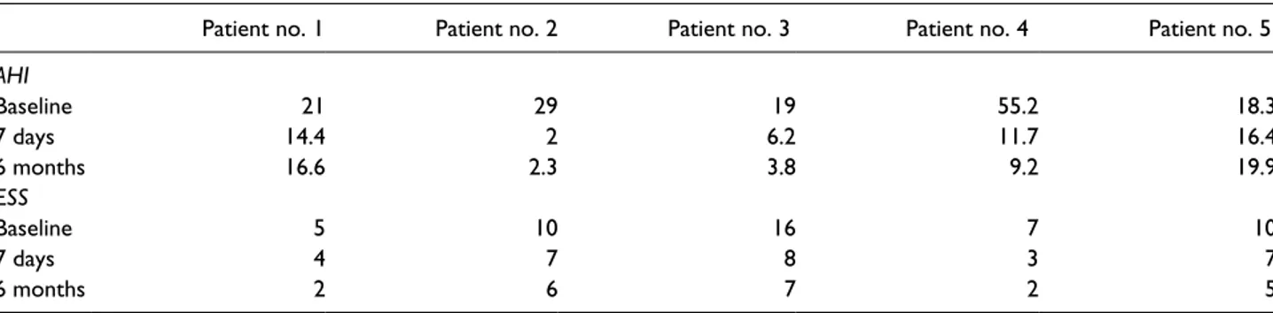
From the evaluation of the SF36 questionnaire, five cooperative patients (Table 1) showed good adapta-bility to the treatment and good compliance; one
patient, corresponding to No. 1, showed good com-pliance after 6 months of treatment, reporting how-ever an inconvenience of use, excess of saliva, pain at the temporomandibular joint, and embarrassment. AHI
Patients 1–4 showed reduction in the AHI in the polysomnographic exam. In patient 5 support with CPAP was recommend.
Daytime sleepiness
The assessment of the ESS, in both the short and long term, shows a reduction of daytime sleepiness in patients 1–4. The assessment of the Epworth scale of the data related to daytime sleepiness is the same.
Not compliant patients
Three of the eight selected patients were not com-pliant to treatment, however, although not present in controls after 6 months, it was possible to study their short-term assessment (at 7 days), that showed the results reported in Table 2 on AHI and ESS.
Moreover, as reported in Table 3, the values of cephalometric analysis has revealed substantial changes in the retropharyngeal air space. The Ba-S-PNS angle, the cephalometric parameter that can evaluate the horizontal position of the hard and soft palate, which is often the main cause in airway obstruction, increased in all patients by an average of 11.1% (± 10.32). The increase of this value, probably, can be attributed to the decreased vol-ume and tropism of soft palate, which follows the reduction of episodes of apnea and of snoring.
Airway percentage also grew by an average of 10.3% (± 6.43), indicating an increase in the per-centage of the nasopharynx free from adenoids.
Table 1. Assessment in three different time of AHI and Epworth Sleepiness Scale in compliance patients.
Patient no. 1 Patient no. 2 Patient no. 3 Patient no. 4 Patient no. 5
AHI Baseline 21 29 19 55.2 18.3 7 days 14.4 2 6.2 11.7 16.4 6 months 16.6 2.3 3.8 9.2 19.9 ESS Baseline 5 10 16 7 10 7 days 4 7 8 3 7 6 months 2 6 7 2 5
This result is supported and corroborated by changes in a positive sense, D- AD1- PNS, D- AD2- PNS, and D- PTV- AD, which were 15.4% (± 7.44), 9.5% (± 3.31), and 25.4% (± 2.19), respectively.
Discussion
OA therapy is indicated primarily for patients with mild to moderate OSAS, and recent data demonstrate efficacy in some patients with more severe disease.14 The role of OA therapy will expand as newer technologies improve the ability to predict success with treatment prior to initiat-ing therapy as well as monitor compliance with ongoing treatment.
We currently assume that the working mecha-nism of an OA is based on advancement of the mandible and its attached soft tissue structures and musculature, especially the genioglossus muscle, resulting in an increased tone with increased anter-oposterior and lateral dimensions of the upper airway.21,23–26
Bearing the abovementioned considerations in mind, this deterioration in treatment success is possibly due to loosening and adaptation of soft tissue structures and musculature of the upper air-way as a result of long-term overnight mandibular advancement.27
It could be hypothesized that patients with more severe OSAS need to protrude the mandible more extensively to gain the desired effect, in addition to a possible overstretching that nega-tively affects the morphology of the upper airway soft tissue structures and tonus of the muscula-ture. Furthermore, it has been described that the muscle tone of the genioglossus is negatively cor-related with age.21
In our study we used the Thornton Adjustable Positioner type-1, which is a two-piece adjustable appliance. As supported from our data, at short and long term, we can say that the TAP® has a dual effect, because it prevents the pharyngeal lumen reduction, due to the fall back of the tongue owing
Table 2. Assessment in two different time of AHI and
Epworth Sleepiness Scale in not compliant patients.
Patient no. 6 Patient no. 7 Patient no. 8
AHI Baseline 12.1 39 31 7 days 3.9 25.7 16 ESS Baseline 7 12 10 7 days 5 9 8
Table 3. The effects of the OA tested in our study, were evaluated using cephalometric analysis conducted on radiographs,
performed before treatment and after 6 months.
Patient no. 1 2 3 4 5 Mean SD
Ba-N-PNS 1 52 57 48 53 48 51.6 3.38 Ba-N-PNS 2 57 60 63 54 54 57.2 3.66 T2–T1 −3 3 −15 −1 −6 5.6 4.96 Variability (%) −5.4 5.2 −23.4 −1.8 −10.9 11.1 10.32 Airway 1% 54 56 56 49 62 55.4 4.18 Airway 2% 60 57 59 59 70 61.0 4.60 T2–T1 6 1 3 10 8 5.6 3.26 Variability (%) 11.1 1.8 5.4 20.4 12.9 10.3 6.43 D-AD1-PNS 1 25 25 27 23 19 23.8 2.71 D-AD1-PNS 2 27 28 33 25 24 27.4 3.14 T2–T1 2 3 6 2 5 3.6 1.62 Variability (%) 8.0 12.0 22.2 8.7 26.3 15.4 7.44 D-AD2-PNS 1 27 23 28 24 22 24.8 2.32 D-AD2-PNS 2 30 25 32 25 24 27.2 3.19 T2–T1 3 2 4 1 2 2.4 1.02 Variability (%) 11.1 8.7 14.3 4.2 9.1 9.5 3.31 D-PTV-AD 1 19 14 18 15 16 16.4 1.85 D-PTV-AD 2 24 17 23 19 20 20.6 2.58 T2–T1 5 3 5 4 4 4.2 0.75 Variability (%) 26.3 21.4 27.8 26.7 25.0 25.4 2.19
to the gravity and muscular hypotonia and, thanks to the advancing of genioglossus muscle, causes a stiffening in the longitudinal pharyngeal wall, in order to avoid the collapse in lateral direction, reducing AHI index.
In one study the objective and subjective efficacy of the Thornton Adjustable Positioner type-1 was compared with a modified Herbst appliance (IST®) in a 2-year follow-up.28 Although the Thornton Adjustable Positioner type-1 was more effective in lowering the AHI, both appliances seemed to be effective therapeutic devices for OSAS which is con-sistent with our findings. Therefore it should be interpreted, according to the data in the literature,19,21 as a favorable factor, since it can represent the regres-sion of the OSAS disease to a pre-clinical stage.
Reported side effects were considered minor and temporary by the participants, and did not pre-vent use of the appliance. We relied upon feedback from our patients for the data on adequate appli-ance usage.
As no gold standard currently exists for objec-tively recording compliance, this method repre-sented the most practical means of evaluating the device. Patients received a thorough explanation of the purpose of the study, and were instructed to sub-jectively record OA usage as accurately as possible. Other limitations of this study were the small sample size and short evaluation period.
A quality assurance/follow-up of patients who have been given OA for moderate and severe sleep apnea since 2013 is ongoing.
The results from this preliminary report suggest the use of OA, such as TAP®, improved sleep-disor-dered breathing and symptoms and are valid thera-peutic options for selected patients with OSAS provided that the patient tolerates OA wear on a regular basis. Regular controls of the treatment effi-cacy and patient tolerability are recommended. As long as the mandible is advanced continuously dur-ing sleep with an appliance and durdur-ing the treat-ment period, appliance characteristics that enhance comfort during wear and the greater durability of its material should be prioritized.
Currently, many types of OA are commercially available for treating patients with OSAS. Therefore careful comparison of results from the current study with studies in which other types of oral appliances are used is important, as there can be differences in efficacy and patients’ preferences.
In conclusion, future studies with larger sample sizes and longer evaluation periods will be necessary
to further validate the use of the device for objective monitoring of OA.
Acknowledgements
All authors thank Mrs. Loredana Meccariello for her pro-fessional support in bibliographic research.
Declaration of conflicting interests
The author(s) declared no potential conflicts of interest with respect to the research, authorship, and/or publication of this article.
Funding
This research received no specific grant from any funding agency in the public, commercial, or not-for-profit sectors.
References
1. Paiva T and Attarian H (2014) Obstructive sleep apnea and other sleep-related syndromes. Handbook
of Clinical Neurology 119: 251–271.
2. Urquhart D (2013) Investigation and management of childhood sleep apnea. Hippokratia Medical Journal 17: 196–202.
3. American Academy of Sleep Medicine (2012) AASM
Manual for the Scoring of Sleep and Associated Events: Rules, Terminology and Technical Specifications (Version 2.0). Darien, IL: AASM.
4. Marcus CL, Brooks LJ, Ward SD, et al. (2012) Diagnosis and management of childhood obstructive sleep apnea syndrome. Pediatrics 130: e714–e755. 5. Araghi MH, Chen YF, Jagielski A, et al. (2013)
Effectiveness of lifestyle interventions on obstructive sleep apnea (OSA): Systematic review and meta-anal-ysis. Sleep 36: 1553–1562.
6. Myers KA, Mrkobrada M and Simel DL (2013) Does this patient have obstructive sleep apnea? The Rational Clinical Examination systematic review.
Journal of the American Medical Association 310:
731–741.
7. Keymel S, Kelm M and Randerath WJ (2013) Non-CPAP therapies in obstructive sleep apnea: An over-view. Pneumologie 67: 50–57.
8. Pasquali R, Colella P, Cirignotta F, et al. (1990) Treatment of obese patients with obstructive sleep apnea syndrome (OSAS): Effect of weight loss and interference of otorhinolaryngoiatric pathology.
International Journal of Obesity 14: 207–217.
9. Aurora RN, Casey KR, Kristo D, et al. (2010) American Academy of Sleep Medicine. Practice parameters for the surgical modifications of the upper airway for obstructive sleep apnea in adults. Sleep 33: 1408–1413.
10. Ronchi P, Cinquini V, Ambrosoli A, et al. (2013) Maxillomandibular advancement in obstructive sleep
apnea syndrome patients: A restrospective study on the sagittal cephalometric variables. Journal of Oral
& Maxillofacial Research 4: e5.
11. Sullivan CE, Issa FG, Berthon- Jones M, et al. (1981) Reversal of obstructive sleep apnea by continuous positive airway pressure applied through the nares.
Lancet 1: 862–865.
12. Giles TL, Lasserson TJ, Smith BH, et al. (2006) Continuous positive airways pressure for obstruc-tive sleep apnea in adults. Cochrane Database of
Systematic Reviews 3: CD001106.
13. Sutherland K, Vanderveken OM, Tsuda H, et al. (2014) Oral appliance treatment for obstructive sleep apnea: An update. Journal of Clinical Sleep Medicine 10: 215–227.
14. Ferguson KA, Cartwright R, Rogers R, et al. (2006) Oral appliances for snoring and obstructive sleep apnea: A review. Sleep 29: 244–262.
15. Ngiam J, Balasubramaniam R, Darendeliler MA, et al. (2013) Clinical guidelines for oral appliance ther-apy in the treatment of snoring and obstructive sleep apnea. Australian Dental Journal 58: 408–419. 16. Kushida CA, Morgenthaler TI, Littner MR, et al.
American Academy of Sleep (2006) Practice parame-ters for the treatment of snoring and Obstructive Sleep Apnea with oral appliances: An update for 2005. Sleep 29: 240–243.
17. American Academy of Sleep Medicine. Available at: http://www.aasmnet.org/ (accessed 24 May 2015). 18. Smith YK and Verrett RG (2014) Evaluation of a
novel device for measuring patient compliance with oral appliances in the treatment of obstructive sleep apnea. Journal of Prosthodontics 23: 31–38.
19. Freedman N (2014) Improvements in current treat-ments and emerging therapies for adult obstructive sleep apnea. F1000 Prime Reports 6: 36.
20. Phillips CL, Grunstein RR, Darendeliler MA, et al. (2013) Health outcomes of continuous positive
air-way pressure versus oral appliance treatment for obstructive sleep apnea: A randomized controlled trial. American Journal of Respiratory and Critical
Care Medicine 187: 879–887.
21. Doff MH, Hoekema A, Wijkstra PJ, et al. (2013) Oral appliance versus continuous positive airway pressure in obstructive sleep apnea syndrome: A 2-year follow-up. Sleep 36: 1289–1296.
22. Wang Y, Teschler T, Weinreich G, et al. (2003) Validation of microMESAM as screening device for sleep disordered breathing. Pneumologie 57: 734– 740.
23. Schulhof RJ (1998) Consideración de la vía aérea en Ortodoncia. In: Ricketts RM, Berch RW, Gugino CF, et al (eds) Técnica bioprogressiva de Ricketts. Buenos Aires: Panamericana, pp. 360–364.
24. Handelman CS and Osborne G (1976) Growth of the nasopharynx and adenoid development from one to eighteen years. Angle Orthodontist 46: 243–259. 25. Linder-Aronson S and Henrikson CO (1973)
Radiocephalometric analysis of anteroposterior naso-pharyngeal dimensions in 6 to 12 year-old mouth breathers compared with nose breathers. Journal of
Otorhinolaryngology and Related Specialties 35: 19–
29.
26. Gale DJ, Sawyer RH, Woodcock A, et al. (2000) Do oral appliances enlarge the airway in patients with obstructive sleep apnea? A prospective computerized tomographic study. European Journal of Orthodontics 22: 159–168.
27. Ryan CF, Love LL, Peat D, et al. (1999) Mandibular advancement oral appliance therapy for obstructive sleep apnea: Effect on awake calibre of the velophar-ynx. Thorax 54: 972–977.
28. Ghazal A, Sorichter S, Jonas I, et al. (2009) A ran-domized prospective long-term study of two oral appliances for sleep apnea treatment. Journal of Sleep