Decrease in N-Acetylaspartate
Following Concussion May Be Coupled
to Decrease in Creatine
Roberto Vagnozzi, MD; Stefano Signoretti, MD, PhD; Roberto Floris, MD;
Simone Marziali, MD; Massimo Manara, MD; Angela M. Amorini, PhD;
Antonio Belli, MD, PhD; Valentina Di Pietro, PhD; Serafina D’Urso, PhD;
Francesco S. Pastore, MD; Giuseppe Lazzarino, PhD; Barbara Tavazzi, PhD
Objectives: To assess the time course changes in N-acetylaspartate (NAA) and creatine (Cr) levels in the brain
of athletes who suffered a sport-related concussion. Participants: Eleven nonconsecutive athletes with concussive head injury and 11 sex- and age-matched control volunteers Main outcome measures: At 3, 15, 30, and 45 days postinjury, athletes were examined by proton magnetic resonance spectroscopy for the determination of NAA, Cr, and choline (Cho) levels. Proton magnetic resonance spectroscopic data recorded for the control group were used for comparison. Results: Compared with controls (2.18± 0.19), athletes showed an increase in the NAA/Cr ratio at 3 (2.71± 0.16; P < .01) and 15 (2.54 ± 0.21; P < .01) days postconcussion, followed by a decrease and subsequent normalization at 30 (1.95± 0.16, P < .05) and 45 (2.17 ± 0.20; P < .05) days postconcussion. The NAA/Cho ratio decreased at 3, 15, and 30 days postinjury (P< .01 compared with controls), with no differences observed in controls at 45 days postconcussion. Compared with controls, significant increase in the Cho/Cr ratio after 3 (+33%,
P< .01) and 15 (+31.5%, P < .01) days postinjury was observed whereas no differences were recorded at 30 and 45
days postinjury. Conclusions: This cohort of athletes indicates that concussion may cause concomitant decrease in cerebral NAA and Cr levels. This provokes longer time for normalization of metabolism, as well as longer time for resolution of concussion-associated clinical symptoms. Key words: brain vulnerability, concussion, creatine,1H magnetic resonance spectroscopy, mild traumatic brain injury, N-acetylaspartate, sports-related concussion
Author Affiliations: Division of Neurotraumatology and
Neuroradiology, Department of Biomedicine and Prevention, University of Rome “Tor Vergata,” Rome, Italy (Drs Vagnozzi, Floris, Marziali, D’Urso, and Pastore); Division of Neurosurgery, Department of Neurosciences—Head and Neck Surgery, “San Camillo” Hospital, Rome, Italy (Dr Signoretti); Association of Sports Physicians Parma, F.M.S.I., Parma, Italy (Dr Manara); Institute of Biochemistry and Clinical Biochemistry, Catholic University of Rome, Rome, Italy (Drs Amorini and Tavazzi); Division of Biochemistry and Molecular Biology, Department of Biology, Geology and Environmental Sciences, University of Catania, Catania, Italy (Dr Lazzarino); and Neuropharmacology and Neurobiology Section, School of Clinical and Experimental Medicine, College of Medical and Dental Sciences and NIHR Surgical Reconstruction and Microbiology Research Centre, University of Birmingham and University Hospitals Birmingham, Birmingham, United Kingdom (Drs Belli and Di Pietro).
This work has been supported in part by research funds of University of Rome “Tor Vergata,” Catholic University of Rome, and University of Catania. The authors thank Mr Salvatore Meo for his technical assistance in preparing the artwork for the manuscript.
The authors declare no conflicts of interest.
Correspondence: Giuseppe Lazzarino, PhD, Division of Biochemistry and
Molecular Biology, Department of Biology, Geology and Environmental Sci-ences, Viale A. Doria 6, 95125 Catania, Italy ([email protected]). DOI: 10.1097/HTR.0b013e3182795045
C
ONCUSSION is defined as a biomechanically induced brain injury characterized by the ab-sence of gross anatomic damages. Supported by the absence of structural lesions on traditional neuroimag-ing, a general and broadly accepted view is that mild traumatic brain injury (mTBI) is indeed a very fre-quent entity but is not a very serious injury, lead-ing only to transient disturbances, and that no inter-vention other than observation is typically required. However, mTBI triggers molecular changes in neuronal cells involving a complex cascade of neurometabolic alterations1–3that reversibly modify the concentrationsof several low-molecular-weight compounds actively in-volved in functions crucial for cell homeostasis and survival.3,4 Such a cascade of molecular events is
con-sidered as the determinant of the so-called state of metabolic brain vulnerability,5,6 which transiently
ex-poses cerebral tissue to the cumulative effect of a sec-ond mTBI occurring during this particular period.7,8
High-energy phosphates,9,10coenzyme A metabolites,11
ATP catabolites,11 and neurotransmitters,12,13are some
of the substances whose concentrations are temporar-ily affected as a result of a traumatic insult. Of
particular relevance is the finding that N-acetylaspartate (NAA), an abundant brain-specific compound involved in several relevant biological functions including water homeostasis14 and lipid myelin biosynthesis,15 clearly
mirrors TBI-induced changes in ATP, showing the same pattern of decrease and recovery following mTBI and no recovery in severe TBI or repeat mTBIs.8,10,11 The
strict correlation between NAA and ATP ensures that this compound may be used as a valid surrogate marker to monitor cerebral energy metabolism.8,10,11Recently,
particular attention has been given to compounds, non-invasively detectable in vivo by proton magnetic reso-nance (MR) spectroscopy (1H MRS), that may be used
as markers of brain metabolism following mTBI. 1H
MRS allows the routine measurement of NAA, crea-tine (Cr), and choline (Cho) in a single set of spectral acquisition,16although the low magnetic field currently
used in the clinical setting (1.5 or 3 T) does not al-low to resolve the N-acetylaspartatylglutamate signal in the NAA peak17 or the creatine phosphate (CrP) signal
in the Cr peak,17 or to resolve about 10 different
com-pounds containing the choline moiety in their molecule in the Cho peak.17 NAA, Cr and Cho can be
mea-sured by1H MRS either by determining their absolute
values17or by using the metabolite ratios.18When
refer-ring to NAA and Cr, this is of limited relevance since, in the brain tissue, NAA and Cr are about 10 times more concentrated than their respective related compounds
N-acetylaspartatylglutamate and CrP. Therefore, areas of
these 2 peaks can generally be considered as valid mea-surements of NAA and Cr levels. Differently, since the Cho peak is mainly composed of phosphocholine, glyc-erophosphocholine, and phosphatidylcholine, quantifi-cation of this peak area is not in direct correlation with one compound only. However, since these compounds are related to phospholipid metabolism, the Cho peak area is generally considered as a good indicator of the cell membrane turnover,17,19 that of NAA is used as a
marker of energy state and neuronal integrity,8,10,11,17
and that of Cr is thought to be a marker of cellular energy.17
Since the period of brain vulnerability following an mTBI is mainly characterized by evident biochemical, metabolic, and molecular changes,3,5,6,20it is
question-able whether the tests currently adopted in the clinical practice to monitor mTBI patients (neuropsychological tests, balance tests, etc), because of their inability to eval-uate cerebral biochemical changes, are of utility to de-termine the end of brain vulnerability. Although some of them (neuropsychological tests) are largely applied to assess the return of athletes with concussive head in-jury to play,21–23 it has never been demonstrated that
this type of normalization overlaps with the recovery of brain metabolism.
In a constant effort to find valid objective biological parameters for the monitoring of brain metabolic recov-ery after head injury, several researchers, using1H MRS
studies, turned their attention to the evaluation of the changes in NAA in mTBI patients.24,25In two
longitudi-nal studies in which patients were scanned 3 or 4 times to perform the time course of postinjury recovery of brain metabolism, we showed that concussion, an mTBI caused by any type of acceleration-deceleration of the brain, frequently encountered in contact sports, caused a reversible decrease in NAA content of the frontal lobe white matter.26,27In these studies, an inclusion criterion
was the constancy in the Cho/Cr ratio that allowed the semiquantitative determination of NAA relative to both Cr and Cho.26,27Significantly, we observed that the
re-covery of brain metabolism occurred weeks after the resolution of the self-reported postconcussive clinical symptoms.26,27Recently, two1H MRS studies reported
that Cr but not NAA levels were affected by mTBI, with head injury producing a significant increase in Cr levels in different brain areas.28,29
As indicated earlier, in our previous studies, to in-vestigate possible changes in NAA evaluated relatively to Cr and/or Cho, we excluded patients in whom the Cho/Cr ratio did not remain unaltered. In this article, we describe 11 cases of athletes with concussive head in-jury in whom alterations of brain metabolism involved not only NAA but Cr as well.
METHODS Patient selection
After obtaining informed consent according to insti-tutional procedures, 11 nonconsecutive amateur athletes of different sport disciplines who suffered a sport-related concussive head injury (defined as a traumatically in-duced alteration in mental status, not necessarily with loss of consciousness), between June 2008 and Octo-ber 2011, were considered for this study. Patient selec-tion was characterized by the following inclusion crite-ria: (i) Glasgow Coma Scale score 14 or more; (ii) no anatomic lesion at conventional imaging (computed to-mography or magnetic resonance imaging [MRI]); (iii) normal neurological objective examination at the time of enrolment; (iv) the value of the Cho/Cr ratio dif-ferent from that of controls; and (v) the requirement to refrain from further athletic activity up to normalization of brain metabolism.
Athletes (age ranging between 16 and 35 years) under-went an MR scan and a proton spectroscopic examina-tion at 3 days postinjury, followed by 3 addiexamina-tional1H
MRS scans at 15, 30, and 45 days postinjury. Results col-lected from patients with concussive head injury were compared with those obtained from 11 healthy, sex- and
age-matched control volunteers, previously screened to exclude prior head injuries. Any intracranial lesion ob-served on the first MR scan automatically excluded the candidate from the study. Before each MR examination, symptoms were assessed by using the SCAT2, a tool that represents a standardized method of evaluating injured athletes for concussion and can be used in athletes ag-ing 10 years and older. Durag-ing the medical examination, patients were asked for symptoms of mTBI including physical, cognitive, emotional, and sleep disturbances. Resolution was determined by the concordance of the results of self-assessment and SCAT2.
MRI and1H MRS acquisition technique
Semiquantitative analyses of Cr, NAA, and Cho were performed after obtaining proton spectra by a 3-T sys-tem (Philips Intera Achieva, Philips Healthcare, Best, The Netherlands). For conventional MRI studies, T1-and T2-weighted TSE (turbo spin-echo) images were ac-quired in axial, coronal, and sagittal planes, and to rule out even the smallest amount of intracerebral blood, fast field echo T2∗sequences were used. A multichannel coil (8 channels) SENSE-Head-8, with 4-mm slice thickness, 1-mm gap, and an FOV (field of view) of 230 mm, was used for all MRI sequences. Following localized shim-ming and water suppression, the spectroscopic examina-tion was carried out using a PRESS (point resolved spec-troscopy) pulse sequence, with the following settings: TE (echo time)= 144 ms; TR (repetition time) = 2000 ms; spectral bandwidth= 2000; and acquisition cycles = 128. The optimal positioning of the voxel was deter-mined using the MR images acquired on axial, coronal, and sagittal planes to facilitate its 3-dimensional place-ment, adjacent to the cortical-subcortical junction, just anterior to the frontal horn of the lateral ventricle, at the same height of a virtual plane positioned just above the corpus callosum, to include only the white matter of the frontal lobes bilaterally. The choice of this location was made to obtain as homogeneous data as possible. To this end, a spectrum from a single voxel customized to sam-ple a volume of interest of 3.375 cm3 (1.5× 1.5 × 1.5
cm) was finally obtained (acquisition time about 5 min-utes for each voxel). In comparison with the multivoxel technique, the single-voxel technique was preferred to obtain a better resolution of the spectral peaks. In addi-tion, in our previous article, data obtained from different neuroradiological centers and using the single- or multi-voxel technique gave overlapping values.27In follow-up
studies, the exact repositioning of the voxel on the same acquisition plane obtained in the previous MRI study was achieved by using dedicated software (SameScan; Philips Medical Systems, Philips Healthcare, Best, The Netherlands). Postprocessing of spectral data allowed us to calculate the area under the peaks of NAA, Cho, and
Cr, using common criteria for peak integration. In the case of a single, well-defined peak (typically the NAA peak), a valley-to-valley integration was performed to obtain the area under the peaks. In the case of not fully resolved peaks (frequently the Cho and Cr peaks), a hori-zontal baseline between the start of the first peak and the end of the second peak was selected; the grouped peaks were then split by a vertical line, drawn from the median point of the common valley between peaks to the hori-zontal baseline and the area under the peaks calculated. These values were used to determine the metabolite ra-tios NAA/Cho, NAA/Cr, and Cho/Cr.
Statistical analysis
All data analyses and calculations of sample size were performed using the Statistical Package for the Social Sciences Windows version 13.0 (SPSS, Chicago, Illi-nois). Descriptive statistics for quantitative continuous variables were presented as mean± standard deviation. Assumptions of normality were demonstrated using the Kolmogorov-Smirnov test. The homogeneity of the ance was evaluated with the Levene test. Analysis of vari-ance for repeated measures, corrected by Bonferroni, was used to evaluate significant differences among groups. Differences were considered to be statistically significant when P< .05.
RESULTS
Clinical features of athletes with concussive head injury
A total of 55 single-voxel1H MRS studies (11 healthy
control subjects, 11 patients studied at 4 time points) were conducted, and a total of 110 brain spectra were successfully acquired. The duration of each study av-eraged 16± 1 minute, with no complications reported. All patients were admitted as outpatients and discharged within 1 hour from the beginning of the MRI and1H
MRS acquisitions. The clinical features of the cohort of athletes with concussive head injury are reported in Table 1. The mean age of athletes with concussive head injury was 24.6± 6.4 years (8 men, 3 women) and that of controls was 25.9 ± 5.7 years (8 men, 3 women). A complete resolution of symptoms, obtained when the concordance of the results of self-assessment and SCAT2 occurred, was observed at 15.2± 2.6 days postinjury.
1H MRS analysis of brain metabolism in patients with concussive head injury
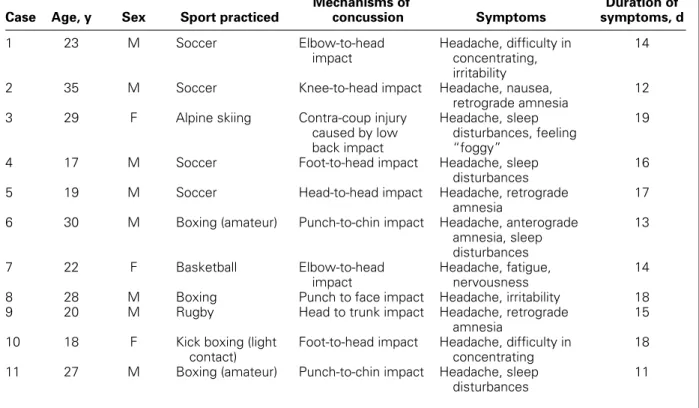
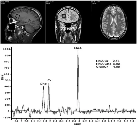
Figure 1 shows the optimal positioning of the voxel and a spectrum recorded in a healthy control subject. The time course of the NAA/Cr ratio determined in 11 patients with head injury at different time points following concussion is illustrated in Figure 2. Different
TABLE 1
Demographic data, sport activity, mechanisms of concussion, and clinical
symptoms of 11 nonprofessional athletes with concussive head injury
Case Age, y Sex Sport practiced
Mechanisms of concussion Symptoms Duration of symptoms, d 1 23 M Soccer Elbow-to-head impact Headache, difficulty in concentrating, irritability 14
2 35 M Soccer Knee-to-head impact Headache, nausea, retrograde amnesia
12
3 29 F Alpine skiing Contra-coup injury caused by low back impact Headache, sleep disturbances, feeling “foggy” 19
4 17 M Soccer Foot-to-head impact Headache, sleep disturbances
16
5 19 M Soccer Head-to-head impact Headache, retrograde amnesia
17
6 30 M Boxing (amateur) Punch-to-chin impact Headache, anterograde amnesia, sleep disturbances 13 7 22 F Basketball Elbow-to-head impact Headache, fatigue, nervousness 14
8 28 M Boxing Punch to face impact Headache, irritability 18 9 20 M Rugby Head to trunk impact Headache, retrograde
amnesia
15
10 18 F Kick boxing (light contact)
Foot-to-head impact Headache, difficulty in concentrating
18
11 27 M Boxing (amateur) Punch-to-chin impact Headache, sleep disturbances
11
from what was previously obtained in patients sustaining similar types of concussion and showing similar clinical conditions,26,27at the time of the first1H MRS scan (3
days postimpact), we recorded a significant increase in the NAA/Cr ratio (2.71± 0.16) with respect to the value of controls (2.18± 0.19; P < .01). This apparent NAA increase was also evidenced 15 days after injury, when the 11 athletes with concussive head injury showed the NAA/Cr ratio of 2.54± 0.21 (+16.5%; P < .01 with respect to the value of controls). Unexpectedly, at the time of the third1H MRS scan (30 days postconcussion),
significant decrease in the NAA/Cr ratio was observed (1.95± 0.16, P < .05 with respect to controls). A value of 2.17± 0.2, not significantly different from that recorded in controls, was measured 45 days after concussion.
Figure 3 illustrates the changes in the NAA/Cho ra-tio during 45 days following concussion. In accordance with our previous observations,26,27 but in contrast to
the apparent increase in NAA suggested by the increase in the NAA/Cr ratio, the NAA/Cho ratio underwent a transient decrease, particularly evident at 3 and 15 days postinjury when mean values of 1. 61± 0.20 and 1.55 ± 0.18 were recorded (mean value of the NAA/Cho ra-tio in controls = 1.94 ± 0.17; P < .01). At the time of the third1H MRS scan, the NAA/Cho ratio slightly
increased (1.70 ± 0.2), although this was still 12.4%
lower than that in controls (P< .01). Recovery of the NAA/Cho ratio was completed 45 days postconcussion when a value of 1.91± 0.19 was recorded (not signifi-cantly different from controls).
Data reported in Figure 4 show the changes in the Cho/Cr ratio observed in 11 athletes with concussive head injury during 45 days of recovery. Different from what was observed in our previous studies, in which the Cho/Cr ratio remained unaltered during the whole ob-servational period after concussion,26,27 in the present
cohort of athletes, we observed a significant increase in the Cho/Cr ratio after 3 (+33%; P < .01) and 15 (+31.5%; P < .01) days postinjury. At the third and fourth1H MRS scans (30 and 45 days postimpact,
re-spectively), no difference in the Cho/Cr ratio was ob-served compared with controls.
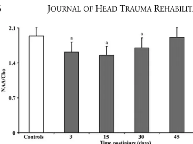
To illustrate more clearly the postinjury phenomenon involving a decrease in NAA and Cr levels and con-stancy of Cho in this cohort of athletes, Figure 5 shows
1H MR spectra both of a representative control and of a
concussed athlete analyzed at 3, 30, and 45 days postin-jury (for graphical reasons, the 15-day spectrum was not reported). A visible decrease in the NAA and Cr peaks at 3 days, with subsequent increase and normalization at 30 and 45 days postconcussion, is evident. Differently, the Cho peak did not show appreciable modifications
Figure 1. 1H magnetic resonance scout image of a healthy volunteer showing the optimal positioning of the single voxel located
adjacent to the cortical-subcortical junction, just anteriorly to the frontal horn of the lateral ventricle, at the same height of a virtual plane positioned just above the corpus callosum, to include only the white matter of the frontal lobes, bilaterally. The proton spectrum shows the peaks corresponding to the metabolites of interest N-acetylaspartate (NAA), creatine (Cr), and choline (Cho). The calculated NAA/Cr and Cho/Cr ratios, relative to this subject, are also indicated.
at any time point, being very similar to that recorded in the control healthy subject (Figure 5).
DISCUSSION
We previously showed that athletes with concussive head injury experience a period of metabolic brain de-rangement, as evidenced by the transient decrease in the NAA/Cr and NAA/Cho ratios.26,27 In both
stud-ies, one of the criteria for inclusion was the constancy of the Cho/Cr ratio since, for the calculation of the relative NAA abundance, it is always necessary to re-fer to an invariant metabolite detectable in the1H MR
spectrum. In these studies, the trends in the NAA/Cr and NAA/Cho ratios after a concussive episode entirely overlapped, thereby supporting the conclusion that Cr and Cho concentrations did not vary (as indicated by the constancy in the Cho/Cr ratio) and that the changes in the NAA/Cr and NAA/Cho ratios were due to a real net decrease in NAA cerebral concentration,26,27that is,
the NAA decrease was merely apparent.
Figure 2. Bar graph showing the metabolite ratios of
N-acetylaspartate/creatine–containing compounds (NAA/Cr)
in controls and patients with concussive head injury. Each bar is the mean value determined in 11 healthy controls and 11 athletes with concussive head injury. Standard deviations are represented by vertical bars. At 3 and 15 days, the NAA/Cr ratio increased by 24.3% and 16.5%, respectively, whereas at 30 days postinjury, it decreased by 10.6%. Normalization was observed 45 days after concussion.aP< .05 with respect to
Figure 3. Bar graph showing the metabolite ratios of
N-acetylaspartate/choline–containing compounds (NAA/Cho) in controls and patients with concussive head injury. Each bar is the mean value determined in 11 healthy controls and 11 athletes with concussive head injury. Standard deviations are represented by vertical bars. At 3, 15, and 30 days postinjury the NAA/Cho ratio decreased by 17.0%, 20.1%, and 12.4%, respectively. Normalization was observed 45 days after concussion.aP< .05 with respect to
controls.
In this study, we found a profound discrepancy be-tween the trends of the NAA/Cr (transient increase and then decrease before normalization) and NAA/Cho (transient decrease) ratios. The incongruity in the trends of these 2 metabolite ratios was accompanied by a tem-porary increase in the Cho/Cr ratio, significantly higher than the value in age-matched healthy controls at 3 and 15 days postinjury, and then normalized 30 days after concussion (Figure 3). According to the aforementioned observations, it can be affirmed that in these athletes, the following observations were made: (i) the fluctuating NAA/Cr ratio (Figure 1) was due to a decrease in NAA
Figure 4. Bar graph showing the metabolite ratios of
choline/creatine-containing compounds (Cho/Cr) in controls and patients with concussive head injury. Each bar is the mean value determined in 11 healthy controls and 11 ath-letes with concussive head injury. Standard deviations are rep-resented by vertical bars. At 3 and 15 days, the Cho/Cr ratio increased by 33.0% and 31.5%, respectively, whereas at 30 and 45 days postinjury, no differences with respect to controls were recorded.aP< .05 with respect to controls.
and a concomitant, more pronounced decrease in Cr levels; (ii) the decrease in the NAA/Cho ratio (Figure 2) was due to a significant decrease in NAA and not due to an increase in Cho levels; (iii) the increase in the Cho/Cr ratio was due to a decrease in Cr levels (Figure 3); (iv) the recovery of Cr was faster than that of NAA, as evidenced by normalization in the Cho/Cr ratio recorded 30 days postconcussion whereas the NAA/Cr ratio was still de-creased (Figure 1 and 3). To corroborate these conclu-sions that clearly explained the apparently contradictory trends of the metabolite ratios, in each athlete, the area under the peak of the spectral signal of Cho did not change at any of the different1H MRS acquisitions and
was similar to that recorded in controls (data not shown). Conversely, the area under the peak of NAA was lower at 3, 15, and 30 days postinjury and that of Cr at 3 and 15 days after impact (data not shown). Even if referred to one representative athlete only, this phenomenon is clearly visible in the spectra reported in Figure 5, which show a decrease and subsequent normalization in the NAA and Cr peaks and no change in the peak of Cho.
One question that can be raised from these results is why these athletes showed a transient decrease in both Cr and NAA levels whereas the other 40 we examined in 2 previous studies26,27 showed decrease in NAA levels
only, with no change in Cr levels. It should be noted that athletes enrolled in the present study differ not only in decrease in Cr levels but also in the longer time necessary for NAA normalization (45 days vs 30 days ob-served previously), as well as for the longer duration of the postconcussive clinical symptoms (15.2± 2.6 days vs 3-7 or 3-15 days recorded previously). Therefore, it appears that this group of athletes suffered from a more severe concussive event, causing longer time in both clinical and metabolic recovery and a more pronounced imbalance of metabolism (change in both NAA and Cr concentrations). On the other hand, using the weight-drop model of closed-head diffuse mTBI,30 we recently
demonstrated that, in addition to NAA and ATP, Cr levels underwent a reversible 44.5% decline, which was accompanied by a less than 15% decline in CrP levels at the same time point (24 hours post-mTBI). Altogether, the sum of Cr + CrP accounted for a 42% decrease at 24 hours postinjury.31 This transient depletion in
the cerebral Cr compound pool was restored after 120 hours of mTBI.31 In the same study, we also showed
that mTBI did not affect the concentration of phos-phatidylcholine, that is, one of the main compounds responsible for the intensity signal of the Cho peak in the1H MR spectrum. These experimental results have
recently been confirmed32and strongly corroborate the
finding reported in the present study, indicating tempo-rary decrease in NAA and Cr levels and no change in Cho levels in our cohort of athletes with postconcussive head injury.
Figure 5. Representative1H magnetic resonance spectra recorded in a healthy control subject and in a concussed athlete at 3,
30, and 45 days postinjury (for graphical reasons, the 15-day spectrum was not reported). Decrease (3 days), recovery (30 days), and subsequent normalization (45 days) in the NAA and Cr peaks and no change in the peak of Cho are clearly visible in the spectra of the postconcussed athlete.
Other authors indicated that patients suffering from mTBI underwent transient increase in Cr levels in the white matter and no change in NAA levels,28,29 in
contrast to the results of the present and previous studies16,26,27,33,34 and also to a conspicuous number
of preclinical studies demonstrating temporary decrease in NAA8,10,11,21 and Cr pool following mTBI.31,32 To
explain their results, these authors concluded that an increase in total Cr (Cr and CrP) levels in the white mat-ter may support a larger pool of high-energy phosphates (CrP and ATP), helping to restore mTBI-induced alter-ation of cell homeostasis through upregulalter-ation of mem-brane pumps and other processes of cellular repair.28,29
Notwithstanding, these conclusions are in contrast with the notion of brain vulnerability,1–3,5–8,10,11,35
which is characterized by a period of metabolic de-pression (hypometabolism), mainly due to mitochon-drial malfunctioning,36–38 particularly affecting NAA
homeostasis39 and ATP supply,8–11,31,39 and also
caus-ing significant depletion in the total Cr pool.31,32
Fur-thermore, given the relative CrP/ATP ratio in the brain tissue, ranging from about 0.8 (from Bryant et al)32
to about 0.33 (from Signoretti et al),31 it seems
im-plausible that CrP may efficiently buffer ATP stores in the case of impaired ATP homeostasis. In tissues
in which the Cr-CrP system has the specific role to buffer the rapid ATP decrease caused by sudden in-crease in the high-energy demand, such as the muscle or cardiac tissues, the CrP/ATP ratio ranges from 3-4 (in muscles) to 1.2-1.5 (in the myocardium). Recent data seem to indicate that the Cr-CrP system in the brain plays an important role in performing the translo-cation of newly synthesized ATP from the mitochon-drial compartment to the cytoplasmic compartment40
and that Cr may act as a neurotransmitter.40,41
There-fore, the postulated increase in total Cr levels follow-ing mTBI reported elsewhere28,29 does not seem to
have the support of either preclinical31,32 or clinical
studies,26,27,33,34 or have a valid biochemical
explana-tion. Conversely, the decrease in total Cr levels ob-served in our group of athletes with concussive head injury may well be explained by the experimentally doc-umented general depression of brain metabolism after mTBI and corresponding to the window of metabolic brain vulnerability. Since Cr homeostasis in the brain is regulated by exogenous Cr source as well as de novo intracellular synthesis, imbalance in cerebral Cr lev-els might be caused either by a decreased activity of the Cr transporter40,42 or by an inhibition of the Cr
A further consideration about this intricate matter of the Cr levels following mTBI26–29 is given by the
preliminary evidence that dietary supplementation of Cr can facilitate recovery of function in TBI.45–47If cerebral
Cr levels were induced to increase following mTBI, it is not clear why exogenously administered Cr would enhance recovery after TBI. On the contrary, positive effects of Cr administration would easily be explained by the notion that TBI is responsible for a decrease (not an increase) in cerebral Cr concentrations and that the exogenous Cr administration may reduce Cr depletion caused by TBI.
Different from those of previous studies,28,29 our
results have been obtained by performing repeated
1H MRS measures on each athlete (4 per athlete), up
to normalization of brain metabolism related to en-ergy supply and mitochondrial functioning. Therefore, as previously reported,26,27the present research offers a
real time course of NAA, Cr, and Cho following mTBI, demonstrating transient cerebral hypometabolism (de-crease in NAA and Cr) corresponding to the window of metabolic brain vulnerability.1–3,5–8,10,11,35 In light of
the data questioning the validity of neuropsychological tests to determine the safe return of athletes to play,48,49
the present results, besides confirming that recovery of brain metabolism occurs much later than disappearance of postconcussive symptoms, once again indicate that
1H MRS is a potent, unique tool with which to monitor
the closure of the window of metabolic brain vulnera-bility following mTBI that may involve important brain metabolites such as NAA and Cr. Furthermore, the very
recent report carried out in a cohort of 24 symptom-free athletes with concussive head injury (as assessed by clinical self-reported symptom resolution, cognitive and clinical balance testing (SCAT2 and Balance Error Scoring System), and clearance from a medical profes-sional for the first stage of aerobic activity) showing significant alterations in brain metabolism (decrease in NAA in the genu but not in the splenium of the corpus
callosum) strongly support the concept that clinical
res-olution is not coincident with normalization of brain metabolism.50
Therefore, the use of1H MRS in athletes affected by
sports-related concussions is highly recommended to evaluate recovery of their cerebral metabolism. Until now, resolution of clinical symptoms has been used as the basis for returning to sports after a concussion. Given the findings of this study, we wonder what a physician, physiotherapist, or trainer would tell a concussed athlete to “go back on the field” after clinical resolution of symptoms, knowing that this athlete still has an abnormal1H MRS result that signifies a cerebral
metabolism marker that has not yet fully recovered. Nevertheless, an important empirical question needing further investigation is whether the sensitivity and specificity of metabolic indices described in this article will provide better risk prediction of a more severe concussion the next time a concussion occurs. Future studies might also help to understand whether different brain areas undergo to metabolic changes similar to those we constantly found by placing voxel in the subcortical region of the frontal lobe.
REFERENCES
1. Giza CC, Hovda DA. The neurometabolic cascade of concussion.
J Athl Train. 2001;36:228–235.
2. Barkhoudarian G, Hovda DA, Giza CC. The molecular pathophysiology of concussive brain injury. Clin Sports Med. 2011;30:33–48.
3. Signoretti S, Vagnozzi R, Tavazzi B, Lazzarino G. Biochemical and neurochemical sequelae following mild traumatic brain injury: summary of experimental data and clinical implications. Neurosurg
Focus. 2010;29:E1–E12.
4. Tweedie D, Milman A, Holloway HW, et al. Apoptotic and be-havioral sequelae of mild brain trauma in mice. J Neurosci Res. 2007;85:805–815.
5. Hovda DA, Badie H, Karimi S, et al. Concussive brain injury pro-duces a state of vulnerability for intracranial pressure perturbation in the absence of morphological damage. In: Avezaat CJJ, van Ei-jndhoven JHM, Maas AIR, Tans JTJ, eds. Intracranial Pressure VIII. New York, NY: Springer-Verlag; 1983:469–472.
6. Hovda DA, Prins M, Becker DP, Lee S, Bergsneider M, Martin NA. Neurobiology of concussion. In: Bailes JE, Lovell MR, Maroon JC, eds. Sports Related Concussion. St Louis, MO: Quality Medical Publishing Inc; 1999:12–51.
7. Laurer HL, Bareyre FM, Lee VM, et al. Mild head injury increasing the brain’s vulnerability to a second concussive impact. J
Neuro-surg. 2001;95:859–870.
8. Vagnozzi R, Signoretti S, Tavazzi B, et al. Hypothesis of the postconcussive vulnerable brain: experimental evi-dence of its metabolic occurrence. Neurosurgery. 2005;57:164– 171.
9. Arun P, Ariyannur PS, Moffett JR, et al. Metabolic acetate ther-apy for the treatment of traumatic brain injury. J Neurotrauma. 2010;27:293–298.
10. Tavazzi B, Signoretti S, Lazzarino G, et al. Cerebral oxidative stress and depression of energy metabolism correlate with severity of diffuse brain injury in rats. Neurosurgery. 2005;56:582–589. 11. Vagnozzi R, Tavazzi B, Signoretti S, et al. Temporal window
of metabolic brain vulnerability to concussions: mitochondrial-related impairment, part I. Neurosurgery. 2007;61:379– 389.
12. Faden AI, Demediuk P, Panter SS, Vink R. The role of excita-tory amino acids and NMDA receptors in traumatic brain injury.
Science. 1989;244:798–800.
13. Katayama Y, Becker DP, Tamura T, Hovda DA. Massive increases in extracellular potassium and the indiscriminate release of gluta-mate following concussive brain injury. J Neurosurg. 1990;73:889– 900.
14. Baslow MH. Brain N-acetylaspartate as a molecular water pump and its role in the etiology of Canavan disease: a mechanistic explanation. J Mol Neurosci. 2003;21:185–190.
15. Arun P, Madhavarao CN, Moffett JR, et al. Metabolic acetate therapy improves phenotype in the tremor rat model of Canavan disease. J Inherit Metab Dis. 2010;33:195–210.
16. Gruber S, Pinker K, Riederer F, et al. Metabolic changes in the normal ageing brain: consistent findings from short and long echo time proton spectroscopy. Eur J Radiol. 2008;68:320–327. 17. Rigotti DJ, Inglese M, Gonen O. Whole-brain N-acetylaspartate
as a surrogate marker of neuronal damage in diffuse neurologic disorders. AJNR Am J Neuroradiol. 2007;28:1843–1849. 18. Garnett MR, Blamire AM, Corkill RG, Cadoux-Hudson TA,
Rajagopalan B, Styles P. Early proton magnetic resonance spec-troscopy in normal-appearing brain correlates with outcome in patients following traumatic brain injury. Brain 2000;123:2046– 2054.
19. Senaratne R, Milne AM, MacQueen GM, Hall GB. Increased choline-containing compounds in the orbitofrontal cortex and hippocampus in euthymic patients with bipolar disorder: a proton magnetic resonance spectroscopy study. Psychiatry Res. 2009;172:205–209.
20. Yoshino A, Hovda DA, Kawamata T, Becker DP. Dynamic changes in local cerebral glucose utilization following cerebral con-cussion in rats: evidence of a hyper and subsequent hypometabolic state. Brain Res. 1991;561:106–119.
21. McCrea M, Guskiewicz KM, Marshall SW, et al. Acute effects and recovery time following concussion in collegiate football players: the NCAA Concussion Study. JAMA. 2003;290:2556–2563. 22. Randolph C, Millis S, Barr WB, et al. Concussion symptom
in-ventory: an empirically derived scale for monitoring resolution of symptoms following sport-related concussion. Arch Clin
Neuropsy-chol. 2009;24:219–229.
23. Schatz P, Pardini JE, Lovell MR, Collins MW, Podell K. Sensitivity and specificity of the ImPACT Test Battery for concussion in athletes. Arch Clin Neuropsychol. 2006;21:91–99.
24. Lin AP, Liao HJ, Merugumala SK, Prabhu SP, Meehan WP III, Ross BD. Metabolic imaging of mild traumatic brain injury. Brain
Imaging Behav. 2012;6:208–223.
25. Difiori JP, Giza CC. New techniques in concussion imaging. Curr
Sports Med Rep. 2010;9:35–39.
26. Vagnozzi R, Signoretti S, Tavazzi B, et al. Temporal window of metabolic brain vulnerability to concussion: a pilot1H-magnetic resonance spectroscopic study in concussed athletes, part III.
Neu-rosurgery. 2008;62:1286–1295.
27. Vagnozzi R, Signoretti S, Cristofori L, et al. Assessment of metabolic brain damage and recovery following mild traumatic brain injury: a multicentre, proton magnetic resonance spectro-scopic study in concussed patients. Brain. 2010;133:3232–3242. 28. Gasparovic C, Yeo R, Mannell M. Neurometabolite
concentra-tions in gray and white matter in mild traumatic brain injury: an 1H-magnetic resonance spectroscopy study. J Neurotrauma. 2009;26:1635–1643.
29. Yeo RA, Gasparovic C, Merideth F, Ruhl D, Doezema D, Mayer AR. A longitudinal proton magnetic resonance spectroscopy study of mild traumatic brain injury. J Neurotrauma. 2011;28:1–11. 30. Marmarou A, Foda MA, van den Brink W, Campbell J, Kita H,
Demetriadou K. A new model of diffuse brain injury in rats, part I: pathophysiology and biomechanics. J Neurosurg. 1994;80:291– 300.
31. Signoretti S, Di Pietro V, Vagnozzi R, et al. Transient alterations of creatine, creatine phosphate, N-acetylaspartate and high-energy phosphates after mild traumatic brain injury in the rat. Mol Cell
Biochem. 2010;333:269–277.
32. Bryant YD, Prins ML, Hovda DA, Harris NG. Ketogenic diet prevents alterations in brain metabolism in young but not adult rats after traumatic brain injury. J Neurotrauma. 2011;28:1813– 1825.
33. Govindaraju V, Gauger G, Manley G, Ebel A, Meeker M, Maudsley AA. Volumetric proton spectroscopic imaging of mild traumatic brain injury. AJNR Am J Neuroradiol. 2004;25:730– 737.
34. Govind V, Gold S, Kaliannan K, et al. Whole-brain proton MR spectroscopic imaging of mild-to moderate traumatic brain injury and correlation with neuropsychological deficits. J Neurotrauma. 2010;27:483–496.
35. Longhi L, Saatman KE, Fujimoto S, et al. Temporal window of vulnerability to repetitive experimental concussive brain injury.
Neurosurgery. 2005;56:354–374.
36. Darwish RS, Amiridze NS. Detectable levels of cytochrome c and activated caspase-9 in cerebrospinal fluid after human traumatic brain injury. Neurocrit Care. 2010;12:337–341.
37. Kilbaugh TJ, Bhandare S, Lorom DH, Saraswati M, Robertson CL, Margulies SS. Cyclosporin A preserves mitochondrial func-tion after traumatic brain injury in the immature rat and piglet. J
Neurotrauma. 2011;28:763–774.
38. Sauerbeck A, Gao J, Readnower R, et al. Pioglitazone attenuates mitochondrial dysfunction, cognitive impairment, cortical tissue loss, and inflammation following traumatic brain injury. Exp
Neu-rol. 2011;227:128–135.
39. Signoretti S, Marmarou A, Tavazzi B, Lazzarino G, Beau-mont A, Vagnozzi R. N-Acetylaspartate reduction as a mea-sure of injury severity and mitochondrial dysfunction follow-ing diffuse traumatic brain injury. J Neurotrauma. 2001;18:977– 991.
40. Beard E, Braissant O. Synthesis and transport of creatine in the CNS: importance for cerebral functions. J Neurochem. 2010;115:297–313.
41. Royes LF, Fighera MR, Furian AF, et al. Neuromodulatory effect of creatine on extracellular action potentials in rat hippocampus: role of NMDA receptors. Neurochem Int. 2008;53:33–37. 42. Tachikawa M, Fujinawa J, Takahashi M, et al. Expression and
possible role of creatine transporter in the brain and at the blood cerebrospinal fluid barrier as a transporting protein of guanidinoac-etate, an endogenous convulsant. J Neurochem. 2008;107:768–778. 43. St ¨ockler S, Isbrandt D, Hanefeld F, Schmidt B, von Figura K. Guanidinoacetate methyltransferase deficiency: the first inborn er-ror of creatine metabolism in man. Am J Hum Genet. 1996;58:914– 922.
44. St ¨ockler S, Schutz PW, Salomons GS. Cerebral creatine deficiency syndromes: clinical aspects, treatment and pathophysiology.
Sub-cell Biochem. 2007;46:149–166.
45. Sullivan PG, Geiger JD, Mattson MP, Scheff SW. Dietary supple-ment creatine protects against traumatic brain injury. Ann Neurol. 2000;48:723–729.
46. Sakellaris G, Kotsiou M, Tamiolaki M, et al. Prevention of com-plications related to traumatic brain injury in children and ado-lescents with creatine administration: an open label randomized pilot study. J Trauma. 2006;61:322–329.
47. Sakellaris G, Nasis G, Kotsiou M, Tamiolaki M, Charissis G, Evangeliou A. Prevention of traumatic headache, dizziness and fatigue with creatine administration. A pilot study. Acta Paediatr. 2008;97:31–34.
48. Randolph C. Baseline neuropsychological testing in managing sport-related concussion: does it modify risk? Curr Sports Med Rep. 2011;10:21–26.
49. Mayers LB, Redick TS. Clinical utility of ImPACT assessment for postconcussion return-to-play counseling: psychometric issues. J
Clin Exp Neuropsychol. 2012;34:235–242.
50. Johnson B, Gay M, Zhang K, et al. The use of magnetic res-onance spectroscopy in the subacute evaluation of athletes re-covering from single and multiple mild traumatic brain injury. J