1
A
A
l
l
m
m
a
a
M
M
a
a
t
t
e
e
r
r
S
S
t
t
u
u
d
d
i
i
o
o
r
r
u
u
m
m
–
–
U
U
n
n
i
i
v
v
e
e
r
r
s
s
i
i
t
t
à
à
d
d
i
i
B
B
o
o
l
l
o
o
g
g
n
n
a
a
DOTTORATO DI RICERCA IN
Scienze Farmacologiche, Tossicologiche, dello
Sviluppo e del Movimento Umano
Ciclo XXIX
Settore Concorsuale di afferenza: 05/G1 Settore Scientifico disciplinare: BIO/14
EPIGENETIC AND TRANSCRIPTIONAL ALTERATIONS IN ALCOHOL USE DISORDER: FOCUS ON BDNF AND OPIOID SYSTEMS IN BRAIN REWARD
AND STRESS CIRCUITS
Presentata da: MARTINA PALMISANO
Coordinatore Dottorato
Relatore
Prof.ssa Patrizia Hrelia
Prof.ssa Patrizia Romualdi
2
ABSTRACT
CHAPTER I. INTRODUCTION 1. DRUG ADDICTION
1.1. OVERVIEW
1.2. BRAIN REWARD SYSTEMS 1.3. BRAIN STRESS SYSTEMS 2. ALCOHOL USE DISORDER (AUD)
2.1. OVERVIEW
2.2. POSITIVE REINFORCEMENT OF ALCOHOL 2.3. NEGATIVE REINFORCEMENT OF ALCOHOL 3. EPIGENETICS
3.1. HISTONE MODIFICATIONS 3.2. DNA METHYLATION
4. EPIGENETICS AND ALCOHOL USE DISORDER 5. RESEARCH AIM
CHAPTER II. EFFECTS OF ACUTE EtOH EXPOSURE ON CLASS I HDACs IN WILD-TYPE AND BDNF (+/-) MICE
1. BDNF
1.1. CORTICOSTRIATAL BDNF AND ALCOHOL 2. HDACs
2.1. HDACs AND ALCOHOL 3. AIM
4. MATERIALS AND METHODS 4.1. ANIMALS
4.2. TREATMENT
4.3. MOTOR COORDINATION: THE ROTA-ROD TEST
4.4. FRACTIONATION OF NUCLEAR-CYTOPLASMIC PROTEINS 4.5. WESTERN BLOT ASSAY
4.6. STATISTICAL ANALYSIS 5. RESULTS
3
5.2. CLASS I HDACs IN THE CPu 5.3. CLASS I HDACs IN THE PFCx 6. DISCUSSION
7. CONCLUSION
CHAPTER III. MODULATION OF DYNORPHIN SYSTEM IN ALCOHOL TOLERANCE AND DEPENDENCE
1. THE DYN/KOP SYSTEM
1.1. THE DYN/KOP SYSTEM IN ALCOHOL USE DISORDER AND STRESS-RELATED DISORDER
2. ANIMAL MODELS TO STUDY THE ALCOHOL USE DISORDER 2.1. MARCHIGIAN SARDINIAN ALCOHOL-PREFERRING RATS 2.2. RAPID ETHANOL TOLERANCE
3. AIM
4. MATERIALS AND METHODS
4.1. msP RATS: CHRONIC INTERMITTENT TWO-BOTTLE FREE CHOICE
4.2. CHRONIC LIQUID DIET MODEL
4.3. RAPID ETHANOL TOLERANCE (RET) MODEL 4.4. RET AND NOR-BNI TREATMENT
4.5. RNA EXTRACTION AND REAL-TIME PCR
4.6. CHROMATIN IMMUNOPRECIPITATION (ChIP) ASSAY 4.7. STATISTICAL ANALYSIS
5. RESULTS
5.1. VOLUNTARY EtOH INTAKE OF msP AND WISTAR RATS 5.2. PDYN AND KOP RECEPTOR GENE EXPRESSION IN msP AND WISTAR RATS
5.3. PDYN AND KOP RECEPTOR GENE EXPRESSION IN CHRONIC LIQUID DIET MODEL
5.4. BLOOD ALCOHOL LEVEL AND ANXIETY-LIKE BEHAVIOR IN THE RET MODEL
5.5. PDYN AND KOP RECEPTOR GENE EXPRESSION IN THE RET MODEL
4
5.6. HISTONE MODIFICATIONS AT PDYN GENE IN THE RET MODEL
5.7. HISTONE MODIFICATION AT KOP RECEPTOR GENE IN THE RET MODEL
5.8. BLOOD ALCOHOL LEVEL AND ANXIETY-LIKE BEHAVIOR IN THE RET MODEL WITH AND WITHOUT NOR-BNI TREATMENT 6. DISCUSSION
7. CONCLUSION
CONCLUSIVE REMARKS AND FUTURE DIRECTION
LIST OF PUBLISHED PAPERS
5
ABSTRACT
Drug addiction is defined as a chronic relapsing disorder of compulsive drug seeking and taking, characterized by a three-stages recurring cycle: binge/intoxication, withdrawal/negative affect and preoccupation/anticipation. Several repetition of this cycle induces allostatic changes in brain reward and stress systems; in particular, allostatic changes represents a combination of the anti-reward system activation and subsequent chronic decrease function of rewards circuits. Although alcohol does not have a specific pharmacologic target, it directly and indirectly interacts with several targets activating the reward pathways. Similar to other drug of abuse, alcohol prolonged exposure and withdrawal induces a decrease of stress buffer system signaling, such nociceptin, neuropeptide Y and brain derived neurotrophic factor (BDNF), and promotes the recruitment of several brain stress systems, such as the corticotropin releasing factor (CRF) and the dynorphin (DYN) / κ opioid (KOP) receptor system. The misbalance of these systems contributes to the negative emotional states (i.e. anxiety, depression) associated with alcohol use disorder (AUD).
A growing body of evidence underlines that neuroplasticity phenomena induced by alcohol and other drugs of abuse involve epigenetic modifications, such as histone modifications, which in turn regulate gene expression. Therefore, in the present study we aimed to investigate epigenetic and transcriptional alterations induced by alcohol in different paradigms of alcohol exposure, in order to identify molecular and functional mechanisms involved in the AUD and the associated negative emotional states.
In the first part, the protein levels of histone deacetylases (HDACs) 1, 2 and 3 belonging to the class I in the caudate putamen (CPu) and prefrontal cortex (PFCx), two areas of the mesocorticostriatal circuitry. In particular, BDNF heterozygous (+/-) mice, which voluntary consume high amount of alcohol, and wild type (WT) animals were acutely
6 injected with EtOH and subsequently molecular analysis was conducted. Results showed that EtOH-treated WT mice has lower protein levels of all HDAC isoforms investigated in the CPu and HDAC 3 in the PFCx, suggesting that EtOH is able to modulate the epigenetic machinery. Moreover, different basal levels of HDACs class I have been detected in the BDNF +/- mice. HDAC 1, 2, 3 protein levels are lower in the CPu as well as HDAC 3 in the PFCx; on the contrary, HDAC 1 and 2 protein levels are significantly higher in the PFCx of BDNF +/- animals. Therefore, BDNF seems to be crucial in regulating epigenetic mechanisms comprising the levels of class I HDACs. Interestingly, genetic manipulation of BDNF has different consequences on HDAC levels in the CPu and PFCx suggesting that BDNF could play different role in distinct brain regions.
In the second part, we focused on the role of DYN/KOP system in different model of alcohol dependence and tolerance. The first model of alcohol dependence is represented by alcohol preferring rats (Marchigian Sardinian alcohol preferring rats, msP) exposed to the chronic intermittent two bottle free-choice paradigm. The gene expression analysis was conducted in the amygdala (AMY) and bed nucleus of stria terminalis (BNST) and revealed that msP animals have higher basal levels of KOP receptor mRNA in the AMY compared to their counterpart Wistar rats. KOP receptor is involved in the alcohol preference and consumption; in fact, KOP knock out animals exhibited low preference for EtOH. Moreover, EtOH-exposed msP rats show a down-regulation of prodynorphin (PDYN) and KOP receptor gene expression in the AMY. The activation of DYN/KOP system has been associated with anxious and depressive signs; therefore, the down-regulation here reported could be related to the attenuation of the anxio-depressive phenotype of the msP rats following alcohol exposure previously
7 reported. Finally, a decrease of KOP receptor mRNA has been detected in the BNST of msP rats following alcohol consumption.
The second model of alcohol dependence investigated is the chronic liquid diet; Sprague-Dawaley rats were fed with EtOH or control liquid diet for 15 days and then one group of EtOH exposed rats underwent 24 hours withdrawal. It has been previously reported that withdrawn animals after the chronic liquid diet exposure showed anxious symptoms. Here, we observed an increase of PDYN and KOP receptor mRNA levels in the AMY of withdrawn rats and a decrease of KOP receptor in the BNST. The opposite regulation of KOP receptor gene expression in the AMY and BNST observed during withdrawal and in msP rats suggests that dysregulation of the KOP receptor in these areas may contribute to the development of the negative emotional state associated to alcohol dependence.
Finally, we investigated a model of rapid tolerance to the anxiolytic effects of EtOH (rapid EtOH tolerance, RET). EtOH was acutely injected and then animals were tested in the elevated plus maze showing anxiolytic-like behavior; however, a second injection of EtOH 24 hours apart does not elicit any anxiolytic effect indicating that animals developed tolerance. It is interesting to note that the development of tolerance is related to the anxiolytic effect exerted by EtOH since no metabolic tolerance, measured as blood alcohol levels, has been observed. Tolerant animals showed an increase of PDYN and KOP receptor mRNA levels in the AMY and no changes in the BNST. Similar alterations of DYN/KOP system in the AMY have been detected in the two alcohol dependence models and RET model, arising the hypothesis that amygdaloid neuronal mechanisms leading to the negative affective consequences of alcohol dependence and rapid tolerance can be analogue. Interestingly, in the present study epigenetic analysis in the AMY revealed that the DYN/KOP system gene expression can be mainly regulated
8 by two histone marks, the trimethylation of lysine 27 and 4 on histone 3 (H3K27me3 and H3K4me3), during acute EtOH exposure and tolerance.
In conclusion, the present thesis provide new information on epigenetic mechanisms involving the BDNF and DYN/KOP systems in the AUD identifying these epigenetic alterations as potential therapeutic targets to treat or prevent alcoholism and alcohol-associated emotional disorders.
9
CHAPTER I. INTRODUCTION
10
1. DRUG ADDICTION
1.1. OVERVIEW
Drug addiction is defined as a “chronic relapsing disorder of compulsive drug seeking
and taking, characterized by a three-stages recurring cycle: binge/intoxication, withdrawal/negative affect and preoccupation/anticipation” (Koob and Le Moal, 1997).
The binge/intoxication stage is the first phase in which drugs of abuse induces dopamine and opioid peptide release in the nucleus accumbens (NAc) (Volkow et al., 2007) exerting rewarding effect and recruiting other areas, such as the dorsal striatum (DS), relevant for the habit formation. The second stage, the withdrawal/negative affect stage, is characterized by loss of function in the reward system, particularly in the NAc, and recruitment of the brain stress system (i.e. the extended amygdala). The occurrence of these phenomena induces subjects to pursue in drug seeking and taking. Finally, the
preoccupation/anticipation stage derives from a disruption of decision-making and
behavioural inhibition mediated by the prefrontal cortex (PFCx) (Koob, 2015).
The repetition of this cycle over time induces allostatic changes in the brain reward and stress system. Allostasis can be defined as “stability through changes” (Koob, 2015); therefore, allostatic state is a state of chronic deviation of the regulatory system from its homeostatic level (Koob and Le Moal, 2001 and 2008) (Figure 1). Therefore, the
allostatic state represents a combination of the anti-reward system activation and
subsequent chronic decrease function of rewards circuits, both leading to the compulsive drug seeking behavior and loss of control in limiting drug intake.
11
Figure 1. Affective response to the presentation of a drug. On the top, there is a schematic representation of the initial experience of drug in a subject with no prior drug history. The a(+)-process represents the positive mood state, while the b(-)-process represents the negative emotional state; an appropriate opponent b-process balancing the activational a-process is hypothesized to retain the homeostatic point. On the bottom, individual with repeated frequent drug use may have a transition to an allostatic state in the brain reward systems and, as a consequence, to addiction. In this case, the opponent b-process does not counterbalance the a-process that shows a residual hysteresis (Koob and Le Moal, 2001).
12
1.2. BRAIN REWARD SYSTEMS
Alcohol and other drug of abuse are able to produce rewarding effect. The role of the mesocorticolimbic and nigrostriatal dopamine (DA) pathway is crucial in mediating drug reward (Wise, 2009). The mesolimbic dopamine pathway is enriched of dopaminergic neurons projecting from the ventral tegmental area (VTA) to cortical and forebrain regions, such as the NAc, the amygdala (AMY) and the PFCx (Di Chiara et al, 2004; Nestler, 2005; Volkow et al, 2004) (Figure 2)., All drug of abuse, interacting with different molecular targets, induce an increase of DA release in the nucleus accumbens (NAc) (Di Chiara and Imperato, 1986). DA and opioid peptide release can induce other neuroadaptations, such as the recruitment of the glutamate-modulated N-methyl-D-aspartate (NMDA) receptors, in glutamatergic projections from the PFCx and the AMY to the VTA and the NAc (Kalivas PW, 2009). These neuroadaptations are responsible for leading to tolerance and withdrawal and triggering drug-associated cue exposure to increase DA levels in the DS, a crucial region in the habit formation processes (Belin D et al., 2013). The subsequent recruitment of cortical-striatal-pallidal-thalamic circuits is important to maintain the strong desire (craving) and the compulsive use of the drug when subjects are exposed to drug-associated cues (Koob, 2015).
13
Figure 2. Brain reward circuit. Major dopaminergic, glutamatergic and GABAergic connections to and from the VTA and NAc in the rodent brain. The dopaminergic projections from the VTA to the NAc, which release dopamine in response to reward-related stimuli, is the primary reward circuit. There are also GABAergic projections from the NAc to the VTA. The NAc receives dense glutamatergic innervation from the medial PFCx, hippocampus (HIPPO) and amygdala (Amy). The VTA receives such inputs from amygdala, lateral dorsal tegmentum (LDTg), lateral habenula (LHb) and lateral hypothalamus (LH). (fromRusso and Nestler, 2013. Nat Rev Neurosci. 14(9):609-25.)
1.3. BRAIN STRESS SYSTEMS
As mentioned above, the withdrawal/negative affect stage is characterized by loss of function in the reward system, particularly in the NAc, and recruitment of the brain stress system. The use of all major drug of abuse causes a dysregulation of the corticotropin-releasing factor (CRF) in the hypothalamic-pituitary-adrenal (HPA) axis and extrahypothalamic nuclei, resulting in an increase of adrenocortitropic hormone, corticosterone and amygdala CRF during acute withdrawal (Olive MF et al., 2002; Rasmussen DD et al., 2000; Roberto M et al., 2010). It has been hypothesized that the HPA axis activation can be an early dysregulation associated to the drug intake that triggers alterations of the extrahypothalamic CRF (Koob and Kreek, 2007; Vendruscolo et al., 2012). Together with the recruitment of the stress systems, during acute and
14 protracted withdrawal the occurrence of anxiety-like responses have been detected; the anxiety-like symptoms are reversed by administration of CRF antagonists (Zorrilla et al., 2014). In particular, the anxiolytic effect induced by CRF antagonists have been localized in the central nucleus of the amygdala (CeA) (Rassnick et al., 1993). Moreover, CRF antagonists are able to block the aversive-like motivational effects elicited by drug withdrawal (Heinrichs et al., 1995; Stinus et al., 2005) and the increase of drug self-administration (George et al., 2007; Greenwell et al., 2009; Specio et al., 2008).
Beside the CRF, the opioid peptide dynorphin (DYN) produces aversive and dysphoric-like behavior and mediates negative emotional states (Wee and Koob, 2010). In fact, the DA and opioid peptide release activates the DYN system, which acting by a feedback mechanism decreases the DA release and contributes to the dysphoric syndrome (Nestler, 2004). Activation of DYN system in the extended amygdala is also responsible for depressive and anxiogenic-like responses to stress during drug withdrawal (Chartoff et al., 2012; Knoll et al., 2007; Land et al., 2008). Moreover, κ opioid (KOP) receptor antagonists block the excessive and compulsive-like drug self-administration (Walker et al., 2010; Wee et al., 2009).
In order to counteract the effects of the pro-stress and pro-negative emotional state system activation, the return to the homeostasis can be facilitated by the emotional buffer system activation; components of the stress buffer system are the neuropeptide Y (NPY), nociceptin (N/OFQ) and endocannabinoids (Koob, 2015).
Therefore, the decrease of the brain reward systems and activation of the brain stress systems produce a negative emotional state that is more than a transient homeostatic dysregulation and it is known as allostatic state (Koob, 2015).
15
Figure 3. Horizontal section of rat brain: the extended amygdala and modulation via brain arousal-stress systems. On the left, a schematic representation of the central division of the extended amygdala with the central nucleus of the amygdala and lateral bed nucleus of the stria terminalis and a transition area in the shell of the nucleus accumbens highlighted. On the right, a description of the brain stress systems and brain stress buffer systems in the extended amygdala. Most of the brain stress or brain stress buffer systems are either local circuits or derived from hypothalamus or brainstem (Koob, 2015).
16
2. ALCOHOL USE DISORDER (AUD)
2.1. OVERVIEW
The alcohol use disorder (AUD) is classified as a “Substance-Related and Addictive Disorder” in the fifth edition of the Diagnostic and Statistical Manual of Mental
Disorders (DSM-5) (American Psychiatric Association, 2013). Although there is
considerable overlap between the DSM-5 and the prior edition (DSM-IV), important differences in the terminology and diagnostic criteria were added.
According the DSM-5, any subject meeting two or more symptoms from the list of 11 criteria during the same 12-month period would receive a diagnosis of AUD. The number of symptoms defines the severity of the AUD as follow:
Mild, in the presence of 2 to 3 diagnostic criteria; Moderate, in the presence of 4 to 5 symptoms; Severe, in the presence of 6 or more symptoms.
In the DSM-IV, there were different criteria for alcohol abuse and alcohol dependence, now integrated under a single disorder, the AUD. In the Figure 4, a comparison of previous and new diagnostic criteria is presented. It is interesting to note that in the DSM-IV, legal problems are listed as a criterion; however, this criterion has been removed in the DSM-5, and the criterion of craving was added.
Based on the Global status report of the World Health Organization (WHO) on alcohol and health 2014, it has been estimated that all over the world in one year 3.3 million deaths result from harmful use of alcohol, representing about the 6 % of all deaths. Moreover, more than 200 disease and injury conditions are caused by the harmful use of alcohol, and again about 5 % of the global burden of disease and injury is attributable to alcohol, measured as disability-adjusted life years. In the young age group (20 – 40
17 years), approximately 25 % of the total early deaths are alcohol-attributable (World Health Organization, 2014).
Alcohol consumption is affected by a variety of individual, societal and environmental factors, such as economic development, culture, availability of alcohol, and the comprehensiveness and levels of implementation and enforcement of alcohol policies. Although one risk factor is not more important than another is, generally the more vulnerabilities a person has, the more likely this person is to develop alcohol-related problems as result of alcohol consumption.
18
Figure 4. Criteria for the diagnosis of AUD. Comparison of diagnostic criteria between DSM-IV and DSM-5.
19
2.2. POSITIVE REINFORCEMENT OF ALCOHOL
Similarly to other drugs of abuse, alcohol is able to activate the reward pathways. In fact, it has been demonstrated that alcohol stimulates the DA release in the VTA-NAc pathway (Di Chiara and Imperato, 1986; Gessa et al, 1985). Pharmacological manipulation of dopamine D1 and D2 receptors was able to reduce alcohol consumption in animals, thus confirming the involvement of the DA system in mediating alcohol positive reinforcement (McBride et al, 1990; Samson et al, 1993). However, in contrast to other major drugs of abuse, alcohol does not have a specific pharmacologic target, but it directly and indirectly interacts with several targets. Alcohol directly interferes with the function of several ion channels and receptors, such as the ionotropic γ-aminobutyric acid (GABA) A receptors, L-type Ca2+ channels, nicotinic acetylcholine receptors (nAChR), metabotropic glutamate receptors (mGluRs), NMDA receptor and 5-hydroxytryptamine 3 (Vengeliene et al., 2008). For example, alcohol inhibits the function of NMDA receptor (Lovinger et al., 1989) and enhances the activity of GABAA receptors (Mihic, 1999). This is further supported by evidence demonstrating
that during alcohol withdrawal GABAergic transmission is decreased and NMDA glutamatergic signaling is increased (Davidson et al., 1995; Roberts et al., 1996; Weiss et al., 1996).
Beside direct effects, alcohol induces a variety of indirect effects on several neurotransmitter/neuropeptide systems. For instance, alcohol can indirectly interact with endogenous opioid system in the mesolimbic pathway, with an increase in endogenous opioid peptide release (Gianoulakis, 1989; Johnson and North, 1992). In particular, beta-endorphin and enkephalins released by alcohol bind to the μ and δ opioid receptor exerting rewarding properties by activation of the mesolimbic DA pathway from the VTA to the NAc; at the same time, alcohol induces the DYN release leading to
20 dysphoric effect (Herz, 1997). These opposing effects are the result of increase and decrease in DA release in the NAc,respectively (Herz, 1997). Although the VTA-NAc circuitry represents a key pathway in the alcohol positive reinforcement, it has been demonstrated that its rewarding properties may be results of the interactions with other brain regions, such as the CeA and ventral pallidum (Heyser et al, 1999; Melendez et al, 2004).
2.3. NEGATIVE REINFORCEMENT OF ALCOHOL
Negative reinforcement has been defined as the process by which removal of an
aversive state increases the probability of a response (Koob, 2015); both
neuroadaptations in the reward circuit and the recruitment of the brain stress systems may concur to the negative reinforcement. The protracted use and abuse of alcohol induce changes in its rewarding effect (i.e. decrease of the DA release) and during alcohol withdrawal GABAergic transmission is decreased and NMDA glutamatergic signaling is increased (Davidson et al., 1995; Roberts et al., 1996; Weiss et al., 1996). In addition, ethanol (EtOH) withdrawal elicits anxiety-like behavior, thus suggesting the recruitment of the brain stress systems. The use of alcohol to alleviate anxiety (named as “relief drinking”) has been observed in human and several animal models of addiction (Sinha et al., 2011; Ciccocioppo et al, 2009; Schank et al, 2012). Dysregulation of CRF system seems to be responsible for anxiety-like behaviour observed in animals underwent acute and prolonged alcohol withdrawal, since CRF receptor antagonists are able to reverse anxiogenic symptoms (Funk et al., 2007; Knapp et al., 2004; Rassnick et al., 1993; Valdez et al., 2002). Similarly, the block of the CRF1 receptor prevents the escalation of voluntary alcohol intake observed in post-dependent
21 animals during withdrawal (Gehlert et al., 2007; Overstreet et al., 2004; Gilpin et al, 2008). Direct injections of CRF receptor antagonists in CeA blocks the EtOH self-administration in dependent rats (Funk et al., 2006) by blocking the increase of GABA release (Roberto et al., 2010).
As mentioned above, the excessive release of DA and opioid peptides induced by drugs of abuse, included alcohol, activates the DYN system. Similar to CRF, the increase of DYN in the CeA inhibits the GABAergic interneurons, leading to excitation of downstream neurons in the bed nucleus of stria terminalis (BNST) (Li et al., 2012; Kallupi et al., 2013). It has been demonstrated that intra-CeA infusions of KOP receptor antagonist prevent the escalation in the alcohol self-administration during both acute withdrawal and protracted abstinence (Kissler & Walker, 2016). Moreover, KOP receptor knock-out mice exhibit less alcohol consumption compared to wild-type (WT) (Kovacs et al., 2005). In contrast, N/OFQ and synthetic NOP receptor agonists are able to block alcohol consumption in a genetically selected alcohol-preferring animal line (Economidou et al., 2008), suggesting an anti-stress role for N/OFQ. Alcohol-preferring rats show high anxiety-like behavior, hypersensitivity to stress and an innate up-regulation of CRF and N/OFQ levels in the CeA (Economidou et al, 2011; Hansson et al, 2006). Similar to N/OFQ, a protective role for NPY has been proposed, since alcohol-preferring animals show low innate levels of NPY (Hwang et al, 2004). Therefore, in the CeA NPY and N/OFQ may reduce GABA release increasing the excitability of CeA interneurons and promoting the GABAergic transmission to the BNST (Koob, 2015).
In addition to these neurotransmitters, the brain-derived neurotrophic factor (BDNF) signaling is involved in anxiety and alcoholism (Pandey et al., 1999; Jeanblanc et al., 2009; Moonat et al., 2010 and 2011). BDNF is a neurotrophic factor which activates the
22 tyrosine kinase B (TrkB) receptor resulting in the cAMP-responsive element binding protein (CREB) phosphorylation and in the upregulation of the CREB-target genes, such as the activity-regulated cytoskeleton-associated (Arc) gene (Messaoudi et al., 2002; Pandey et al., 2008; Moonat and Pandey, 2012). It has been demonstrated that reduced BDNF expression may lead to an increased preference for EtOH (Hensler et al. 2003; McGough et al. 2004). Accordingly, alcohol preferring (P) rats show innate preference for alcohol and low levels of BDNF in the central and medial nucleus of amygdala (CeA and MeA) (Prakash et al., 2008). Moreover, EtOH exposure increases BDNF expression in the DS (Logrip et al., 2008) suggesting that endogenous BDNF contributes to the regulation of EtOH intake (Jeanblanc et al. 2009).
It has been reported that in the CeA and MeA, the alcohol exposure can increase BDNF signaling exerting an anxiolytic effect (Moonat et al., 2011; Pandey et al., 2006). For instance, blocking the BDNF expression in these amygdaloid subregions an increase of voluntary EtOH intake and anxiety-like behaviour was observed (Pandey et al., 2006). Acute EtOH exposure induces anxiolytic effects with an increase of BDNF-Arc signaling and dendritic spine density in the extended amygdala (Pandey et al., 2008). Finally, it has been demonstrated that during withdrawal from chronic EtOH exposure animals exhibited anxiety-like behaviour and reduced BDNF signaling in the CeA and MeA (Pandey et al., 2008).
23
3. EPIGENETICS
In recent years, several study has focused on the epigenetic mechanisms to better understand the molecular mechanisms of human diseases (i.e. cancer, psychiatric and substance use disorders) and find new therapeutic targets.
In the 1940s, Waddington was the first scientist referring to epigenetics as “the process
by which the genotype gives rise to the phenotype”(Waddington, 1942). Epigenetics is currently defined as the study of changes in gene expression which occur in the absence
of mutation, but are mitotically inheritable (Morange, 2002). Changes in DNA
sequences and the complex relation between genotype and phenotype (included in the Waddington’s definition) are lacking in the current definition of epigenetics. It is interesting to note that in the 1960s the approach to the complex relationships between genotype and phenotype was transformed by the advent of molecular biology (Jacob & Monod, 1961). In fact, a single relation was replaced by a dual relation: one between gene and protein, and one between protein and phenotype.
The term epigenetics refers to chemical modifications (e.g., covalent addition or removal of groups) of the proteins around which the DNA is wrapped (i.e., histone proteins) and the direct addition of methyl groups (i.e., methylation) to the DNA sequence (Murrell et al., 2013). These epigenetic mechanisms do not act as a single epigenetic mark, but act in concert to remodel the structure of the chromatin (i.e., the protein–DNA complex). The epigenetic marks can be deposited, removed or recognized by specific protein domains present in different proteins, thus regulating the access of the transcriptional machinery to the DNA and, consequently, the gene expression (Murrell et al., 2013; Jenuwein & Allis, 2001). Beside histone modifications and DNA methylation, new epigenetic mechanisms are emerging, such as the non-coding RNA and the short microRNAs (Khalil et al., 2009).
24
3.1. HISTONE MODIFICATIONS
The nucleosome is composed of an octamer core of four histones (H3, H4, H2A, H2B) around which DNA is wrapped and it is the chromatin fundamental unit. Histone modifications contribute in regulating the chromatin state, making it more or less accessible to transcription factors (Kouzarides, 2007). Modifications include acetylation, methylation, phosphorylation, ubiquitination, SUMOylation, citrullination and ADP-ribosylation occuring mainly at lysine (K) amino acid residue located in the tails of histones, particularly H3 and H4, but H2A and H2B can be modified as well (Kouzarides, 2007).
The most studied modifications are histone acetylation and methylation. In particular, histone acetylation is the result of the activity of two enzyme classes: the histone acetyltransferases (HATs) and the histone deacetylase (HDACs) (Struhl, 1998). The acetylation of the lysine residues is generally associated with the chromatin transcriptional active state (Strahl & Allis, 2000). In fact, in the presence of many acetyl groups the chromatin is relaxed and accessible to the transcription factors (i.e. euchromatin), resulting in increased gene transcription; conversely, when few acetyl groups are added to histone tails, the chromatin is condensed and the access to transcriptional proteins is prevented (i.e. heterochromatin), resulting in gene silencing (Strahl & Allis, 2000). The main acetylation sites include K9, K14, K18 and K23 on the H3 tail (Thorne et al., 1990). It has been noticed that the space between those acetylatable lysines is regular and, interestingly, this space periodicity is reminiscent of that of a α-helix (3,6 residues) (Strahl & Allis, 2000). Acetylation of specific lysine residues is also associated with biological processes apart from transcription; during DNA replication, H3 and H4 are involved in replicating chromatin processes (Turner & O'Neill, 1995). For example, the H4 acetylation sites (K5 and K12) are highly
25 conserved, while the H3K9 acetylation seems to have a more dominant role in histone deposition and chromatin assembly (Sobel et al., 1995).
The addition of methyl groups is mostly hosted on H3 and H4, particularly in K4, K9 and K27 (Strahl et al., 1999). Histone methylation differs from histone acetylation for several aspects; first, the lysine residue can accept one, two, or three methyl groups to form mono-, di-, or trimethylated products. Moreover, based on the specific lysine residue involved and the amount of methyl groups added, histone methylation can be associated to transcriptional active or silent state (Strahl & Allis, 2000). For example, the mono- and tri-methylation of H3K4 is related to gene transcription activation, whereas di- and tri-methylation of H3K9 and H3K27 are considered repressive markers. Moreover, the mono-methylation of H3K9 and H3K27 modulates the gene transcription in an opposite way compared to di- and tri-methylation on these same residues (Kouzarides, 2007). Finally, it is interesting to note that methylation can also occur on arginine (R) residues of H3 and H4; however, differently from what observed for K residues, R methylation serves for transcriptional activation only (Berger, 2007).
26
Figure 5. Schematic representation of the most important histone modifications.
Covalent modifications occur at the aminoacidic residues on the histone tails. Most common chemical modifications are the addition or the removal of acetylation, methylation, phosphorylation and ubiquitination groups. These chemical alterations mostly involved the aminoacid lysine (K) on histone H2A, H2B, H3 and H4. However, other aminoacids, as serine (S) and arginine (R), can be modified.
27
3.2. DNA METHYLATION
In eukaryotic cells, the DNA methylation involves the 5-position of cytosine bases and is generally associated with the repressive state of chromatin (Klose and Bird, 2006; Bird and Wolffe, 1999). Methylated cytosine bases prevent the association between DNA-binding factors and their DNA recognition sequences (Watt and Molloy, 1988); consequently, gene expression can be inhibited. Moreover, protein recognizing the methyl-CpG (such as the methyl-CpG-binding proteins, MBPs) indirectly elicit the repression of gene expression by recruiting other co-repressors (Boyes and Bird, 1991; Jones et al., 1998).
DNA methyltransferase (DNMT) enzymes are responsible for the DNA methylation; in mammals, DNMT family includes four isoform: DNMT1, DNMT3A, DNMT3B, and DNMT3L (Subramaniam et al., 2014). DNMTs exert their action with associated factors, such as the polycomb proteins, and in the presence of the methyl donor S-adenosyl-methionine (Robertson, 2001). In addition to their catalytic action, DNMTs can have a non-enzymatic role in transcriptional silencing (Fuks et al., 2001;Bachman et al., 2001); in fact, DNMTs biochemically interact with HDACs and histone methyltransferases mediating the gene silencing (Fuks et al., 2003 and 2001; Bachman et al., 2001).
Methylated DNA sequence can be found in the promoter as well as in the body of the gene, both resulting in reduced gene expression; interestingly, DNA methylation can also interact with the RNA polymerase II reducing its occupancy over the gene body and consequently interacting with DNA elongation (Hsieh, 1997).
28
4. EPIGENETICS AND ALCOHOL USE DISORDER
As mentioned, both genetic and environmental factors can play a crucial role in the manifestation of alcohol addiction. It has been demonstrated that epigenetic modifications can contribute to cellular adaptations in the brain leading to alcohol tolerance and dependence (Krishnan et al., 2014). In fact, neuroplasticity phenomena induced by alcohol and other drugs of abuse involve epigenetic modifications (i.e. histone modifications, DNA methylation and non-coding RNAs) which in turn regulate gene expression (Moonat and Pandey, 2012; Robison and Nestler, 2011). In addition to neuroplasticity, liver and gastrointestinal system can be subjected to epigenetic changes induced by alcohol exposure (Shukla and Lim, 2013); moreover, an important role of the epigenome has been ascertained in the fetal alcohol spectrum disorders (Perkins et al., 2013; Resendiz et al., 2013). Since alcohol exerts potent effects on the brain at the cellular and molecular level, the early life exposure can affect epigenetic regulation of several genes involved in imprinting, neural and glial development, cell cycle regulation and nervous system growth (Haycock and Ramsay, 2009; Hicks et al., 2010; Liu et al., 2009; Zhou et al., 2011). Similarly, during adolescence the alcohol consumption may interfere with epigenetic processes inducing long-lasting functional changes and alcohol-related psychopathologies later in life (Kyzar et al., 2016).
Epigenetic changes induced by alcohol exposure can play an important role in the development of the negative dysphoric state associated to the AUD. It has been shown that acute EtOH induces anxiolytic effects associated with chromatin transcriptional active state while repeated EtOH exposure followed by withdrawal causes chromatin condensation and increases anxiety-like behaviour (Moonat et al., 2013; Sakharkar, Zhang, et al., 2014; You et al., 2014). Studies using the HDAC inhibitors are indicating that these effects can be mediated by histone acetylation/de-acetylation mechanisms
29 proposing the HDACs as an attractive therapeutic target (Pandey et al., 2008; Moonat et al., 2013; Sakharkar et al., 2014).
Human studies showed several changes in the global DNA methylation; interestingly, a hypomethylation in the brain while a hypermethylation in blood cells have been reported suggesting a cell type specificity of the DNA methylation profile (Tulisiak et al., 2016). Moreover, alcohol seems to induce different changes in DNA methylation based on the genomic location. For instance, normally high methylated intergenic regions are generally less methylated in the alcoholic brain (Ponomarev et al., 2012) and this condition may be related to a deficiency in methyl donors (Ponomarev, 2013). On the contrary, promoter regions and gene bodies show different and gene specific patterns of methylation (Manzardo et al., 2012; Wang et al., 2016). Different patterns of CpG methylation have been identified on the prodynorphin (PDYN) single-nucleotide polymorphisms (SNPs) in the dorsolateral PFCx of human alcoholics, representing a risk factor for developing AUD (Taqi et al., 2011).
Considering all these evidence, the epigenetic studies are indicating the enzymes responsible for epigenetic alterations as potentially promising therapeutic targets to treat or prevent alcoholism and alcohol-associated emotional disorders.
30
5. RESEARCH AIM
As mentioned, several neurotransmitters, such as opioid peptides and BDNF, have been identified to play a crucial role in the development of the AUD, including the associated negative emotional states. Understanding the role of these systems in alcoholism can contribute to develop new therapeutic approaches that could reduce the alcohol intake and prevent the relapse.
In the recent years, epigenetic studies are indicating that alcohol induces chromatin remodeling which in turn regulates the expression of several genes. Particularly, it has been found that the enzymes responsible for the epigenetic modifications (i. e. HDACs and DNMTs) are deeply involved in the alcohol-induced neuroplasticity phenomena. The aim of the present dissertation is to investigate epigenetic and transcriptional alterations induced by alcohol in different paradigms of alcohol exposure, in order to identify molecular and functional mechanisms involved in the AUD and the associated negative emotional states.
First, we focused on the HDACs role in a model of BDNF transgenic mice. In particular, we will discuss about the role of the corticostriatal BDNF in regulating AUD mechanisms and then we will provide an overview on the HDACs and their involvement in alcohol addiction. We will present the picture of the HDACs protein levels in the caudate putamen (CPu) and PFCx of animals with low BDNF levels, either in basal conditions and following acute alcohol exposure.
Then, given the role of the DYN system in mediating negative emotional states associated to alcohol tolerance and dependence (Wee and Koob, 2010), we explored the role of DYN system in the AMY and the BNST.
In addition, we will focus on two different model of alcohol addiction: one is represented by alcohol preferring rats, which show an innate preference for EtOH and
31 anxious and depressive phenotype; the second one refers to a model of rapid EtOH tolerance to the alcohol anxiolytic effect.
32
CHAPTER II.
EFFECTS OF ACUTE EtOH EXPOSURE ON CLASS I HDACs IN WILD-TYPE AND BDNF (+/-) MICE
33
1. BDNF
BDNF is a growth factor belonging to the family of neurotrophins (Park and Poo, 2013). BDNF is a CREB-target gene; the phosphorylated CREB (pCREB) binds a specific region (the Ca2+ response element, CRE) in the BDNF gene sequence resulting
in the BDNF transcription (Tao et al., 1998). BDNF is subjected to transcriptional modifications encoding for splice different variants, which share a common coding region but have different segments of the 5ʹ and 3ʹ UTRs (Kendall et al., 2000). It has been reported that rat neurons produce BDNF transcripts with either a short or a long 3ʹ UTR, and the long 3ʹ UTR form seems to be directed to dendrites, where BDNF protein synthesis may occur (Lau et al., 2010). BDNF is synthesized as a precursor, namely proBDNF, which can be processed into mature BDNF (mBDNF) and both these forms can be detected in the central nervous system (CNS) (Fahnestock M. et al. 2001). Moreover, it mainly binds the tropomyosin-related kinase B (TrkB) receptor (Soppet et al. 1991), but it was demonstrated that the proBDNF also interacts with the p75NTR receptor (Rodriguez-Tébar et al., 1990).
In mouse and rat, BDNF mRNA and protein is already detectable during embryonic development and is highly expressed in the hippocampal neurons during adulthood (Ernfors et al., 1990; Hofer et al., 1990; Kawamoto et al., 1996; Conner et al., 1997). Different BDNF mRNA isoforms are controlled by multiple promoters and the expression of these isoforms is tissue-specific. For instance, BDNF exon I mRNA is highly expressed in several CNS region of rats and mice, except for the cerebellum where low levels of this transcript were found (Aid et al., 2007).
Epigenetic mechanisms responsible for the chromatin remodeling can also plays an important role in the regulation of BDNF gene expression. In fact, using inhibitors of different HDAC classes it has been demonstrated that HDACs can differently modulate
34 the BDNF transcription (Koppel and Timmusk, 2013). Rat cultured neurons exposed to the class II HDACs inhibitors show a rapid upregulation of BDNF mRNA levels, suggesting that class II HDACs are involved in transcriptional regulation of BDNF (Koppel and Timmusk, 2013). Moreover, HDAC 2, which belongs to the class I, has been shown to bind BDNF promoters I, II and IV (Gräff et al., 2012; Guan et al., 2009), suggesting that class I HDAC isoforms can also play an important role in BDNF mRNA transcription (Fig. 6).
Figure 6. Inhibitors of class I and II HDACs induce BDNF mRNA expression in cultured neurons. (A) BDNF mRNA expression in primary rat cortical neurons treated with different concentrations of HDAC inhibitors: suberoylanilide hydroxamic acid (SAHA) is a class I/II inhibitor, MS-275 is class I specific inhibitor and MC1568 is selective for class II. All three inhibitors are able to induce BDNF mRNA transcription (adapted from Koppel and Timmusk, 2013).
35 BDNF is a key factor in regulating neuronal development, neuroprotection, synaptic plasticity and learning and memory (Castren, 2004; Cowansage et al., 2010; Lu et al., 2008; Minichiello, 2009). In contrast, dysfunction of BDNF activity has been implicated in several neuropsychiatric disorders (Autry and Monteggia, 2012; Castren, 2014), such as depression (Duman and Li, 2012), schizophrenia (Buckley et al., 2007), anxiety (Andero et al., 2014) and drug abuse (Ghitza et al., 2010).
1.1. CORTICOSTRIATAL BDNF AND ALCOHOL
Depending on the drug of abuse and the neuronal circuitry implicated, BDNF differently regulates the drug self-administration. For instance, BDNF infusion into the VTA or NAc increased cocaine sensitization, self-administration and reinstatement of cocaine-seeking (Horger et al., 1999; Lu et al., 2004; Graham et al., 2007). In contrast, infusion of BDNF into the medial prefrontal cortex (mPFCx) of animals exposed to cocaine reduced the later cocaine seeking (Berglind et al., 2007; Hearing et al., 2008; Sadri-Vakili et al., 2010).
It has been reported that BDNF has a protective action towards the excessive and uncontrolled intake of alcohol. In fact, innate low BDNF expression may predispose rats to higher alcohol intake; genetically selected alcohol-preferring (P) rats display high alcohol preference and intake (Li et al., 1987) and low levels of BDNF protein in the NAc, CeA, MeA and BNST compared to non-preferring (NP) rats (Yan et al., 2005; Prakash et al., 2008). Similarly, CREB heterozygous (+/-) mice express reduced levels of BDNF and exhibit higher alcohol preference than WT mice (Pandey et al., 2004). Acute administration of alcohol significantly increases BDNF mRNA levels in the DS of C57BL/6J mice (McGough et al., 2004). Similarly, the increase of BDNF mRNA
36 was observed in the DS following both the limited and chronic moderate self-administration of alcohol (McGough et al., 2004; Logrip et al., 2009; Jeanblanc et al., 2009). Accordingly, the acute alcohol treatment increases BDNF mRNA expression in striatal primary neurons, resulting in the protein translation and secretion and in the activation of TrkB receptor (Logrip et al., 2008). It is interesting to note that the alcohol-induced BDNF increase is regionally and substance specific, since no changes have been observed in the NAc (Logrip et al., 2009; McGough et al., 2004) and following sucrose consumption (Logrip et al., 2009). In contrast, escalation in alcohol intake induces no alteration of BDNF mRNA expression in the DS (Logrip et al., 2009). This lack of BDNF increase in the DS can contribute to enhance the drinking behavior suggesting a disruption of the BDNF protective mechanism observed during moderate and limited access (Logrip et al., 2015). In the same experimental paradigm, a long-lasting decrease of BDNF mRNA levels has been observed in cortical regions (Logrip et al., 2009). In agreement with these findings, prolonged voluntary intake of high alcohol amount decreases BDNF expression in the mPFCx and BDNF levels are directly correlated with the alcohol amount consumed (Darcq et al., 2014).
Systemic administration of RACK1, a protein that increases BDNF levels (He et al., 2010; Neasta et al., 2012), significantly reduced the alcohol intake in the two bottle choice free-choice paradigm (Jeanblanc et al., 2006; McGough et al., 2004). Similarly, direct infusion of Tat-RACK1, a RACK1 protein expressed with a Tat sequence allowing the transduction across the blood-brain barrier (Schwarze et al., 2000), into the DS reduces the operant alcohol self-administration (Jeanblanc et al., 2006). Conversely, the effect of Tat-RACK1 on alcohol drinking is abolished in BDNF +/- mice or following the Trk inhibitor K252a treatment (McGough et al., 2004; Jeanblanc et al., 2006). Therefore, these data suggest that BDNF in the DS functions as a negative
37 regulator of alcohol intake, maintaining moderate levels of alcohol consumption (Logrip et al., 2015) (Fig. 7). This role seems to be specific in the dorsolateral striatum, since the reduction of endogenous BDNF levels in this subregion, via RNAi, significantly elevates alcohol self-administration (Jeanblanc et al., 2009).
Beyond the striatum, BDNF exerts the protective action on alcohol intake in other brain regions. In the AMY, BDNF is able to repress both anxiety-like behavior and alcohol intake (Pandey et al., 2006), suggesting that amygdaloid BDNF can regulate the anxiety-induced alcohol consumption. Indeed, the infusion of an antisense oligonucleotide repressing the BDNF expression in the CeA and MeA significantly increases both anxiety-like behavior and alcohol intake, which can be rescued by BDNF infusion (Pandey et al., 2006).
In conclusion, the corticostriatal BDNF seems to play a crucial role in the regulation of alcohol consumption, maintaining a moderate intake and driving the transition from moderate to high intake when BDNF levels in the mPFCx are reduced. However, other brain regions are involved in mediating BDNF protective mechanisms in alcohol addiction.
38
Figure 7. Schematic representation for BDNF action in the DS. Moderate levels of alcohol stimulate the BDNF transcription and translation in the DS. Then, secreted BDNF activates its receptor TrkB, which in turn decreases the alcohol consumption (adapted from Logrip et al., 2015).
39
2. HDACs
Chromatin remodeling is essential in regulating gene transcription. As mentioned in the paragraph 2.1, increased levels of histone acetylation are associated with increased transcriptional activity, whereas decreased levels of acetylation are associated with repression of gene expression (Strahl and Allis, 2000). The acetylation steady-state level of histones is the result of the balance between opposing activities of two classes of enzymes, the HATs and HDACs (Struhl, 1998).
Two protein families with HDAC activity have been identified, the SIR2 family consisting of NAD+-dependent HDACs, and the classical HDAC family; classical
HDACs are subdivided into two different classes, the class I and class II (de Ruijter AJ et al., 2003). The class I HDACs comprises the HDAC 1, 2, 3 and 8 and are mostly localized in the nucleus. In particular, the localization of HDAC 1 and HDAC 2 are exclusively nuclear (Johnstone, 2002) while HDAC3 has both nuclear and cytoplasm localization(Yang et al., 2002). Finally, HDAC8 has been demonstrated to be localized in the nucleus (Van den Wyngaert et al., 2000). Class II HDACs includes several HDAC isoforms (HDAC 4, 5, 6, 7, 9, 10 and 11) that might be involved in cellular differentiation and developmental processes (Morris and Monteggia, 2013).
The HDAC enzymes remove the acetyl group from the lysine residues of the histones; the catalytic domain is formed by a stretch of ~ 390 conserved amino acids (Finnin, et al., 1999). Removal of an acetyl group occurs via a charge-relay system consisting of two adjacent histidine residues, two aspartic residues and one tyrosine residue and the presence of the Zn2+ ion (Finnin, et al., 1999). Inhibitors of the HDACs displace the
Zn2+ ion resulting in the dysfunction of the charge-relay system. For example, the
40 phenyl group that give it the optimal conformation to fit into the active site and potently inhibit the HDACs activity (Finnin, et al., 1999).
In the present dissertation, we will focus on the class I HDACs. HDAC 1 and HDAC 2 are highly similar enzymes (Li et al., 2002) and display activity within a complex of proteins that bind DNA, such as NuRD and Co-REST (Zhang et al., 1999). Both deacetylase activity and complex formation are regulated by HDAC 1 and HDAC 2 phosphorylation, with an increased activity when these enzymes are phosphorylated and a decreased in the presence of hypophosphorylation (Galasinski et al., 2002; Pflum et al., 2001). HDAC 3 is most closely related to HDAC 8 and even if it shares structural and functional features with other class I HDACs, HDAC 3 can exist in multisubunit complexes that are different from other known HDAC complexes (de Ruijter AJ et al., 2003). HDAC3 is able to form oligomers in vitro and in vivo with other HDACs, such as HDAC 4, 5 and 7 (Fischle et al., 2001; Yang et al., 2002). Finally, HDAC 8 has been recently discovered and is not well known the specific co-repressor complex regulating its action (Buggy et al., 2000).
The involvement of HDACs in several pathologies have been described and the pharmacological inhibition of these enzymes has been proposed as an effective treatment of some cancers (Dokmanovic et al. 2007; Lane and Chabner, 2009). Recently, several studies have focused on the HDAC role in psychiatric disorders, including stress-related disorders and addiction, suggesting the HDAC inhibitors as potential therapeutic agents (Covington et al., 2009; Pandey et al., 2008; Renthal and Nestler, 2008; Tsankova et al., 2007).
41
2.1. HDACs AND ALCOHOL
It has been shown that acute and chronic EtOH exposure induce histone acetylation of several genes (D’Addario et al., 2013; Finegersh and Homanics, 2014). Acute alcohol induces anxiolytic-like effect and decreases the HDACs activity in the AMY resulting in a global increased acetylation of H3K9 and H4K8 but not H3K14 (Moonat et al., 2013; Pandey et al., 2008). It has been reported that the development of rapid tolerance to anxiolytic effects of ethanol is associated with HDAC-induced histone modifications (H3K9 and H4K8) and changes in NPY expression in the CeA and MeA (Sakharkar et al., 2012). Similarly, sensitized animals exhibit a reduction in the striatal HDAC activity following acute ethanol treatment and an increase of H4 acetylation specifically in the core of the NAc (Botia et al., 2012). Moreover, the non-specific HDAC inhibitor sodium butyrate (NaBut) can prevent and reverse the ethanol-induced behavioral sensitization and the gene expression alterations, such as the BDNF mRNA changes in the striatum and PFCx (Legastelois et al., 2013).
In recent years, the role of specific HDAC isoforms in alcohol dependence and exposure has emerged. Results on primary monocyte-derived dendritic cells from alcohol users show that class I HDACs gene expression and protein levels are significantly higher than control subjects (Agudelo et al., 2016). In addition, HDAC 2 expression is increased by alcohol in a dose-dependent manner (Agudelo et al., 2011). Using the selective class I HDACs inhibitor MS-275, a decrease motivation to consume EtOH and relapse has been showed, suggesting that class I HDACs can be a therapeutic target in alcohol addiction (Jeanblanc et al., 2015). Alcohol-preferring (P) rats innately show high levels of HDAC 2 protein and an increase in the total HDAC activity particularly in the CeA and MeA (Moonat et al., 2013). Similarly to what observed following the ethanol treatment, the intra-amygdala injection of the siRNA to
42 knockdown HDAC 2 attenuates the anxiety-like behavior, and increases the BDNF and Arc promoter acetylation in P rats (Moonat et al., 2013). Moreover, P rats treated with TSA, a HDAC inhibitor, has an attenuation of the anxiety-like behavior and a decrease of the amygdaloid nuclear HDAC activity and HDAC 2 protein levels (Sakharkar et al., 2014). Taken together, all these results suggest that HDAC 2 is deeply involved in regulating the alcohol drinking behavior, particularly in the AMY. The involvement of HDAC specific isoform in other brain areas remains quietly unexplored; recently it has been demonstrated that chronic EtOH-treated mice exhibit a decrease of the HDAC 1, 2, 5 and BDNF mRNA levels in the HIPPO (Stragier et al., 2015). Accordingly, a reduction of HDAC 1 mRNA levels and the translocation of HDAC 1/4 proteins from nuclear to cytosolic compartment has been observed in the rat HIPPO and entorhinal cortex following EtOH exposure (Zou and Crews, 2014).
Finally, the involvement of HDACs has been also documented during withdrawal conditions; after chronic EtOH exposure, 24 hours withdrawal induces an increase of the anxious symptoms (Pandey, 2003; Pandey et al., 2008; You et al., 2014) and the HDAC activity, and a decrease of H3K9 and H4K8 acetylation levels in the AMY (Pandey et al., 2008).
43
3. AIM
As described above, BDNF have been implicated in the development of alcohol addiction; in particular, the BDNF-signaling in DS plays a pivotal role in modulating alcohol intake. Moreover, EtOH exposure can modulate chromatin remodeling, affecting the histone acetylation/deacetylation mechanisms. Class I HDACs inhibition influences the BDNF expression and attenuates the alcohol drinking behavior and the withdrawal-associated anxiety symptoms. However, the role of the HDAC specific isoforms in EtOH-related phenomena remains to be deepened.
Based on these premises, we aimed to investigate the protein levels of HDAC 1, 2 and 3 in the CPu and PFCx, two areas belonging to the mesocorticostriatal circuitry. In particular, wild type (WT) and BDNF +/- mice, which voluntary consume high amount of EtOH, were acutely injected with EtOH and subsequently tested to the rota-rod; after that, animals were sacrificed and molecular analysis was conducted.
44
4. MATERIALS AND METHODS 4.1. ANIMALS
Animals were housed at temperature and humidity controlled conditions under a cycle of 12 h light/dark (lights on at 7:00 A.M.). Food and water were available ad libitum. Mice were allowed to settle down for one week before starting the experiments. Experiments were conducted in agreement with the European Communities Council Directive of 24 November, 1986 (86/609/EEC) and Italian National (Ministry of Health, Italy) laws and policies (authorization number 139/2012-B). The study received the approval of the “Ethic Scientific Committee for the Animal Experiments” of the University of Bologna.
Male and female BDNF+/+ and BDNF+/− mice were used to generate BDNF +/- and WT littermate control mice (Korte et al., 1995). The specific genotype was ascertained by PCR analysis on DNA from the tail using the following primers: BD2A
GTGTCTATCCTTATGAATCGCC; BKO-1 ATAAGGACGCGGACTTGTACA;
3’NEO GATTCGCAGCGCATCGCCTT.
4.2. TREATMENT
A total of 24 animals (12 BDNF +/- and 12 WT mice) were used. For the rota-rod test, 6 BDNF+/− and 6 WT mice intraperitoneally (i.p.) received cumulative injections of 20% EtOH in saline at the dose of 0.5 g/kg, every five minutes. The doses injected were five, for a total cumulative dose of 2.5 g/kg. After each injections, mice were place on the rota-rod and tested for motor coordination.
For the Western blot analysis, BDNF+/− and WT mice (n = 6 for each group) were i.p. treated with an acute injection of 20% EtOH in saline at the dose of 2 g/kg, or saline as
45 vehicle. Mice were sacrificed 1 hours after EtOH treatment and the CPu and PFCx were rapidly dissected out, frozen on dry ice and stored at –80 ◦C until analysis.
4.3. MOTOR COORDINATION: THE ROTA-ROD TEST
BDNF+/− and WT mice were tested on the rota-rod in order to evaluate possible differences due to the genotype and to observe alterations of the animal coordination in response to a cumulative EtOH dose. The training period lasted for seven days during which mice were daily placed on a 3 cm diameter rota-rod apparatus (Ugo Basile Srl, Italy) and trained to run for 1 minute at 20 rpm. On the test day, the baseline performance was recorded and then mice were i.p. injected with a total cumulative dose of 2.5 g/kg EtOH. The latency to fall from the rota-rod was recorded after each injection three times for animal.
4.4. FRACTIONATION OF NUCLEAR–CYTOPLASMIC PROTEINS
For the protein analysis a second batch of BDNF+/− and WT mice (n = 6 for each group) were i.p. treated with a single 2 g/kg injection of 20% EtOH in saline. One hour later, animals were killed and CPu and PFCx were collected. Nuclear and cytoplasmic protein fractions were extracted from the dissected brain regions using the NE-PER®
Nuclear and Cytoplasmic Extraction Reagents kit (Thermo Scientific) according to the manufacturer’s protocol. Briefly, tissues were homogenized in an appropriate volume of CER I buffer and protease inhibitor cocktail (PIC) and then incubated on ice for 10 minutes. The samples were centrifuged at maximum speed for 5 minutes and then the supernatant (cytoplasmic fraction) was collected. The pellet was suspended in NER
46 buffer, vortexed and incubated 10 minutes on ice for several times. Finally, the samples were centrifuged at maximum speed and the supernatant (nuclear extract) was transferred. Both the cytoplasmic and the nuclear fractions were stored at – 80°C. The protein concentration was determined using Pierce® BCA protein assay kit (Thermo
Scientific).
4.5. WESTERN BLOT ASSAY
The same amount of proteins (20 g) for each samples was mixed with an appropriate volume of 2X sodium dodecyl sulphate (SDS) loading buffer and boiled for 5 minutes. Then, the proteins were loaded and separated on 8–16% Precise Tris–Glycine Gels (Thermo Scientific) and transferred to 0.4 m nitrocellulose membranes (Bio-Rad). Membranes were blocked with 5% non-fat dry milk in TBS-T (Tris-buffered saline with 1% Tween-20) for 60 min and then incubated with the specific antibody overnight at 4°C. Accordingly to the datasheet of the antibodies, each antibody was diluted in the 5% non-fat dry milk and TBS-T as follow: HDAC 1 (65 kDa, 1:1000; cod. no. 06-720 Millipore), HDAC 2 (55 kDa, 1:700; cod. no. ab16032 Abcam) and HDAC 3 (50 kDa, 1:900; cod. no. ab16047 Abcam), glyceraldehyde 3-phosphate dehydrogenase (GAPDH) (36 kDa, 1:2000; cod. no. MAB374 Millipore) and Actin (43 kDa, 1:1000; cod. no. A2066 Sigma). GAPDH and Actin were used as reference proteins for the cytoplasmic and nuclear fractions, respectively. Membranes were washed three times with TBS-T and incubated for 1 h at room temperature with a horseradish peroxidase-linked anti-rabbit secondary antibody (1:3000, cod. no. NA934V GE Healthcare UK Ltd). Immunoreactive bands were visualized using the Pierce® ECL Western blotting Substrate (Thermo Scientific). The intensities of the bands were quantified by
47 densitometry, using a molecular analysis software system (Bio-RAD ChemiDocTM MP Imaging System).
4.6. STATISTICAL ANALYSIS
Data were analyzed by two-way ANOVA. F-values reaching significance (p < 0.05) were further analyzed by Bonferroni post-hoc test. Statistical analysis was performed using the GraphPad Prism software version 5 (GraphPad Software, San Diego, CA, USA) and results are reported as the mean of values ± SEM.
48
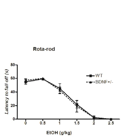
5. RESULTS
5.1. MOTOR COORDINATION
The cumulative doses of EtOH progressively affects motor coordination in both BDNF +/- and WT mice (Figure 8). Since no significant differences were observed, it can be assumed that the genotype has no effect on EtOH-induced motor coordination impairment.
Figure 8. EtOH treatment progressively impairs motor performance of WT and BDNF+/− mice. The latency to fall was recorded after each single EtOH injection (up to a total cumulative dose of 2.5 g/kg). Data are presented as mean ± SEM (n = 6 mice per group; WT vs BDNF+/−).
49
5.2. CLASS I HDACs IN THE CPu
Acute EtOH induces a significant reduction of nuclear HDAC 1 protein levels in the CPu of WT mice (WT EtOH-treated group = 13.33 ± 2.80 vs WT vehicle group = 100 ± 7.64; p < 0.001) (Figure 9a). Moreover, an innate high difference in the nuclear HDAC 1 levels has been detected between the WT and BDNF +/- mice, with lower protein levels in the BDNF+/− animals (WT vehicle group = 100 ± 7.64 vs BDNF+/− vehicle group = 26.96 ± 5.35; p < 0.001) (Figure 9a). Interestingly, EtOH treatment does not induce changes of the nuclear HDAC 1 levels in the BDNF+/− mice (Figure 9a). A significant genotype × treatment interaction has been also reported (F(1,20) = 66.76; p <
0.0001). Finally, the cytoplasmic amount of HDAC 1 was very low and we could not adequately quantify it.
Similar to what observed for HDAC 1, BDNF +/- mice innately exhibit lower levels of nuclear HDAC 2 compared to WT mice (WT vehicle group = 100 ± 6.97 vs BDNF+/− vehicle group = 27.60 ± 4.99; p < 0.001) (Figure 9b). EtOH induces a decrease of nuclear HDAC 2 protein levels in the CPu of WT mice (WT EtOH-treated group = 23.32 ± 3.27 vs WT vehicle group = 100 ± 6.97; p < 0.001) whereas no changes in the BDNF+/− mice (Figure 9b). In addition, a significant interaction of genotype × treatment has been observed (F(1,20) = 52.35; p < 0.0001). Finally, the CPu cytoplasmic
content of HDAC 2 is significantly reduced in the BDNF+/− EtOH-treated mice (BDNF+/− EtOH-treated = 46.87 ± 3.09 vs BDNF+/− vehicle group = 132.19 ± 28.10; p < 0.01) (Figure 9c).
Accordingly to the HDAC 1 and 2 results, differences of basal HDAC 3 levels have been observed in the CPu of BDNF+/− compared to WT mice (WT vehicle group = 100 ± 5.50 vs BDNF+/− vehicle group = 28.33 ± 6.00; p < 0.001) (Figure 9d). WT EtOH-treated mice has a reduction of nuclear HDAC 3 protein levels compared to WT
50 vehicle-treated animals (WT EtOH-treated = 23.30 ± 6.97 vs WT vehicle group = 100 ± 5.50; p < 0.001), but no changes have been observed in the BDNF +/- EtOH-treated mice (Figure 9d). As observed for HDAC 1 and 2, a significant genotype × treatment interaction has been also reported for HDAC 3 (F(1,20) = 37.62; p < 0.0001). Finally, in
the cytoplasmic fraction, BDNF+/− mice exhibit lower basal level of HDAC 3 protein compared to WT animals (WT vehicle group = 100 ± 16.63 vs BDNF+/− vehicle group = 49.11 ± 13.17; p < 0.05) (Figure 9e).
51
Figure 9. Protein levels of HDAC class I in the CPu: Western blot analysis. Nuclear (N) and cytoplasmic (C) contents of HDAC 1, 2 and 3 after acute EtOH i.p. (2 g/kg) or vehicle (Veh) in WT and BDNF+/− mice. The HDAC 1, 2 and 3 protein levels were assessed using specific antibodies compared to Actin (nuclear fraction) and GAPDH (cytoplasmic fraction). Data are presented as mean ± SEM (n = 6 mice per group) and analyzed by two-way ANOVA (*** p < 0.001; ** p < 0.01; * p < 0.05). In the upper right panel, representative immunoblots of HDAC 1, 2 and 3 were reported.