Long-Term Disease-Free Survival of Patients with
Radically Resected Thymomas
Relevance of Cell-Cycle Protein Expression
Tommaso Claudio Mineo,M.D.1 Vincenzo Ambrogi,M.D.1 Davide Mineo, M.D.1 Alfonso Baldi,M.D.2
1Department of Thoracic Surgery, Tor Vergata University, Rome, Italy.
2Department of Biochemistry and Biophysics “F. Cedrangolo,” Section of Anatomic Pathology, Sec-ond University, Naples, Italy.
This study was performed within the Research Fellowship Program Dottorato di Ricerca in Tec-nologie e Terapie Avanzate in Chirurgia, appointed by Tor Vergata University, and supported by grants “Progetto di Ricerca di Ateneo” ex-60%, 2003 and in part by “Progetto ricerca finalizzata Ministero della Salute-Regione Lazio 2002.”
Address for reprints: Vincenzo Ambrogi, M.D., De-partment of Thoracic Surgery, Chirurgia Toracica, Policlinico Universita Tor Vergata, Viale Oxford 81, 00133, Rome, Italy; Fax: (011) 390620902881; E-mail: [email protected]
Received March 9, 2005; revision received May 23, 2005; accepted June 14, 2005.
BACKGROUND.Despite radical surgical resection, thymomas often recur. The ob-jective of the current retrospective study was to investigate the prognostic rele-vance of the expression of cell-cycle proteins in these neoplasms to formulate a possible therapeutic surveillance strategy for the prevention of recurrence.
METHODS.The authors retrospectively reviewed the main clinicopathologic factors, including the World Health Organization (WHO) classification, of patients with thymoma who had undergone radical surgical resection. Specimens were studied using immunohistochemistry and the expression of cell-cycle proteins (i.e., p21, p27, and p53) was assessed. Univariate and multivariate analysis of predicting survival prognostic factors were performed.
RESULTS.The authors analyzed 88 patients with thymoma who underwent radical surgical resection at the study institution. According to the Masaoka staging system, 41 patients had Stage I disease, 31 patients had Stage II disease, and 16 patients had Stage III disease. There were 24 tumor recurrences (27.3%), 4 of which were local, 16 of which were distant intrathoracic, and 4 of which were extratho-racic. The second radical resection provided a disease-free survival rate that was similar to the first. Only Masaoka stage (P⫽0.001), WHO classification (P⫽0.001), high expression of p53 (P⫽0.03), and low expression of p21 (P⫽0.02) and p27 (P⫽0.001) were found to be correlated with a reduced disease-free survival. Low p27 expression was found to be the most significant predictive factor of a short disease-free survival (P⫽0.001), especially when associated with low p21 expres-sion and high p53 expresexpres-sion (P⫽0.0001).
CONCLUSIONS.Long-term disease-free survival in thymoma patients treated with radical surgical resection was found to be correlated with Masaoka stage, WHO classification, and expression of cell-cycle proteins, with the latter found to be the most significant predictive factor. Functional cooperation between cell-cycle pro-teins might constitute another level of regulation in tumor growth. More careful surveillance should be adopted whenever there is negative cell-cycle protein ex-pression. Cancer 2005;104:2063–71. © 2005 American Cancer Society.
KEYWORDS: thymomas, thoracic surgery, Masaoka staging, WHO classification, cell-cycle proteins.
C
omplete surgical resection is recognized worldwide as thetreat-ment of choice for thymomas1and has been reported to be the
best predictor of a long disease-free survival.2– 4 Nevertheless, the
prevention of postsurgical recurrences remains a heated topic of
discussion. The current staging system proposed by Masaoka et al.5,6
has proven quite effective in predicting survival. However, even
early-stage disease occasionally recurs.7–9 Several histologic classifications
for thymoma have been published to date,10 –12but to our knowledge
© 2005 American Cancer Society DOI 10.1002/cncr.21433
their prognostic significance is still controversial.13–15
The new World Health Organization (WHO)
classifica-tion16demonstrated that histologic subtype might be
an independent prognostic factor.17,18Strobel et al.19
recently proposed a therapeutic algorithm related to tumor stage, WHO histotype, and completeness of surgical removal, thus suggesting adjuvant therapy in the case of Stage III disease or a B2-3 histotype. Nev-ertheless, postoperative therapeutic strategy
contin-ues to be debated,20,21 and prospective studies are
warranted.
Significant progress has been made in under-standing the molecular and cellular pathogenesis of
neoplasms.22 One area that has been the focus of
much research is the control of the cell-cycle. A key role is played by cell-cycle kinases, relatively small proteins that are regulated by their arrangement in a multimeric complex with larger proteins, called “cyc-lins” because of their cyclic expression and degrada-tion during the cell-cycle. Cell-cycle kinase/cyclin complexes are negatively modulated through their in-teraction with a family of small proteins called
cell-cycle kinase inhibitors, namely p21 and p27.23,24 The
p53 tumor suppressor gene also is involved in cell-cycle checkpoints, acting as a transcription factor for several cell-cycle regulatory proteins, including the
p21 gene.25 To our knowledge, few of the factors
volved in regulating cell-cycle control have been in-vestigated to date in thymomas.
The objective of the current study was to compare the prognostic specific weight of traditional clinico-pathologic factors with the expression of p53, p21, and p27 cell-cycle proteins in a large series of patients who underwent radical surgical resection for thymoma and who had an adequate long-term follow-up to select the most suitable patient category for stricter clinical surveillance and incidental adjuvant therapy.
MATERIALS AND METHODS
Patients
Between January 1987 and December 2004, 97 consec-utive patients with thymoma underwent surgery in the Department of Thoracic Surgery at the University of Rome Tor Vergata. Several clinicopathologic features were studied retrospectively: patient gender and age
(cutoff age of ⱖ 50 yrs), the presence of myasthenia
gravis (MG), surgical notes (including surgical ap-proach and radical surgical resection), postoperative complications, pathologic issues, postoperative ther-apy, pattern of disease recurrence, and available long-term follow-up information.
Staging was based on surgical and pathologic
cri-teria as described by Masaoka et al.5,6and was
retro-spectively performed at the time of the review of the
surgeon’s pathology and surgical notes. The morpho-logic classification of the thymomas was conducted
according to the specifications of Bernatz et al.,11
Marino and Muller-Hermelink,12 and the WHO.16All
specimens were reevaluated retrospectively by the same pathologist (A.B.).
An essential prerequisite for inclusion in the study was the complete surgical resection of the thymoma, implying total thymectomy with excision of the medi-astinal fatty tissue between both phrenic nerves and evidence of a free surgical resection margin, with no macroscopic or microscopic residual tumor. Invasion of the great mediastinal vessels resulted in absolute exclusion from the study. Disease recurrence was de-fined as any evidence of tumor, such as regrowth of tumor in the mediastinum, pleural dissemination, or pulmonary metastasis, as detected by imaging or bi-opsy during follow-up. Patients were contacted by telephone. In cases in which patients could not be reached, their primary care physician or neurologist was contacted to update their care. Patients with in-complete follow-up information were excluded from the study.
Immunohistochemistry
We were able to retrieve surgical specimens from 79 patients in the study group in which cell-cycle analysis was performed. Briefly, sections from each specimen
were cut at 3–5 m, mounted on glass, and dried
overnight at 37 °C. All sections were then deparaf-finized in xylene, rehydrated through a graded alcohol series, and washed in phosphate-buffered saline. This buffer was used for all subsequent washes and for dilution of the antibodies. Tissue sections were heated twice in a microwave oven for 5 minutes each at 700 watts in citrate buffer (pH of 6.0) and we then pro-cessed with the standard streptavidin-biotin-immu-noperoxidase method (Dako Universal Kit; Dako
Cor-poration, Carpinteria, CA). Mouse monoclonal
antibodies (Santa Cruz Biotechnology, Santa Cruz, CA), specific for p27 (mouse monoclonal antibody 1641) and p21 (mouse monoclonal antibody sc-6246), were used at a 1:100 dilution, while a monoclo-nal antibody specific for p53 (D01; Dako Corporation) was used at a 1:500 dilution. All the primary antibodies were incubated for 1 hour at room temperature. Dia-minobenzidine was used as the final chromogen, and hematoxylin was used as the nuclear counterstain. Negative controls for each tissue section were per-formed, leaving out the primary antibody. Positive controls included in each experiment consisted of tis-sue previously shown to express the antigen of inter-est. One pathologist (A.B.) evaluated the staining pat-tern of the three proteins separately and scored the
protein expression in each specimen by scanning the entire section and estimating the percentage of tumor cell nuclei staining. All immunoreactive nuclei were regarded as positive, regardless of the intensity of staining.
Statistical Analysis
All data were statistically analyzed using SPSS software
(SPSS威 9.05 for Windows; SPSS Inc., Chicago IL).
Pre-liminary descriptive analysis was performed for main epidemiologic and pathologic variables to identify anomalous data.
Interdependence among factors (clinicopatho-logic parameters and cell-cycle protein expression) was assessed using the chi-square test and the Fisher
exact test. A P value ⬍ 0.05 was considered to be
statistically significant in two-tailed tests.
Thereafter, every prognostic variable was corre-lated for risk of disease recurrence and of death.
Ac-cording to our previous report,26a dichotomized
scor-ing system was used, with p53, p21, and p27
expression in⬎ 5% of the tumor cells defined as high
expression and used as a cutoff point. Survival analysis was performed according to the Kaplan–Meier method. Thymoma recurrence and death attributable to cancer or noncancer causes were considered as the final event. The statistical significance of the
differ-ences in survival distribution among the prognostic groups was evaluated by the log-rank test. The Cox proportional hazards model was applied to the multi-variate survival analysis only for those factors found to be significant on univariate analysis.
RESULTS
Clinicopathologic Findings
The study population consisted of 88 patients (42 men and 46 women), with a median age of 48.5 years (range, 16 –78 yrs). The main clinicopathologic char-acteristics of the patients are summarized in Table 1. All patients underwent a surgical approach through a total longitudinal median sternotomy; in 5 patients (6%) we were obliged to perform a second access through a lateral thoracotomy to permit radical surgi-cal resection. Four procedures that began for diagnos-tic purposes as videothoracoscopy were converted into open access. Radical thymectomy also included
infiltrated areas of the pericardium (n⫽ 10 patients),
lung (n ⫽ 4 patients), and phrenic nerve (n ⫽ 2
pa-tients). The mean surgical time was 118⫾ 37 minutes.
No intraoperative or postoperative mortality occurred. Ten patients (11.3%) developed postoperative
compli-cations: pneumonia (n ⫽ 4 patients), respiratory
fail-ure because of MG crisis (n ⫽ 2 patients), surgical
wound infections (n⫽ 5 patients), and prolonged air
TABLE 1
Epidemiology and Main Clinico-pathogic Features
No. of patients (%) P value Median age in yrs (range) P value Gender ratio M/F P value Presence of MG (%) P value Median follow-up in mos (range) P value
Staging system (Masaoka et al.5,6 )
I 41 (46.65) NS 50 (16–72) NS 15/20 0.05 10 (24.3) 0.01 79 (3–204) NS
II 31 (35.2) 48 (18–72) 17/20 13 (41.9) 78 (7–215)
III 16 (18.42) 43.5 (25–78) 10/6 11 (68.87) 55.5 (7–120)
Neoplasm histotype (Bernatz et al.11 )
Spindle 11 (12.5) NS 53 (21–72) NS 4/7 NS 6 (17.6) NS 66 (18–159) NS
Mixed 35 (39.87) 54 (16–78) 15/20 15 (42.98) 91 (2–215)
Lymphocytic 27 (30.69) 44 (17–73) 14/13 9 (33.3) 67 (3–180)
Epithelial 15 (17.40) 46 (15–72) 9/6 4 (26.7) 54 (7–166)
Immunohistochemical (Marino and Muller-Hermelink12 )
Medullary 19 (21.46) NS 44 (21–71) NS 8/11 NS 7 (36.8) NS 118 (3–199) NS
Mixed 21 (23.8) 38 (16–71) 9/12 8 (38.1) 79 (12–215)
Cortical 48 (54.5) 50.5 (17–78) 25/23 19 (39.56) 59 (7–166)
WHO classification (Rosai et al.16 ) A 9 (10.2) NS 33 (21–72) NS 3/6 0.03 3 (33.3) NS 62 (3–144) NS AB 35 (397.89) 54 (16–78) 16/19 14 (40.0) 91 (7–215) B1 27 (30.67) 50 (18–73) 12/15 10 (37.0) 79 (12–180) B2 15 (17.1) 46 (17–72) 10/5 6 (40.0) 54 (7–166) B3 2 (2.3) 43.5 (42–45) 1/1 1 (50) 41 (17–65) Total 88 48.5 (16–78) 42/46 34 (38.6) 71 (3–215)
leak (n⫽ 3 patients). The mean hospital stay was 5.3 ⫾ 0.7 days. Adjuvant therapy was administered in all patients with Stage III disease (according to the
Masaoka staging system5,6). The median follow-up
pe-riod was 70 months (range, 3–216 mos).
All the cell-cycle-associated proteins examined were present in the nuclei of tumor cells, although a small proportion of the cells displayed cytoplasmic immunoreactivity in addition to nuclear staining.
Interdependence Analysis
Clinicopathologic factors were distributed in the study group according to findings in the literature. No sig-nificant differences were noted with regard to length of follow-up or patient age and gender distribution within the different categories of the study group. No significant interdependence was found between stage of disease and histotypes (data not shown). The cell-cycle checkpoint proteins were analyzed with respect to the detailed clinicopathologic information available for all patients in this cohort. No correlations were found with the other clinical features, such as patient age, patient gender, clinical tumor stage, and all his-tologic classifications. A mild but significant correla-tion has been identified between p27 expression and traditional prognostic factors. These results are sum-marized in Table 2.
Pattern of Disease Recurrence
Only late clinical stage and WHO classification were found to be correlated with the probability of disease recurrence and a shorter overall survival, whereas dif-ferent histologic classifications, the male-to-female
ra-tio, patient ageⱖ50 years, and the surgical approach
used (sternotomy vs. thoracotomy) were not. The presence of MG was found to be correlated signifi-cantly with a higher rate of disease recurrence
(P⫽0.01), without affecting the survival. There were 24
(27.3%) tumor recurrences: local in 4 patients, distant intrathoracic in16 patients, and extrathoracic in 4 pa-tients. The median time to disease recurrence was 36 months (range, 3–78 mos). Treatment of the recur-rence consisted of 14 radical and 2 incomplete surgical reresections, whereas 8 patients were treated with other therapies (chemotherapy alone in 4 patients and combined chemoradiotherapy in 4 patients). The overall 5-year survival rate from the time of disease recurrence was 26%. Radical surgical reresection was associated with a significantly higher survival com-pared with patients treated with other therapies (5-year survival rates of 73% and 0%, respectively). The second disease-free interval was not found to be sta-tistically significantly different from that reported after the first surgery (Fig. 1).
Four patients died from thymoma spread after first intrathoracic disease recurrence (three patients) TABLE 2
Interdependence between Clinico-pathologic Features and Cell-Cycle Protein Expression High p53 expression P value Low p21 expression P value Low p27 expression P value Staging system (Masaoka- et al.5,6
)
I 18 NS 16 NS 14 0.03
II 7 10 12
III 6 8 9
Neoplasm histotype (Bernatz et al.11 )
Spindle 3 NS 3 NS 4 NS
Mixed 15 17 15
Lymphocytic 6 7 8
Epithelial 7 7 8
Immunohistochemical (Marino and Muller-Hermelink12 )
Medullary 4 NS 5 NS 3 0.05
Mixed 11 10 11
Cortical 16 19 21
WHO classification (Rosai et al.16 ) A 3 NS 3 NS 2 0.03 AB 16 18 17 B1 4 5 6 B2 7 7 9 B3 1 1 1
NS: not significant; WHO: World Health Organization. FIGURE 1.Disease-free survival (DFS) after the first recurrence versus global
or distant recurrence (one patient); two of these pa-tients had MG. The median time from disease recur-rence to death was 20.5 months.
Survival According to the Pathologic Staging
According to the Masaoka pathologic staging
sys-tem,5,641 patients had Stage I disease, 31 patients had
Stage II disease, and 16 patients had Stage III disease. The 5-year and 10-year disease-free survival rates were 91% and 87%, respectively, for patients with Stage I disease; 91% and 65%, respectively, for patients with Stage II disease; and 23% and 21%, respectively, for
patients with Stage III disease (P⫽0.001). At the same
time, the 10-year overall survival rate was 100% for patients with Stage I disease, 90% for patients with Stage II disease, and 75% for patients with Stage III disease (Fig. 2).
Survival by Histologic Classification
Using the system of Bernatz et al.,11we classified 11
spindle, 35 mixed, 27 lymphocytic, and 15 epithelial histotypes; using the system of Marino and
Muller-Hermelink,12 we classified 19 medullary, 21 mixed,
and 48 cortical thymomas. No classification disclosed a statistically significant difference in survival for ei-ther overall or disease-free survival.
According to WHO criteria, there were 9 patients (10%) with subtype A tumors, 35 patients (40%) with subtype AB tumors, 27 patients (31%) with subtype B1 tumors, 15 patients (17%) with subtype B2 tumors, and 2 patients (2%) with subtype B3 tumors. The 10-year disease-free survival rate was 100% for patients with subtype A tumors, 73% for those with subtype AB tumors, 65% for those with subtype B1 tumors, 43% for patients with subtype B2 tumors, and 0% for pa-tients with subtype B3 tumors, with a significant dif-ference noted among the 5 patient populations
(P⫽0.001). Conversely, the 10-year overall survival
re-vealed only a mild difference (Fig. 3).
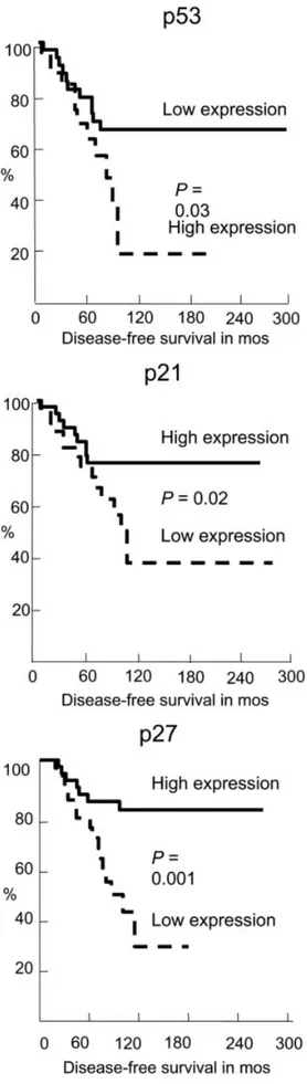
Survival by Cell-Cycle Protein Expression
p53 was found to be highly expressed in 31 cases out of 79 available samples (39.2%), and p21 and p27 were low-expressed in 34 (43.0%) and 35 samples (44.3%), respectively. No correlation was noted between cell-cycle protein expression and disease recurrence (data not shown). Disease-free survival appeared to be in-fluenced by high expression of p53, and low expres-sion of p21 and p27. The 5-year and 10-year disease-free survival rates were 61% and 19%, respectively, for
patients with high expression of p53 (P⫽0.03), 77%
and 40%, respectively, for patients with low expression
of p21 (P⫽0.02), and 71% and 33%, respectively, for
patients with Masaoka Stage III disease (P⫽0.001)
(Fig. 4).
Multivariate Analysis of Survival
Using multivariate analysis, we matched Stage I pa-tients versus Stage non-I, A-AB-B1 versus B2-B3 WHO histotypes, and single cell-cycle protein ex-pression. The most relevant negative predictor of
FIGURE 2.Overall survival and disease-free survival according to the stage of disease.
disease-free survival was found to be low p27
ex-pression (P⫽0.001). Furthermore, dichotomizing
the most negative expression of cell-cycle protein expression (i.e., the combination of low p27 expres-sion with high p53 and low p21 expresexpres-sion vs. all other combinations), we obtained an even more
significant result (P⫽ 0.0001). The data are reported
in Table 3.
DISCUSSION
The natural history of thymomas remains unpredict-able; they may recur independently of classic clinico-pathologic factors and despite radical surgical resec-tion, which is still considered the treatment of
choice.1,9This difficulty complicates the planning of a
structured postoperative therapeutic strategy. In fact, to our knowledge multiple studies have failed to dem-onstrate the precise role of adjuvant therapy, espe-cially after radical surgical resection at early stages of disease. Since 1985, adjuvant radiotherapy has been
performed by Monden et al.,27who proposed it even
for patients with Stage I disease. The results with this procedure generally were good but not completely satisfactory; after total surgical resection, approxi-mately 5% of the irradiated patients still developed
disease recurrence. Dziuba and Curran28
recom-mended postoperative radiation therapy for Stage II disease in the case of transgression of the tumor through the capsule, despite macroscopically com-plete surgical resection. Conversely, two recent stud-ies21,29found that the addition of adjuvant
radiother-apy did not significantly alter rates of local or distant disease recurrence in patients with Stage II thymomas who had undergone radical surgical resection. Fur-thermore, even after complete surgical resection, postoperative irradiation has demonstrated only mar-ginal benefits in patients with Stage III disease, with
significant morbidity reported.20
Many studies to date have demonstrated that the probability of disease recurrence is not a simple mat-ter of tumor progression according to stage of disease, but histologic subtypes also may represent an
inde-pendent prognostic factor.9,17–19Therefore, the
thera-peutic strategy should be redefined also according the histologic subtype of the tumor.
The WHO histologic typing of tumors of the
thy-mus first was published in 1999.16Some studies have
reported that the WHO classification appears to have
clinical and prognostic value.17,18,30 More recently,
Strobel et al.19proposed a therapeutic algorithm to be
tested prospectively that includes WHO classification, radicalness of the surgical resection, and clinical stage
of disease. Furthermore, Inoue et al.22suggested that
the WHO histotypes are somewhat correlated with genetic alterations; A-AB histotypes are biologically distinct from the others and B2-B3 thymomas form a continuum, with evidence of tumor progression.
On the basis of analogous studies performed in
patients with lung carcinoma,26 we hypothesized an
active role of genomic expression for thymoma growth
FIGURE 3.Overall survival and disease-free survival according to the World Health Organization (WHO) classification.
and progression. The ability of a cell to control its own replication is very important for the maintenance of the structure and functions of the organ it belongs to and, in final analysis, of the organism it is a part of. Currently, several pathologies are connected to an altered control of cellular replication and, among these, cancer is one of the most studied. We therefore analyzed the expression of three key proteins involved in cell-cycle checkpoints in a large series of well char-acterized thymoma patients.
When we examined the correlation between clin-icopathologic data and the expression of cell-cycle proteins, we found a slightly positive correlation be-tween classic disease recurrence prognosticators and p27 expression, suggesting a possible role for this pro-tein in the progression of this disease.
When we investigated, using univariate analysis, the correlation between the expression of different proteins and survival, we found that all the cell-cycle proteins analyzed had a statistically significant corre-lation with disease-free survival. On the multivariate analysis, the most significant parameter found to in-fluence the disease-free survival was p27 and this sig-nificance became even more evident when we use the combination of high p53 expression and low p21 and p27 expression.
Taking into account the complicated functional network constituted by the cell-cycle regulator pro-TABLE 3
Multivariate Cox Regression Analysis of Disease-Free Survival in Thymomas Patients
RR of
recurrence 95% CI P value
Masaoka stage
Stage I 1 —
Stages II and III 1.7 0.796–2.007 0.05
WHO classification A-AB-B1 1 — B2–B3 12.8 1.17–140.7 0.03 p27 expression High 1 — Low 3.49 1.44–6.73 0.001
High p53 expression, low p21 expression, and low p27
expression
Absent 1 —
Present 2.18 1.01–5.61 0.0001
RR: relative risk; 95% CI: 95% confidence interval; WHO: World Health Organization.
Š
FIGURE 4.Disease-free survival according to expression of cell-cycle pro-teins.
teins, it appears evident that knowledge of the level of expression of these factors, and their coregulation, may be important in predicting patient clinical re-sponse to simple surgical therapy. Nevertheless, tar-geting multiple checkpoint proteins may represent a good therapeutic strategy for the development of new molecular treatments for lung carcinoma. In fact, it is clear that functional cooperation between different cell-cycle inhibitor proteins constitutes another level of regulation in cell growth control and tumor sup-pression. Therefore, it could be possible to hypothe-size that silencing more then one cell-cycle regulator in the same tumor cell, acting at either the RNA or protein level, could achieve more effective results. This could be obtained through the use of several technologies such as antisense oligonucleotides, dom-inant-negative constructs, or antibodies able to block the action of a specific protein. The data presented in the current study support this hypothesis and strongly suggest further research aimed at investigating the simultaneous expression of numerous cell-cycle regu-lators in patients with thymoma.
We acknowledge several limitations to the current study. First, the retrospective nature of the study, which is necessary because of the relative rarity of the disease, unless performing multicentric analysis. Sec-ond, the length of follow-up in the current study was not long enough for a disease with low malignancy. Thymoma has a known potential to recur beyond 10 years and our median follow-up time was 70 months; however, this follow-up was similar to that used in
many recent studies.19,21,29Another potential concern
is that the selection of patients to receive radiotherapy was based only on their having Stage III disease, re-gardless of the histologic classification of the tumor. This bias restricted potential adjuvant benefit to pa-tients per se affected with a more aggressive disease, which is one of the purposes of postoperative therapy. Long-term disease-free survival in patients with thymoma who are treated with radical surgical resec-tion appears to be correlated with the Masaoka stage of disease, the WHO classification, and cell-cycle gene expression proteins, with the latter found to be the prevalent factor on multivariate analysis. This sup-ports the theory that functional cooperation between different cell-cycle inhibitor proteins constitutes an-other level of regulation in cell growth control and tumor suppression. Finally, analysis of cell-cycle reg-ulators, such as p27, should be included in ongoing prospective studies to determine a more precise pre-dictor of disease recurrence and to adopt a stricter surveillance program.
REFERENCES
1. Wilkins KB, Sheikh E, Green R, et al. Clinical and pathologic predictors of survival in patients with thymoma. Ann Surg. 1999;230:562–572.
2. Maggi G, Casadio C, Cavallo A, Cianci R, Molinatti M, Ruffini E. Thymoma: results of 241 operated cases. Ann Thorac
Surg. 1991;51:152–156.
3. Blumberg D, Port JL, Weksler B, et al. Thymoma: a multi-variate analysis of factors predicting survival. Ann Thorac
Surg. 1995;60:908 –913.
4. Regnard J-F, Magdeleinat P, Dromer C. Prognostic factors and long-term results after thymoma resection-a series of 307 patients. J Thorac Cardiovasc Surg. 1996;112:376 –384. 5. Masaoka A, Monden Y, Nakahara K, Tanioka T. Follow-up
study of thymomas with special reference to their clinical stages. Cancer. 1981;48:2485–2492.
6. Masaoka A, Yamakawa Y, Hiwa H, et al. Thymectomy and malignancy. Eur J Cardiothorac Surg. 1994;8:251–253. 7. Shimosato Y. Controversies surrounding the
subclassifica-tion of thymoma. Cancer. 1994;74:542–544.
8. Lardinois D, Rechsteiner R. Prognostic relevance of Masaoka and Muller-Hermelink classification in patients with thymic tumors. Ann Thorac Surg. 2000;69:1550 –1555. 9. Nakagawa K, Asamura H, Matsuno Y, et al. Thymoma: a
clinicopathologic study based on the new World Health Organization classification. J Thorac Cardiovasc Surg. 2003; 126:1134 –1140.
10. Rosai J, Levine GD. Tumors of the thymus. In: Harlan I, Ferminger MD, editors. Atlas of tumor pathology, 2nd series. Fascicle 13. Washington, DC: The Armed Forces Institute of Pathology, 1976:1–221.
11. Bernatz PE, Harrison EG, Clagett OT. Thymoma: a clinico-pathologic study. J Thorac Cardiovasc Surg. 1961;42:424 – 444.
12. Marino M, Muller-Hermelink HK. Thymomas and thymic carcinoma. Relation of thymoma epithelial cells to the cor-tical and medullary differentiation of thymus. Virchows Arch
A Pathol Anat Histopathol. 1985;407:119 –149.
13. Kornstein MJ. Thymoma classification–my opinion. Am J
Clin Pathol. 1999;112:304 –307.
14. Suster S, Moran CA. Thymoma classification. The ride of the Valkyries? Am J Clin Pathol. 1999;112:308 –310.
15. Harris NL, Muller Hermelink HK. Thymoma classification. A siren’s song of simplicity. Am J Clin Pathol. 1999;112:299 – 303.
16. Rosai J, Sobin LH. Histological typing of tumors of the thymus. In: World Health Organization, editors. Interna-tional histological classification of tumors, 2nd ed. Berlin: Springer-Verlag, 1999:1– 65.
17. Chen G, Marx A, Wen-Hu C, et al.. New WHO classification predicts prognosis of thymic epithelial tumors. A clinico-pathologic study of 200 thymoma cases from China. Cancer. 2002;95:420 – 429.
18. Kondo K, Yoshizawa K, Tsuyuguchi M, et al. WHO histologic classification is a prognostic indicator in thymoma. Ann
Thorac Surg. 2004;77:1183–1188.
19. Strobel P, Bauer A, Puppe B, et al. Tumor recurrence and survival in patients treated for thymomas and thymic squa-mous cell carcinomas: a retrospective analysis. J Clin Oncol. 2004;22:1501–1509.
20. Ogawa K, Uno T, Toita T, et al. Postoperative radiotherapy for patients with completely resected thymoma. A multi-institutional retrospective review of 103 patients. Cancer. 2002;94:1405–1413.
21. Singhal S, Shrager JB, Rosenthal DI, LiVolsi VA, Kaiser LR. Comparison of stages I-II thymoma treated by complete resection with or without adjuvant radiation. Ann Thorac
Surg. 2003;76:1635–1642.
22. Inoue M, Starostik P, Zettl A, et al. Correlating genetic ab-errations with World Health Organization-defined histology and stage across the spectrum of thymomas. Cancer Res. 2003;63:3708 –3715.
23. Sherr CS. Cancer cell cycles. Science. 1996;274:1672–1677. 24. Grana X, Reddy EP. Cell cycle control in mammalian cells:
role of cyclins, cyclin dependent kinases (CDKs), growth suppressor genes and cyclin-dependent kinase inhibitors.
Oncogene. 1995;11:211–219.
25. Kirsch DG, Kastan MB. Tumor-suppressor p53: implications for tumor development and prognosis. J Clin Oncol. 1998; 16:3158 –3168.
26. Esposito V, Baldi A, Tonini G, et al. Analysis of cell-cycle regulator proteins in non-small cell lung cancer. J Clin
Pathol. 2004;57:58 – 63.
27. Monden Y, Nakahara K, Iioka S, et al. Recurrence of thy-moma: clinicopathological features, therapy, and prognosis.
Ann Thorac Surg. 1985;39:165–169.
28. Dziuba SJ, Curran WJ Jr. The radiotherapeutic management of invasive thymomas. Chest Surg Clin N Am. 2001;11:457– 466.
29. Mangi AA, Wright CD, Allan JS, et al. Adjuvant radiation therapy for stage II thymoma. Ann Thorac Surg. 2002;74: 1033–1037
30. Okumura M, Ohta M, Tateyama H, et al. The WHO Health Organization histologic classification system reflects the oncologic behavior of thymoma. Cancer. 2002;94:624 – 632.