-1-
U
NIVERSITÀD
EGLIS
TUDID
IM
ESSINAT
ESI DID
OTTORATO DIR
ICERCA INB
IOLOGIAA
PPLICATA EM
EDICINAS
PERIMENTALEC
URRICULUM INM
EDICINAS
PERIMENTALEXXXIII CICLO
SSD BIO/10
Absence of Formyl Peptide Receptor (FPR)-1
improves the outcome in mouse models
of acute and chronic inflammation
Candidata:D
OTT.
SSARAMONA
D’AMICO
Tutor: Ch.ma Prof.ssa ROSANNA DI PAOLA
Coordinatore:
Ch.ma Prof.ssa Nunziacarla Spanò
-2-
SUMMARY
INTRODUCTION ... 6
CHAPTER 1: N-formyl peptide receptors (FPRs)... 8
1.1 Introduction ... 8
1.2 N-Formyl Peptide Receptor Family ... 9
1.3 Ligands for FPRs ... 10
1.4 Expression of FPRs in immune and non-immune cells ... 12
1.5 FPRs in the inflammatory response ... 14
1.6 Focus on FPR1 ... 16
1.6.1 FPR1 signal transduction ... 17
1.6.2 Mouse Fpr-1 Receptors ... 19
CHAPTER 2: Bronchiolitis obliterans (BO) ... 21
2.1 Introduction ... 21
2.2 Etiology, epidemiology and incidence ... 22
2.3 Pathophysiology ... 23
2.4 Pharmacotherapy ... 25
2.4.1 Non-Transplant Bronchiolitis Obliterans ... 25
2.4.2 Post-Transplant Bronchiolitis Obliterans ... 26
2.3.3 Alternative strategies ... 28
CHAPTER 3: Traumatic Brain Injury (TBI) ... 30
3.1 Introduction ... 30
3.2 Etiology, epidemiology and incidence ... 31
3.3 Pathophysiology ... 32
3.2.1 TBI and Neuroinflammation ... 33
3.4 Pharmacotherapy ... 35
3.4.1 Therapeutic strategies in pre-clinical studies ... 35
3.4.2 Therapeutic strategies in clinical trials ... 36
-3-
CHAPTER 4: Aim ... 39
4.1 Targeting FPR-1 as an emerging pharmacological strategy for inflammatory diseases ... 39
CHAPTER 5: Materials and methods ... 41
5.1 Materials and methods for BOS study ... 41
5.1.1 Animals ... 41
5.1.2 Experimental Model of Tracheal Transplantation ... 41
5.1.3 Experimental Groups ... 41
5.1.4 Histopathology examination ... 42
5.1.5 Mast Cell Evaluation ... 42
5.1.6 Western Blot Analysis ... 43
5.1.7 Immunohistochemical Analysis ... 44
5.1.8 Terminal Deoxynucleotidyl Nick-End Labeling (TUNEL) Assay ... 45
5.2 Materials and methods for TBI study ... 46
5.2.1 Animals ... 46
5.2.2 Induction of Experimental Traumatic Brain Injury (TBI) ... 46
5.2.3 Experimental groups ... 46
5.2.4 Histopathology examination ... 47
5.2.5 Assessment of Lesion Volume ... 47
5.2.6 Western Blot Analysis ... 48
5.2.7 Bromodeoxyuridine (BrdU) Treatment ... 48
5.2.8 Immunohistochemical Analysis... 48
5.2.9 Myeloperoxidase activity ... 49
5.2.10 ELISA analysis of IL-1 and IL-18 ... 49
5.2.11 Behavioural testing ... 49
5.2.11.1 Open Field ... 49
5.2.11.2 Social interaction test ... 50
5.2.11.3 Novel object recognition Test ... 50
-4-
5.3 Materials ... 52
5.4 Statistical Evaluation ... 52
CHAPTER 6: RESULTS ... 53
6.1 Results for BOS study... 53
6.1.1 Histopathology evaluation and mast cell density in IL-1β/IL-18 KO, Casp-1 KO, and Fpr-1 KO mice ... 53
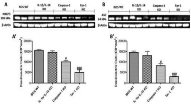
6.1.2 Effects of the absence of IL-1β/IL-18, Casp-1, and Fpr-1 on the NF-kB pathway 57 6.1.4 Effects of the absence of IL-1β/IL-18, Casp-1, and Fpr-1 on Nitrotyrosine formation and PARP activation ... 59
6.1.5 Effects of the absence of IL-1β/IL-18, Casp-1, and Fpr-1 on apoptosis ... 61
6.1.6 Effects of the absence of IL-1β/IL-18, Casp-1, and Fpr-1 on grow factors expression ... 63
6.1.7 Effects of the absence of IL-1β/IL-18, Casp-1, and Fpr-1 on the MAPK pathway 65 6.2 RESULTS FOR TBI STUDY ... 67
6.2.1 Effects of absence of Fpr-1 on histological alteration 24 h after TBI ... 67
6.2.2 Effects of absence of Fpr-1 on neutrophils accumulation 24 h after TBI ... 69
6.2.3 Effects of absence of Fpr-1 on MAPK pathway 24 h after TBI ... 71
6.2.4 Effects of absence of Fpr-1 on COX-2 and prostaglandin expression 24 h after TBI ... 73
6.2.5 Effects of absence of Fpr-1 on NF-κB pathway 24 h after TBI ... 75
6.2.6 Effects of absence of Fpr-1 on inflammasome components 24 h after TBI ... 77
6.2.7 Effects of absence of Fpr-1 on cytokines expression and oxidative stress activation 24 h after TBI ... 79
6.2.8 Effects of absence of Fpr-1 on astrocytes activation 24 h after TBI ... 81
6.2.9 Effects of absence of Fpr-1 on microglia activation 24 h after TBI ... 83
6.2.10 Effects of absence of Fpr-1 on severity of tissue damage 4 weeks following TBI 85 6.2.11 Effects of absence of Fpr-1 on COX-2 expression 4 weeks following TBI ... 87
6.2.12 Effects of absence of Fpr-1 on iNOS and prostaglandin expression 4 weeks following TBI ... 89
6.2.13 Effects of absence of Fpr-1 on cell proliferation 4 weeks following TBI ... 91 6.2.14 Effects of absence of Fpr-1 on behavioral performance 4 weeks following TBI . 93
-5-
6.2.15 Effects of absence of Fpr-1 on self-renewal and neurogenesis 4 weeks following
TBI ... 95
CHAPTER 7: Discussion ... 97
CHAPTER 8: Conclusion ... 104
-6-
INTRODUCTION
Formyl peptide receptors (FPRs) are a family of G protein-coupled receptors, whose principal function is to detect the presence of pathogen-associated molecules or harmful endogenous ligands, including also typical biomarkers of immune activation and inflammation [1, 2]. In humans, the FPR family is constituted by FPR1, FPR2, and FPR3, all three functional receptors encoded by three genes, while in rodents, three genes encode functional receptors and six genes encode orphan receptors [3]. In particular, FPR1 was the first neutrophil chemoattractant receptor to be characterized biochemically [4] and has been shown to be a key player in innate immunity and host defence [5]. FPR1 is principally expressed on immune cells with chemotactic or phagocytic activity like monocytes, neutrophils and macrophages [6, 7]. Although FPR1 is expressed in many types of immune cells, studies on FPR1 functions mostly focus on neutrophils. In particular, once the receptor is activated, neutrophils migrate toward the site of injury, initiating the inflammatory response by releasing a number of proinflammatory mediators, thus amplifying the immune response [8]. Moreover, it is expressed also on non-phagocytic and non-mobile sentinel cells like endothelial cells, epithelial cells, neurons and glia [3, 9]. Additionally, FPRs family is able to bind several structurally distinct ligands including endogenous and bacterial derived peptides, lipids and small non-peptide molecules [2]. In details, the principal agonists of FPR1 are mitochondrial and pro-inflammatory bacterial N-formylpeptides, but also bind anti-inflammatory agonists lipoxin A4 (LXA4) and annexin-A1 (ANXA1) [5, 10]. These ligands for FPR1 were detected during inflammatory processes and may stimulates numerous responses such as phagocytosis, chemotactic migration, degranulation and free oxygen species production [11]. Furthermore, inflammatory cells expressing FPRs, once they are recruited at the lesion site, are activated and trigger multiple pathways, e.g., the increase of gene transcription, the assembly of intracellular pro-inflammatory complexes, and the release of reactive oxygen species (ROS) and nitric oxide [12].
-7-
FPRs represents a family of interesting pharmacological targets for the management of organ-specific or systemic inflammatory conditions [11, 13, 14], such as arthritis, inflammatory lung and inflammatory bowel disease, ischemia-reperfusion (I/R) injury, sepsis, and wound healing. High levels of FPR expression also has found in the central (CNS) and peripheral (PNS) nervous system [15, 16]; in particular their implication in the process of neurogenesis and neuronal differentiation. Thus, FPRs may exert a distinctive role in the response to pathogens because they signal at two levels; firstly at the level of CNS to alert the host of dangers and secondly at the level of the immune system by starting a protective inflammatory response [7].
Despite promising findings from in vitro studies, at present, there has been few animal experimentation targeting FPR pathway for inflammatory resolution or neuronal regeneration. In recent years, the critical roles of FPR receptors in disease progression are increasingly recognized, thanks also to the availability of genetically engineered mouse strains deficient in one or more Fprs [6]. Studies with Fpr knockout (KO) mice had been demonstrated that deletion of the Fpr gene reduced tissue damage and inflammation in several experimental models, such as endometriosis, colitis and depression [17-19].
Therefore, targeting FPRs has strong potential, and once supported by a greater knowledge of the functional role of FPR receptors, may bring about novel options in treatment of acute and chronic inflammation.
-8-
CHAPTER 1: N-formyl peptide receptors (FPRs)
1.1 Introduction
FPRs are a family of chemoattractant receptors (GPCRs) coupled to G-protein and play a critical role on host defence and modulation of inflammation [1]. This family of receptors is so-called since initial studies identified the chemotaxis activity of receptor-mediated neutrophil, induced by peptides containing formylmethionine [20, 21]. Initially, the researchers hypothesized that the presence of a formylmethionine group in ligand was an essential factor binding to FPR receptors. Since the natural sources of these peptides are derived from bacterial or mitochondrial proteins, it was identified that FPRs evolved to promote the recruitment of phagocytic leukocytes to loci of infection or damage [22]. When it was discovered that these formylated molecules might derive from both bacterial and endogenous mitochondrial fonts, important evidence was supplied for the endosymbiotic theory of mitochondrial evolution from primitive bacteria [23]. This was further confirmed when endogenous formylated peptides emitted from the mitochondria of necrotic cells were shown to stimulate the recruitment of monocytes that contribute to inflammatory process through interaction with FPR receptors [24]. Additionally to cell chemotaxis, some researchers [20] stated that FPR activation also induced the release of lysosomal enzymes by phagocytic cells in pathologic site, further facilitating the removal of pathogens and damaged tissue detritus. Later, investigators showed that myeloid FPR activation induced pro-inflammatory cytokines and superoxide production [25, 26]. Recently, biological functions of the FPR family have extended to non-myeloid settings, where additional roles from tissue homeostasis to inflammatory responses have been identified [1], indicating the adaptive nature of FPR receptors, and their distinct functionality relative to the cellular context in which they express themselves.
-9- 1.2 N-Formyl Peptide Receptor Family
In humans, FPR family comprises three members: FPR1, FPR2, and FPR3, which all recognise a wide range of endogenous and exogenous ligands [27, 28]. The activation of FPRs receptors causes their homo- or hetero-dimerization, which in turn depends on the exact ligand to which they bind [29, 30]. In this way, FPRs can have both pro and anti-inflammatory properties on immune cells. In rodents, FPR (mFPR or Fpr) family consists of at least 8 gene including Fpr1, Fpr2, Fpr-rs1, Fpr-rs3, Fpr-rs4, Fpr-rs5, Fpr-rs6, and Fpr-rs7 [31]. Fpr1 is considered as an orthologue of human FPR1, while Fpr2 is structurally and functionally similar to human FPR2 [32]. The mouse equivalent of human FPR3 is not well described. Since Fpr2 shares a human ligand FPR3 [33], it was proposed that Fpr2 act as a counterpart of both FPR2 and FPR3. The other 6 murine Fpr genes are expressed in leukocytes, but the identity of their encoded receptors remain unknown [26].
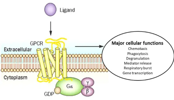
FPRs are protein molecules consisted of residues of 350 or 351 aminoacids [34]. They have similarity in the structure of the membrane: seven transmembrane domains connected via three extracellular and three intracellular loops, the N_terminal end directed into the extracellular space and the C_terminus into the cytoplasm (Fig.1). The extracellular domains are responsible primarily for detection ligands and their access to the structural core. The structural core, consisting of seven transmembrane domains, binds the ligands and transfers a signal into cell due to its conformational changes. Intracellular domains bind to G_proteins, arrestins, receptor kinases coupled with G-proteins, and other systems of the cytoplasm [35]. FPR1 is the first neutrophil GPCR to be identified, cloned and sequenced [36]. All three receptors are clustered on chromosome 19q13.3 and share important homology of the sequence [31]. FPR1 shares 69% of identity of the aminoacids with FPR2 and 56% with FPR3, while FPR2 and FPR3 share 83% of the identity [5].
-10-
Figure 1. Structure of FPRs
1.3 Ligands for FPRs
FPRs are distinguished for their promiscuity; indeed, they are able to bind a large number of distinct ligands including endogenous and bacterial derived peptides, lipids and small non-peptide molecules. This unusual ligand diversity has resulted in the FPRs being labelled as pattern recognition receptors [3].
FPR1 and FPR2 were initially identified due to their capacity to recognize N-formyl-methionine and other oligopeptides, of which the most studied is N-formyl- N-formyl-methionine-leucyl-phenylalenine (fMLF) [34]. Formylated peptides are the products of degradation of bacterial or the organism’s own mitochondrial proteins and serve as strong chemoattractants. FPR1 or FPR2 activation by chemotactic agonists provokes a cascade of signalling events leading to cell migration, increased phagocytosis, release of mediators and new gene transcription [6, 37]. Depending on the concentration, formylated peptides activate various phagocytic processes, such as chemotaxis, secretory degranulation, and respiratory burst; all these reactions being necessary for defence against bacterial infections. Formylated peptides are the main agonists of FPR1; besides, FPR1 can bind with endogenous proteins such as cathepsin G [38]. In addition to bacteria-derived formyl peptides, mitochondria also contain
-11-
formyl peptides that can be released upon pathological injury. The mitochondrial formyl peptides act as mitochondrial DAMPs. They are recognized by FPR1 and lead to the activation of neutrophils and subsequent migration of the cells into the injured area [39]. During cell death or tissue damage, diverse DAMPs are released into environment. A well-characterized DAMP is cathelicidin. It contains a C-terminal cationic antimicrobial domain and mature peptides show very potent antimicrobial activity [40].
Formylated peptides also bind to FPR2, but with lower affinity. FPR2 receptors can bind to various ligands including serum amyloid A (SAA) and LXA4 [37]. Specifically, these ligand-specific interactions cause either proinflammatory or anti-inflammatory effects [41, 42], which is important to prevent the evolution of inflammatory process into chronic form. For example, ANXA1 and LXA4 trigger FPR2 activation to inhibit leucocyte recruitment, enhance apoptosis of neutrophils and stimulate efferocytosis of macrophages [43, 44]. On contrary, once bind to FPR2, SSA induce proinflammatory responses and migration of neutrophils at inflammatory sites [45].
The function of FPR3 receptor remains poorly understood compared to that of other members of the FPR family. FPR3 is highly phosphorylated (signal for inactivation and internalization of the receptor) and mainly localized into small intracellular vesicles [46], in contrast to its counterpart receptors located on the cell surface. This indicates that after binding its ligands, FPR3 easily internalizes and thus can act as a "decoy" receptor to minimize the binding of its agonists to other receptors [6]. It is noteworthy that FPR3 does not bind formylated chemoattract peptides and nor shares ligands with FPR1 and FPR2. Few specific ligands have been identified; in particular F2L is the most specific ligand for FPR3 [47] but its biological role in vivo is still to be identified. Therefore, FPR3 might have its own unique and specific function.
-12-
1.4 Expression of FPRs in immune and non-immune cells
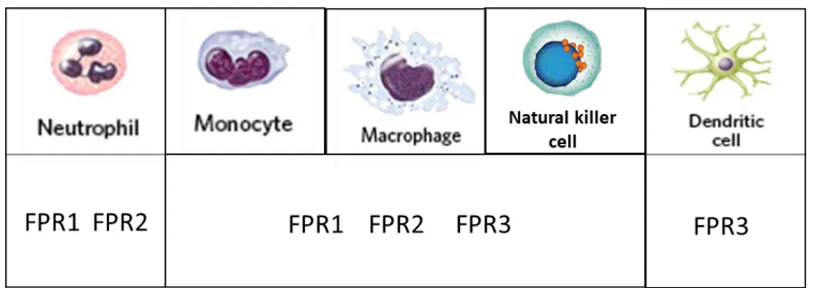
FPRs are primarily expressed in leukocytes (Fig. 2) including neutrophils, monocytes/macrophages, natural killer (NK) cells, and dendritic cells (DCs) [6, 31, 36]. Human neutrophils express FPR1 and FPR2, but not FPR3 [38]. In neutrophils, the activation of FPRs causes chemotactic migration of the cells, phagocytic activity and produces ROS, which increase innate defence activity [31, 36]. Human monocytes, macrophages and NK cells express all three receptors [38]. In monocytes and macrophages, activation of FPRs also induces chemotactic migration of the cells, and regulates defence activity against invading pathogens. NK cells activated by FPR agonists produce interferon-gamma (IFN-γ) and show cytolytic activity, suggesting potential functional roles in anti-tumour or viral activity [48].
Immature DCs express FPR1 and FPR3, while mature DCs express FPR3, but not FPR1 and FPR2 [49]. In addition to innate immune cells, adaptive immune cells express FPRs [50]. In details, recently, FPR2 has recently been observed in naive CD4+ T cells, human tonsillar follicular helper T cells, Th1 cells, Th2 cells, and Th17 cells [50, 51].
Figure 2. FPRs in immune cells
Notably, FPRs are also expressed in a several non-immune cells (Table 1) suggesting a wider range of biological function of these receptors. Endothelial cells and progenitor endothelial cells express FPR2 [52, 53], as do synovial fibroblasts [54] and keratinocytes [55]. The latter mediate cell proliferation and proinflammatory response. FPRs are expressed also in
-13-
intestinal epithelial cells [56], as do in glial cells [57], which function similar to macrophages in the brain. In details, FPR1 is most highly expressed in cells of immune system and bone marrow, although it abundantly expressed also in cells of lungs, brain, and gastrointestinal tract [9]. FPR2 tends to be ubiquitously expressed compared to the others receptors. The expression is predominantly found in cells of immune system, bone marrow, GI tract, female organ tissues, and endocrine glands, but lower levels of expression were also found in cells of brain, liver, gallbladder, and pancreas [9]. FPR receptors also expressed in some malignant human tumours cells and respond to bacterial or endogenous agonists by increased growth and motility. For example, FPRs expressed by human gastric cancer cells facilitate epithelial-mesenchymal transition, migration, cell proliferation and resistance to apoptosis [58]. Besides their expression in various types of cells, FPRs are known for promiscuity of ligand, by interacting with both damage-associated chemotactic molecular patterns (DAMPs) and pathogen-associated chemotactic molecular patterns (PAMPs).
Cells FPR1 FPR2 FPR3
Endothelial cells X X Not clear Endothelial progenitor
cells
X
Intestinal epithelial cells X X Keratinocytes X X
Hepatocytes X
Mesenchymal stem cells (MSCs)
X
Synovial Fibroblasts X
Glial cells X X
-14-
Summing up briefly, the expression of FPR receptors is highest in sentinel innate cells with chemotactic or phagocytic activities such as neutrophils, monocytes, macrophages and dendritic cells. However, FPRs are also expressed in non-phagocytic and ‘‘immobile’’ sentinel cells such as endothelial cells, mucosal epithelial cells and glia. In all these cells, FPRs exert a ‘‘sentinel role’’ by sensing pathogens present in the microenvironment and by favouring repair upon inflammation and damage.
1.5 FPRs in the inflammatory response
The family of FPR has evolved as chemoattractant receptors that help the host in countering bacterial infections, by facilitating the migration of phagocytes into site of bacterial invasion [14]. FPRs signalling has been described to modulate the survival and the phagocytic activity of infiltrating cells [13]. During an acute inflammatory response, leukocytes migrate towards an increasing concentration gradient (range from nM to µM concentrations) of chemotactic factors [14]. In addition, the discovery of FPRs in several cell types and tissues, besides to immune system cells, and the existence of several endogenous FPR ligands contributed to a wider view of FPR functions. Therefore, there is accumulating evidence that FPRs have distinctly different functions beyond simple pathogen recognition.
Studies with cultured cells, preclinical models and clinical samples have positioned FPR receptors at pivotal checkpoints from initiation of the inflammatory response to return to homeostasis (Fig. 3). FPR ligands are ubiquitous in the context of inflammation and act as danger signals [13]. Not surprisingly, they control all phases of the inflammatory response starting from sensing chemotactic signals up to tissue repair, by modulating leukocyte migration and clearance inflammatory cells. There have been detailed studies on the expression of FPRs in human cells and tissues. Indeed, immunoactivity of FPRs was observed in fibroblasts, hepatocytes, astrocytes, autonomic nervous system neurons, lung, endothelial cells, testis, uterus, ovary, kidney, stomach and colon [8, 59-61]. Inflammatory
-15-
disorders, amyloidosis, Alzheimer’s disease, prion disease, diabetes and obesity are critically influenced by FPRs [62]. Additionally, the capacity of FPR receptors to recognise endogenous ligands seems to be fundamental for the regulation of non-infectious inflammation and regeneration of tissue [14].
While the studies addressing the function of FPRs have largely been conducted with experimental models, a growing number of clinical studies reported close association of altered patterns of expression of FPRs or some of their ligands with human diseases [13]. These observations are important due to their potential causal or diagnostic significance and pave the way for the development of new therapies. In context of diagnostic biomarkers, elevated SSA levels were recognized as prognostic marker for acute coronary artery disease, rheumatoid arthritis and acute chronic obstructive pulmonary disease (COPD) [13, 45, 63, 64]. Another example is the presence in peripheral blood of elevated levels of FPR1 mRNA, which could be used as a biomarker for small cell and non-small cell lung cancers [65]. The therapeutic potential of FPR family has been recognized for some time, however the wide variety of their ligands makes it difficult to develop therapeutic approaches as indicated by preclinical study results. The most rewarding avenue to pursue can be arguably the development of compounds that can counteract the actions of proinflammatory ligands without impairing host defence or resolution programs, or even stimulating resolution mechanisms.
-16-
Figure 3. Main inflammatory responses
1.6 Focus on FPR1
Among the three members known in humans, most studies focused on FPR1. As said previously, these receptors are expressed by immune cells and transduce chemotactic signals to trigger cell migration, angiogenesis and generation of ROS as well as tissue repair [66]. FPR1 has an ambivalent role in pathogenic processes [67]. In some diseases, FPR1 has a positive effect. For example, in the case of infections by Escherichia coli or Listeria monocytogenes in mice, a genetic deficiency of Fpr-1 compromises pathogen elimination, increasing therefore mortality. Indeed, during bacterial infection, FPRs induce migration of phagocytic cells towards the site of infection and promotes the elimination of pathogens [68, 69]. In cancers that develop on Fpr-1 KO mice, the recruitment of DCs in the tumour bed (not neutrophils, as in many other circumstances) is reduced in response to chemotherapy, thus compromising the antitumor immune response needed to be effective in chemotherapy [70].
FPR1 has several endogenous ligands including ANXA1, cathepsin G (CTSG) and N-formylated peptides contained in mitochondria. In vivo experiments revealed that only the deletion of the gene coding for ANXA1 compromised the capacity of dying cancer cells to
-17-
induce anticancer immune responses [67]. Thus, ANXA1 appears to be the ligand of FPR1 relevant of cancer immunosurveillance.
In sharp contrast, FPR1 has negative effects in many other diseases, suggesting its neutralization might be considered as a potential therapeutic intervention. For example, Fpr-1 KO mice are protected from Yersinia pestis infection, in line with the finding that FPRFpr-1 acts as the receptor for these pathogens, which represent the causative agent of human plague [71]. Moreover, Fpr-1 KO mice subjected to I/R injury to heart showed inflammation reduction, apoptosis of cardiomyocytes and ventricular remodelling, accompanied by the inhibition of the mitogen-activated protein kinases (MAPK) pathway [72]. Similarly, FPR1 plays a negative role in celiac disease, a highly prevalent autoimmune condition that can be attenuated but not cured by a gluten-free diet. FPR1 promotes migration of neutrophils towards the gut after exposure to gliadin (gluten pathogenic component) [73]. Acute endotoxin-induce lung injury is also attenuated in Fpr-1 KO mice associated with reduced local neutrophils recruitment [74]. At the same way, Fpr-1 KO mice are protected from cigarette smoking-induced lung emphysema, accompanied with a significant reduction in migration of neutrophils and macrophages to the lung after smoke exposure [75]. Indeed, patients with COPD show elevated expression of FPR1 on peripheral neutrophils and in bronchoalveolar fluids. Finally, Fpr-1 KO mice did not develop pulmonary fibrosis after intratracheal bleomycin injection and failed to recruit neutrophils into the impaired lungs [66].
1.6.1 FPR1 signal transduction
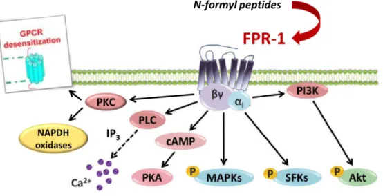
The expression of FPR1 receptor, expressed constitutively on surface of quiescent neutrophil, is rapidly up-regulated in response to a large range of proinflammatory stimuli. In response to these stimuli, specific receptors on the neutrophil surface are activated to trigger several downstream signalling pathways. For example, FMLP, a strong chemoattractant that induces neutrophil activation, can bind to FPR1 [26]. After binding,
Gi--18-
type G-protein is activated, following which the conversion of GDP to GTP induces the dissociation of α-subunits from βδ-subunits. Later, βδ-subunits activate both the phosphoinositide 3-kinase (PI3K) gamma and phospholipase C (PLC) beta signalling cascades. The latter are involved in the release of intracellular calcium stores. As the βδ-subunits activate and pull PI3K gamma toward the plasma membrane, the activities of Src-like tyrosine kinases are increased, further triggering MAPK signalling pathways. p38 MAPKs and Erk principally influence chemotaxis and FPR1-mediated transcriptional activity [76]. Chemoattractants also activate protein kinase C and trigger the assembly of NADPH oxidases to produce ROS. Moreover, cAMP is a vital secondary messenger for several cellular physiological functions. It can down-regulate immune responses, such as respiratory burst and degranulation, particularly in FMLP-activated neutrophils [77]. Raf serine/threonine kinases are major signal transducers of diverse extracellular stimuli that activate the MAPK signaling pathways. The receptor-mediated activation of the small GTPase Ras recruits Raf to the plasma membrane where Raf kinase activity is regulated [78]. The Raf-MEK-Erk signaling pathway is a protein kinase cascade that regulates cell growth, proliferation, and differentiation in response to growth factors, cytokines, and hormones [79].
Another important feature of the receptor is its desensitization upon ligand stimulation, preventing further stimulation [80]. When FPR1 is activated by a cognate ligand, it is phosphorylated by GPCR kinases, inducing the linking of arrestin molecules, which prevent further binding to G proteins, leading to its inactivation and internalization. The important inflammatory actions initiated by activation of FPR1, such as chemotaxis, ROS production and granule release, are underscored by its high expression in myeloid cells as neutrophils, monocytes and macrophages [81].
-19-
Figure 4. FPR-1 signal transduction
1.6.2 Mouse Fpr-1 Receptors
FPR1 has been identified in several species, like horse, rabbits, and rodents, with substantial differences in functional responses to formylated peptides [31]. In comparison to the three FPRs described in humans, the mouse genome encodes multiple FPR receptors from chromosome 17A3.2 [82]. Fpr1 is the murine orthologue of human FPR1, sharing 77% homology, expression on similar cell types, and induction of the same effects of neutrophil chemotaxis, degranulation, cytokine production and phagocytosis [82]. Despite the relatively high sequence homology of FPR1 between mice and humans, and the structure of the intracellular domain is highly conserved, there are distinct differences in the affinity of murine Fpr1 for fMLF, which is around 100 times less than that of its human counterpart [83]. This difference probably is attributed to modifications in the folding of the transmembrane and extracellular domains, as determined by the apposition of multiple non-contiguous residues [84]. While differences in the affinity of a receptor to prototypic E. coli derived fMLF are defined, it should be noted that murine Fpr1 remains a high-affinity receptor for other bacterial formylated peptides, in particular to Listeria monocytogenes, Staphylococcus aureus and mitochondria-derived formylated peptides [83]. Although knowledge of such differences is necessary, inferences from mouse disease models remain
-20-
valid and have significantly advanced our understanding of FPR biology and neutrophil function in both the physiological and pathophysiological states to date.
-21-
CHAPTER 2: Bronchiolitis obliterans (BO)
2.1 Introduction
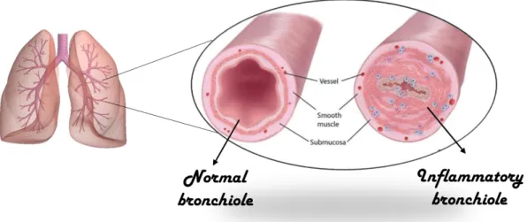
Bronchiolitis obliterans (BO) is a chronic irreversible obstructive pulmonary disease which results in obstruction of small airways [85]. Three main BO entities are distinguished: post infectious BO (PIBO), BO post lung transplantation, and BO after bone marrow transplantation (BMT) or hematopoietic stem cell transplantation (HSCT). All three entities are distinct, but exhibit similar histopathological features and development pathways [86]. BO is characterized by inflammation and fibrosis of the terminal and respiratory bronchioles leading to narrowing and/or total obliteration of the airway lumen following injury to the lower respiratory tract [86]. Depending on whether or not the fibrous proliferation is concentric or eccentric, the small airways can be partially or fully obstructed and the damage may extend beyond the epithelium and into the submucosa and/or adventitia [87].
There has been many confusion about the term bronchiolitis obliterans. The first descriptions on the characteristics of the BO coincide with what is today called bronchiolitis obliterans organizing pneumonia (BOOP) [88, 89]. Although the very similar terminology of BO and BOOP, these disease have very different histopathology. BOOP is a distinct histopathologic disease with bronchiolar intraluminal polyps consisting of fibroblasts rich in mucopolysaccharides that may extend into alveoli and alveolar ducts, contributing to organizing pneumonia. In fact, bronchiole involvement can be relatively minor [90]. Unlike BOOP, BO is a uniquely bronchiolar disease, lung parenchyma or the distal alveoli are not affected [91]. Instead, the bronchiolar submucosa is circumferentially thickened by collagenous scarring. This results in alteration of the airway structure and lumen narrowing. Additionally, Myers and Colby [92] suggested that BO is divided in two categories: proliferative and constrictive. Differentiation between the two categories was felt to be of clinical significance. The proliferative BO is characterized by obstruction of lumen of airways by polyps of granulation tissue. The constrictive form is characterized by
-22-
peribronchial fibrosis and partial or complete narrowing of lumen [86]. Bronchiolitis obliterans syndrome (BOS) refers to a form of constrictive bronchiolitis seen in transplant recipients, mainly lung or hematopoietic cell transplants [93, 94]. More recently, BOs has gained prominence as the principal cause of lung transplant failure beyond the immediate post-transplant period.
Figure 5. Airway lumen in normal condition and during BOS
2.2 Etiology, epidemiology and incidence
The etiology is often not determined with certainty, perhaps due to the fact that BO can be diagnosed well after the acute phase of disease. Infection remains the most commonly identified etiology of non-transplant BO, with adenovirus being the most prevalent infectious agent [95, 96], followed by Mycoplasma pneumonia [97]. Currently, the most common cause of BO is post-transplant BO, particularly after lung transplantation [98]. However, it have been identified several risk factors which predispose BO to develop more rapidly. Main among these is the occurrence of acute rejection [86].
The incidence of nontransplant BO is difficult to estimate due to its rarity. However, in a large retrospective review shown a prevalence in this selected sample of 0.6%. By contrast, post-transplant BO is relatively common and accounts for the majority of lung graft failures
-23-
outside of the immediate post-transplant recovery period [98]. Long-term survival of lung transplant patients is lower than for other solid organs transplantations, with five-year survival of about 50 percent. BOS remains the mainly reason of death after 1 year post-transplant (approximately 40% of paediatric deaths and 27% of adult deaths), but not only, it often affects more of 50% recipients within 5 years of transplantation [99].
2.3 Pathophysiology
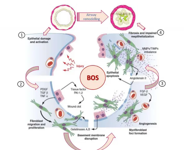
The pathogenesis of BO has not been completely understood. Since BO is a common cause of death after lung transplantation, a significant amount of effort has been expended trying to understand the pathogenesis of BO. BOS in lung transplant patients is characterized as a persistent reduction in forced expiratory volume in one second (FEV1), attributable to a combination of lung injury and chronic immune rejection of the transplant [100]. BO can be an injury response of repeated or chronic insults to the airway epithelium [101, 102]. Damage of epithelium seems important for development of pathology. Indeed, marked lymphocytic infiltration with loss of epithelium precedes complete fibrous obliteration of the graft airway, in a heterotopic tracheal allograft rat model [103]. Reseeding the grafts with epithelial cells significantly mitigates the obstructive changes, emphasizing the importance of disruption of epithelial layer to the development of BO [104]. In an interesting animal study, progression to BO was prevented by reepithelialization of the donor graft with recipient-derived epithelium, reinforcing the hypothesis that immune-mediated injury to the epithelium is key to BO development [105, 106]. Epithelial damage with a loss of integrity of the basement membrane could allow access to the mucosa of the airway by lymphocytes. Indeed, in the majority of biopsies of post-infectious BO was detect rises in both CD4 and CD8 lymphocytes [107]. Infiltrating CD4+ and CD8+ T-lymphocytes release a variety of inflammatory cytokines, notably Th1 cytokines such as interferon-γ and interleukin-2 (IL-2) [108]. As part of this inflammatory response, fibroblasts and myofibroblasts lay down collagenous scar tissue as reported by increases in profibrotic factors (24) and procollagen I
-24-
and III mRNA in BO animal models [109]. The accumulation of fibroblasts and myofibroblasts together to collagen deposition results in granulation tissue and eventually a fibrous scar that obstructs bronchioles (Fig. 6).
Recently there has been recognition of the importance of microvascular changes in BO development. Angiogenesis and vascular remodelling are critical aspects of the fibroproliferative process [110]. Additionally, transplant patients with BO show significant increases in airway microvascular vessel counts as opposed to patients without BO [111]. In rat tracheal allografts, overexpression of vascular endothelial growth factor (VEGF) results in luminal occlusion. This obstructive mechanism appears to operate through platelet-derived growth factor (PDGF) as inhibition of this pathway with imatinib (a PDGF tyrosine kinase inhibitor) prevents luminal occlusion [112]. However, the exact role of VEGF in human BO pathology still remains unclear. The etiology of BO in non-transplant patients is less clear but, similar to the post-transplant situation, is likely the result of a chronic inflammatory insult to the airway epithelium. Comparing an animal model of toxin-induced BO to a tracheal transplant model, both groups of animals show similar increases in cytokines including TGF-β and interferon-γ [113]. The histology of the BO is identical in the two models. However, the rapid increase in osteopontin seen after toxicant injury but not seen in the transplant model suggests that although the mechanisms leading to BO are similar, they are not necessarily identical. Instead, BO probably represents a “final common pathway” following severe and/or repeated airway injury [114]. This injury induces a primarily lymphocytic, T-cell–mediated inflammatory response that includes fibroproliferation leading to the classic circumferential scar formation and progressive obliteration of the airway lumen [107].
-25-
Figure 6. Pathophysiology of BOS
2.4 Pharmacotherapy
2.4.1 Non-Transplant Bronchiolitis Obliterans
The natural history of nontransplant BO is difficult to determine due to the varied etiologies and to relatively small numbers of affected patients. The most common form of nontransplant BO is post-infectious BO. No specific treatment exists for post-infectious BO. The use of both inhaled and oral corticosteroids is controversial. In some patients have been documented benefits, but lung inflammation can continues despite using on the steroids [115]. The use of bronchodilators also may be controversial in a pathology characterized as irreversible airway obstruction. However, regular use of bronchodilators can be helpful in some patients [116]. Patients with severe obstruction, hypoxia, and functional impairment may require lung transplantation.
The most common collagenvascular disease leading to BO is rheumatoid arthritis (RA) [117]. It is often unclear whether this is a direct result of RA or a result of the drugs used to
-26-
treat it, such as penicillamine or gold salts. Unlike post-infectious BO, which can stabilize or even have some remission, most patients with RA-associated BO die within 1 to 5 years [118, 119].Treatment with corticosteroids or other immunosuppressive agents has been disappointing. A small clinical trial showed daily use of erythromycin relieve or stabilize the symptoms [120].
2.4.2 Post-Transplant Bronchiolitis Obliterans
The greatest challenge in treating of BO was the attempt to halt its development, especially in patients receiving lung transplants. Despite improvements in immediate survival after transplantation, the ability to either prevent BO or slow down the development of BO was only marginal success. When BOS is diagnosed, the mainstay of therapy is optimization of the immunosuppressive treatment of the patient. More intensive immunosuppression carries important risks, like infection and malignancy. The traditional immunosuppressive regimen has consisted in cyclosporine, azathioprine and corticosteroids [121].
There has been evidence that tacrolimus and mycophenolate mofetil (MMF) may provide superior prevention and treatment of BO in lung-transplant patients. Tacrolimus is a calcineurin inhibitor similar to cyclosporine. It acts to inhibit lymphokine production by helper and cytotoxic T-lymphocytes [122]. Tacrolimus presumably affects BO by more effectively regulating the inflammation of the graft, which is the causal event that leads to BO. In vitro studies on cultured CD4+ T-lymphocytes show that tacrolimus is at least 100 times more potent than cyclosporine in inhibiting cytokine secretion [123]. Early prospective clinical trials in humans comparing tacrolimus to cyclosporine as part of post–lung transplant maintenance therapy demonstrated fewer episodes of acute rejection and development of BO [123]. The data supporting the superiority of tacrolimus (vs cyclosporine) is sufficiently convincing to prompt a strong trend among lung-transplant centers toward the use of tacrolimus as part of the post-transplant maintenance immunosuppressive therapy [94].
-27-
In addition, observational data from the Registry of the International Society for Heart and Lung Transplantation shows that the combination of tacrolimus and MMF has the lowest overall rate of rejection [121]. MMF is a purine synthesis antagonist (inosine monophosphate dehydrogenase inhibitor) that suppresses proliferation of T- and B-cells [124]. In prospective studies evaluating MMF versus azathioprine as part of maintenance immunosuppressive therapy, no substantial differences were noted in two different groups regarding the frequency of acute rejection or the progression of BO up to 3 years of follow-up [125]. However, MMF does appear better tolerated by patients than azathioprine. MMF has the lowest risk of acute rejection episodes in association with tacrolimus and has become the preferred purine synthesis antagonist (MMF: 46% of lung transplant patients at 1 year; azathioprine: 30%) [121].
Numerous other agents have been identified as treatments for BO. Steroids have always had a role in treat post-transplant BO, including high-dose pulse methylprednisolone and inhaled fluticasone propionate. Corticosteroids should be given early while the disease process is in the developing phase before airway fibrosis is complete [121, 126].
Inhaled therapies allow to delivering high doses of drugs to the airway while reducing systemic absorption and side effects. Currently, inhaled steroids are not recommended routinely but can be useful in individual patients. Other inhaled medications being explored include inhaled cyclosporine [127]. In a randomized controlled study of aerosolized cyclosporine started within 6 weeks of lung transplantation, significant improvements in overall survival and BOS-free survival were noted [128].To confirm the benefits of inhaled cyclosporine, further trials involving a greater number of patients are clearly needed. Additionally, it has been demonstrated that macrolide antibiotics have anti-inflammatory properties, which have been useful in the treatment of diffuse panbronchiolitis and cystic fibrosis. Recent trials of azithromycin in post-transplant patients have suggested similar
-28-
benefits. Azithromycin improves FEV1 and decreases airway neutrophilia in patients with reversible neutrophilic inflammation [129, 130].
Development of bronchiolar fibrosis is an important part of the pathology of BO. Antifibrotic drugs carry the promise to curb this feature of BO. The antifibrotic drug pirfenidone was able to prevent the onset of obstructive airway changes in a mouse model of BO and may serve as a model for future drugs development in humans [131]. The mammalian target of rapamycin (mTOR) is a downstream protein kinase of the PI3K–Akt signalling pathway [132]. mTOR may drive or contribute to fibromuscular proliferation. Antagonism of mTOR with medications such as sirolimus and everolimus may be beneficial. A pilot study of post-transplant BO patients added sirolimus to a calcineurin inhibitor and prednisone and demonstrated stabilization of pulmonary function but at the cost of significant adverse effects [133]. Everolimus used for maintenance therapy instead of azathioprine results in less episodes of acute rejection and BOS at 12 months post-transplant. At 24 months posttransplant, the BOS effect is lost and only the decline in acute rejection remains [134]. Similar to sirolimus, the side effects of the drug are substantial. The dosing and indications for the use of sirolimus and everolimus need to be more fully explored before they may become part of routine treatment.
2.3.3 Alternative strategies
Unfortunately, some patients have little or no response to conventional therapy and there is a need for alternative strategies; one such approach is extracorporeal photopheresis (ECP). ECP involves removing leukocytes from the blood, treating them with ultraviolet radiation in the presence of 8-methoxypsoralen (8-MOP), and returning the leukocytes to circulation. Although the exact mechanism are unclear, the process appears to preferentially induce apoptosis of activated immune cells, which are phagocytized after reinfusion, generate tolerogenic antigen presenting cells, and expand regulatory T cells [135]. A recent meta-analysis of non-invasive BO therapies found evidence that association of ECP with
-29-
established immunosuppression can stabilize lung function better than only standard therapy [94].
Total lymphoid irradiation (TLI) is another form of intensive immunosuppression typically reserved for patients with rapidly progressing disease, as it causes important side effects like pancytopenia and infection. The procedure entails irradiation of the major lymphatic beds of the body, thus supplying intense and unspecific immunosuppression [136]. Initial experience with this technology showed promising results in patients with cardiac and renal transplants. Some reports suggest that TLI reduces the rate of decline in lung function in patients with progressive BOS [87].
For patients who fail the therapies discussed above, retransplantation remains the only effective therapy. Given the paucity of suitable lung donors, retransplantation is a limited therapy for chronic rejection [137].
Nevertheless, these suboptimal outcomes following retransplantation highlight the need for more effective ways to treat BOS.
-30-
CHAPTER 3: Traumatic Brain Injury (TBI)
3.1 Introduction
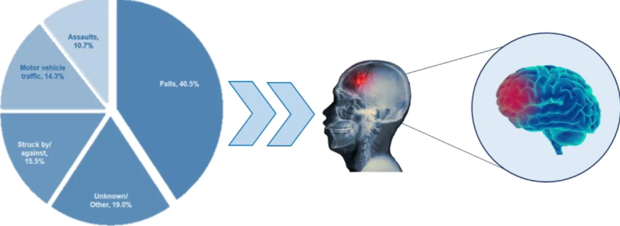
Traumatic brain injury (TBI) is defined as damage to the brain sustained after the application of external mechanical force that causes temporary or permanent functional or structural damage to the brain [138]. Brain injury can be mild, moderate and severe. It is not a distinct entity but a heterogeneous group of pathologies that are initiated by diverse mechanisms and have different survival consequences. Additionally, TBI can be typically classified as closed or penetrating [138]. Closed head injury typically describe automobile accidents, falls and assaults, while a penetrating head injury is generally caused by stab wounds or gunshot (Fig.7). Regardless of origin, TBI sufferers experience a series of symptoms related the injury: confusion, dizziness and occasionally loss of consciousness (mainly in severe injury) [139]. Also after the initial TBI has been treated and resolved, about 70–80% of patients experience long-lasting symptoms, like changes in personality and behaviour, including depressive-like behaviours and anxiety [140, 141]. It has been well demonstrated that TBI represents a process, which once initiated, can extend either silently or symptomatically to neurodegeneration. This may lead to early onset of Parkinson’s disease (PD), dementia, and other degenerative disorders [142-144]. In particular, TBI is an important risk factor for development of Alzheimer’s disease (AD) or earlier AD onset [145]. Moreover, TBI is associated with the development of chronic traumatic encephalopathy (CTE) in athletes, especially following repeated concussive TBI [146].
In the last decades, TBI mechanisms have been investigated through numerous experimental models e (i.e., controlled cortical impact, overpressure blast injury and the fluid percussion models), which display histological, physiological and neurological changes similar to those observed in clinical brain injury [147]. Although animal models do not replicate all the physiological, anatomical, and neurobehavioral characteristics of human TBI, they are
-31-
essential to clarify underlying injury mechanisms and to assess the safety and efficacy of novel therapies prior to clinical trials.
Figure 7. Main causes of TBI
3.2 Etiology, epidemiology and incidence
As mentioned previously, the major causes of TBI are falls, motor-vehicle traffic incidents or assaults (Fig.7). A variable proportion of TBIs occur during sports or as a result of war wounds.
TBI represents an important public health concern in USA and worldwide, based on incidence, prevalence, healthcare resource utilization, resulting death and disability, and total economic cost [148]. In the United States, 1.7 million people suffer from TBI each year, with about 235,000 patients needing hospitalization and specialized healthcare. In 2000, there were 10,958 TBI diagnoses. In 2015, this number jumped to 344,030, with rising data in the following years. Statistically, the Center for Disease Control and Prevention (CDC) has estimated that annually, about 1.5 million Americans survive a TBI [146]. Mortality is approximately 3% for all severities of TBI, but morbidity is more difficult to estimate [149]. However, these statistics underestimate the incidence of TBIs, because most head injuries are mild and are often overlooked by the medical profession. Furthermore, these statistics do not account for all individuals who have not receive medical treatment,
-32-
had outpatient or office-based visits, or who received care in federal structures (i.e., persons serving in the U.S. military or receiving care at Veterans Affairs hospitals) [150].
According to the latest data from the CDC, rates of TBI are highest among babies aged 0 to 4 and children aged 15 to 19. Older adults aged 75 and over also have a high TBI prevalence and they represent the highest rate of hospitalizations and death associated to TBI [149]. Moreover, according to epidemiological literature, TBI is more frequent in males than females. Males were 1.4 times more likely to have TBI; indeed, they had an approximate annual incidence of TBI of 998,176 compared to 693,329 for females. Furthermore, the rate of TBI in males was highest in all age groups [149].
3.3 Pathophysiology
The pathological and inflammatory features of TBI vary based on the severity (mild vs. moderate vs. severe) and duration (single vs. repetitive) of the injury [151, 152], as well as conditions such as age, gender, genetics, and medications [153].
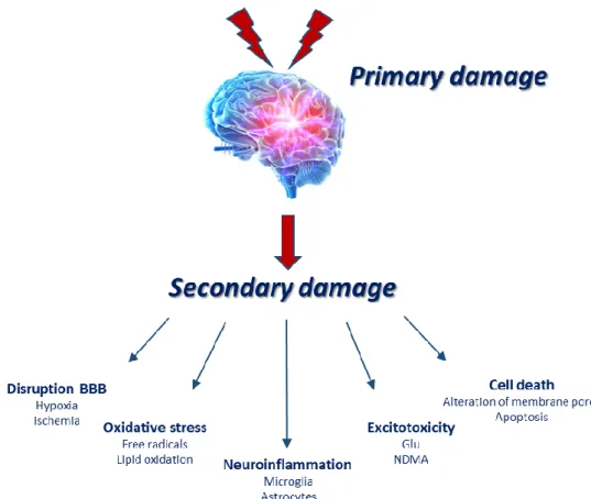
TBI instigates complex pathological mechanisms involving a wide variety of molecular and cellular pathways. Brain damage associated to TBI can be divided into two phases (Fig. 8). First, an initial primary damage phase occurs at the moment of insult, as a direct consequence of physical and mechanical impact on the brain. This may involve contusion, brain swelling, diffuse axonal injury and intracranial haemorrhage, and eventually contribute to immediate cell death [154]. This is followed by a prolonged secondary phase involving cascades of biological mechanisms triggered at the time of trauma, which can last over much longer periods, from days to many weeks [138]. This delayed phase, triggered by numerous molecular and cellular responses initiated in an attempt to potentially restore cell homeostasis of the injured tissue, is not particularly well regulated and can often lead to an exacerbation of primary damage, neurodegeneration and cell death [155, 156]. Hallmarks of the secondary response can include breakdown of blood-brain barrier (BBB), glutamate excitotoxicity, oxidative stress and neuroinflammation [147].
-33-
Behaviourally, these alterations manifest as post-traumatic headache, depression, individuality changes, anxiety, aggression, and deficits in attentiveness, cognition, sensory processing, and communication [157, 158]. A TBI in a patient may result in coma or death, seizures, or cognitive and behavioural disabilities.
3.2.1 TBI and Neuroinflammation
Neuroinflammation occurs at both primary (acute) and secondary (chronic) stages of TBI [138]. It seems responsible for both adverse and beneficial effects; indeed, neuroinflammation may cause to primary and secondary damage but, at the same time, facilitate tissue repair. In this regard, cellular inflammatory responses are activated in the injury site after the primary insult, with the purpose to repair the injured tissue; however, the excessive production of cytokines may become an important factor for TBI pathological progression [159]. The development of neuroinflammation after TBI involves a complex mechanism of cumulative changes occurring within the brain. After TBI, multiple types of quiescent glial cells of are rapidly triggered through a process called “reactive gliosis”. In turn, the activation of microglia induce astrocytic activation by producing and releasing inflammatory mediators, which in turn act on neurons and surrounding cells [147]. These pro-inflammatory mediators not only affect surrounding neurons and glia but additionally recruit peripheral immune cells, such as neutrophils, lymphocytes and macrophages into brain [159]. Nevertheless, in the chronic phase, excessive production of inflammatory mediators triggers secondary cell death contributing to injury in the brain circuit. Different mechanisms of secondary cell death drive brain damage. Among these, excitotoxicity is a process characterized by increased levels of neurotransmitters and glutamate in the synaptic space, which stimulate the surrounding nerve cells N-methyl-d-aspartate (NMDA) and a-amino-3-hydroxy-5-methyl-4-isoxazolepropionic acid (AMPA) receptors [160]. These receptors remain activated, favouring the influx of both sodium and calcium ions into cells [161]. In cytosol, a high concentration of calcium ions determines the activation of protein
-34-
phosphatases, phospholipases, and proteases. These activations can damage DNA, membranes and proteins. Furthermore, overexcitement of glutamate receptors stimulates the production of nitrogen oxide (NO), free radicals, and pro-death transcription factors. High ROS levels cause lipoperoxidation of the cellular membrane, leading to mitochondrial dysfunction and oxidizing proteins, which may cause the alteration in the structure of membrane pores [138].
Although elaborate cellular interactions are involved in TBI physiopathology, activation of microglia and subsequent recruitment of macrophages constitute the main inflammatory response of the immune system, 48–72 h after TBI. Moreover, although the peripheral immune response mitigates within 2–3 weeks, microglia and macrophages activation persists for a long time, for months to years after the initial injury [162, 163]. Thus, dysregulated activation of microglia/macrophages can not only exacerbate lesion pathology, but can also result in chronic bystander tissue damage [164].
-35- 3.4 Pharmacotherapy
As the primary damage that represents the direct mechanical insult, can not be mended, therapeutic targets focus on the secondary injury. The major contributor to secondary damage is the neuroinflammation mainly characterized by chronic microglia and astrocytes activation, secretion of pro-inflammatory cytokines and oxidative stress. So far, the preclinical and clinical studies have primarily focused on neuroprotective approaches with the purpose to prevent and reduce secondary injury in brain after TBI. It was reported that it is fundamental to start the therapeutic interventions immediately after TBI, in particular within 4 h of injury, in order to achieve the best promising neuroprotective outcome [138].
3.4.1 Therapeutic strategies in pre-clinical studies
Many preclinical studies have tested therapeutic efficacy of drugs in animal TBI models by targeting secondary injury mechanisms including corticosteroids, calcium channel blockers, N-methyl D-aspartate (NMDA) receptor antagonist, excitatory amino acid inhibitors, free radical scavengers and growth factors.
In particular, corticosteroids have been used as treatment for head injuries since at least three decades because they appear to reduce intracranial pressure (ICP) after TBI [165]. Corticosteroid examples include dexamethasone and methylprednisolone.
Synthetic agonists of peroxisome proliferator-activated receptor (PPAR) also used as an anti-inflammatory, therapeutic agents for TBI [166]. Fenofibrate, a PPAR-α receptor agonist, reduces cerebral edema, inflammation and oxidative stress by reducing behavioural deficits following TBI induction [167]. Rosiglitazone and pioglitazone, other PPAR-α receptor agonists, decrease microglial activation, histological and behavioural alteration and increase neuroprotective antioxidant proteins, induced after TBI [168].
Another approach to TBI treatment is blocking glial proliferation by inhibiting the cell cycle.
Through cyclic-dependent kinases (CDKs) inhibition, flavopiridol is able to reduce lesion volume and promote sensorimotor cognition and the recovery after TBI [169]. Roscovitine,
-36-
another inhibitor of cell cycle, also modulates CDK and showed moderate neuroinflammation and neurodegeneration after injury [170].
In addition, N-acetylcysteine (NAC) could act as an anti-inflammatory drug, especially in mild TBI, apparently through its antioxidant capacity [171]. Indeed, in animal models of TBI, NAC showed a potent antioxidant activity, decreasing markers of oxidative stress and increasing glutathione levels. Moreover, NAC was able to decrease activation of NF-κB, thus decreasing pro-inflammatory cytokines levels [172]. NAC also reduces lesion volume and BBB breakdown after TBI [173]. Importantly, the safety and potential therapeutic efficacy of NAC was effectively evaluated in a phase I randomized clinical trial [174].
3.4.2 Therapeutic strategies in clinical trials
Some therapeutic strategies for TBI management have already advanced into clinical trials. Erythropoietin (EPO), a physiological protein, plays an important role in stimulating the differentiation, maturation and survival of hematopoietic progenitor cells. While EPO and its receptor (EPOR) are weakly expressed in normal brain, their expression is greatly increased in neuronal progenitor cells, neurons, glia and cerebrovascular endothelial cells in response to multiple types of cell damage. EPO demonstrated potential neuroprotective effects in most experimental models of TBI [175]. However, in clinical trial with patients with severe TBI, the administration of EPO failed to improve outcomes at six months [176]. Thus, although EPO has demonstrated neuroprotective proprieties in preclinical studies, its effectiveness as a medical strategy is questionable.
Statins, potent inhibitors of cholesterol biosynthesis, promote the recovery following TBI. Many of the pleiotropic effects of statins are cholesterol independent, such as improvement of endothelial function, antioxidant properties, inhibition of inflammatory responses, immunomodulatory actions, regulation of angiogenesis, neurogenesis and synaptogenesis [138]. Such effects target pathways that affect the acute as well as chronic phases of brain damage. For example, simvastatin inhibits the activation of caspase-3 and apoptosis of cells,
-37-
thereby increasing neuronal survival after TBI. Moreover, it increases expression of several growth factors, induces neurogenesis and controls the restoration of mental function in rats after TBI [177]. Atorvastatin administration after TBI significantly decreases neurological functional deficits and enhances neuronal survival [178]. Additionally, it induce synaptogenesis and angiogenesis in the boundary zone of the lesion and in the CA3 regions of the hippocampus in rats subjected to TBI. However, Food and Drug Administration reported cognitive side effects associated with statins treatment [138]. Given these contradictory results, further clinical trials are necessary to validate the neuroprotective effects of treatment with statins.
Progesterone is a steroid produced in the brain, besides being synthesized in reproductive organs and adrenal glands. Progesterone has pleiotropic effects, and thus has several candidates for mechanisms of action with regard to its potential therapeutic efficacy in TBI [179]. Multiple pre-clinical models of TBI have demonstrated neuroprotective properties of progesterone and have shown that it enhances behavioural and functional outcomes, decreases cerebral edema, apoptosis, pro-inflammatory cytokines, and other inflammatory markers, and prevents neuronal cell death [180]. Progesterone has also demonstrated clinical improvement in two phase II randomized, controlled trials. Despite positive results from preclinical studies and phase II clinical trials, two phase III clinical trials on progesterone treatment in acute TBI ended with negative data [181, 182]; therefore, the results continue to fail in TBI clinical trials.
3.4.3 Alternative strategies
Besides pharmacological therapies for TBI, innovative developments based on preclinical results are focused on the practice of biologics (e.g., gene therapy, stem cells, microRNA, peptide therapy, exogenous growth factors). Neural and mesenchymal stem cell therapy shows neuroregenerative and neurorestorative potential [183]. A recent study explored the association of stem cells with drug therapies to overcome the limits related to stem cell
-38-
transplantation. To date, erythropoietin, statins and progesterone have shown the most promising results for the endogenous stem-cells-mediated repair [184].
Furthermore, growth factors attract considerable attention for their neuroprotective and neuroregenerative efficiency. In particular, it has been shown that VEGF, human fibroblast growth factor 2 (FGF2), and nerve growth factor (NGF) improve neuronal survival when accompanying transplanted stem cells in injured models [185]. In particular, VEGF and FGF2 improve functional outcomes, while NGF reduces brain edema and production of beta-amyloid in animal models of TBI. TBI usually affects brain functions, for example executive actions, attention, memory, cognitive grade and language skills [138].
Neuropsychological rehabilitation (NR) is aimed at reducing cognitive, emotional and behavioural deficits induced by TBI. Additionally, neurotherapy can promote neuroplasticity [186]. Additionally, transcranial magnetic stimulation (TMS) as a way of non-invasive direct modulation of neuronal activity seems to be efficient for treatment of TBI [187]. Nevertheless, more researches are needed.
-39-
CHAPTER 4: Aim
4.1 Targeting FPR-1 as an emerging pharmacological strategy for inflammatory diseases
As described previously, FPR1 can trigger several signaling pathways for immune reactions during inflammation. Formyl peptides bind to FPRs on neutrophils, activate Gi proteins, and induce the G-α subunit to dissociate from the G-βγ subunits. Gβγ subunits mediate downstream responses such as calcium influx, PI3K, PLC, Akt and MAPK. The phosphorylation of ERK, JNK, and p38 MAPK mediate FPR1-mediated transcriptional activity and chemotaxis, and it promotes a chemokine/chemoattractant-triggered function as a pro-inflammatory response [188]. Upon the activation of these signals, neutrophils respond with chemotaxis, migration, translocation, phagocytosis, respiratory burst, and degranulation [76]. Moreover, activation of FPR1 has been shown to trigger activation of the NF-kB pathway. NF-kB is a chief regulator of inflammation, it controlling various cellular processes such as apoptosis, cell proliferation, the secretion of cytokines, and oxidative stress [189]. In a normal condition, it is bound by the inhibitor protein IkB-α, which sequestered it into the cytoplasm. Several external stimuli induce the degradation of IkB-α, releasing NF-kB from the complex and allowing migration into the nucleus. Here, it activates the transcription of target genes involved in the enhancement of the inflammatory process, including the NLRP3 inflammasome. Inflammasomes are cytosolic protein complexes implicated in the induction of innate immune/inflammatory response [190]. At present, five inflammasomes have been identified; of these, NLR Family Pyrin Domain Containing 3 (NLRP3) is the most studied due of its possible involvement in several human diseases [191]. This complex contains NLRP3, a NOD-like receptor that is a sensor for inflammasome activation, and an apoptosis-associated speck-like protein containing a CARD complex (ASC), through which it binds pro-caspase. Pro-caspase, in turn, is cleaved in caspase-1 (Casp-1), which is a protease involved in apoptosis. It also controls the inflammatory
-40-
response by release of cytokines. Indeed, Casp-1 is responsible of cleavage of pro-interleukin (IL)-1β and pro-IL-18 into the respective active cytokines [191]. Both cytokines induces signal transduction cascades in inflammatory pathways in several cell types.
The therapeutic potential of FPRs modulation has been confirmed in experimental models of acute or chronic inflammatory diseases, such as colitis and endometriosis [17, 18].
In light of the above, we investigated the effects of Fpr-1 deletion in two different models of inflammation in vivo: BOS in chronic and TBI in acute and chronic.
Considering that BOS is a disease of inflammatory nature, we performed an experimental study to explore the molecular and cellular processes involved in airway repair and regeneration through FPR and NLRP3 pathways. Additionally, several studies have found that FPR1 was expressed in neurons [16]. In this regard, in the second study, we decide to investigate the effect of genetic deficiency of Fpr-1 in mice subjected to TBI from the early stage of acute inflammation to neurogenesis 4 weeks after injury.
-41-
CHAPTER 5: Materials and methods
5.1 Materials and methods for BOS study 5.1.1 Animals
IL-1β/IL-18 double KO mice were obtained from Arturo Zychlinski (Max Planck Institute, Berlin, Germany), while Casp-1 KO mice were obtained from The Jackson Laboratory (Bar Harbor Maine, USA). Fpr-1 KO mice on the C57BL/6 genetic background and C57BL/6 animals, used as WT controls, were acquired from Envigo (Milan, Italy). All mice located in a controlled environment and provided with standard rodent chow and water. Animals were housed separately by genotype, with five mice per cage. The University of Messina Review Board for the care of animals approved the research (9 February 2017, 137/2017-pr). Animal care was in conformity with current legislation for the protection of animals used for scientific purposes (Directive 2010/63/EU).
5.1.2 Experimental Model of Tracheal Transplantation
Tracheas were transplanted as previously described [192]. Briefly, the trachea was removed from donor mice by an anterior middle incision. The resected trachea was immediately placed in ice-cold phosphate-buffered saline (PBS) with streptomycin sulfate (100 µg/mL) and penicillin-G sodium (100 U/mL) (Life Technologies). The receptor was subjected to inhalation anaesthesia with isoflurane in a titrated dose to reach analgesia with spontaneous breathing, and a 0.5 cm horizontal incision was made in the dorsal suprascapular area. Then, the donor trachea was sewn and the overlying skin was closed. The grafts were collected 4 weeks after transplantation.
5.1.3 Experimental Groups
Mice were casually allocated into following groups (n = 10):
The BOS WT group: mice were subjected to tracheal transplantation as described above.