EXTENDED REPORT
Anti-chromatin antibodies in systemic lupus
erythematosus: a useful marker for lupus nephropathy
R Cervera, O Viñas, M Ramos-Casals, J Font, M García-Carrasco, A Sisó, F Ramírez,
Y Machuca, J Vives, M Ingelmo, R W Burlingame
. . . . Ann Rheum Dis 2003;62:431–434
Background:Anti-chromatin antibodies have recently been described in patients with systemic lupus erythematosus (SLE) and it has been suggested that their presence is associated with lupus nephritis. Objective:To assess the prevalence and clinical associations of these antibodies in SLE.
Methods:The presence of anti-chromatin antibodies in 100 patients with SLE was investigated by an enzyme linked immunosorbent assay (ELISA). To determine the specificity of these antibodies, 100 patients with primary Sjögren’s syndrome, 30 with primary antiphospholipid syndrome (APS), 10 with systemic sclerosis, and 100 normal controls were also tested.
Results:Positive levels were detected in 69/100 (69%) patients with SLE. In contrast, they were found in only 8/100 (8%) of those with primary Sjögren’s syndrome, in 1/10 (10%) with systemic sclerosis, in 2/30 (7%) with primary APS, and in none of the 100 healthy controls. Patients with anti-chromatin antibodies had a twofold higher prevalence of lupus nephropathy than those without these antibodies (58% v 29%, p<0.01). A significant correlation was found between the levels of chromatin anti-bodies and disease activity score as measured by the European Consensus Lupus Activity Measurement (ECLAM; p=0.011).
Conclusions:The measurement of anti-chromatin antibodies appears to be a useful addition to the laboratory tests that can help in the diagnosis and treatment of SLE. These antibodies are both sensitive and specific for SLE, and are a useful marker for an increased risk of lupus nephritis.
S
ystemic lupus erythematosus (SLE) is the prototypic autoimmune connective tissue disease. It may affect any organ of the body and display a broad spectrum of clini-cal and immunologiclini-cal manifestations. However, it is now thought that patients with SLE can be divided into more homogeneous subsets of pathogenic, therapeutic, and prog-nostic significance.1The presence of certain autoantibodies is one of the factors associated with some symptoms of the disease and aids in the classification of patients with SLE into specific subsets. This is the case for antiphospholipid antibodies, which are clearly associated with the development of thrombotic events and obstetric morbidity,2
anti-ribonuclear protein (RNP) antibod-ies that are markers of myositis and Raynaud’s phenomenon,3
and anti-SSA/Ro that are associated with congenital heart abnormalities in newborn infants.4
One of the most severe events in the course of SLE is the development of glomerulonephritis,5
and much effort has been spent to find a useful and early marker of this complication. Anti-dsDNA antibodies are often associated with lupus nephritis,6
and the presence of anti-dsDNA is a hallmark of SLE.7 8
However, evidence has accumulated in recent years that anti-chromatin autoantibodies are correlated even better with lupus nephritis than anti-dsDNA.9–12
Chromatin is the native histone-DNA complex found in the nucleus of eukaryotic cells, and it is organised into a repeating series of nucleo-somes. Anti-chromatin-chromatin immune complexes can bind to the glomerular basement membrane in vivo.13
Chromatin (or nucleosomes) is an antigen for T and B cells from patients with SLE.12 14
Additionally, chromatin anti-bodies are a ubiquitous feature of murine lupus,15
and are nec-essary but not sufficient for the development of glomerulo-nephritis in one strain of mouse.16 It was found that
chromatin always preceded the appearance of anti-dsDNA antibodies in two strains of mice, and the suggestion
was made that anti-dsDNA antibodies were a subset of anti-chromatin antibodies.15
In this study we investigated the prevalence of both anti-dsDNA and anti-chromatin antibodies in a large series of patients with SLE in order to assess their clinical significance and, particularly, their value as a marker of lupus nephropathy. To determine the specificity of these antibodies for SLE, a large number of patients with other connective tissue diseases were tested for anti-chromatin antibodies. It was also possible to follow the antibodies over time from serial bleeds of some patients with SLE.
PATIENTS AND METHODS
Patients and controls
Clinical and laboratory features of 100 consecutive and unse-lected patients (93 female, seven male; mean (SD) age 37 (14) years, range 11–70 years) with SLE were prospectively studied. All fulfilled four or more of the 1982 American College of Rheumatology (ACR) revised criteria for the classification of SLE.8
The disease control groups consisted of 100 patients with primary Sjögren’s syndrome (classified according to the Euro-pean criteria),1730 with primary antiphospholipid syndrome
(APS) (categorised according to the preliminary criteria for the classification of APS),18
and 10 with systemic sclerosis (classified according to the ACR preliminary criteria).19
The normal control group consisted of 100 healthy blood donors from the blood bank of our hospital.
. . . .
Abbreviations:ACR, American College of Rheumatology; APS, antiphospholipid syndrome; CI, confidence interval; ECLAM, European Consensus Lupus Activity Measurement; ELISA, enzyme linked
immunosorbent assay; OR, odds ratio; SLE, systemic lupus erythematosus See end of article for
authors’ affiliations
. . . .
Correspondence to: Dr R Cervera, Servei de Malalties Autoimmunes, Hospital Clínic, Villarroel 170, 08036-Barcelona, Catalonia, Spain; [email protected] Accepted 25 September 2002 . . . . 431 www.annrheumdis.com
Definition of clinical features and disease activity The patients had been attending our institute either as in- or outpatients between 1999 and 2000. All had documented medical histories and underwent a medical interview as well as a routine general physical examination by a qualified internist. A serum sample from each patient was collected for the immunological tests. Clinical and serological characteris-tics of all these patients were collected in a protocol form. Salient features included in this protocol were: (a) gender, (b) age, (c) laboratory features, and (d) clinical manifestations at the time blood was drawn. Information collected in the proto-col forms was transferred to a computerised database program. The study was performed according to the principles of the Declaration of Helsinki.
The clinical manifestations evaluated in this protocol were defined according to the recommendations of the ACR glossary committee.20
Specifically, nephropathy was consid-ered when patients presented (a) persistent proteinuria >0.5 g/day or greater than 3+ if measurement was not performed; or (b) cellular casts (may be red cell, haemoglobin, granular, tubular, or mixed); or (c) otherwise unexplained rise in serum creatinine >75µmol/l. Renal biopsies were reviewed by two pathologists and categorised according to the modified classi-fication proposed by the World Health Organisation21
: type I—normal kidney; type II—mesangial glomerulonephritis (presence of mesangial deposits with mesangial hypercellu-larity); type III—focal proliferative glomerulonephritis (in-flammatory changes affecting some glomeruli but leaving other unaffected); type IV—diffuse proliferative glomerulo-nephritis (virtually all glomeruli show inflammation); type V—membranous nephropathy (diffuse generalised thickening of the capillary wall and predominant intramembranous and/or subepithelial electrodense deposits, without inflamma-tory changes).
Disease activity was assessed by the European Consensus Lupus Activity Measurement (ECLAM).22
Detection of autoantibodies
Anti-chromatin antibodies of the IgG isotype were measured by a commercial semiquantitative enzyme linked immuno-sorbent assay (ELISA; INOVA Diagnostics Inc, San Diego, CA) according to the manufacturer’s instructions. Antinuclear antibodies were determined by indirect immunofluorescence using triple tissue cryostat sections (liver-stomach-kidney) and Hep-2 cells as substrate (Euroimmun). Anti-dsDNA anti-bodies were determined by Farr’s ammonium sulphate precipitation technique (Amerlex, Trinity Biotech, Ireland).23
Antibodies to extractable nuclear antigens of the IgG isotype, including Ro(SSA), La(SSB), U1-snRNP, and Sm were detected by ELISA (Captia, Trinity Biotech, Ireland). Rheuma-toid factor was detected by nephelometry (Behring). Anticar-diolipin antibodies of the IgG and IgM isotypes were measured by an ELISA, as previously described.23
The lupus anticoagulant activity was detected by coagulation assays, fol-lowing the guidelines of the International Society on Throm-bosis and Haemostasis (Scientific Subcommittee on Lupus Anticoagulants/Phospholipid Dependent Antibodies).24
Statistical analysis
Conventionalχ2and Fisher’s exact tests were used for
analys-ing qualitative differences, and Student’s t test was used for comparison of means, assuming similar variance in independ-ent samples. A value of p<0.05 was taken to indicate signifi-cance. When several independent variables appeared to have statistical significance in the univariate analysis, a logistic regression test was performed for multivariate analysis to rule out possible confounding variables. Only those variables showing statistical significance in the multivariate analysis were considered significant in this study. The odds ratio (OR) was calculated for assessing the risk of appearance of each
variable. A lower limit of the 95% confidence interval (CI) that exceeded 1.0 was taken to indicate statistical significance in the case of positive association and an upper limit lower than 1.0 in the case of negative association. Results of the analysis of continuous variables are indicated as mean (SD). Linear regression analysis was performed for comparison of continu-ous variables. This statistical analysis was performed by the SPSS and STATCALC programs using the information stored in the database program.
RESULTS
Prevalence of anti-chromatin antibodies
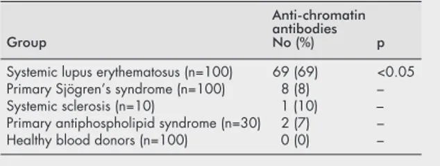
Positive levels of anti-chromatin antibodies (>20 U) were detected in 69/100 (69%) patients with SLE. In contrast, they were found in only 8/100 (8%) patients with primary Sjögren’s syndrome, in 1/10 (10%) patients with systemic sclerosis, in 2/30 (7%) patients with primary APS, and in 0/100 (0%) healthy blood donors (table 1).
Relationship between anti-chromatin antibodies and clinical features
Fifty two (52%) patients with SLE had clinical evidence of lupus nephropathy at the time of the protocol study. Renal biopsies performed at the time of clinical diagnosis of renal involvement had disclosed type IV lesions in 19 patients, type III in 15, type II in 11, and type V in 7. Forty two of the patients with nephropathy were positive for anti-chromatin antibod-ies, yielding a sensitivity of these antibodies for lupus nephropathy of 81% and a specificity of 39%. Patients with anti-chromatin antibodies had a twofold higher prevalence of lupus nephropathy than those without these antibodies (58%
v 29%, p<0.01; OR=3.4, 95% CI 1.3 to 9.3). The mean level of
anti-chromatin antibodies in patients with lupus nephropathy was 68 U and in patients without nephropathy 42 U (p<0.01). No differences in the prevalence of the other clinical mani-festations were found among patients with and without anti-chromatin antibodies (table 2).
Table 1 Prevalence of anti-chromatin antibodies in systemic autoimmune diseases and controls
Group
Anti-chromatin antibodies No (%) p Systemic lupus erythematosus (n=100) 69 (69) <0.05 Primary Sjögren’s syndrome (n=100) 8 (8) – Systemic sclerosis (n=10) 1 (10) – Primary antiphospholipid syndrome (n=30) 2 (7) – Healthy blood donors (n=100) 0 (0) –
Table 2 Prevalence of active SLE clinical manifestations in patients with and without
anti-chromatin antibodies. Results are shown as No (%)
Manifestations Anti-chromatin antibodies p Positive (n=69) Negative(n=31) Cutaneous 30 (43) 14 (45) NS Arthritis 22 (32) 10 (32) NS Serositis 2 (3) 0 (0) NS Haematological 4 (6) 2 (6) NS Nephropathy 40 (58) 9 (29) <0.01 Central nervous system 1 (1) 0 (0) NS Thrombosis 1 (1) 0 (0) NS NS, not significant.
432 Cervera, Viñas, Ramos-Casals, et al
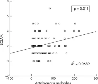
A significant correlation was found between the levels of anti-chromatin antibodies and disease activity score as measured by ECLAM (p=0.011, R2
=0.0689) (fig 1). Relationship between anti-dsDNA antibodies and clinical features
Positive levels of anti-dsDNA antibodies (>15 U) were found in 55 (55%) patients with SLE but in none of the other groups of patients. Thirty nine of the 52 patients with nephropathy were positive for anti-dsDNA, yielding a sensitivity of these antibodies for lupus nephropathy of 75% and a specificity of 63%. Patients with anti-dsDNA antibodies were also found to have a higher prevalence of lupus nephropathy than those without these antibodies (71% v 32%, p<0.001; OR=5.4; 95% CI 2 to 14.8). No differences in the prevalence of the other clinical manifestations were found among patients with and without anti-dsDNA antibodies.
Relationship between anti-chromatin antibodies and anti-dsDNA and other serum autoantibodies
Forty nine patients had positive levels of both anti-dsDNA and anti-chromatin antibodies (30/ 49 (61%) had lupus nephropa-thy), but 20 patients had anti-chromatin without anti-dsDNA
antibodies (8/20 (40%) had lupus nephropathy), while only six had anti-dsDNA without anti-chromatin antibodies (3/6 (50%) had lupus nephropathy). Additionally, a significant cor-relation was found between the levels of chromatin anti-bodies and those of anti-dsDNA antianti-bodies (p<0.0001, R2
=0.189) (fig 2).
Serial determinations of both chromatin and anti-dsDNA antibodies in three patients who developed lupus nephropathy during the study disclosed that anti-chromatin antibodies appeared before anti-dsDNA antibodies in one of them (fig 3).
No correlation was found between anti-chromatin antibod-ies and the presence of antinuclear antibodantibod-ies, antiphospho-lipid antibodies, antibodies to extractable nuclear antigens, or rheumatoid factor.
DISCUSSION
Lupus nephropathy is a common complication of SLE that can greatly influence the prognosis. Clearly, the mortality rate is higher for patients with SLE with nephritis than in those without renal disease,5 22 25
and some 10–60% of patients with SLE with nephritis (depending on genetic, socioeconomic, and treatment differences) eventually develop end stage renal fail-ure that requires dialysis or transplantation.26
Therefore, much effort has been spent in finding a useful and early marker of this complication. In this study we found that anti-chromatin antibodies were clearly associated with the presence of lupus nephritis with a sensitivity of 81%, which was slightly higher than the 75% sensitivity of anti-dsDNA antibodies. Addition-ally, a significant correlation was found between the levels of anti-chromatin antibodies and disease activity.
Most studies have found that anti-chromatin/nucleosome antibodies are quite sensitive and specific for SLE,9–12 27
but a few have found a high prevalence in other diseases.11 28 29
In the current study anti-chromatin showed a sensitivity of 69% in the patients with SLE. There was a specificity of 100% in the normal group, and 92% in the disease control group. These results agree with the group of studies showing both high sensitivity and specificity. Also in agreement with these stud-ies, chromatin showed a higher prevalence than anti-dsDNA in patients with SLE. There are three reasons that some studies showed high anti-chromatin reactivity in patients with systemic sclerosis. In one case whole nucleo-somes were used that contained small amounts of the Scl-70 antigen. The patients with systemic sclerosis who were anti-Scl-70 positive were low positive on this preparation.29
. In another case, the antigen was denatured H2A plus denatured H2B added to DNA (personal communication).28
In this case the sera from patients with systemic sclerosis were reacting with epitopes on the denatured histones that did not reconsti-tute into their native conformations. In a third study11
a cut off between positive and negative for anti-chromatin was chosen
Figure 1 Correlation between the levels of anti-chromatin antibodies and SLE disease activity score as measured by ECLAM.
Figure 2 Correlation between the levels of anti-chromatin antibodies and those of anti-dsDNA antibodies.
Figure 3 Serial determinations of both anti-chromatin and anti-dsDNA antibodies in a patient who developed lupus nephropathy during the study period, showing that anti-chromatin antibodies appeared before anti-dsDNA antibodies.
Anti-chromatin antibodies in SLE 433
that was two standard deviations above the average of a non-disease group. Because a non-non-disease group does not display a Gaussian distribution of binding, this was not the appropriate statistical method to choose a cut off (it was too low). When H1-stripped chromatin or nucleosome core particles are used as the antigen, and an appropriate cut off between positive and negative is used, virtually no patients with systemic scle-rosis are positive for anti-chromatin antibodies.27 29
In two studies using ELISA to measure anti-dsDNA and anti-chromatin antibodies in both human and murine lupus, no samples were positive for anti-dsDNA and negative for anti-chromatin antibodies,9 15
suggesting that anti-dsDNA were a subset of anti-chromatin. In this study, using the Farr assay to measure anti-dsDNA antibodies, six samples were anti-dsDNA positive but anti-chromatin antibody negative. The most likely explanation is that some antibodies recognise structures of DNA that can occur in protein-free DNA in solu-tion but do not occur in the DNA wrapped around the histones in chromatin that is bound to the solid phase of the ELISA plate. None the less, in this study at least one patient did develop chromatin antibodies before developing anti-dsDNA. Additionally, 49 of the 55 anti-dsDNA positive samples were also anti-chromatin positive. Both these findings are consistent with the concept that most of the anti-dsDNA antibodies in patients with SLE are a subset of anti-chromatin antibodies.
The measurement of anti-chromatin antibodies appears to be a useful addition to the laboratory tests that can help in the diagnosis and treatment of SLE. These antibodies are both sensitive and specific for SLE, and are a useful marker for an increased risk of lupus nephritis. Because a positive LE cell test was often correlated with more severe symptoms in lupus, and anti-chromatin (previously called antideoxyribonucleoprotein) antibodies are the main antibody causing LE cell formation29
—together with anti-histone H1 antibodies30
—it is not surprising to find correlations between disease and anti-chromatin. Additionally, the finding that immune com-plexes comprising chromatin and anti-chromatin can deposit in the glomerular basement membrane of the kidney,13and
that anti-chromatin antibodies are a necessary component for the development of glomerulonephritis in one strain of mouse,16
provides theoretical evidence that anti-chromatin antibodies can have pathological properties in some patients with SLE.
. . . . Authors’ affiliations
R Cervera, M Ramos-Casals, J Font, M García-Carrasco, A Sisó, F Ramírez, M Ingelmo,Department of Autoimmune Diseases, Institut Clínic d’Infeccions i Immunologia (ICII), Hospital Clínic, Institut d’Investigacions Biomèdiques August Pi i Sunyer (IDIBAPS), Barcelona, Catalonia, Spain
O Viñas, Y Machuca, J Vives,Laboratory of Immunology, Institut Clínic d’Infeccions i Immunologia (ICII), Hospital Clínic, Institut d’Investigacions Biomèdiques August Pi i Sunyer (IDIBAPS), Barcelona, Catalonia, Spain R W Burlingame,Department of Research and Development, INOVA Diagnostics Inc, San Diego, CA, USA
REFERENCES
1 Cervera R, Khamashta MA, Font J, Sebastiani GD, Gil A, Lavilla P,et al. Systemic lupus erythematosus: clinical and immunological patterns of disease expression in a cohort of 1000 patients. Medicine (Baltimore) 1993;72:113–24.
2 Gharavi AE, Harris EN, Asherson RA, Hughes GRV. Anticardiolipin antibodies: isotype distribution and phospholipid specificity. Ann Rheum Dis 1987;46:1–6.
3 Van Venrooij WJ, Sillekens PTG. Small nuclear RNA associated proteins: autoantigens in connective tissue diseases. Clin Exp Rheumatol 1989;7:635–45.
4 Askanase AD, Friedman DM, Copel J, Dische MR, Dubin A, Starc TJ,et al. Spectrum and progression of conduction abnormalities in infants born to mothers with anti-SSA/Ro-SSB/La antibodies. Lupus 2002;11:145–51. 5 Cervera R, Khamashta MA, Font J, Sebastiani GD, Gil A, Lavilla P,et al.
Morbidity and mortality in systemic lupus erythematosus. A multicenter prospective study of 1,000 patients. Medicine (Baltimore)
1999;78:167–75.
6 Tan EM, Schur PH, Carr RI, Kunkel HG. Deoxybonucleic acid (DNA) and antibodies to DNA in the serum of patients with systemic lupus erythematosus. J Clin Invest 1966;45:1732–40.
7 Tan EM. Antinuclear antibodies: diagnostic markers for autoimmune diseases and probes for cell biology. Adv Immunol 1989;44:93–151. 8 Tan EM, Cohen AS, Fries JF, Masi AT, McShane DJ, Rothfield NF,et al.
The 1982 revised criteria for the classification of systemic lupus erythematosus. Arthritis Rheum 1982;25:1271–7.
9 Burlingame RW, Boey ML, Starkebaum G, Rubin RL. The central role of chromatin in autoimmune responses to histones and DNA in systemic lupus erythematosus. J Clin Invest 1994;94:184–92.
10 Amoura Z, Piette J-C, Bach J-F, Koutouzov S. The key role of nucleosomes in lupus. Arthritis Rheum 1999;42:833–43.
11 Amoura Z, Koutouzov S, Chabre H, Cacoub P, Amoura I, Musset L,et al. Presence of antinucleosome autoantibodies in a restricted set of connective tissue diseases. Antinucleosome antibodies of the IgG3 subclass are markers of renal pathogenicity in systemic lupus erythematosus. Arthritis Rheum 2000;43:76–84.
12 Bruns A, Bläss S, Hausdorf G, Burmester GR, Hiepe F. Nucleosomes are major T and B cell autoantigens in systemic lupus erythematosus. Arthritis Rheum 2000;43:2307–15.
13 Kramers C, Hylkema MN, van Bruggen MC, van de Lagemaat R, Dijkman HB, Asmann KJ,et al. Anti-nucleosome antibodies complexed to nucleosomal antigens show anti-DNA reactivity and bind to rat glomerular basement membrane in vivo. J Clin Invest 1994;94:568–77. 14 Mohan C, Adams S, Stanik V, Datta SK. Nucleosome: a major
immunogen for pathogenic autoantibody-inducing T cells of lupus. J Exp Med 1993;177:1367–81.
15 Burlingame RW, Rubin RL, Balderas RS, Theofilopoulos AN. Genesis and evolution of antichromatin autoantibodies in murine lupus implicates T-dependent immunization with self antigen. J Clin Invest
1993;91:1687–96.
16 Morel L, Blenman KR Croker BP, Wakeland EK. The major murine systemic lupus erythematosus susceptibility locus, SLE1, is a cluster of functionally related genes. Proc Natl Acad Sci USA 2001;98:1787–92. 17 Vitali C, Bombardieri S, Moutsopoulos HM, Balestrieri G, Bencivelli W, Bernstein RM,et al. Preliminary criteria for the classification of Sjögren’s syndrome. Results of a prospective concerted action supported by the European Community. Arthritis Rheum 1993;36:340–7.
18 Wilson WA, Gharavi AE, Koike T, Lockshin MD, Branch DW, Piette JC, et al. International consensus statement on preliminary classification criteria for definite antiphospholipid syndrome. Report of an international workshop. Arthritis Rheum 1999;42:1309–11.
19 Subcommittee for scleroderma criteria of the American Rheumatism Association diagnostic and therapeutic criteria committee. Preliminary criteria for the classification of systemic sclerosis (scleroderma). Arthritis Rheum 1980;23:581–90.
20 American Rheumatism Association glossary committee. Signs and symptoms. Dictionary of the Rheumatic Disease 1982;1:1–80. 21 Grishman E, Gerber MA, Churg J. Patterns of renal injury in systemic
lupus erythematosus: light and immunofluorescence microscopic observations. Am J Kid Dis 1982;2(suppl 1):135–41.
22 Vitali C, Bencivelli W, Isenberg DA, Smolen JS, Snaith ML, Sciuto M,et al. Disease activity in systemic lupus erythematosus: report of the Consensus Study Group of the European Workshop for Rheumatology Research. I. A descriptive analysis of 704 European lupus patients. Clin Exp Rheumatol 1992;10:527–39.
23 Cervera R, Font J, López-Soto A, Casals F, Pallarés L, Bové A,et al. Isotype distribution of anticardiolipin antibodies in systemic lupus erythematosus. Prospective analysis of a series of 100 patients. Ann Rheum Dis 1990;49:109–13.
24 Exner T, Triplett DA, Taberner D, Machin SJ. Guidelines for testing and revised criteria for lupus anticoagulants. SSC subcommittee for the standardization of lupus anticoagulants. Thromb Haemost 1991;65:320–2.
25 Rosner S, Ginzler EM, Diamond HS, Weiner M, Schlesinger M, Fries JF, et al. A multicenter study of outcome in systemic lupus erythematosus. II. Causes of death. Arthritis Rheum 1982;25:612–17.
26 Steinberg AD, Steinberg SC. Long-term preservation of renal function in patients with lupus nephritis receiving treatment that includes
cyclophosphamide versus those treated with prednisone only. Arthritis Rheum 1991;34:945–50.
27 Hmida Y, Schmit P, Gilson G, Humbel RL. Failure to detect antinucleosome antibodies in scleroderma: comment on the article by Amouraet al. Arthritis Rheum 2002;46:280–2.
28 Wallace DJ, Lin HC, Shen GQ, Peter JB. Antibodies to histone (H2A-H2B)-DNA complexes in the absence of antibodies to
double-stranded DNA or to (H2A-H2B) complexes are more sensitive and specific for scleroderma-related disorders than for lupus. Arthritis Rheum 1994;37:1795–7.
29 Schlumberger W, Daehnrich C, Suer W, Frahm S Stoecker W. Autoantibodies against nucleosomes are pathognomonic for SLE - a 2nd generation ELISA shows no reactivity with sera from scleroderma patients. In: Conrad K, Fritzler M, Meurer M, Sack U, Shoenfeld Y, eds. Proteomics to molecular epidemiology: relevance of autoantibodies. Dresden: Pabst Science Publishers, 2002:639–40.
30 Schett G, Rubin RL, Steiner G, Hiesberger H, Muller S, Smolen J. The lupus erythematosus cell phenomenon. Comparative analysis of antichromatin antibody specificity in lupus erythematosus cell-positive and -negative sera. Arthritis Rheum 2000;43:420–8.
434 Cervera, Viñas, Ramos-Casals, et al