Abstract. Several natural antioxidants, including antho-cyanins, have been reported to have chemotherapeutic activity
in vivo and in vitro. The aim of the present study was to
delin-eate the anti-proliferative activity and the cytodifferentiation properties mediated by cyanidin-3-O-β-glucopyranoside (C3G) treatment in the DU145 and LnCap human prostatic cancer cell lines. C3G produced anti-proliferative effects through activation of caspase-3 and induction of p21 protein expression. The reduced cell viability was associated with a clear increase of DNA fragmentation in both cell lines after C3G treatment. Since LnCap and DU145 exhibited differences in sensitivity to C3G treatment, the redox state of these cells was further investigated by estimating the levels of ROS and GSH. C3G antioxidant activity was confirmed only in DU145 cell line. Treatment with C3G increased the levels of tumor suppressor P75NGFR, indicating a possible role of C3G in the acquisition of a normal-like cell phenotype. Results reported in the present study demonstrate that C3G, the most abundant anthocyanin in diet, may represent a new approach and highly effective strategy in reducing carcinogenesis. C3G may be considered a new therapeutic agent with both anti-proliferative and pro-differentiation properties.
Introduction
Cancer is a major public health problem in developed countries and mortality has been increasing, in spite of the enormous amount of research and rapid developments that have proceeded in the past decade. Prostate cancer (PC) is the most common cancer in men aged >50 years. PC repre-sents one of the leading causes of cancer-related mortality in Western countries (1-3) and it is rapidly increasing in Asia (4).
Prostate and early-stage PC depend on androgens for growth and survival. However, in advanced stages of PC, growth and development of epithelial cells become refractory to androgen effects and cells grow in an uncontrolled manner (5).
The main treatment of PC remains androgen ablation therapy; however, even though >80% of PC responds to this therapy, almost all of these cases relapse in less than a decade and become refractory to treatment (3). Therefore, prevention of PC is especially important. The field of chemoprevention, using natural substances to prevent cancer, has become increas-ingly studied in recent years. A diet rich in fruits and vegetables has been reported to reduce the risk of common types of cancer and may prove useful in cancer prevention. Moreover, since less-differentiated tumors become resistant to a wide variety of cytotoxic drugs, considerable attention has been focused on chemoprevention with natural compounds as a new and alternative approach to cancer control. Epidemiological studies have shown the ability of dietary compounds to act epigeneti-cally against cancer cells and to influence an individual's risk of developing cancer (6). Several natural antioxidants, in particular polyphenols, have been reported to exhibit chemotherapeutic activity both in vivo and in vitro (7-12).
Differentiation therapy is a recent experimental approach aimed to compel malignant cells to resume the process of maturation, differentiating into more mature or normal-like cells (13,14). Tumor reversion by differentiation inducing compounds seems to be an extremely attractive anticancer therapy. Numerous drugs derive their antitumor activity from the ability to induce apoptosis or differentiation. The first differentiation agent found to be successful was all-trans-retinoic acid in the treatment of acute promyelocytic leukemia (15,16). Our previous study demonstrates that ellagic acid, the most prevalent polyphenol in pomegranate, displays anti-proliferative and pro-differentiation properties in two prostate cancer cell lines (17,18).
Several studies have also focused on determining the pharmacological profile of a flavonoid of the anthocyanin class, cyanidin-3-O-β-glucopyranoside (C3G), which is widely spread throughout the plant kingdom and it is present in both fruits and vegetables of human diets (19,20). One of the richest dietary source of C3G is represented by pigmented orange (blood orange) typically growing in Sicily, Italy (21) as well as in Florida (22). In addition, C3G was found in its intact
Cyanidin induces apoptosis and
differentiation in prostate cancer cells
VALERIA SORRENTI1, LUCA VANELLA1, ROSARIA ACqUAVIVA1, VENERA CARDILE2, SALVATORE GIOFRè3 and CLAUDIA DI GIACOMO1
Departments of 1Drug Science, Biochemistry Section and 2Bio-Medical Sciences, Physiology Section, University of Catania, I-95125 Catania; 3Department of ‘Scienze del Farmaco e Prodotti per la Salute’, University of Messina, I-98168 Messina, Italy
Received March 24, 2015; Accepted May 11, 2015 DOI: 10.3892/ijo.2015.3130
Correspondence to: Dr Luca Vanella, Department of Drug Science, Biochemistry Section, University of Catania, Viale A. Doria 6, I-95125 Catania, Italy
E-mail: [email protected]
Besides showing a remarkable ability to reduce oxidative damage mediated by reactive oxygen species (ROS), even more effectively than other natural antioxidants (25,26), C3G seems to be able to induce various modifications in different tumor cell lines and in particular in human colon carcinoma cells in vitro (27), as well as in rat colorectal cancer in vivo (28). It has also been reported that C3G induces differentiation of HL-60 promyelocytic cells into macrophage-like cells and granulocytes (29) and revert human melanoma cells from a proliferating to a differentiated state (30).
In this study, we investigated the effect of C3G on prolif-eration and differentiation of the androgen-sensitive (LnCap) and of the androgen-independent (DU145) prostate cancer cell lines. To investigate the capacity of C3G to induce differ-entiation, receptor of nerve growth factor (p75NGFR) levels were evaluated. Anti-carcinogenic properties of C3G were also evaluated by measuring the levels of proteins involved in apoptotic pathway such as caspase-3 and p21. In order to better understand the effects of C3G, DNA fragmentation was monitored by Comet assay. Moreover, since commonly used radio-therapeutic and chemotherapeutic drugs act influencing ROS levels, the ability of C3G to modulate ROS production was also investigated. Finally, given the implication of gluta-thione in cell growth (31), the hypothesis of its implication in the mechanism of C3G was also tested by measuring levels of non-proteic thiol groups (RSH) as an index of GSH content. Materials and methods
Cell culture conditions. Human prostate cancer LnCap cells
were purchased from American Type Culture Collection (Manassas, VA, USA) and grown in RPMI-1640 medium supplemented with 10% fetal bovine serum (FBS), 0.1% streptomycin-penicillin, 1% L-glutamine, 1% sodium pyru-vate and 1% glucose. DU145 cells (human prostate carcinoma, epithelial-like cell line) were purchased from American Type Culture Collection and grown in DMEM supplemented with 10% fetal calf serum (FCS), 0.1% streptomycin-penicillin, 1% L-glutamine and 1% non-essential amino acids. Cells were incubated at 37˚C in a 5% CO2 humidified atmosphere and
maintained at subconfluency by passaging with trypsin-EDTA (Gibco, NY, USA).
Cell viability. LnCap and DU145 cells were seeded at a
concen-tration of 2x10⁵ cells per well of a 96-well, flat-bottomed 200-µl
microplate. Cells were incubated at 37˚C in a 5% CO2 humidified
atmosphere and cultured for 48 h in the presence and absence of different concentrations (3-200 µM) of C3G (Polyphenols Laboratories, Sandnes, Norway). Four hours before the end of the treatment time, 20 µl of 0.5% 3-(4,5-dimethylthiazol-2-yl)-2,5-diphenyltetrazolium bromide (MTT) in phosphate-buffered saline (PBS) was added to each microwell. After incubation with the reagent, the supernatant was removed and replaced with 100 µl DMSO. The amount of formazan produced is proportional to the number of viable cells present. The optical density was measured using a microplate spectrophotometer reader (Thermo Labsystems Multiskan, Milano, Italy) at
λ=570 nm. Results are expressed as percentage of viability.
Lactic dehydrogenase release. Lactic dehydrogenase (LDH)
activity was measured spectrophotometrically in the culture medium and in the cell lysates by analyzing the decrease in NADH absorbance at λ=340 nm during the pyruvate-lactate transformation, as previously reported (32). Cells were lysed with 50 mM Tris-HCl and 20 mM EDTA pH 7.4 plus 0.5% sodium dodecyl sulfate, further disrupted by sonication and centrifuged at 13,000g for 15 min. The assay mixture (1 ml final volume) for the enzymatic analysis contained 33 µl of sample (5-10 µg of protein) in 48 mM PBS pH 7.5 plus 1 mM pyruvate and 0.2 mM NADH. The percentage of LDH released was calculated as percentage of the total amount, considered as the sum of the enzymatic activity present in the cell lysate and in the culture medium. The optical density was measured using a Hitachi U-2000 dual beam spectrophoto-meter (Hitachi, Tokyo, Japan).
Immunocytochemistry. Experiments were carried out as
described by Sigala et al (33). Specifically DU145, and LnCap cells, both untreated and C3G-treated, were plated on poly-l-lysine-treated coverslips. Forty-eight hours later, cells were fixed for 5 min at -20˚C in methanol and washed in PBS. Endogenous peroxidases were inactivated at room tempera-ture for 30 min with 1% hydrogen peroxide solution. Cells were permeabilized in PBS containing 10% normal donkey serum and 0.2% Triton X-100 and incubated with a 1:200 dilu-tion of p75NGFR mouse monoclonal antibody (NeoMarkers, Freemont CA, USA). After extensive washes (3 times), the donkey anti-mouse biotinylated secondary antibody (Dako S.p.A., Milano, Italy) was added. Signal detection was carried out with the ABC kit (Dako). Omission of the primary antibody (not shown) and replacement of the primary antibody with goat normal serum were also used to define the non-specific signal.
Western blotting. DU145 and LnCap cells were cultured for
48 h in the presence or absence of different concentrations of C3G and then suspended in 25 mM Tris-buffered saline, pH 8.5, containing 100 mM NaCl (Sigma-Aldrich, St. Louis, MO, USA), 7 mM mercaptoethanol (Merck KGaA, Darmstadt, Germany) and a protease inhibitor cocktail (1:1,000) (Sigma-Aldrich). The pellet was then sonicated for 3 cycles of 5 sec. The whole lysate was collected to evaluate caspase-3 and p21 expression by western blot analysis. Briefly, 50 µg of lysate were loaded on a 10% SDS-PAGE and transferred to a nitrocellulose membrane (Bio-Rad Laboratoires, Hercules, CA, USA). The membranes were blocked with 3% fat-free milk in 10 mM Tris-HCl (pH 7.4), 150 mM NaCl and 0.05% TBST buffer, at 4˚C for 2 h and then incubated with a 1:1,000 dilu-tion of the primary antibody overnight at room temperature, with constant shaking. Primary polyclonal antibodies directed against caspase-3 and p21 were purchased from Cell Signaling Technology, Inc. (Danvers, MA, USA). The membranes were then washed with TBS and probed with horseradish peroxidase-conjugated donkey secondary anti-mouse and anti-goat IgG at a dilution of 1:5,000. Chemiluminescence detection was performed with the ECL plus detection kit (Amersham, Biosciences Piscataway, NJ, USA) according to
the manufacturer's instructions. Western blots were quantified by densitometric analysis performed after normalization with
α-tubulin (Santa Cruz Biotechnology, Santa Cruz, CA, USA). Results were expressed as arbitrary units (AU).
ROS measurement. Determination of ROS was performed
by using a fluorescent probe 2',7'-dichlorofluorescein diacetate (DCFH-DA) as previously described (17). The fluorescence [corresponding to the oxidized radical species 2',7'-dichlorofluorescein (DCF)] was monitored spectrofluoro-metrically (excitation, λ=488 nm; emission, λ=525 nm). The total protein content was evaluated for each sample, and the results are reported as percentage increase in fluorescence intensity/mg protein with respect to control (untreated) cells. Protein concentration was measured according to the method described by Bradford (34).
DNA analysis by Comet assay. The presence of DNA
frag-mentation was examined by single-cell gel electrophoresis (Comet assay) as previously reported (14). Briefly, 0.8-1x10⁵
cells were mixed with 75 µl low melting agarose, 0.5%, and spotted on slides. The ‘minigels’ were maintained in lysis solution (1% N-laurosil-sarcosine, 2.5 M NaCl, 100 mM Na2EDTA, 1% Triton X-100, 10% dimethyl sulfoxide, pH 10.0)
for 1 h at 4˚C, denatured in a buffer (300 mM NaOH, 1 mM Na2EDTA) for 20 min and finally electrophoresed in the same
buffer at 13 V for 60 min. At the end of the run, the minigels were neutralized in 0.4 M Tris-HCl, pH 7.5, stained with 100 µl ethidium bromide (2 µg/ml) for 3 min and scored using a Leika fluorescence microscope (Leika, Wetzlar, Germany) interfaced with computer software (Cayman Sarin, Florence, Italy).
RSH evaluation. Since GSH is the principal and the most
abundant non-proteic thiol in mammalian cells, the deter-mination of RSH is indicative of cell GSH content. For RSH determination, DU145 and LnCap cells were cultured for 48 h in the presence or absence of different concentrations of C3G and then washed in PBS. The pellets were then suspended in 0.25 M Tris buffer, containing 20 mM EDTA, pH 8.2.
RSH levels were measured in 200 µl of cell suspension as previously described (35). Briefly, this spectrophotometric
assay is based on the reaction of thiol groups with 2,2-dithio-bis-nitrobenzoic acid (DTNB) in absolute ethanol to give a colored compound absorbing at λ=412 nm. Since the DTNB method is strongly affected by pH, the possibility of avoiding acids (trichloroacetic or sulfosalicylic acid) to precipitate proteins represents a remarkable advantage to ensure the accu-racy of the assay. We then carried out the removal of proteins with an excess of absolute ethanol, followed by centrifugation at 3000 x g for 10 min at room temperature. Each value repre-sents the mean ± SEM of three experimental determinations for each sample. Results were expressed as nanomoles per milligram of protein.
Statistical analysis. Each experiment was repeated at a
minimum of three times in triplicate. Statistical analysis was performed by using one-way analysis of variance followed by Dunnett's post hoc test for multiple comparisons with control. All statistical analyses were performed using the statistical soft-ware package SYSTAT, version 9 (Systat, Evanston IL, USA). A value of p<0.05 was considered to be statistically significant. Results
Effect of C3G on cell viability and lactic dehydrogenase release. To investigate the effect of C3G on proliferation of
human prostate cancer cell line, a concentration-response graph (3-200 µM) was established using a 48-h exposure time. Two human prostatic cell lines were evaluated for viability using the MTT assay. As seen in Fig. 1, LnCap cells were significantly more sensitive to C3G treatment as compared to DU145. C3G treatment of LnCap decreased cell vitality in a concentration-dependent manner. Under the same experi-mental conditions C3G displayed no cytotoxic effect against normal human epithelial cells (data not shown).
Based on these results, C3G concentrations of 25, 50 or 100 µM were used for further experiments. LDH release was measured to evaluate the presence of cell necrosis as a result of cell disruption. As seen in Table I, LDH release in treated cell did not differ from controls after C3G exposure to different concentration (25, 50 and 100 µM) for 48 h. Thus suggesting that C3G treatment induced apoptotic cell death.
Figure 1. Cell growth inhibition, determined using MTT assay, of androgen-insensitive (DU145) and androgen-sensitive (LnCap) human prostate cancer cells untreated and treated with C3G at different concentrations (3-200 µM) for 48 h. Stock solution of the compound was prepared in DMSO. Data are the means ± SEM from three experiments in triplicate. *p<0.05 versus DU145 untreated cells. #p<0.005 versus LnCap untreated cells.
Effect of C3G on p75NGFR levels. As previously described
in our study, p75NGFR expression is downregulated in less differentiated prostate cancer cell line (14) and may represent a potential tumor suppressor gene in prostate cancer. To validate this hypothesis, the expression of p75NGFR in tumor epithe-lial cells was studied utilizing a fluorescence microscopy. The level of p75NGFR expression was significantly lower in the less differentiated cancer cell lines (DU145 and LnCap) compared to the hypertrophic cell line BPH-1 (14). Tumor epithelial cells cultured for 48 h in the presence of C3G (25, 50 and 100 µM), showed a significant increase in the green fluorescence intensity (Fig. 2).
Effect of C3G on caspase-3 and p21 expression. To
investi-gate the possibility that activation of caspase-3 and p21 could account for the decrease in cell proliferation and activation of the apoptotic pathway, we assessed the levels of these proteins after 48 h of culture in the presence of C3G at different concentration (25, 50 and 100 µM). Fig. 3 reports immunoblot and densitometric analysis of cleaved caspase-3 and p21 expression in untreated and C3G-treated cells. Our results showed that C3G treatment resulted in activation of caspase-3, as demonstrated by a significant increase of its cleaved form, exclusively in the DU145 cells, whereas p21 expression was increased (p<0.05) only in the C3G-treated LnCap cells. It
Figure 2. Light microscopy immunofluorescence analysis of p75NGFR distribution. Representative images of p75NGFR fluorescent intensity in DU145 and LnCap untreated and treated with C3G at three different concentrations (25, 50 and 100 µM) for 48 h.
Figure 3. Representative western blotting (A) and densitometry analyses (B) of caspase-3 and p21 protein expression in cultured DU145 and LnCap cells untreated and treated with C3G at different concentration (25, 50 and 100 µM). Results represent the means ± SEM of three experiments performed in triplicate. *p<0.05, significant result of C3G vs DU145 control; #p<0.05, significant result of C3G vs LnCap control.
should be noted that the expression of p21 in the DU145 cell line was not detectable.
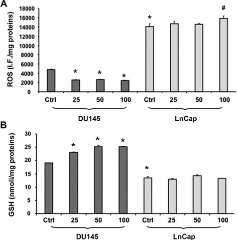
Effect of C3G on ROS and RSH levels. Since LnCap and
DU145 exhibited differences in sensitivity to C3G treat-ment, we further investigated the redox state of these cells by estimating ROS and RSH levels under both the control and treated conditions. ROS levels were determined using the fluorescent probe, DCFH-DA. The intensity of
fluores-cence was proportional to the levels of intracellular oxidant species. As shown in Fig. 4A, LnCap cells displayed a significant increase of basal ROS levels compared to DU145. Results confirmed the ROS-scavenging activity of C3G in DU145 cells, thus causing a dose-dependent decrease of ROS production following C3G treatment. Interestingly, exposure of LnCap cells to both 25 and 50 µM C3G did not affect ROS production, whereas cell treatment with 100 µM resulted in a significant increase in ROS levels compared to the untreated cells. In agreement with lower ROS levels, DU145 cells showed significantly higher basal GSH content than that found in LnCap cells (Fig. 4B). In addition to that, following C3G treatment, DU145 showed a significant (p<0.05) addi-tional increase of GSH levels. C3G had no effect on GSH levels in LnCap cells.
DNA fragmentation induced by treatment with C3G. In order
to investigate the capability of C3G to induce DNA damage, the typical Comet assay was performed on DU145 and LnCap after C3G exposure to different concentrations (25, 50 and 100 µM) for 48 h. Results obtained showed that DNA frag-mentation is clearly increased in DU145 and LnCap after treatment with C3G.
In fact, the DNA damage is extensive, as indicated by the large comet tail. As seen in Fig. 5, C3G exposure significantly increased DNA damage in a dose-dependent manner.
Table I. Cytotoxicity was determined by the LDH release assay in untreated and treated cultured cancer epithelial cells for 48 h with C3G.a DU145 LnCap (% LDH release) (% LDH release) Control 13.24±0.13 12.65±0.31 C3G 25 µM 13.04±0.26 11.32±0.53 C3G 50 µM 10.83±0.44 13.6±0.02 C3G 100 µM 13.12±0.01 12.53±0.11
aValues are presented as the mean ± SEM of three different experi-ments.
Figure 4. (A) Intracellular oxidants in DU145 and LnCaP cells untreated and treated for 48 h with C3G at different concentrations (25, 50 and 100 µM). Values represent the means ± SEM of three experiments performed in triplicate. (B) Thiol groups in DU145 and LnCap cells untreated and treated for 48 h with C3G at different concentrations (25, 50 and 100 µM). Thiol groups are expressed as nmol/mg protein. Values represent the means ± SEM of three experiments performed in triplicate. *p<0.005, significant result vs. untreated DU145 cells. #p<0.05, significant result vs LnCap control.
Discussion
Chemoprevention involves the administration of natural (8,18) or synthetic agents (36,37) designed to suppress the development or progression of cancer. Since less differenti-ated tumors become resistant to a wide variety of cytotoxic drugs, considerable attention has focused on chemoprevention with natural compounds as a new and alternative approach to cancer control (38). Epidemiological evidence indicates that polyphenolic compounds in diets are protective against cancer, and C3G is abundant in many fruits such as pigmented orange, berry fruits and grapes. In order to study the anti-proliferative and pro-differentiating properties of C3G, we have chosen two prostate tumor cells, known to have differential chemo-therapeutic response, namely, LnCap and DU145. LnCap and DU145 cells were chosen as prostate cancer experimental model, since these cell lines corresponds to different stages of prostate cancer. Indeed, the DU145, an androgen-independent prostate cancer cell line, is considered more invasive and less differentiated as compared to the androgen-dependent prostate cancer cell line LnCap. The loss of androgen receptor (AR), due to promoter methylation, represents an important hallmark of DU145 cell line (39). The growth and develop-ment of prostate cancer is initially androgen-dependent and androgen depletion therapy has been extensively explored as a strategy for prostate cancer prevention. Although androgen depletion is a valid strategy for the prevention and therapy of androgen-dependent PC, additional approaches are needed for advanced stages of PC that are androgen-independent. Our study shows that C3G reduced the number of viable prostate cancer cells after 48 h of exposure when compared to the vehicle control cells. Although C3G induced apoptosis in both LnCap and DU145 cells, these latter were much less sensitive than LnCap cells. Evaluation of DNA damage by Comet assay, widely considered a versatile and highly effective tool in bio-monitoring DNA integrity, shows that C3G, both in LnCap and DU145 cells, as shown in other cancer cell lines (40), induced DNA damage as evidenced by large comet tails. In addition, in the present report we demonstrate that C3G treatment exerts pro-apoptotic effects by activating caspase-3 and increasing
p21 protein expression. As shown in Fig. 3, C3G induces both caspase-independent and caspase-dependent apoptotic cell death. C3G caused a significant increase of p21 expression, a negative regulator of the cell cycle, only in LnCap cells. In contrast, C3G caused activation of caspase-3 only in DU145 cells. These results suggest an apoptotic cell death, character-ized by formation of apoptotic bodies, without an increase in cellular membrane breakage as demonstrated by no significant changes in LDH release observed in C3G-treated cells when compared to untreated cells. It should be noted that, while LnCap cells highly express p21, this protein is downregulated in DU145 cells. Cell cycle inhibitor p21 has been identified as one of the effector proteins upregulated by AR in order to achieve its tumor suppressor function. AR mRNA and protein levels detected in DU145 cell line are much lower than LnCap, an AR-positive cell line (39). Furthermore, DU145 cells are not able to engage AR to drive the expression of p21 and subsequently induce growth inhibition (41). DU145 cells lack necessary cofactors in order to achieve AR tumor suppressor. This may explain why DU145 are more resistant to C3G treat-ment.
The present study also demonstrates the possibility of modulating the growth of tumor cells through a new strategy. Cancer is a reversible state characterized by altered matura-tion in which the transformed phenotype can be suppressed by cytostatic agents and by the pharmacological induction of differentiation towards a benign form. Thus, compounds like polyphenols, capable of inducing differentiation (14,42) may represent potential chemotherapeutic agents. We show for the first time that C3G, the most abundant anthocyanin in diet, inhibits cell growth/cell viability resulting in the reversion of both LnCap and DU145 cells from a proliferating to a differ-entiated state. Analysis of p75NGFR expression showed that it is decreased in PC and that the extent of p75NGFR loss is related to PC progression (43). Thus, p75NGFR has been proposed as a tumor suppressor molecule that mediates the anti-proliferative effects of NGF. Our data show that C3G treatment increases p75NGFR expression, confirming the role of C3G in the acquisition of a normal-like cell pheno-type.
Figure 5. DNA strand break analysis performed on DU145 and LnCap cells treated with C3G for 48 h. Representative images of cancer cells untreated and treated with C3G at three different concentrations (25, 50 and 100 µM). A large comet tail is visible in DU145 and LnCap treated cells, indicating DNA strand breaks.
ROS are known to be associated with many diseases by activating different signaling transductional/transcriptional pathways. Regardless of the mechanism, altered production of radical species may either sustain the life of tumor or cause its death. ROS are associated not only with initiation, but also with promotion and progression in the multistage carcinogenesis mode (44). Cancer cells often exhibit a high level of intrinsic oxidative stress due to an increased forma-tion of ROS. In light of the important role of ROS in tumor progression, we measured the levels of ROS in both DU145 and LnCap cell lines. Our results indicate that the anti-prolif-erative/cytotoxic activity exhibited by C3G was sustained by decreased endocellular ROS level in DU145 cells confirming the scavenging activity of C3G. On the other hand, surpris-ingly, C3G increased ROS levels in LnCap cells, but only at the highest concentration, suggesting that the same compound act differently in two different stages of prostate cancer. Cells employ many enzymatic and non-enzymatic antioxidants to counter the effect of ROS and to rebalance cell homeostasis. One of the most versatile protectors of such antioxidants is GSH. GSH performs several important physiological functions such as inactivation of oxygen-derived highly reactive species, detoxification of various types of xenobiotics and maintenance of the oxidative status of other antioxidants. The GSH content in cancer cells is particularly relevant in regulating mutagenic mechanisms, DNA synthesis, growth, and multidrug and radiation resistance. Cancer cell lines containing low GSH levels have been demonstrated to be much more sensitive than control cells to the effect of irradiation (45). Elevated GSH levels are observed in many malignant cells, and this makes the neoplastic tissues more resistant to chemotherapy (31). This is further confirmed by the fact that DU145 cells show significantly higher basal RSH content than that found in LnCap cells suggesting a reducing type of redox environ-ment in DU145 cells compared to that of LnCap. The slight reduction in RSH observed in LnCap cells and its significant increase in DU145 cells further supports the results obtained by measuring ROS. An attractive hypothesis to explain the different RSH levels found in the present study might be just related to the higher replication rate of more malignant cells. Intracellular GSH levels fluctuate throughout the cell cycle depending on the phase of the cell cycle; elevated intracellular GSH levels have been observed upon cell cycle entry, followed by a significant reduction during M phase (46). Whether such differential cellular responses are cell type specific or related to alterations in mechanisms involved in GSH homeostasis are interesting questions that warrant further study. In conclusion, induction of apoptosis and differentiation are two promising cancer prevention strategies, as well as therapy. C3G has drawn increasing attention because of its potential anticancer properties but little is known about the effects of C3G on prostate cancer. We demonstrate that C3G, the most abundant anthocyanin in diet, displays anti-proliferative and pro-differ-entiation properties in two different prostate cancer cell lines. Our results provide a new perspective in the development of novel strategies for the prevention and treatment of PC through consumption of C3G in an appropriate cancer prevention diet. It should be noted that, C3G is active at concentrations corre-sponding to those achieved with food intake (range of µM) and without any toxicity for non-tumorigenic cells.
References
1. Ferlay J, Parkin DM and Steliarova-Foucher E: Estimates of cancer incidence and mortality in Europe in 2008. Eur J Cancer 46: 765-781, 2010.
2. American Cancer Society: Prostate Cancer Statistics 2013. http:// www.cancer.org. Accessed May 20, 2013.
3. American Society of Clinical Oncology Prostate Cancer Statistics: http://www.cancer.net/prostate. Accessed May 20, 2013.
4. Ito K: Prostate cancer in Asian men. Nat Rev Urol 11: 197-212, 2014.
5. Arnold JT and Isaacs JT: Mechanisms involved in the progres-sion of androgen-independent prostate cancers: It is not only the cancer cell's fault. Endocr Relat Cancer 9: 61-73, 2002.
6. Chen J and Xu X: Diet, epigenetic, and cancer prevention. Adv Genet 71: 237-255, 2010.
7. Kang NJ, Shin SH, Lee HJ and Lee KW: Polyphenols as small molecular inhibitors of signaling cascades in carcinogenesis. Pharmacol Ther 130: 310-324, 2011.
8. Weng CJ and Yen GC: Chemopreventive effects of dietary phytochemicals against cancer invasion and metastasis: Phenolic acids, monophenol, polyphenol, and their derivatives. Cancer Treat Rev 38: 76-87, 2012.
9. Russo A, Piovano M, Lombardo L, Vanella L, Cardile V and Garbarino J: Pannarin inhibits cell growth and induces cell death in human prostate carcinoma DU-145 cells. Anticancer Drugs 17: 1163-1169, 2006.
10. Yim D, Singh RP, Agarwal C, Lee S, Chi H and Agarwal R: A novel anticancer agent, decursin, induces G1 arrest and apoptosis in human prostate carcinoma cells. Cancer Res 65: 1035-1044, 2005.
11. Cardile V, Scifo C, Russo A, Falsaperla M, Morgia G, Motta M, Renis M, Imbriani E and Silvestre G: Involvement of HSP70 in resveratrol-induced apoptosis of human prostate cancer. Anticancer Res 23C: 4921-4926, 2003.
12. Russo A, Berretta M, Cardile V, Lombardo L, Vanella L, Troncoso N, Garbarino J, Ignazio B and Li Volti G: Pharmaco-logical induction of heme oxygenase-1 reduces KB cell viability: Role of carbon monoxide. J Carcinog & Mutagen 2014, http:// dx.doi.org/10.4172/2157-2518.S8-009. Accessed April 16, 2014. 13. Waxman S: Differentiation therapy in acute myelogenous
leukemia (non-APL). Leukemia14: 491-496, 2000.
14. Vanella L, Barbagallo I, Acquaviva R, Di Giacomo C, Cardile V, Abraham NG and Sorrenti V: Ellagic acid: Cytodifferentiating and antiproliferative effects in human prostatic cancer cell lines. Curr Pharm Des 19: 2728-2736, 2013.
15. Breitman T, Collins SJ and Keene B: Terminal differentiation of human promyelocytic leukemic cells in promyelocytic leukemic cells in primary culture response to retinoic acid. Blood 57: 1000-1004, 1981.
16. Ohno R, Asou N and Ohnishi K: Treatment of acute promyelo-cytic leukemia: Strategy toward further increase of cure rate. Leukemia 17: 1454-1463, 2003.
17. Vanella L, Di Giacomo C, Acquaviva R, Barbagallo I, Cardile V, Kim DH, Abraham NG and Sorrenti V: Apoptotic markers in a prostate cancer cell line: Effect of ellagic acid. Oncol Rep 30: 2804-2810, 2013.
18. Vanella L, Di Giacomo C, Acquaviva R, Barbagallo I, Li Volti G, Cardile V, Abraham NG and Sorrenti V: Effects of ellagic Acid on angiogenic factors in prostate cancer cells. Cancers (Basel) 5: 726-738, 2013.
19. Wang H, Race EJ and Shrikhande AJ: Characterization of antho-cyanins in grape juices by ion trap liquid chromatography-mass spectrometry. J Agric Food Chem 51: 1839-1844, 2003.
20. Seeram NP, Cichewicz RH, Chandra A and Nair MG: Cyclooxygenase inhibitory and antioxidant compounds from crabapple fruits. J Agric Food Chem 51: 1948-1951, 2003. 21. Amorini AM, Fazzina G, Lazzarino G, Tavazzi B, Di Pierro D,
Santucci R, Sinibaldi F, Galvano F and Galvano G: Activity and mechanism of the antioxidant properties of cyanidin-3-O-beta-glucopyranoside. Free Radic Res 35: 953-966, 2001.
22. Lee HS: Characterization of major anthocyanins and the color of red-fleshed Budd Blood orange (Citrus sinensis). J Agric Food Chem 50: 1243-1246, 2002.
23. Matsumoto H, Inaba H, Kishi M, Tominaga S, Hirayama M and Tsuda T: Orally administered delphinidin 3-rutinoside and cyanidin 3-rutinoside are directly absorbed in rats and humans and appear in the blood as the intact forms. J Agric Food Chem 49: 1546-1551, 2001.
1999.
25. Ichikawa H, Ichiyanagi T, Xu B, Yoshii Y, Nakajima M and Konishi T: Antioxidant activity of anthocyanin extract from purple black rice. J Med Food 4: 211-218, 2001.
26. Kähkönen MP and Heinonen M: Antioxidant activity of antho-cyanins and their aglycons. J Agric Food Chem 51: 628-633, 2003.
27. Briviba K, Abrahamse SL, Pool-Zobel BL and Rechkemmer G: Neurotensin-and EGF-induced metabolic activation of colon carcinoma cells is diminished by dietary flavonoid cyanidin but not by its glycosides. Nutr Cancer 41: 172-179, 2001.
28. Hagiwara A, Miyashita K, Nakanishi T, Sano M, Tamano S, Kadota T, Koda T, Nakamura M, Imaida K, Ito N, et al: Pronounced inhibition by a natural anthocyanin, purple corn color, of 2-amino-1-methyl-6-phenylimidazo[4,5-b]pyridine (PhIP)-associated colorectal carcinogenesis in male F344 rats pretreated with 1,2-dimethylhydrazine. Cancer Lett 171: 17-25, 2001.
29. Fimognari C, Berti F, Nüsse M, Cantelli-Forti G and Hrelia P: Induction of apoptosis in two human leukemia cell lines as well as differentiation in human promyelocytic cells by cyanidin-3-O-beta-glucopyranoside. Biochem Pharmacol 67: 2047-2056, 2004.
30. Serafino A, Sinibaldi-Vallebona P, Lazzarino G, Tavazzi B, Rasi G, Pierimarchi P, Andreola F, Moroni G, Galvano G, Galvano F, et al: Differentiation of human melanoma cells induced by cyanidin-3-O-β-glucopyranoside. FASEB J 18: 1940-1942, 2004.
31. Traverso N, Ricciarelli R, Nitti M, Marengo B, Furfaro AL, Pronzato MA, Marinari UM and Domenicotti C: Role of gluta-thione in cancer progression and chemoresistance. Oxid Med Cell Longev 2013: 972913, 2013.
32. Vanella L, Di Giacomo C, Acquaviva R, Santangelo R, Cardile V, Barbagallo I, Abraham NG and Sorrenti V: The DDAH/NOS pathway in human prostatic cancer cell lines: Antiangiogenic effect of L-NAME. Int J Oncol 39: 1303-1310, 2011.
33. Sigala S, Tognazzi N, Rizzetti MC, Faraoni I, Missale C, Bonmassar E and Spano P: Nerve growth factor induces the re-expression of functional androgen receptors and p75(NGFR) in the androgen-insensitive prostate cancer cell line DU145. Eur J Endocrinol 147: 407-415, 2002.
34. Bradford MM: A rapid and sensitive method for the quantita-tion of microgram quantities of protein utilizing the principle of protein-dye binding. Anal Biochem 72: 248-254, 1976.
Antiproliferative effect of oleuropein in prostate cell lines. Int J Oncol 41: 31-38, 2012.
36. Romeo R, Giofrè SV, Garozzo A, Bisignano B, Corsaro A and Chiacchio MA: Synthesis and biological evaluation of furopy-rimidine N,O-nucleosides. Bioorg Med Chem 21: 5688-5693, 2013.
37. Romeo R, Giofrè SV, Carnovale C, Campisi A, Parenti R, Bandini L and Chiacchio MA: Synthesis and biological evalu-ation of 3-hydroxymethyl-5-(1H-1,2,3-triazol) isoxazolidines. Bioorg Med Chem 21: 7929-7937, 2013.
38. Calabrese V, Cornelius C, Trovato A, Cavallaro M, Mancuso C, Di Rienzo L, Condorelli D, De Lorenzo A and Calabrese EJ: The hormetic role of dietary antioxidants in free radical-related diseases. Curr Pharm Des 16: 877-883, 2010.
39. Renis M, Calandra L, Scifo C, Tomasello B, Cardile V, Vanella L, Bei R, La Fauci L and Galvano F: Response of cell cycle/stress-related protein expression and DNA damage upon treatment of CaCo2 cells with anthocyanins. Br J Nutr 100: 27-35, 2008. 40. Alimirah F, Chen J, Basrawala Z, Xin H and Choubey D: DU-145
and PC-3 human prostate cancer cell lines express androgen receptor: Implications for the androgen receptor functions and regulation. FEBS Lett 580: 2294-2300, 2006.
41. Litvinov IV, Antony L, Dalrymple SL, Becker R, Cheng L and Isaacs JT: PC3, but not DU145, human prostate cancer cells retain the coregulators required for tumor suppressor ability of androgen receptor. Prostate 66: 1329-1338, 2006.
42. Hagiwara Y, Kasukabe T, Kaneko Y, Niitsu N and Okabe-Kado J: Ellagic acid, a natural polyphenolic compound, induces apoptosis and potentiates retinoic acid-induced differentiation of human leukemia HL-60 cells. Int J Hematol 92: 136-143, 2010.
43. Yang Z, Chen H, Huo L, Yang Z, Bai Y, Fan X, Ni B, Fang L, Hu J, Peng J, et al: Epigenetic inactivation and tumor-suppressor behavior of NGFR in human colorectal cancer. Mol Cancer Res 13: 107-119, 2015.
44. Panayiotidis M: Reactive oxygen species (ROS) in multistage carcinogenesis. Cancer Lett 266: 3-5, 2008.
45. Meister A: Glutathione deficiency produced by inhibition of its synthesis, and its reversal; applications in research and therapy. Pharmacol Ther 51: 155-194, 1991.
46. Shaw JP and Chou IN: Elevation of intracellular glutathione content associated with mitogenic stimulation of quiescent fibro-blasts. J Cell Physiol 129: 193-198, 1986.