Università degli Studi di Catania
Scuola Superiore di Catania
International PhD
in
Translational Biomedicine
XXVI cycle
Relationship between extracellular matrix (ECM) components
and mineralization in bone marrow stromal cells
Giusy Villaggio
Coordinator of PhD Tutor
Prof D.F. Condorelli Prof. F. Sinatra
INDEX
Thesis purpose
1. Introduction………pag. 5
1.1 The extracellular matrix………...5
1.2 Mesenchymal stem cells………...10
1.2.1 Characteristics of MSCs in vitro………12 1.2.2 Differentiation of MSCs in vitro………15 1.2.3 Clinical applications of MSCs………19 a) Systemic delivery………..19 b) Gene therapy……….21 c) In situ transplantation………22
1.3 Biomaterials for tissue engineering………..23
2. Materials and methods ………32
2.1 Mesenchymal Stem Cell Culture……….32
2.2 Cell-free extracellular matrix preparation………32
2.3 Osteogenic differentiation medium………..33
2.4 MTT assay………34
2.5 Scanning Electron Microscopy (SEM)……….34
2.5.1 X-Ray Microanalysis………..34
2.6 α5 integrin and microfilaments immunofluorescence……….34
2.7 Histochemistry……….35
2.7.1 Alkaline phosphatase staining and measurement………..35
2.7.2 Alizarin Red staining and measurement………35
2.8.1 RNA isolation………..35
2.8.2 cDNA synthesis………..36
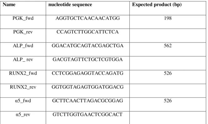
2.8.3 Reverse Transcriptase-Polymerase Chain Reaction (RT-PCR)………36
3. Results………39
3.1 Analysis of hBMSCs on ECM coatings and TCP in growth medium………..39
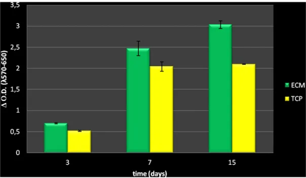
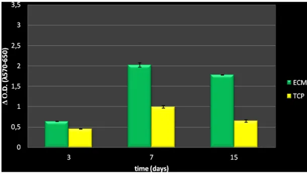
3.1.1 Cell survival and proliferation………...39
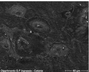
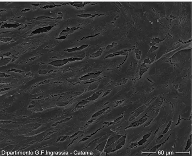
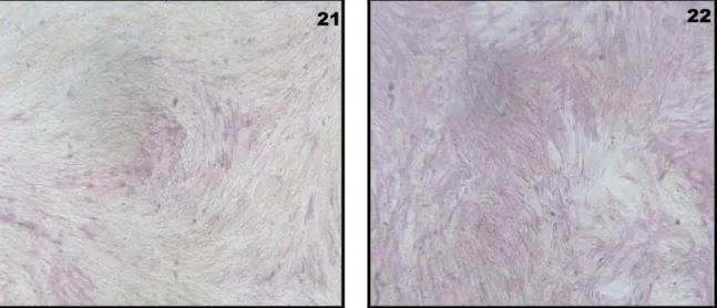
3.1.2 Morphological investigations………41
3.1.3 Integrin α5and Microfilaments: cell adhesion to substrate………44
3.1.4 Alkaline phosphatase activity and staining measurement……….46
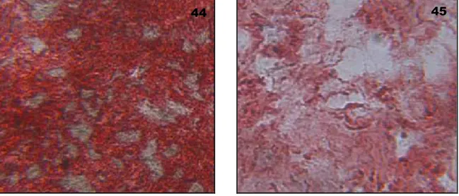
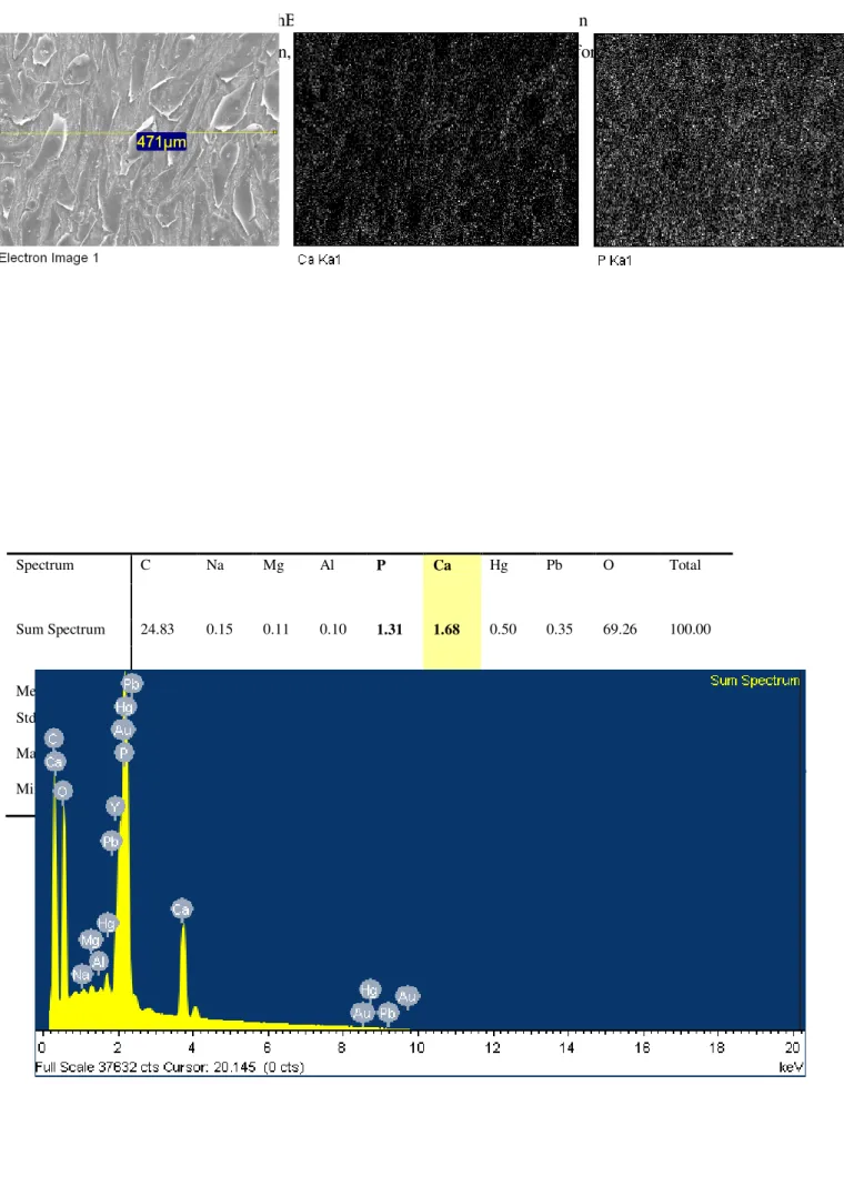
3.1.5 Calcium content revealed by Alizarin Red staining and measurement and X-Ray microanalysis………..48
3.2 Analysis of hBMSCs on ECM coatings and TCP in osteogenic medium………..50
3.2.1 Cell survival and proliferation………50
3.2.2 Morphological investigations……….52
3.2.3 Integrin α5and Microfilaments: cell adhesion to substrate……….55
3.2.4 Histochemistry………57
a) Alkaline phosphatase activity and staining measurement………..57
b) Alizarin Red staining and measurement………..60
3.2.5 X-Ray microanalysis………..62
3.2.6 Osteogenic markers and α5 adhesion molecule gene expression……….65
4. Discussion………..66
5. Conclusions………...70
THESIS PURPOSE
The relations between cells and extracellular matrix seem to orchestrate tissue organization by regulating cell functions during fetal development and throughout normal adult life. Mesenchymal stem cells naturally reside within an extracellular matrix (ECM), which is a biological scaffolding material consisting of structural and functional molecules. Besides providing structural support to cells, the ECM is a dynamic microenvironment that also plays a role in modulating cell survival, migration, proliferation, and differentiation. Thus, focusing on the innate ability of the native ECM to better modulate cell behavior, the coating of synthetic biomaterials with cell-derived decellularized extracellular matrices is a promising approach to confer bioactivity to otherwise inert materials and direct the fate of host or transplanted cells in tissue engineering applications. Furthermore, mesenchymal stem cells (MSCs) are under investigation for possible uses in the production of decellularized matrix-coated substrates due to their high proliferative potential, ability to differentiate toward multiple lineages and extensive matrix production. My research activities regarded the adhesion and proliferation of human bone marrow stem cells grown on cell free extracellular matrix and the influence that these matrices have on the maintenance of cell stemness and biological functions, and their role during the induction of MSCs osteogenic differentiation.
1. INTRODUCTION
1.1 The extracellular matrix
Recently, it has become increasingly evident that the extracellular matrix (ECM) is an important component of the cellular niche within all animal tissues and organs, and provides not only essential physical scaffolding for the cellular constituents but also critical biochemical and biomechanical cues to initiate and sustain cellular functions such as tissue morphogenesis, differentiation and homeostasis (Kresse H and Schonherr E, 2001; Daley WP et al., 2008). The importance of the ECM is vividly illustrated by the wide range of syndromes, from minor to severe, arising from genetic abnormalities in ECM proteins; indeed, any inherited or acquired structural defect, such as a single amino acid substitution, and/or metabolic disturbance in the ECM, may cause cellular and tissue
alterations that may lead to the development or progression of a disease (Jarvelainen Het al., 2009).
The extracellular matrix consists of a variety of proteins and glycoproteins secreted locally and assembled in an organized network in close association with the surface of the cell responsible for their production. Although, fundamentally, the ECM is composed of water, proteins and polysaccharides, each tissue has an ECM with a unique composition and topology, which is generated during tissue development through a dynamic and mutual, biochemical and biophysical dialogue between the various cellular components (e.g. epithelial, fibroblast, adipocyte, endothelial elements) and the evolving cellular and protein microenvironment. Indeed, the physical, topological, and biochemical composition of the ECM is not only tissue-specific, but it is also markedly heterogeneous (Leitinger B and Hohenester E, 2007; Xian X et al., 2010).
The connective extracellular matrix is often most abundant in cells which surround and determine the physical properties of the tissue. The connective tissues form the scaffolding in vertebrates, but the amount present in the various organs varies considerably, – from the cartilage and bone, containing the highest percentage, to the brain and spinal cord, where they are only minor constituents. The variations in the relative quantities of different types of matrix macromolecules and the way they are organized give rise to a surprising diversity of forms, each adapted to the functional requirements of the tissue in question. The matrix can become calcified to form the hard structures of bones and teeth, transparent to form the corneal stroma or its molecules can form a
parallel structure to give tendons their enormous tensile strength (Adams JC and Watt FM, 1993;
Geiger B et al., 2001).
The extracellular matrix is composed of two main classes of macromolecules: glycosaminoglycans (GAGs) which are usually joined covalently to proteins in the form of proteoglycans (PGs), and fibrous proteins including collagens, fibronectins, elastins and laminins, which have both structural and adhesive functions (Schaefer L and Schaefer RM, 2010; Alberts B et al., 2007).
Proteoglycans (PGs) consist of glycosaminoglycan (GAG) chains which, with the exception of hyaluronic acid, are covalently linked to a specific protein core. The GAG chains are unbranched polysaccharide chains composed of repeating disaccharide units, [sulfated N-aceltylglucosamine or N-acetylgalactosamine and D-glucuronic or L-iduronic acid] which can be divided further into sulfated (chondroitin sulfate, dermatan sulfate, heparan sulfate and keratan sulfate) and non-sulfated (hyaluronic acid) GAGs (Schaefer L and Schaefer RM, 2010). These molecules are extremely hydrophilic and, consequently, take highly extended conformations which are essential for hydrogel formation, and the matrices they form, are able to resist highly compressive forces.
There are several classification criteria of proteoglycans based on location, composition and
function of the GAG chains. However, it is difficult to identify a single structure recurring within
this class of macromolecules (Ruoslahti E, 1988); indeed there are different types of proteoglycans and they can also undergo modifications (substitution of sugar residues, phosphorylation,
sulphation)during cell survival, adapted to different biological needs. A classification according to
their core proteins, localization and GAG composition indentifies three main PGs families: small leucine-rich proteoglycans (SLRPs), modular proteoglycans and cell-surface proteoglycans (Iozzo RV and Murdoch AD, 1996; Iozzo RV, 1998).
Proteoglycan molecules have a wide variety of functions reflecting their unique buffering, hydration, binding and force-resistance properties; as a consequence, genetic diseases (congenital stromal dystrophy of the cornea, dyssegmental dysplasia, Schwartz-Jampel syndrome) have been linked to mutations in PG genes (Schaefer L and Schaefer RM, 2010; Kresse H and Schonherr E, 2001). PGs fill the majority of the extracellular interstitial space within the tissue forming a gel-like
highly hydrated " basal substance" in which the fibrous proteins are embedded (Jarvelainen Het al.,
2009). The polysaccharidic gel resists compressive forces on the matrix while allowing rapid diffusion of nutrients, metabolites and hormones between blood vessels and tissue cells (Bishop JR
et al., 2007).
SLRPs, such as decorin, biglycan and lumican, have been involved in multiple signaling pathways including binding to and activation of epidermal growth factor receptor (EGFR), insulin-like growth factor 1 receptor (IGFIR) and low-density lipoprotein-receptor related protein 1 (LRP1), regulation of inflammatory response reaction, and binding to and activation of TGFβ (Goldoni S and Iozzo RV, 2008; Iozzo RV and Schaefer L, 2010 ; Schaefer L and Schaefer RM, 2010). Also, SLRPs seems to be engaged in binding collagens and in the modulation of fibrillogenesis, thus regulating the assembly of the interstitial matrix and its three-dimensional configuration according to the mechanical load that the tissue must sustain. For example, it was shown that decorin, in particular, when bound to collagen is capable of sequestering cytokines at the level of the ECM, exerting a role in the control of cell proliferation. Specifically, it inhibits the action of TGF (Yamaguchi Y et al.,
1990) and binds the EGF receptor, hindering its phosphorylation (Patel S et al., 1998; Santra M et
al., 2002).
Modular PGs are a heterogeneous group characterized by the assembly of various protein modules in an elongated and often highly glycosylated structure. They can modulate cell adhesion, migration and proliferation (Schaefer L and Schaefer RM, 2010). Basement membrane modular PGs (perlecan, agrin and collagen type XVIII) have a dual function as pro- and anti-angiogenic factors (Iozzo RV et al., 2009).
Cell-surface PGs (syndecans and glypicans), mainly with heparan sulfate chains, are also involved in the control of cell proliferation, acting as co-receptors since many growth factors bind with high affinity particular domains of the heparan sulfate chains on the one hand, and with their respective signaling receptors on the other (Kresse H and Schonherr E, 2001; Schaefer L and Schaefer RM, 2010).
Collagen is the most important fibrous protein within the extracellular matrix. As the main component of skin and bone, it is the most abundant protein in mammals, accounting for 25% of the total protein mass in these animals. To date, 28 different types of collagen have been identified in vertebrates (Gordon MK and Hahn RA, 2010).The structural unit of collagen is made of tropocollagen, a protein formed by three polypeptide chains wound spirally by hydrogen and covalent bonds to one another, and forming a triple-stranded helix. The tropocollagen molecules, synthesized within the cell as soluble precursors (procollagen), are associated longitudinally (head-to-tail) and in parallel, with an arrangement staggered, giving rise to the collagen fibrils. After exocytosis, the procollagen is cut and becomes collagen (Prockop DJ and Kivirikko KI, 1995); the majority of these molecules can assemble into supramolecular complexes, such as fibrils and networks, depending on the type of collagen. Fibrous collagens form the backbone of the fibril bundles within the interstitial tissue stroma, whereas network collagens are incorporated into the basal membrane (BM) (Frantz C et al., 2010).
Collagen fibrils form structures which provide tensile strength, regulate cell adhesion, support chemotaxis and migration, and direct tissue development. The diameters and organization of these molecules vary according to the tissue considered; moreover, although within a given tissue, collagen fibers are generally a heterogeneous mix of different types, one type of collagen usually predominates (Rozario T and DeSimone DW, 2010). Cells can adjust the arrangement of the collagen molecules after secretion, guiding the formation of fibrils in close association with the plasma membrane. For example, synthesis of collagen type I involves a number of enzymatic posttranslational modifications (Gordon MK and Hahn RA, 2010; Myllyharju J and Kivirikko KI, 2004), mainly the hydroxylation of proline and lysine residues, the glycosylation of lysine and the cleavage of N- and C-terminal propeptides. Following their cleavage, collagen fibrils are strengthened by the covalent crosslinking between the lysine residues of the constituent collagen
molecules by lysyl oxidases (LOX) (Myllyharju J and Kivirikko KI, 2004; Robins SP, 2007). Furthermore, since the spatial organization of collagen fibrils partly reflects their interaction with other molecules of the ECM, cells can influence this organization by secreting, together with fibrillar collagens, different types and amounts of other matrix macromolecules (Prockop DJ and Kivirikko KI, 1995).
Elastin is another major ECM protein, its fibers provide recoil to tissues that undergo repeated stretches and, importantly, its elongation is crucially limited by tight association with collagen fibrils (Wise SG and Weiss AS, 2009). Secreted tropoelastin (elastin precursor) molecules assemble into fibers and become highly crosslinked to one another via their lysine residues by members of the lysyl oxidase (LOX) enzyme family, which include LOX and LOXL (Lucero HA and Kagan HM, 2006). Furthermore, elastin fibers are covered by glycoprotein microfibrils, mainly fibrillins, which are essential for their integrity (Wise SG and Weiss AS, 2009).
A third fibrous protein, fibronectin (FN) is intimately involved in directing the organization of the interstitial ECM and, additionally, has a crucial role in mediating cell adhesion and function (Vakonakis I and Campbell ID, 2007). It is also important for cell migration during development and has been implicated in cardiovascular diseases and tumor metastases (Rozario T and DeSimone DW, 2010; Tsang KY et al., 2010). FN is a glycoprotein consisting of two polypeptide chains linked by disulphide bonds near the C-terminus. It has several binding sites to other FN dimers, to collagen, to glycosaminoglycans (GAGs), to heparin and also a sequence Arg-Gly-Asp (RGD) implicated in the interaction with specific receptors on the cell membrane; indeed, the molecules of fibronectin assemble into fibrils only on cell surface in a process guided by additional proteins, especially integrins (Schwarzbauer JE and Lichtman JW, 1999; Pankov R and Yamada KM, 2002). The fibrils of fibronectin can be strongly stretched over its resting length and usually are aligned with adjacent stress fibers of intracellular actin. These, indeed, promote the assembly of secreted molecules of fibronectin into fibrils influencing their orientation (Smith ML et al., 2007). The interactions between the extracellular fibronectin fibrils and intracellular actin filaments across the cellular plasma membrane are mediated mainly by transmembrane adhesion integrin. Cell contraction through the actomyosin cytoskeleton generates tension on fibronectin matrix resulting in FN fibril stretching. This mechanism causes the expure of cryptic binding sites on fibronectin molecules allowing them to bind one another, (Leiss M et al., 2008; Mao Y and Schwarzbauer JE, 2005; Vakonakis I and Campbell ID, 2007) as well as the further exposure of integrin binding sites within the molecule which results in pleiotrophic changes in cellular behavior and implicate FN as an extracellular mechanoregulator (Smith ML et al., 2007). Thus, actin cytoskeleton and the resulting integrin clustering promote FN fibril polymerization and matrix assembly.
Integrins are heterodimeric proteins composed of two trans-membrane subunits, α and β, not covalently linked. Each subunit consists of a large extracellular domain, a trans membrane domain
and a small cytoplasmic domain. To date 16 α and 8 β subunits have been identified and, depending on their combination, integrin binds specific proteins of the ECM. For example, α1β1 binds collagen, while α5β1 and αvβ3 are receptors for fibronectin and vitronectin respectively. On the one hand, integrins anchor the cell to the extracellular matrix proteins, on the other hand they bind proteins of the cytoskeleton (Anselme K, 2000).
The site of cell adhesion to the extracellular matrix via integrins is called "focal contact", and it regulates cellular behaviors by, for example, applying stronger traction forces to the substrate during cell migration. In cultured fibroblasts, there are remarkable differences between the ‘classical’ focal contacts – oval shaped, peripheral structures, regulated by the small G-protein Rho, enriched in activated αvβ3-integrin, paxillin, vinculin and tyrosine-phosphorylated proteins – and ‘fibrillar adhesions’, which are elongated or dot-like, central structures containing α5β1-integrin, tensin and parvin/actopaxin and attached to fibronectin fibrils. In addition, recent studies have shown, by using an antibody-chase technique, that whereas αvβ3 integrin remains in focal contacts,
α5β1 integrin continuously translocates from peripheral focal contacts towards the cell center,
forming fibrillar adhesions which indicates that this process plays a major role in fibronectin fibrillogenesis (Pankov et al., 2000; Zamir E and Geiger B, 2001;Cukierman E et al., 2001).
Furthermore, a recent study by Roca-Cusachs P et al. (2009), on mouse embryonic fibroblasts has
indicated the differential function of integrin species in adhesion processes. High matrix forces were found to be primarily resisted by clustered α5β1 integrins, while less stable αvβ3 integrin binding was shown to initiate mechanotransduction, resulting in a reinforcement of the integrin-cytoskeleton interactions. Indeed, these integrins have been identified as key regulators of osteoblast proliferation and differentiation (Biggs MJP and Dalby MJ,2010). Since the cytoskeleton can exert forces that are able to direct the ECM macromolecules, which, in turn, can organize the cytoskeleton of cells they came into contact with, the ECM can theoretically propagate order from cell to cell, creating structures oriented on a large scale. ECM components together with integrin receptors on the cell surface can be viewed as intricate nanodevices allowing cells to physically organize their 3D environment, as well as to sense and respond to various types of mechanical stress (Geiger B et al., 2001).
1.2 Mesenchymal stem cells
The concept of mesenchymal cells has achieved wide popularity and the studies involving these cells are undergoing a rapid development. Despite the rapid growth of this field and the vast potential applications of mesenchymal stem cells from a scientific and medical point of view, uncertainties remain with respect to the defining characteristics of these cells, including their potency and self-renewal.
The history of mesenchymal stem cells originated at the end of the 19th century as a hypothetical
assumption to explain the ability of certain tissues, such as blood, skin, etc., to regenerate for the lifetime of an organism even though they are made of short-lived cells. In these classical studies, the identification of stem cells as discrete cellular entities, resulting from the development of methods to isolate stem cells candidates, led the German pathologist Cohnheim JF in 1867 to suggest the presence of nonhematopoietic stem cells in bone marrow. His work showed that bone marrow could be the source of fibroblasts depositing collagen fibers as part of the normal process of wound repair (Prockop DJ, 1997). Around the same period, Goujon EJ (1869) demonstrated that autologous bone marrow (BM) transplanted into heterotopic anatomical sites formed de novo ectopic bone and marrow (Bianco P et al., 2008).
Despite the importance of this first classical evidence, the ultimate proof of an innate osteogenic potential of the BM and, the idea of a “mesenchymal” stem cell came from the pioneering experiments of Tavassoli and Crosby in the 1960s. While investigating the significance of the specific localization of hematopoiesis in bone, they transplanted bone-less fragments of bone marrow into heterotopic sites, and observed the orderly formation of heterotopic bone at the graft site. This experiment revealed that bone marrow includes an entity, unknown at the time, endowed with the ability to generate histology-proven bone tissue (Tavassoli M and Crosby WH, 1968). However, because these tests were carried out with entire fragments of bone-free BM, the precise identity of any cell candidate to be the osteogenic progenitor could not be defined. Evidence that bone marrow contains cells able to differentiate into other mesenchymal cells, as well as fibroblasts, is now well-known, and was demonstrated in a series of studies by Friedenstein and colleagues in the 1960s and 1970s. They proved that the osteogenic potential repeatedly revealed by the previous classical experiments of heterotopic transplantation, was attributable to a subpopulation of BM cells, entirely distinguishable from the well-known hematopoietic stem cells (HSCs). Indeed, when whole bone marrow was placed in plastic culture dish, cells rapidly adhered to plastic, and medium changed 4 hours later, only removed most of the non adherent, hematopoietic stem cells. The remaining non-phagocytic, adherent cells seemed heterogeneous, but most were spindle-shaped and formed loci of two to four cells which, after an initial lag of 2-4 days, began to divide rapidly, with
population doubling time depending on the donor and the initial seeding density. Following several culture passaging, the adherent cells became more fibroblastic in appearance (Friedenstein AJ et al., 1968). Friedenstein and his coworkers (1970) also highlighted another feature; specifically, they showed that cells, when grown in culture at low density, were able to form single-derived colonies designed as colony-formed unit fibroblastic CFU-F. When transplanted in vivo, strains derived from a single cell were able to generate a variety of fully differentiated connective tissues including bone, cartilage, adipose tissue, fibrous tissue and myelosupportive stroma. In other words, they realized that all these connective tissues had a single ancestral progenitor which Friedenstein and Owen (1987, 1988) called “osteogenic stem cell” or, later, “bone stromal stem cell”.
Their observations were confirmed by other research groups throughout the 1980s (Ashton BA et
al., 1980; Castro-Malaspina H et al., 1980), although the implications of these findings were
initially appreciated merely in experimental hematology and only later for their relevance to bone biology and diseases. The innovative idea of the presence of nonhematopoietic stem cells in BM was accepted worldwide only after a study carried out and published by Pittenger et al.(1999). Actually, the repeatedly validated concept of Friedenstein and colleagues set a limit: the putative stem cell they isolated was a progenitor of all the skeletal tissues excluding all the mesodermal derivatives and, furthermore, it was located only in bone marrow. Subsequently, Caplan (1991) and Pittenger (1999), on the basis of Friedenstein’s work, coined the widely used term “Mesenchymal stem cell” (MSC), and proclaimed that MSC was a common ancestor not only of skeletal tissues, but also of ‘‘mesenchymal’’ tissues, meaning substantially all nonhematopoietic derivatives of mesoderm: although found in bone marrow, it also resided in all tissues of postnatal organisms. As reported by Bianco et al. (2006 and 2008) the idea of a “mesenchymal stem cell” in postnatal tissues was easily accepted due to the acclaimed recent isolation of human embryonic pluripotent cells in culture, but remained essentially unverified because it was only demonstrable with a heterothopyc transplant of a single cell-derived colony. Hence, the term “skeletal stem cells” was suggested to refer to bone marrow-derived multipotent stromal cells with an in vivo demonstrable differentiative potential.
To date MSCs have been isolated not only from bone marrow but also from many other tissues and organs, including adipose tissue (Zuk PA et al., 2002), umbilical cord blood, placental tissue, liver, spleen, testes, menstrual blood (Rossignoli F et al., 2013), amniotic fluid, pancreas (Karaoz E et al., 2010), synovial membrane, dermis, dental pulp (Shi S et al., 2005) and periosteum. Furthermore, MSC-like cells have been isolated from pathological tissues (e.g. rheumatoid arthritis) and express bone morphogenetic protein receptors (Marinova-Mutafchieva L et al., 2000).
Even though very few direct comparisons have made between MSCs isolated from different sources thus far (Kern S et al., 2006; Rebelatto CK et al., 2008), these studies agree that these cells show no significant differences in their morphology and immune phenotype, but they are heterogeneous in
their distinct success rates of isolation, proliferation and differentiation potential. Particularly, these reports demonstrate that bone marrow is thought to be the most available and abundant reservoir of MSCs as well as the major source for these precursor cells, which populate other adult tissues and organs (Prockop DJ, 1997). Adipose tissue-derived MSCs are considered an interesting alternative because they are abundantly distributed and easily accessible, but they have a lower osteogenic potential (Niemeyer P et al., 2010). Overall, MSCs account for a small fraction in bone marrow and other tissues; the exact frequency is difficult to calculate due to the different methods of collection and separation. However, the frequency in human bone marrow has been estimated to be approximately 0.001-0.01% of the total nucleated cells, and therefore about 10-fold less abundant than haematopoietic stem cells (Pittenger MF et al., 1999). Furthermore, the frequency of MSCs declines with age, from 1/10.000 nucleated marrow cells in a newborn to about 1/100.000 nucleated marrow cells in a 80 year old person (Caplan AI, 1994).
1.2.1 Characteristics of MSCs in vitro
The growing interest in the potential of MSCs has resulted in an exorbitant increase of scientific publications in a short time. However, the studies performed on MSCs used different isolation and expansion methods, as well as different approaches to cell characterization.
To allow for an easier comparison between the results of these studies and facilitate progress in the field, the Mesenchymal and Tissue Stem Cell Committee of the International Society for Cellular Therapy (ISCT), as reported in Dominici M et al. (2006), has proposed three criteria to define MSCs. First, MSCs must be adherent to plastic when maintained in standard culture conditions. Second, MSC populations (≥/95%) must be positive for several antigens such as CD105 ((known as endoglin), CD73 (known as ecto 5’ nucleotidase) and CD90 (also known as Thy-1). Since new surface markers may be identified in future studies leading to changes in these criteria, the ISCT recommends that the expression of hematopoietic antigens should not be used as a further requirement to identify the MSC. In other words, these cells should not have the expression (≤ 2%) of hematopoietic antigens such as CD45 (a pan-leukocyte marker), CD34 (marks primitive hematopoietic progenitors and endothelial cells),CD14 or CD11b (expressed on monocytes and macrophages),CD79a or CD19(markers of B cells) and HLA class II. Third, the cells must be able to differentiate at least into osteoblasts, adipocytes and chodroblasts under standard in vitro differentiating conditions. The differentiation can then be demonstrated by well-accepted staining protocols (Dominici M et al., 2006).
However, it should be said that the criteria introduced by the ISCT have several limitations. For example, although the ability of MSCs to adhere to plastic surfaces is accepted to define these cells,
also pre-B-cell progenitors and granulocytic/monocytic precursors show plastic adherence (Phinney DG et al., 1999). Moreover, adherent cells capable of density-independent growth are found in a number of non hematopoietic tissues, such as periosteum and dental pulp, and probably in all connective tissues, and are also called CFU-Fs. In addition, not all cells within a given population are stem cells (Bianco P et al., 2008).
Several similar works strongly suggest that MSCs and isolated clones are heterogeneous not only with respect to their self-renewal ability but also to their multi-potentiality (Bianco P et al., 2001). Indeed, the concept of self- renew is often erroneously confused with the ability of a cell to give rise to a broad, long-term proliferation in culture, which is the number of the population doublings. Instead, self-renewal is to be understood as the ability of a stem cell to reconstruct in vivo a stem cell compartment with propriety and phenotype identical to the starting population, so that the cell maintains the stem cell pool while generation progenies undergo clonal expansion and differentiation. While self-renewal was widely confirmed for hematopoietic stem cells, recently it has been demonstrated also for bone marrow in the work of Sacchetti et al. (2007), which showed that BM stem cells can self-renew since they can be successfully explanted like cells expressing MCAM (marker that identifies all of the clonogenic stem cells), grown through several population doublings and then transplanted to recreate a compartment of identical cells in vivo while generating heterotopic “ossicles” (a shell of cortical bone with a cavity containing hematopoietic tissue). Sacchetti et al. also stated that BM MSCs can be directly identified with a specialist type of mural cells, also called pericytes, found in the sinusoid walls and long known as adventitial reticular cells, which act as organizers and regulators of the hematopoietic microenvironment/niche (Bianco P et
al., 2011).
The concept of multy-potentiality opens an additional controversy arising from the common place that BMSCs can give origin to all tissues of mesodermal origin. A differentiation assay, able to unequivocally demonstrate this feature, should be conducted through the use of clonal populations of cells; it must exclude the use of artificial factors stimulating the differentiation or factors which reprogram cell fate, such as bone morphogenetic proteins (BMPs), because spontaneous differentiation potential and responsiveness to reprogramming are equally important biological characteristics of a given cell, and yet they are radically distinct conceptually and experimentally (Bianco P et al., 2006; Bianco P et al., 2008). Finally, differentiation must be unequivocal, that is ideally coinciding with the generation in vivo of histological verified tissue and not only based on the expression of a number of tissue-specific proteins or mRNA (Sacchetti B et al., 2007; Bianco P
et al., 2001).
In keeping with their anatomical origin, BMSCs have an osteogenic imprinting, but they are non-differentiated osteogenic progenitors (Satomura K et al., 2000), which is suggested not only by in vivo experiments but also by the constitutive expression of the marker regulator of skeletogenesis
Runx2/Cbfa1; osteogenic commitment directed by Cbfa1 occurs upstream of the ontogeny of marrow stromal cells, which are the precursors of osteogenic cells. These cells retain expression of Runx2/Cbfa1, possibly as an inheritance of their osteogenic origins, but they remain capable of multy-differentiation so that osteogenic fate is not mandatory (Sacchetti B et al., 2007 ; Bianco P and Pamela Gehron Robey, 2000 ).
With a view to the possible clinical applications of mesenchymal stem cells, it should be noted that their phenotype (indicated as MHC I +, MHC class II-, CD40-, CD80-, CD86-) is considered to be non-immunogenic; studies report that transplants in allogeneic host do not require to use of
immunosuppressive drugs (Vater C et al., 2011).Di Nicola et al. (2002) described MSCs as having
immunosuppressive properties, and, specifically, that MSCs can modulate T-cell functions including cell activation; indeed when autologous or allogeneic BMSCs were added to T cells stimulated with dendritic cells (because these cells are considered professional antigen-presenting cells capable of modulating T-lymphocyte activation) and mitogens, a significant dose-dependent reduction of T-cell proliferation was evident. Also, by neutralizing monoclonal antibodies, these authors indicated transforming growth factor β1 and hepatocyte growth factor as the mediators of BMSC influence.
Many works have also shown that MSCs have immunomodulatory properties impairing maturation and function of dendritic cells (Jiang X et al., 2005; Aggarwal S and Pittenger MF, 2005), and that human MSCs inhibit in vitro human B-cell proliferation, differentiation, and chemotaxis (Corcione A et al., 2006). Even though, the mechanisms by which these cells exert their immunosuppressive function are still unclear, it is probable that they involve both cell-to-cell contact and soluble factors in antigen specific or non-specific manners (Yagi H et al., 2010). Despite some disagreement, there is evidence that these in vitro observations may translate to the in vivo setting; in particular, autologous and allogeneic MSCs therapeutic potential has been investigated as a new therapeutic strategy for T cell-mediated diseases such as graft-versus-host disease (GVHD) (Toubai T et al.,
2009), Crohn’s disease (Forbes GM et al., 2013) and the prevention of organ transplantation
rejection (Casiraghi F et al., 2008).
Besides the immunomodulatory ability, another reason for us to believe in useful future applications of MSCs in cell therapy is the evidence that these cells are able to act as homing agents. Homing is the mechanism whereby exogenous MSCs migrate from circulation into damaged tissues, possibly in response to signals that are up-regulated in case of injury, and once arrived, they can exert local functional effects. Caplan AI (2007) was referring to the ability of MSCs to home to injured tissues or to participate in the injury response by providing a broad array of paracrine factors as their ‘‘trophic” activity. However, several concerns for an overall clinical approval remain at present; indeed while the homing of leukocytes to sites of inflammation has been studied in depth, the mechanisms of progenitor cell homing to sites of ischemia or injury are still poorly understood.
Moreover, starting from the niche hypothesis proposed by Schofield R in 1978, aimed to describe the physiologically limited microenvironment supporting stem cells, many works have been directed to support this idea by means of a variety of coculture experiments in vitro and by bone marrow transplantation, in which the niche is first “emptied” through irradiation or drug treatments (Dexter TM et al., 1976; Dexter TM et al., 1977; Moore KA , et al., 1997).
Recent reports attempting to clarify the identity of the niche components and their localization have revealed an emerging role of bone marrow stromal cells and osteoblasts as stem cell niches, which would be able to act as organizers for the hematopoietic microenvironment within bone marrow (Sacchetti B et al., 2007; de Barros APDN et al., 2010). As consequence, attention has shifted from osteoblasts and endothelial cells (the former to be referred to as niche in bone marrow) to MSCs or osteoprogenitors, as providers of niche regulating hematopoietic stem cells while able to maintain their undifferentiated state (Omatsu Y et al., 2010; Bianco P, 2011). In addition, these data demonstrate that specific perturbations in osteolineage cells can induce complex hematological disorders indicating the central role that individual cellular elements of ‘stroma’ can play in tissue homeostasis (Raaijmakers MH et al., 2010). This is the first example of the interplay between two different systems of stem/progenitor cells that functionally interact in the regulation of hematopoiesis and bone physiology (Mendez-Ferrer S et al., 2010).
This discovery, which is fascinating for its biological meaning, entails a new point of view on applicative translational approaches involving the use of bone marrow stem cells. Whereas it has been demonstrated that osteoprogenitors, which constitute a bone marrow microenvironment component, express all the genes implicated in a putative niche effect (Bianco P et al., 2008), attempts have been made to manipulate the HSC niche using regulators of the physiology of osteogenic lineage such as parathyroid hormone (whose daily treatment is a clinically approved method for increasing osteoblast functions) in order to optimize physiological interactions leading to homing and engraftment of transplanted HSCs (Calvi LM et al., 2003; Adams GB and Scadden DT, 2008). Genetic alteration make osteoprogenitors capable of directing an aberrant kinetics of HSC self-renewal, leading to myelodysplasia and leukemogenesis (Raaijmakers MH et al., 2010). Thus, control of hematopoietic physiology by bone marrow stromal cells opens highly innovative prospects for understanding and targeting hematopoietic diseases (Lane SW et al., 2009).
1.2.2 Differentiation of MSCs In vitro
A broader understanding of the molecular mechanisms driving the differentiation of these cells should significantly facilitate their use in clinical applications. The development of mesenchymal progenitors along with an osteogenic, chondrogenic and adipogenic linage occurs especially under the influence of chemical stimuli, for example dexamethasone, transforming growth factor β3 and
insulin, which is accompanied by profound changes in morphology, proliferation, gene expression, and molecular signaling events (Jaiswal N et al., 1997; Mackay AM et al., 1998; Jaiswal RK et al., 2000). Cellular differentiation is induced by cues in the environment immediately surrounding cells; however, the underlying mechanisms governing mesenchymal stem cell phenotype in vitro and in
vivo are not yet completely understood. Many of the soluble factors known to influence hMSC
differentiation have been identified.
The classical method for osteogenic differentiation of MSCs in vitro involves incubating a confluent monolayer of MSCs with combinations of Dexamethasone (Dex), beta-glycerophosphate (β- GP) and ascorbic acid 2-phosphate (Asc- 2-P) for several weeks. When exposed to osteogenic medium hMSCs transform their shape from fibroblastic to cuboidal, produce extracellular matrix mainly composed of collagen type I, and, at a later stage, deposit calcium phosphate as hydroxyapatyte crystals which can be stained positively by alizarin red and von Kossa techniques (Bruder SP et al., 1997). This medium also triggers a series of molecular events including the activation of signal transduction pathways and expression of osteogenic marker genes such as Runt-related transcription factor-2 (Runx-2) which, in turn, influences the expression of bone-specific genes, such as osterix (Osx), collagen type 1 alpha-1 (Col1a1), osteocalcin (OC) and bone sialoprotein (BSP), by binding to their promoters (Kern B et al., 2001; Nakashima K et al., 2002; Higuchi C et al., 2002). Generally, Runx-2, ALP, Col1a1, transforming growth factor-beta 1
(TGF-β1), osteonectin (ON) and bone morphogenetic protein-2 (BMP-2), are known to be early markers
of osteoblastic differentiation, whereas OC and osteopontin (OPN) are expressed later in the differentiation process (Spector JA et al., 2001; Long MW, 2001). Dexamethasone is a synthetic glucocorticoid and has been reported to be an essential requirement for osteoprogenitor cell differentiation in MSCs (Leboy PS et al., 1991; Herbertson A and Aubin JE, 1995). While MSCs cultured in basal medium without osteogenic supplements express increased levels of ALP, they fail to express mineralized ECM as well as other osteogenic markers such as Col1 (Hildebrandt C et al., 2009).
Although the precise mechanisms of action of Dex on stem cell differentiation and skeletal function are unknown, it is thought to induce transcriptional effects. In rat osteoblast-like cells, for instance, Dex induces transcription of BSP by binding on the glucocorticoid response element (GRE) in the promoter region of the BSP gene (Ogata Y et al., 1995). In addition, Dex improves the expression of the β-catenin-like molecule TAZ (transcriptional coactivator with PDZ-binding motif) and of integrin α5, both of which promote osteoblastic differentiation of MSCs by activating Runx-2-dependent gene transcription (Hong D et al., 2009; Hamidouche Z et al., 2009). While glucocorticoids clearly induce osteoblast differentiation under certain conditions, in supraphysiological amounts they have deleterious effects on bone, resulting in inhibition of the osteoblast function. In a study by Walsh et al (2001) MSCs were cultured in the presence and
absence of Dex at concentrations between 10 pM and 1 µM for up to 28 days. They demonstrated that at a physiological concentration (10 nM), Dex had no effect on the adhesion of hBMSCs or on their subsequent proliferation, but enhanced their osteogenic differentiation and further maturation. However, at a supraphyfisiological concentration, the effects of Dex on the osteogenic recruitment and maturation of cells and their progeny were maintained albeit with the disadvantage of a decrease in cell number. The authors suggested that a decrease in proliferation of the osteogenic precursors, but not in their differentiation, is likely to be a key factor in the genesis of glucocorticoid-induced osteoporosis.
Furthermore, glucocorticoids may suppress bone growth in vivo (Ng PC et al., 2002), which may limit their usefulness for repairing bone in situ. Cheng and coworkers (2000) hypothesized that the detrimental effect of glucocorticoids on bone derived, at least in part, from decreased integrin matrix interactions. They demonstrated that Dex exhibited time-dependent regulation on the expression of αvβ3 and αvβ5 integrins in normal human osteoblastic cells. Short-term (two days) exposure to Dex increased the levels of αvβ3 and αvβ5 on the surface, cell adhesion to osteopontin and vitronectin, whereas long-term (8 days) exposure to Dex decreased the expression of integrins and inhibited the cell adhesion to matrix proteins. Response to this agent is biphasic and concentration-dependent, and varies according to the length of exposure (Aubin JE, 1998, 2001). In addition, at high concentration of Dex, proliferation seems to be negatively affected, mainly due to the inhibitory effect of glucocorticoids on collagen (type I and IV) synthesis through a direct effect on the collagen gene promoter and appears also to have a post-transcriptional effect on procollagen mRNA content (Weiner FR et al., 1987). However, when MSCs are cultured in the presence of ascorbic acid, the effects of glucocorticoids on collagen production are markedly masked (Vater C
et al., 2011).
Recent studies have shown that in the presence of Asc-2-P MSCs upregulate genes related to cell cycle and mitosis, whereas absence of Asc-2-P leads to reduced ALP expression and inhibition of calcium accumulation (Fernandes H et al., 2009). Usually, concentrations ranging from 50 to 500 µM are used to induce osteogenic phenotype of MSCs (Song I et al., 2009; Pytlík R et al., 2009). Furthermore, for matrix mineralization the presence of both calcium and phosphate ions is essential.
β-GP, which is enzymatically hydrolyzed by alkaline phosphatase, serves as a crucial source of
inorganic phosphate (Chang YL et al., 2000). Chung et al. (1992) showed that osteoblast-like cells in culture medium containing β-GP undergo mineralization, lactate production, increased ALP activity, as well as protein and phospholipid synthesis, indicating enhanced osteogenic differentiation. Usually 5–10 mM beta-glycerophosphate is used for osteogenic differentiation of MSCs (Hildebrandt C et al., 2009; Chen M et al., 2009).
In addition, combinations of vitamin D3 (vit D3), transforming growth factor-beta (TGFβ) and bone morphogenetic proteins (BMPs), are also used for osteogenic differentiation. Through interaction
with a nuclear receptor, vitamin D in its active form [1α,25-dihydroxyvitamin D3(1,25-D3)] has been shown to stimulate the expression of bone-related transcription factors, i.e. Runx-2 and Osx, in addition to osteoblast differentiation markers, such as ALP, Col1a1, OC and OPN (Maehata et al., 2006). Although vit 1,25-D3 synergized with both Dex and bone morphogenetic protein-2 promoting expression of osteoblastic markers, it was unable to induce matrix mineralization alone (Jørgensen NR et al., 2004; Fromigué O et al., 1997). In contrast to this, Jaiswal et al. (1997) reported that Dex may reduce vitamin D receptor expression in osteoblastic cells (Jaiswal N et al., 1997), leading to a reduced uptake ability for 1,25-D3 and, therefore, decreased expression of differentiation markers such as OC. TGF-β1 influences cell growth and plays an essential role in the control of bone formation by modulating the synthesis and degradation of several bone matrix components, e.g. collagen type 1 and non-collagenous proteins (Centrella M et al., 1987, 1991). Notably, although TGF-β1 stimulates the expression of Runx-2, it inhibits osteoblast differentiation in the late stages (Fromigué O et al., 1997). Finally, BMPs are also members of the TGF-superfamily and can, in contrast to TGF-β1, induce ectopic bone formation in developed tissues (Hogan BL, 1996; Holleville N et al., 2003). Recent reports investigating the role of BMPs in osteogenesis (Diefenderfer DL et al., 2003; Knippenberg M et al., 2006) have shown that it may have species-specific effect in vitro; in both mice and rats, BMPs promote osteoblast differentiation (Cheng H et al., 2003; Osyczka AM et al., 2004). Interestingly, there is body of evidence that BMP-2, BMP-4 and BMP-7 fail to induce osteogenic differentiation of human MSCs (Diefenderfer DL et al., 2003; Osyczka AM et al., 2004). In contrast, several studies have demonstrated that in cells of the osteoblast lineage, these BMPs are capable of inducing expression of ALP, Col1a1, OPN, BSP and other non-collagenous proteins found in bone (Hildebrandt C et al., 2009; Cheng H
et al., 2003; Lecanda F et al., 1997; Locklin RM et al., 2001). There is a clear need to better understand the molecular mechanisms that control osteogenesis in MSCs. Moreover, insoluble cues affecting cellular differentiation arise largely from cellular binding to ECM proteins but the mechanisms linking ECM binding to osteogenic differentiation, especially in hMSC, are still largely unknown.
The adipogenic differentiation is enhanced by incubating MSC cultures with dexamethasone, insulin, isobutyl methyl xanthine, and indomethacin. Thus, an accumulation of lipid rich vacuoles occurs within cells, which express adipocyte-specific peroxisome proliferation- activated receptor
γ2 (PPARγ2), lipoprotein lipase (LPL), and the fatty acid-binding protein-4 (FABP4/aP2).
Eventually, the lipid vacuoles could combine and fill the cells. Accumulation of lipid in these vacuoles is assayed histologically by oil red O staining (Vater C et al., 2011).
To promote chondrogenic differentiation, MSCs are centrifuged to form a pelleted micromass and cultured in the presence of transforming growth factor-β (Mackay AM et al., 1998). The cell pellets develop a multilayered, matrix-rich morphology, and histological analysis shows strong staining
with toluidine blue, thus indicating an abundance of glycosaminoglycans within the extracellular matrix (Kopen GC et al., 1999). The cells also produce type II collagen, which is typical of articular cartilage (Pittenger MF et al., 1999). It has also been demonstrated that, when treated with 5-azacytidine and amphotericin B, MSCs differentiate into myoblasts that fuse into multinucleated myotubes (Wakitani S et al., 1995).
In addition, differentiation into neuron-like cells expressing markers typical for mature neurons has been reported (Woodbury D et al., 2000; Kohyama J et al., 2001). However, Hofstetter and colleagues (2002) established that these neuron-like cells lack voltage-gated ion channels necessary for generation of action potentials; but, when delivered into the injured spinal cord of animals rendered paraplegic, MSCs survive well and form nerve fiber-permissive tissue bridges across areas
of debris which are associated with a degree of long-term functional improvement.Therefore, these
cells may not actually be classified as true neurons but a beneficial effect on the function of target organs has often been observed (Phinney DG and Prockop DJ, 2007; Picinich SC et al., 2007). Further studies have also demonstrated that MSCs can also differentiate, under appropriate in vitro conditions, to form tenocytes and cells of visceral mesoderm (endothelial cells) (Pittenger MF et al., 1999; Reyes M et al., 2001).
1.2.3 Clinical applications of MSCs
Recognition of the broad growth, the phenotypic characteristics and differentiation potential of marrow stromal cells and the ease with which they can be obtained and increased in number has opened the door to at least three classes of clinical applications.
a) Systemic delivery
The first and perhaps most ambitious use for the mesenchymal stem cells would be to reconstitute some or all of the tissue to cure diseases by systemic delivery. A large number of studies were carried out on animal models (Pereira RF et al., 1998; 95: Hou Z. et al., 1999). Barbash IM and colleagues (2003) transfused labeled rodent BM-MSCs in rats subjected to myocardial infarction (MI) by direct left ventricular cavity infusion and intravenous infusion; they found that intravenous delivery of BM-MSCs is limited by the entrapment of the donor cells in the lungs, with a small amount of engrafting in the heart, but much smaller than after direct delivery into the ventricle.
However, previously Gao et al.(2001) had found that treatment with vasodilator sodium nitroprusside administered prior to cell infusion decreased the number of cells entrapped.
MSCs were also used to treat lung injury in mice: in their study Ortiz and colleagues (2003) demonstrated that murine MSCs home to lung in response to injury, adopt an epithelium-like phenotype, and reduce inflammation and collagen deposition in lung tissue of bleomycin treated mice, representing a model of pulmonary fibrosis. Despite evidence from animal models of the ability of stromal cells to colonize the target damaged organs once infused into the circulation is still missing, human bone marrow transplant (BMT) has already been attempted. Horwitz and colleagues (1999) administered systemically cultured MSCs after ablative chemotherapy to treat children with severe deforming osteogenesis imperfecta (OI), a disease in which osteoblasts produce defective type I collagen, which leads to osteopenia, multiple fractures, bone deformities, and shortened stature. Three months later they reported new dense bone formation, an increase in total body bone mineral content, growth velocity, and reduced frequency of bone fracture in all patients. Although there was an engraftment of 1–2% bone cells (estimated by ex vivo culture of recipient bone and bone marrow cells) and clinical improvements were evaluated over time, the clinical controls and the histological data lack in accuracy (Bianco P and Robey PG, 2000). In addition, increasing the time after infusion slowed down growth rate while bone mineral content continued to increase; as a consequence, it was hypothesized that additional therapy using isolated hMSCs, without marrow ablative chemotherapy, would enhance the responses after BM transplantation. Therefore, culture-expanded hMSCs were infused into children who had previously undergone conventional BMT. As a result, five out of six patients showed engraftment in one or more sites, including bone, skin, and marrow stroma, and had an acceleration of growth velocity during the first 6 months following infusion (Horwitz EM et al., 2002). However, in both these works the authors failed to give sufficient evidence of the presence of donor cells and, since myeloablation apparently enhance osteogenic activity in several animal models, it remains to be determined whether clinical improvement was caused by the replacement of host osteoblasts with the administrated donor cells (Bianco P and Robey PG, 2000; Docheva D et al., 2007). In other words, evidence for a biologically significant effect of the systemic infusion of bone marrow stromal cells is not available. The major limitation of this application depends on the commonplace that bone marrow stromal cells transplantation can take place using the same principles and procedures as transplantation of hematopoietic cells, which is most widely accepted. Although it was claimed that during BMT a small number of donor stromal cells could be found in the receiver, the majority of evidence indicates that marrow stromal cells are not transplanted during this procedure (Simmons PJ et al., 1987; Agematsu K et al., 1991).
The main point to consider is that this technique is based on the few accredited biological characteristics of hematopoietic stem cells which are completely different from those of stromal
cells. Whereas HSCs are known to circulate and pass the sinusoidal wall in the marrow via selective cell-cell interactions which allow them to locate in the extravascular compartment, circulating progenitors of the stromal system have not been identified conclusively (Luria EA et al., 1971). Even assuming that such cells exist, there is little doubt that non circulating locally resident progenitors fabricate the majority of skeletal tissues during both development and postnatal growth. Another pertinent point is that while HSCs can replenish the whole hematopoietic system in a few weeks, renewal in an adult skeleton is markedly slower and much more complex. It requires 15 years and entails the creation of a complex physical structure whose precise spatial layout reflects an equally precise timing of events over a period of years. Consequently, we would expect replacement of skeletal tissue with infused BMSCs to occur over longer timescales compared to rapidly self-renewing tissues, even though issues related to efficient cell delivery and systemic engraftment were resolved (Bianco P et al., 2001; Bianco P and Robey PG, 2001). Considering all the foregoing, systemic transplantation must follow precise guidelines and prove the homing ability of viable donor derived cells in the receiver as well as their presence in bone and bone marrow. Likewise, these cells must be shown to be competent for engraftment, generating a differentiated progeny in the recipient’s marrow which must be sufficient to influence, in turn, tissue function. Finally, and to avoid the occurrence of any potential danger to humans, it must be proved that these cells produce the desired biological effect in appropriate animal preclinical models before clinical trials for these procedures are performed, although preliminary clinical studies are already underway (Bianco P and Robey PG, 2000).
b) Gene therapy
Due to their poor immunogenicity, MSCs may be also ideal carriers to deliver genes into the tissues of interest for gene therapy applications; this is probably the most difficult challenge. Several approaches have been examined and used to introduce exogenous DNA into MSCs to use them in tissue regeneration therapies. A popular option consists in viral transduction, particularly using adenovirus mediated gene transfer, which is able to generate stable cell clones with high efficiency and low cell mortality. For example, in the work of Chamberlain JR et al. (2004) an adenovirus vector was used successfully to disrupt dominant-negative mutant (COL1A1) collagen type I gene in MSCs from individuals with brittle bone disorder, osteogenesis imperfecta, where it acted as bone-repairer. However, the safety problems associated with viral transduction have led scientists to search for alternative non-viral gene delivery approaches. Traditional transfection methods, such as calcium phosphate coprecipitation, microinjection, lipofection, and electroporation, have had little success in delivering plasmid DNA into MSCs, usually resulting in less than 1% transfection efficiency and high cell mortality (Song L et al., 2004). These methods have therefore proved to not be suitable for producing sufficient amount of engineered human stromal cells for gene delivery and
transplantation. Furthermore, usual regulation of expression of a desired gene in these cells appears to be difficult, and transgenes which are expressed successfully in standard, continuous, or immortalized cell lines cannot be used directly for in vitro models using human cells, let alone for clinical applications (Bianco P and Robey PG, 2000).
c) in situ transplantation
Finally, in situ transplantation is the most easily implemented use of marrow stromal cells and generally involves their osteogenic potential for the reconstruction of localized bone defects. The advantage provided over other existing methods (the use of uncultured marrow or biomaterials) is the hypothetical full biological compatibility provided by a device composed entirely of cells, which might overcome the limits in the size and shape of defects to be repaired (Bianco P and Robey PG, 2000). A number of preclinical studies in animal models have strongly supported the feasibility application of marrow cell grafts for orthopedics (Krebsbach PH et al., 1997; Gazit D et
al., 1999; Kon E et al., 2000) and led to preliminary studies in humans (Granchi D et al., 2010).
Besides osteoblastic cells, cardiomyocytes have been reported another possible target of stromal cell manipulation and transplantation. For instance, several researchers have used BMSCs to repair the infarcted myocardium (Orlic D et al. 2001 a;2001 b). Also, Hofstetter and colleagues (2001) injected MSCs into the spinal cords of rats rendered paraplegic one week after the injury. They found that MSCs formed robust bundles which bridged the epicenter of the injury guiding regeneration through the spinal cord lesion, thus promoting recovery. This phenotypic shift is explicable by the plasticity characterizing the bone marrow stromal system, which distinguishes it from the hematopoietic one, because its cells are able to differentiate into elements which are not phenotypically related to the cells in their tissue of origin (Bianco P et al., 2001).
Generally, the cells of connective tissues are characterized by a slow turn-over and most are exposed to an abundant extracellular matrix (ECM) that helps maintain their differentiated phenotype, but the marrow stroma is perhaps the only connective tissue with a remarkable paucity of ECM, which may in part explain the facility with which these cells can pass from one phenotype to another (Bianco P and Robey PG, 2000). Nonetheless, the ideal ex vivo expansion conditions, the number of cells required for the regeneration of a volume of bone and the composition and structure of the ideal carrier are still under investigation.
1.3 Biomaterials for tissue engineering
The recent advances in stem cell biology and recognition of their unique properties have opened important prospects about their applications in tissue and organ disorders repairs.
For over 50 years patients suffering from diseased and injured organs have often been treated with organ transplants. This practice has been in use since 1954 when Murray successfully transplanted a kidney from one identical twin to another. It was the first entire organ to be replaced in a human. Several years later, Murray performed an allogeneic kidney transplant from a non-genetically identical patient to another. This transplant, which overcame the immunologic barrier, marked a new era in medicine and opened the door for the use of transplantation as therapy for different organ systems (Murray JE et al., 1976). Since current medicine has increased human life expectancy, the aging population has grown, as has the need for donor organs, because aging organs are generally more prone to failure. As organ transplants became increasingly widespread, the most significant problem related to them was the shortage of available organs. Furthermore, patients fortunate enough to receive a donor organ are at risk of pathogen transfer and acute or chronic rejections, and even if they does not occur, immunosuppressive therapy is still needed throughout the patients’ lives, which also entails associated morbidity and many unknown variables in the process of new organ maturation and development (Badylak SF et al., 2012). To overcome these difficulties, physicians and scientists are searching for new techniques as alternatives to organ transplantation.
In the 1960s, a natural evolution occurred whereby researchers began to combine new devices and materials with cell biology, thus creating a new field which is now termed “tissue engineering”. The most common concept in tissue engineering is the creation of a living device, combining a scaffold or a matrix, living cells and/or bioactive factors (such as growth factors or other biological molecules) in order to restore, maintain or improve injured tissue or organ functions (Langer R and Vacanti JP, 1993). The cell based nature of tissue engineering, not necessarily stem cell based, serves to specify, and distinguish it from ‘guided tissue regeneration’ in which a scaffold is designed to support regeneration solely by cells residing at the site of its transplantation (Stock UA and Vacanti JP, 2001). Since the fields of stem cells, cell transplantation, cloning and tissue engineering all have the common aim of living tissues and organs regeneration, in 1999, William Haseltine, the then scientific founder and chief executive officer of Human Genome Sciences, coined the expression “regenerative medicine”, to group all these fields together under one term. In recent years, a variety of different biomaterials have been investigated for their scaffolding ability in tissue engineering. The scaffold supports cell colonization, migration, growth and differentiation and often guides the development of the required tissue or acts as a drug delivery vehicle (Hutmacher DW and Garcia AJ, 2005). For this use, materials have to fulfill some
fundamental requirements. They have to be biodegradable to allow replacement by regenerated tissue; they must be immune-compatible, and they must neither be toxic nor release toxic substances when they are degraded. Besides these features, matrices formed from biomaterials must have distinct properties with regard to the desired kind of tissue (Ehnert S et al., 2009).
In the past, synthetic biomaterials such as ceramics, bioglass and metals were introduced to replace or rebuild diseased tissues or parts in the human body thus opening a new field of research that led to the development of a wide array of devices for human use (Bose S et al., 2012). Although these devices were capable of providing structural support, they typically lacked the innate capacity to actively modulate cell phenotype, making it difficult to effectively control cell behavior in vitro, and not allowing the complete restoration of the original tissue (Olson JL et al., 2011). Subsequently, the use of synthetic polymers as scaffold has greatly impacted the advancement of modern medicine; in particular, polymeric biomaterials, such as polylactide (PLA), polyglycolide (PGA), and poly (lactide-co-glycolide) (PLGA), –which are biodegradable–, are especially advantageous because they can be broken down and removed after they have served their function, thus providing a variety of clinical applications such as surgical sutures and implants (Ulery BD 2011).
However, while a number of synthetic polymer-based scaffolds possess desirable physical properties, because they are biodegradable and capable of providing structural support (Chan G and Mooney DJ, 2008), many of them lack biocompatibility and cannot be used for the delivery and subsequent cellular growth, particularly given the intrinsic challenges in maintaining the viability and biological functions of the transplanted cells at the disease-compromised tissue site (Noth U, 2010). In addition to their biological inertness which makes them unable to actively modulate cell phenotype in vitro, the presence of acidic moieties, residual catalysts, and microscale particulates accompaning degradation limited their clinical application (Williams DF, 2008).
The need to provide signals to cell populations in vitro in order to direct their responses, as well as, the improved understanding of the interactions between the cells and their micro environments have led scientists to focus on the role of the extracellular matrix. The ECM, an important component of the cellular niche in a tissue, plays a central role in regulating the maintenance and behavior of progenitor cells via physical interactions with cell surface proteins and modulation of soluble growth factor (Chen XD, 2010; Guilak F et al., 2009). Traditionally, ECM proteins, such as collagens and fibronectin, were perceived as the ECM scaffold with a mainly structural role, but now they are known to control many different functions such as cell proliferation, growth, cell survival, migration and differentiation (Fernandes H et al., 2009). Therefore, the possibility of taking advantage of the ECM potential has become of great interest for tissue engineering scientists who have begun to focus on developing novel biomaterial surfaces which are better able to direct cell phenotype by mimicking the in vivo cellular environment.