Impact of asymptomatic genital tract
infections on in vitro Fertilization (IVF)
outcome
Susanna RicciID1,2*, Stefano De Giorgi1, Elisa Lazzeri1, Alice Luddi3, Stefania Rossi3, Paola Piomboni3,4, Vincenzo De Leo3,4, Gianni Pozzi1,2
1 Laboratory of Molecular Microbiology and Biotechnology (LA.M.M.B.), Department of Medical
Biotechnologies, University of Siena, Siena, Italy, 2 Bacteriology Unit, Siena University Hospital, Siena, Italy,
3 Department of Molecular and Developmental Medicine, University of Siena, Siena, Italy, 4 Centre for
Diagnosis and Treatment of Couple Sterility, Siena University Hospital, Siena, Italy
Abstract
Background
Infertility is estimated to affect approximately 9–30% of reproductive-aged couples. Several conditions involving one or both partners may contribute to infertility. The aim of this study is to evaluate the role of asymptomatic genital tract infections in the outcome of In Vitro Fertili-zation (IVF) in couples with infertility.
Methods
A total of 285 infertile couples were enrolled in the study. Vaginal/endocervical swabs and semen samples were collected and subjected to microbiological analysis. Spermiograms were carried out on semen specimens, and lactobacilli were quantified in vaginal swabs. Data were associated with IVF results and analysed by using non parametric tests and mul-tivariate analysis.
Results
Microbiological analysis showed that 46.3% of couples presented with an asymptomatic genital tract infection. Spermiogram results showed a significantly diminished motility of sperm cells in samples positive to microbiological testing compared to negative specimens.
Enterococcus faecalis was the most prevalent species (11.6%) in positive semen samples
and was found to negatively affect both sperm morphology (p = 0.026) and motility (p = 0.003). Analysis of genital swabs from females showed that the presence of E. faecalis (p<0.0001), Escherichia coli (p = 0.0123), Streptococcus agalactiae (p<0.0001), and
Gard-nerella vaginalis (p = 0.0003) was significantly associated to reduced levels of vaginal
lacto-bacilli. Association of microbiological data with IVF outcome showed that 85.7% of IVF+ couples was microbiologically negative, while IVF was successful in just 7.5% of couples infected with E. faecalis and/or U. urealyticum and/or M. hominis (p = 0.02).
a1111111111 a1111111111 a1111111111 a1111111111 a1111111111 OPEN ACCESS
Citation: Ricci S, De Giorgi S, Lazzeri E, Luddi A, Rossi S, Piomboni P, et al. (2018) Impact of asymptomatic genital tract infections on in vitro Fertilization (IVF) outcome. PLoS ONE 13(11): e0207684.https://doi.org/10.1371/journal. pone.0207684
Editor: Riccardo Manganelli, University of Padova, Medical School, ITALY
Received: August 3, 2018 Accepted: November 5, 2018 Published: November 16, 2018
Copyright:© 2018 Ricci et al. This is an open access article distributed under the terms of the
Creative Commons Attribution License, which permits unrestricted use, distribution, and reproduction in any medium, provided the original author and source are credited.
Data Availability Statement: All relevant data are within the manuscript and its Supporting Information files.
Funding: The study was supported by the Italian Ministry of Education, University and Research (Ministero dell’Istruzione, dell’Università e della Ricerca; MIUR), ‘Progetti di Rilevante Interesse Nazionale’ (PRIN) 2012 to GP.
Competing interests: The authors have declared that no competing interests exist.
Conclusions
The results show the negative impact of E. faecalis on sperm quality and the association of definite bacterial pathogens with reduced levels of vaginal lactobacilli. The presence of E.
faecalis and/or U. urealyticum and/or M. hominis in genital samples of infertile couples is
predictive for a negative outcome of IVF.
Introduction
Infertility is a medical condition that is appraised to affect between 9% to 30% of reproductive-aged couples worldwide [1]. In 2009, the International Committee for Monitoring Assisted Reproductive Technology (ICMART) and the World Health Organization (WHO) have defined ‘infertility’ as the failure to achieve a clinical pregnancy after 12 months of regular unprotected sexual intercourse [2]. Different pathological conditions affecting one or both partners may participate in infertility, including infections of the urogenital tract. Chronic or not appropriately treated infections are generally regarded as more critical for infertility com-pared to acute infections. Infections of the female genital tract may concern the vagina, the cer-vix, the uterus, or the tubal/pelvic area. Ascending infections are considered the most relevant for infertility as they can cause pelvic inflammatory disease and salpingitis which can eventu-ally lead to tissue adhesions and tubal damage [3]. Male infections account for about 15% of total male infertility [3,4], leading to qualitative and quantitative sperm alterations [5]. Micro-bial pathogens present in semen can directly and indirectly impact on sperm quality and func-tion [5]. Bacteriospermia may be accompanied by leukocytospermia, although its clinical relevance in male infertility is controversial [6–8].
Various genital pathogens have been implicated in infertility with different degrees of statisti-cal significance. Infections caused byNeisseria gonorrohoeae, Chlamydia trachomatis, Trepo-nema pallidum, human immunodeficiency virus (HIV), and mumps are relevant for infertility.
In particular,N. gonorrohoeae can influence both male and female fertility, while C. trachomatis
can affect sperm motility and viability, but it is particularly dangerous for the female where it can lead to tubal infertility [3,4,9]. Both Gram-positive and Gram-negative bacteria can alter sperm function.Enterococcus faecalis has been associated with oligozoospermia and
teratozoos-permia [5], whereasEscherichia coli has been shown to induce apoptosis in sperm cells and
reduce their motility [5,10].Ureaplasma urealyticum and Mycoplasma hominis are more
com-monly isolated from the genital tract of women and are considered as relevant for female infertil-ity [3,11,12]. Nonetheless, both species were found to affect sperm motility and vitality [5,10,13]; additionally,M. hominis has been associated with abnormal sperm morphology [14] andU. urealyticum has been shown to damage nuclear chromatin with possible implications for
embryo development [15]. On the contrary, neitherMycoplasma genitalium nor Ureaplasma parvum could be correlated to male infertility based on a recent meta-analysis [16], whileM. gen-italium has lately been appraised in relation to cervicitis and pelvic inflammatory disease [17].
The health status of the female genital tract is largely related to the presence of a normal vaginal microbiota [18]. The composition of the vaginal microbiota can profoundly influence all stages of female reproduction, starting from conception, throughout pregnancy until birth. Bacterial vaginosis (BV) is a vaginal microbiota disorder occurring when the normal flora, pri-marily composed ofLactobacillus spp., is reduced and replaced by mostly anaerobic
microor-ganisms. BV is regarded as a dysbiosis of the vaginal microbiota and is associated to a heterogeneous cluster of pathogens rather than a single etiologic agent. The list of BV agents
continues to enlarge and includesGardnerella vaginalis, Atobopium vaginae, M. hominis, and
different species ofPrevotella, Porphyromonas, Mobiluncus, Sneathia, Peptoniphilus, Anaero-coccus and Clostridium [19]. Recent epidemiological evidence indicate that BV may be sexually transmitted, suggesting that the male partner may serve as a reservoir for infection and re-infection [20]. Several studies have reported that BV is prevalent among infertile women, espe-cially those with infertility due to tubal/pelvic factors [21]. In contrast, aerobic vaginitis (AV) has been described as an inflammatory condition in which aLactobacillus-based microbiota
shifts to a microbiota dominated by enterobacteria, staphylococci, streptococci, and entero-cocci [22]. These disorders of the normal vaginal microbiota have been associated to increased risk of miscarriage and preterm birth [21,23–25].
Assisted Reproductive Technology (ART) consists of all procedures that includein vitro
handling of human oocytes and sperm cells or embryos with the purpose of establishing a pregnancy [2]. Among thein vitro fertilization (IVF) approaches, introduction of the
intracy-toplasmic sperm injection (ICSI) procedure in the early 1990s represented a major break-through in reproductive medicine. ICSI, initially preferred to other techniques to overcome male factor infertility, is now also employed for advanced maternal age and idiopathic infertil-ity. Currently, ICSI reckons for approximately 70–80% of total ART cycles, thus representing the most commonly used ART treatment [26].
Despite the fact that genital tract infections are recognized to affect human fertility, there are still no consensus guidelines available on the microbiological management of infertile cou-ples undergoing IVF treatment. In the present study, 285 infertile coucou-ples were tested for the presence of asymptomatic infections of the genital tract before being subjected to IVF, and results were associated with the outcome of IVF.
Materials and methods
Patients
A total of 285 couples, consecutively attending for 3 years the Centre for Diagnosis and Treat-ment of Couple Sterility at Siena University Hospital, were enrolled in the study. All couples presented with fertility disorders and were subjected to different medical examinations prior to undergoing IVF procedures. Among the medical tests, the presence of genital tract patho-gens in semen and vaginal/endocervical swabs and the quality of semen were evaluated. None of the couples had signs or symptoms of genital infection. Written informed consent was obtained from each patient. The local ethical committee CEAVSE (Comitato Etico Area Vasta Sud Est) approved conduction of the study.
Samples
Semen and vaginal/endocervical specimens were obtained about two months before IVF pro-cedure. Semen was collected by masturbation after 3–5 days of sexual abstinence and subjected to spermiogram according to WHO guidelines [27]. Male patients were given instructions to perform semen collection after accurate genital hygiene and discard of urine first void. For each female patient, both a vaginal and an endocervical swab were collected using sterile cot-ton swabs (FL Medical, Padova, Italy). All samples were sent to the laboratory of clinical microbiology for analysis.
Detection and identification of cultivable pathogens
Standard bacteriological culture methods were used to detect genital pathogens from semen specimens and vaginal swabs. Semen was used directly upon arrival at the laboratory, while
vaginal swabs were immersed in 1 ml of saline solution (0.9% NaCl) for 15 min prior to testing. Vaginal swabs were employed to assess for the presence of vaginal pathogens along with lacto-bacilli (see below). Selective and differential solid media (Oxoid, Milan, Italy) were used for cul-tivable microorganisms, including gram-positive cocci, gram-negative bacteria, lactobacilli, anaerobes, and fungi. Species identification was carried out by using Matrix Assisted Laser Desorption Ionisation-Time Of Flight (MALDI-TOF) VITEK MS (Biome´rieux Italia S.p.A., Florence, Italy) coupled with the Myla software v2.0 with a cut-off identification � 99%. Semen samples were considered positive if bacterial concentrations were � 5x10³cfu/ml according to WHO guidelines recommending 103cfu/ml as the cut-off value for ‘significant bacteriospermia’ [27,28]. Vaginal swabs were regarded as positive at viable counts � 105cfu/swabs.
Vaginal lactobacilli
Quantitative analysis of lactobacilli in vaginal swabs was carried out on selective media for lac-tobacilli (Rogosa agar, Oxoid) and anaerobes (Schaedler agar, Oxoid). Presence of laclac-tobacilli was regarded as ‘normal’ when cfu counts were � 104cfu/swab, and ‘low’ at values < 104cfu/ swab. Assay detection limit was 102cfu/swab.
Detection and identification of non-cultivable pathogens
Genital tract pathogens with fastidious growth requirements or non-cultivable, includingC. trachomatis, U. urealyticum, M. hominis, N. gonorrhoeae, Trichomonas vaginalis and HSV,
were searched out both in semen and vaginal/endocervical swabs by Real-Time PCR. Endocer-vical swabs were the specimen of choice for searchingU. urealyticum, C. trachomatis and N. gonorrhoeae, whereas vaginal swabs were assessed for the presence of M. hominis, T. vaginalis
and HSV. As abovementioned, swabs were immersed in 1 ml of saline solution (0.9% NaCl) for 15 min prior to analysis. All genital samples were heat-inactivated (85˚C for 10 min) and subjected to automated DNA extraction using the MagNA Pure LC DNA Isolation Kit III (Roche Diagnostics GmbH, Mannheim, Germany) and the MagNA Pure LC machinery (Roche Diagnostics) according to the manufacturer’s instructions. For PCR reactions, 2μl of DNA, 10 pmol of each primer (S1 Table) and 5 pmol of TaqMan probe (Roche Diagnostics) were used. Samples were transferred into a 96-multiwell plate (Roche Diagnostics), placed in the Light Cycler 480 (Roche Diagnostics) and programmed for 40 cycles of amplification (denaturation at 95˚C for 15 sec, annealing and extension at 60˚C for 1 min). Samples were considered positive with an average CTvalue of 38. The initial target copy number in clinical
samples was determined based on external standard curves specific for each microorganism. Standard curves generated by 10-fold dilutions of control DNA were linear over a range of 5 log units with an efficiency of 1.747 and a slope of -4.129. Detection limits were between 10 and 103target copies/μl of sample, depending on the efficiency of primer pairs and Taqman probe and the quality of biological samples.
Semen analysis
Specimens were analysed according to WHO guidelines [27]. After liquefaction of the ejacu-late, sperm concentration (number of sperm cells/ml), progressive and total motility were determined. Eosin Y test was used to detect necrotic sperm cells. Morphological examination of the specimens was carried out by counting 200 spermatozoa/sample and evaluating mor-phological abnormalities of sperm organelles (nucleus, acrosome, tail). Values of concentra-tion, motility and morphology of sperm cells were considered altered if placed below the fifth percentile of the reference population [27]. Samples were evaluated as leukocytospermic from counts of 106leukocytes/ml of semen, according to WHO [28]. Leukocytes in semen
specimens were counted by using the method of Politch [29]. Briefly, H202(0.0375%) was
added to 4 ml of benzidine stock solution (0.0125% in 50% ethanol, w/v; Sigma-Aldrich, Milano, Italy). Tenμl of seminal fluid were mixed with 20 μl of freshly prepared benzidine-H2O2solution. After 5 min, 160μl of phosphate buffered saline (PBS) were added, and
peroxi-dase-positive (round brown cells) and peroxidase-negative (unstained) cells were counted using a Mackler chamber and a phase-contrast microscope.
IVF procedure
Multiple follicle growth was obtained by using recombinant Follicle Stimulating Hormone (rFSH) at a dose of 150–300 IU based on the ovarian response as evaluated by hormone serum levels and ultrasound examination. As soon as the dominant follicle reached 14 mm in diame-ter, a Gonadotropin Releasing Hormone (GnRH) antagonist was administered daily. Ovula-tion was induced by injecOvula-tion of human chorionic gonadotropin (hCG) when at least three follicles of size > 16 mm were present in the ovaries. Oocyte pick-up was scheduled 34–36 h after hCG injection. Oocytes were fertilized by the ICSI procedure, and embryo transfer (ET) was performed 3–5 days after IVF. Serum levels of hCG were determined at day 14 post-ET, and the presence of a gestational sac was evaluated by transvaginal ultrasound examination at week 7 of gestation.
Statistical analysis
Statistical analysis was performed using the software GraphPad Prism 5.0. Analysis of microbi-ological results was carried out by evaluating each infectious agent as a distinct unit, except for
Candida species and Enterobacteriales other than E. coli which were analysed as two microbial
groups. Results of spermiogram and microbiological analysis of semen samples were analysed by using the Mann-Whitney test. The Chi square (χ2
) test (with Yates’ correction or Fisher exact test) was employed to assess the association between the presence of genital pathogens and vaginal lactobacilli in vaginal/endocervical swabs. The association of genital tract patho-gens in infertile couples with the outcome of IVF was analysed by using theχ2
test (with Yates’ correction or Fisher test). Multivariate analysis was chosen to identify single infectious agents or groups associated as independent risk factors to IVF outcome. Ap value lower than 0.05
was regarded as significant.
Results
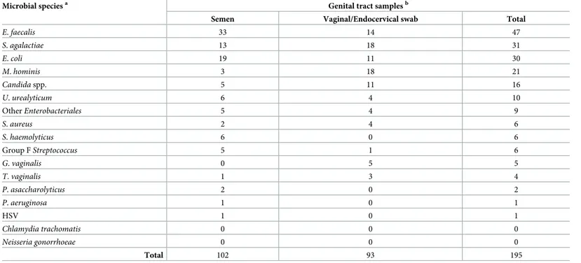
In this study, 285 infertile couples were tested for the presence of genital tract pathogens prior to undergoing IVF treatment. Microbiological analysis was conducted on a total of 855 genital sam-ples, of which 285 semen specimens, 285 vaginal swabs and 285 endocervical swabs. A total of 195 clinical strains belonging to 25 different microbial species was detected in the samples tested (Table 1).E. faecalis represented the most common finding with a prevalence of 24.1% (47/195).
Other frequently identified microbial species includedS. agalactiae (15.9%), E. coli (15.4%), M. hominis (10.8%), Candida spp. (8.2%), and U. urealyticum (5.1%) (Table 1). Co-presence of two different pathogens was detected in 14 semen specimens and in 16 vaginal/endocervical swabs, while the simultaneous presence of 3 pathogens was observed in 3 samples. It should be noted that neitherC. trachomatis nor N. gonorrhoeae were found in genital tract samples.
Prevalence and aetiology of genital tract infections in infertile couples
Microbiological results showed that 29.1% (83/285) of males and 26.3% (75/285) of females were found positive to at least one genital pathogen. A total of 132 couples (46.3%) was positive
for at least one pathogen in at least one of the two partners. Of the 132 positive couples, the male partner only was positive in 57 cases, the female only was positive in 49, and both part-ners were positive in 26 couples (Fig 1). Out of these 26 couples, 16 shared at least one genital pathogen between partners. The most prevalent microbial pathogens found in infected couples includedE. faecalis (32.6%), E. coli (22%), S. agalactiae (20.5%), M. hominis (13.6%), Candida
spp. (9.8%),Enterobacteriales other than E. coli (6.8%), and U. urealyticum (6.8%) (Fig 2).M. hominis and T. vaginalis were more prevalent in females, G. vaginalis was not found in males,
andS. haemolyticus was not detected in females (Fig 2).
Impact of genital tract pathogens on semen quality
A total of 285 semen specimens were analysed, of which 12 samples were azoospermic. Out of 273 samples, 72 (26.4%) had oligozoospermia (concentration < 15x106/ml), 65 (23.8%) showed asthe-nozoospermia (total motility < 40%), 62 (22.7%) exhibited leukocytospermia (leukocyte
counts � 106cell/ml) and 28 (10.3%) presented with teratozoospermia (typical morphology < 4%). Percent sperm motility in samples with leukocytospermia (total motility = 46±17; progressive motility = 43±18) was significantly lower compared to that of specimens with normal counts of seminal leukocytes (total = 56±17; progressive = 53±18), (p = 0.0003).
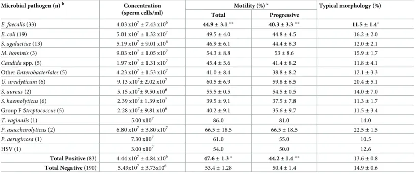
Spermiogram results showed that both total (p = 0.012) and progressive (p = 0.0098)
motil-ity were significantly diminished in samples positive to microbiological testing compared to negative specimens (Table 2). Out of all pathogens identified, onlyE. faecalis was found to
sig-nificantly alter semen parameters. Concentration, motility, and typical morphology of sperm
Table 1. Prevalence of microbial species in genital tract samples from infertile couples.
Microbial speciesa Genital tract samplesb
Semen Vaginal/Endocervical swab Total
E. faecalis 33 14 47 S. agalactiae 13 18 31 E. coli 19 11 30 M. hominis 3 18 21 Candida spp. 5 11 16 U. urealyticum 6 4 10 OtherEnterobacteriales 5 4 9 S. aureus 2 4 6 S. haemolyticus 6 0 6 Group FStreptococcus 5 1 6 G. vaginalis 0 5 5 T. vaginalis 1 3 4 P. asaccharolyticus 2 0 2 P. aeruginosa 1 0 1 HSV 1 0 1 Chlamydia trachomatis 0 0 0 Neisseria gonorrhoeae 0 0 0 Total 102 93 195 a
Candida spp., C. albicans, C. parapsilosis, C. glabrata and C. krusei; other Enterobacteriales, K. pneumoniae, K. oxytoca, P. mirabilis, M. morganii, E. aerogenes, and C. kroserii.
b
A total of 855 genital samples (285 semen specimens, 285 vaginal and 285 endocervical swabs) from 285 males and 285 females were subjected to microbiological analysis. Both a vaginal and an endocervical swab were collected from each female patient. Endocervical swabs were used to search forU. urealyticum, C. trachomatis andN. gonorrhoeae, whereas vaginal swabs were employed to test for all other pathogens.
cells were reduced inE. faecalis-positive compared to E. faecalis-negative samples (Table 2). Differences in total (p = 0.005) and progressive (p = 0.003) motility and in typical morphology
(p = 0.026) of sperm cells between semen cultures positive and negative for E. faecalis were
sta-tistically significant (Table 2). Out of 62 samples with leukocytospermia, 49 (79%) were microbiologically negative (Table 3). Although leukocytospermia was higher in negative (25.8%) than in positive (15.7%) semen samples, differences were not significant (Table 3).
The presence of
E. faecalis, E. coli, S. agalactiae, and G. vaginalis is
associated to reduced levels of vaginal lactobacilli
Genital swabs from 285 females were assessed for the presence of both genital tract pathogens and vaginal lactobacilli. A highly significant association (p<0.0001) between reduced amounts
of vaginal lactobacilli (< 104cfu/swab) and the presence of genital tract pathogens was found in the female study population (Fig 3A). When analysis was applied to each individual patho-gen, a statistically significant association was observed between reduced levels of lactobacilli
Fig 1. Venn diagram of genital tract infections in infertile couples. A total of 285 infertile couples were enrolled in the study and subjected to
microbiological analysis to search for genital tract pathogens in semen specimens and vaginal/endocervical swabs prior to IVF. One hundred and thirty-two (46.3%) couples were positive for at least one pathogen in one or both partners. The male was positive in 57 cases (20%; blue) and the female in 49 cases (17.2%; red). The intersection of blue and red regions represents the couples (n = 26; 9.1%) where both partners were positive for at least one genital tract pathogen. The external set comprises all the couples included in the study.
and the presence ofE. faecalis (p<0.0001), E. coli (p = 0.0123), S. agalactiae (p<0.0001), and G. vaginalis (p = 0.0003), (Fig 3B). It is worth noting thatG. vaginalis, a key bacterial species of
BV, was detected only in swabs with decreased load of vaginal lactobacilli.
Infection with
E. faecalis and/or U. urealyticum and/or and M. hominis is
predictive of a negative IVF outcome in infertile couples
To investigate whether specific genital tract pathogens could be associated to IVF failure, microbiological results were correlated to IVF outcomes. IVF success was slightly higher in non-infected than in infected couples. Microbiological data indicated that specific pathogens (E. faecalis, U. urealyticum, M. hominis, G. vaginalis, E. coli) were more prevalent in
unsuccess-ful (IVF-) than successunsuccess-ful (IVF+) couples, however, no significant differences were calculated when each pathogen was tentatively associated with IVF outcome. Therefore, analysis was per-formed by examining couples positive for groups of genital tract pathogens after sequential exclusion of the pathogens that seemed not to affect IVF outcome. The microbial group consti-tuted ofE. faecalis, U. urealyticum, M. hominis, G. vaginalis, and T. vaginalis was more
preva-lent in IVF- than IVF+ couples, but differences were not significant (p > 0.05, χ2test with Yates’ correction). Elimination ofT. vaginalis showed that prevalence of the microbial group
was significantly higher in IVF- (36.3%) compared to IVF+ (16.7%) couples (p = 0.03, χ2test with Yates’ correction). Finally, by further excludingG. vaginalis, the smallest infectious group
significantly associated with IVF failure includedE. faecalis and/or U. urealyticum and/or M. hominis (Table 4). Analysis of the IVF+ couples showed that 30/35 (85.7%) were negative to microbiological testing, whereas out of the couples infected withE. faecalis and/or U.
Fig 2. Prevalence of genital tract pathogens in infertile couples. Tornado graph showing the number of clinical strains of different microbial pathogens found in genital tract samples from male (blue bars) and female (red bars) patients. For each pathogen, the number of positive couples is also shown. Pathogens are listed according to their prevalence in infected couples.Enterobacteriales other than E. coli included Klebsiella pneumoniae, Klebsiella oxytoca, Proteus mirabilis, Morganella morganii, Enterobacter aerogenes, and Citrobacter kroserii. Candida spp. included C. albicans, C. parapsilosis, C. glabrata, and C. krusei.
urealyticum and/or M. hominis, just 5/67 (7.5%) obtained a successful IVF (p = 0.02, χ2test with Yates’ correction;Table 4). Interestingly, among the IVF- couples positive for this micro-bial group,E. faecalis and U. urealyticum were found in approximately 90% of cases, whereas M. hominis was detected in all the couples with a poor IVF outcome.
Discussion
Infertility is an ongoing challenge throughout the world and is increasingly being considered not only as a private matter but also as a public health burden. ART has allowed to overcome certain issues, however, rates of conception are still low [30]. Several factors can participate in infertility, including genital tract infections. However, no consensus guidelines are available yet on microbiological evaluation of infertile couples prior to undergoing IVF.
Table 2. Correlation of genital tract pathogens with semen parameters in infertile malesa.
Microbial pathogen (n)b Concentration (sperm cells/ml)
Motility (%)c Typical morphology (%) Total Progressive E. faecalis (33) 4.03 x10⁷ ± 7.43 x10⁶ 44.9 ± 3.1�� 40.3± 3.3�� 11.5± 1.4� E. coli (19) 5.01 x10⁷± 1.32 x10⁷ 49.5± 4.0 44.8± 4.5 16.2± 2.0 S. agalactiae (13) 5.19 x10⁷± 9.01 x10⁶ 46.9± 6.1 44.4± 6.3 12.0± 2.1 M. hominis (3) 9.03 x10⁷ ± 1.05 x10⁷ 54.3 ± 8.8 53± 8.6 15.9± 1.7 Candida spp. (5) 1.97 x10⁷ ± 1.31 x10⁷ 45.4 ± 5.6 41.4± 8.2 11.8± 4.1 OtherEnterobacteriales (5) 4.23 x10⁷± 1.53 x10⁷ 41.0± 8.4 38.8± 8.2 12.1± 3.3 U. urealyticum (6) 9.13 x10⁷ ± 2.02 x10⁷ 60.5 ± 6.9 59.8± 6.5 20.4± 5.1 S. aureus (2) 5.15 x10⁷ ± 9.50 x10⁶ 55.5 ± 0.5 54.5± 0.5 14.0± 7.0 S. haemolyticus (6) 2.39 x10⁷ ± 1.39 x10⁷ 39.5 ± 9.1 37.5± 7.8 11.3± 1.7 Group FStreptococcus (5) 2.28 x10⁷± 9.81 x10⁶ 40.2± 9.1 35.6± 9.7 11.5± 3.4 T. vaginalis (1) 5.00 x10⁷ 86.0 81.0 14.0 P. asaccharolyticus (2) 6.80 x10⁷ ± 3.80 x10⁷ 66.5 ± 18.5 66.5± 18.5 22.5± 1.5 P. aeruginosa (1) 7.30 x10⁷ 61.0 55.0 10.5 HSV (1) 3.00 x10⁷ 54.0 50.0 12.6 Total Positive (83) 4.44 x10⁷ ± 4.84 x10⁶ 47.6 ± 1.3� 44.2± 1.4�� 13.6± 0.8 Total Negative (190) 5.49x10⁷ ± 3.73x10⁶ 53.4 ± 1.28 50.4± 1.4 14.9± 0.6
aAll data are represented as mean
± SEM.
bNumbers (in brackets) of genital tract pathogens identified in 273 semen specimens. Twelve patients were excluded because azoospermic. Total positive (n = 83),
number of males positive for at least one pathogen. Total negative (n = 190), number of males negative to microbiological testing.
cTotal motility, sperm cells moving in all directions; progressive motility, sperm cells moving along a straight line.
�Statistical analysis (Mann-Whitney test) was performed against the group of negative males. Significant differences are in bold:�,p < 0.05;��,p < 0.01.
https://doi.org/10.1371/journal.pone.0207684.t002
Table 3. Association between the presence or absence of genital tract pathogens and leukocytospermia in semen samples.
Pathogensa Semen samplesb p valuec
Absence of leukocytospermia (%) Presence of leukocytospermia (%) Total
Absence 141 (74.2) 49 (25.8) 190 0.09
Presence 70 (84.3) 13 (15.7) 83
Total 211 62 273
aMicrobial pathogens were searched in a total of 273 semen samples after exclusion of 12 specimens from azoospermic males. bSemen samples were considered as leukocytospermic when leukocyte counts were � 106/ml.
cχ2test with Yates’ correction.
The present study focused on the impact of asymptomatic genital tract infections on couple fertility. Main findings are: (i) approximately half of the couples was diagnosed with a genital tract infection, (ii)E. faecalis had a significantly negative impact on sperm motility and
mor-phology, (iii) the presence ofE. faecalis, E. coli, S. agalactiae, and G. vaginalis in females was
Fig 3. Association between genital tract pathogens and vaginal lactobacilli in infertile females. Both a vaginal and an endocervical swab were collected from each female patient enrolled in the study. Endocervical swabs (n = 285) were used to search forU. urealyticum, C. trachomatis and N. gonorrhoeae, while vaginal swabs (n = 285) were simultaneously tested for all other pathogens and vaginal lactobacilli. Lactobacilli were quantified on selective solid media, and counts < 104cfu/swab were regarded as reduced levels of lactobacilli. A. Contingency table reporting the number of swabs negative (-) or positive (+) for at least one genital tract pathogen in relation to the number of swabs with normal (+) or reduced (-) levels of lactobacilli. B. Contingency table showing the number of swabs positive forE. faecalis, E. coli, S. agalactiae, and G. vaginalis that presented with either normal (+) or low (-) levels of lactobacilli. The χ2test with Yates’ correction was used for all cases except forG. vaginalis (Fisher exact test). For each of the above pathogen, statistical analysis was performed against the negative samples with normal (n = 171) or reduced (n = 39) levels of vaginal lactobacilli.
https://doi.org/10.1371/journal.pone.0207684.g003
Table 4. Association between the presence ofE. faecalis and/or U. urealitycum and/or M. hominis and IVF outcome in infertile couples.
Pathogens IVF outcome p valueb
IVF- couples (%) IVF+ couples (%) Total
Absencea 111 (78.7) 30 (21.3) 141 0.02 E. faecalis, and/or U. urealitycum, and/or M. hominis 62 (92.5) 5 (7.5) 67 Total 173 35 208 a
Results refer to all the couples negative to microbial testing (n = 153) after exclusion of 12 couples where the male partner was azoospermic.
bχ2
test with Yates’ correction.
significantly associated to reduced levels of vaginal lactobacilli, (iv) the presence of the group
E. faecalis, U. urealyticum, and M. hominis in infertile couples was significantly associated to
IVF negative outcome.
All the infertile couples enrolled in the study were asymptomatic for genital tract infections, but nearly half (46.3%) resulted positive to microbiological testing. As to our knowledge no data are available in the literature on the prevalence of asymptomatic genital tract infections in naturally fertile couples, it is difficult to estimate the impact of asymptomatic infections on fer-tility. The microbial species identified in our study are mostly colonizers of the male anterior urethra and coronal sulcus or the vaginal milieu [31,32], but are also responsible of urinary tract (UTI), genital and systemic infections. The prevalence of pathogens was different between males and females, with certain microbial species showing a clear predominance in males (E. faecalis, S. haemolyticus, Group F Streptococcus) or females (M. hominis, Candida
spp.,G. vaginalis, T. vaginalis). E. faecalis and M. hominis/S. agalactiae were the most
fre-quently detected microbes in males and females, respectively.M. hominis is a common
endo-symbiont ofT. vaginalis, which acts not only as a protective niche but also as a ‘Trojan horse’
to transmit the bacteria to the human genital tract [33]. In our case, although the prevalence of the protozoan in infected females was low (4%), 66.7% of theT. vaginalis-positive vaginal
swabs also containedM. hominis. Out of all pathogens detected in infected couples, E. faecalis
was the most common (32.6%) and was shown to adversely affect couple fertility.
Genital infection and inflammation can impact on male fertility in several ways, including deterioration of spermatogenesis, impairment of sperm functions, generation of ROS leading to DNA fragmentation or oxidative protein modification, production of sperm antibodies, and obstruction of the seminal tract [4]. The relationship between bacteriospermia, leukocytosper-mia and semen parameters in infertile men is still debated. Some studies showed a detrimental influence of microbial pathogens or/and leukocytes on semen quality [5,7,10,34–37], while others did not observe any effect [6,38,39]. In this study, 29.1% of semen specimens was posi-tive to microbiological analysis, which is in accordance with literature data reporting that rates of bacteriospermia in infertile men can fluctuate from 15 to 60% [5–7,36,39]. The most preva-lent species wasE. faecalis, which was significantly associated to reduced motility and altered
morphology of spermatozoa, suggesting that enterococci may have a direct negative influence on semen quality as previously published [5,6,36]. In contrast, significant sperm abnormalities were not observed inE. coli- and S. agalactiae-positive samples, the second and third most
commonly isolated species. Some authors have shown thatE. coli induces alterations in
human spermatozoa, resulting in reduced motility, altered acrosomal function, and decreased vitality [5,35,40]. Nonetheless, the fact that those data mostly originate fromin vitro studies
based on large bacterial concentrations that will unlikely be reached duringin vivo infection,
may explain our negative result onE. coli. Leukocytospermia is generally considered as a
marker of inflammation with a poor diagnostic value for genital tract infections [6,7,41]. Sev-eral factors independent of infectious challenges have been associated to elevated seminal leu-kocytes, including ageing, medications, smoking, alcohol and drug abuse [7,8]. In our study, leukocytospermia was not significantly associated with the presence of microbial pathogens in semen (p = 0.09), as evidenced by the fact that 79% of leukocytospermic samples were
microbi-ologically negative as reported before [42]. This finding, which is not unexpected
[6,7,41,42,43], probably indicates that the presence of potential pathogens in semen does not necessarily lead to a full-fledged inflammatory response with substantial leukocyte recruit-ment. The hypothesis may be especially valid in asymptomatic patients as those enrolled in the present study. Moreover, the time of semen collection could be another relevant factor to explain our data since bacteria and leukocytes may not be present simultaneously in semen samples as described [44].
BV, AV and abnormal vaginal flora (decrease/absence of lactobacilli) have been reported to affect pregnancy rate and outcome [24]. A recent systematic review and meta-analysis showed that BV is associated with female infertility, preclinical pregnancy loss and preterm birth, although it does not seem to impact on conception rates [21]. Prevalence of BV in infertile women varies considerably from 10% to 45% depending on the study population and diagnostic criteria [21,25,45,46]. Diagnosis of BV can be performed based on clinical, microscopic, and (cultural and/or molecular) microbiological criteria [19]. The Nugent scoring system was the first standardized method founded on classification and enumeration of ‘morphotypes’ in Gram-stained vaginal smears [47]. Despite being long recognized as the gold standard for BV diagnosis, the Nugent system has several limitations [19]. In the current study, we chose a com-bined culture- and molecular-based diagnostic approach for detection and quantification of both pathogens and lactobacilli in genital swabs. Microbiological analysis showed that the pres-ence of vaginal lactobacilli was generally associated to abspres-ence of genital pathogens; conversely, detection ofE. faecalis, E. coli, S. agalactiae, and G. vaginalis was significantly associated with
reduced levels of vaginal lactobacilli.G. vaginalis is strongly linked to BV [19], whereasE. coli, S. agalactiae, and E. faecalis are not typical BV-defining microorganisms but are instead common
agents of UTI and AV. Absence of lactobacilli, besides being a key feature of BV, has also been correlated to AV [22,48] and recurrent UTI byE. coli [49] and other uropathogens [50], under-lining the protective function exerted by lactobacilli against urogenital infections. In contrast, the presence ofM. hominis, another recurrent species in BV, was not significantly associated to
decreased numbers of vaginal lactobacilli in this study population. It should also be noted that a small percentage (18.6%) of females negative to microbiological testing had low amounts of lac-tobacilli, indicative of either a healthy microbiota not dominated byLactobacillus spp. [18] or a transitional stage in the dynamic shifts of the vaginal microbial ecosystem [51]. The outcome of IVF has been associated with the composition of the vaginal microbiota on the day of ET [52], and a microbiota exclusively composed by lactobacilli is considered the most promising sce-nario for successful IVF [53]. In our case, only 12.8% of patients with reduced quantity of lacto-bacilli belonged to the IVF+ couples, yet again emphasizing the key role played by a
Lactobacillus-dominated microbiota for a positive outcome of IVF.
In the current study, IVF negative outcome was slightly more elevated in infected than non-infected couples, but differences were not significant. However, specific pathogens were more frequently found in IVF- couples, and hence a definite infectious group (E. faecalis and/
orU. urealyticum and/or M. hominis) was identified that significantly correlated with IVF
fail-ure (p = 0.02). It should be noted that G. vaginalis was initially included in the group
(p = 0.031), in accordance with a recent prospective study reporting the association of G. vagi-nalis and A. vaginae with low pregnancy rates in IVF patients [25]. Identification of a group of infectious agents, rather than a single microbial species, is not unexpected, as the above patho-gens can be found as agents of polymicrobial genital tract infections [19,50,54].M. hominis has
been shown to synergistically cooperate withG. vaginalis in BV [19,55] and is a frequent sym-biont ofT. vaginalis in trichomoniasis and BV [33,56], whileE. faecalis can be detected in
poly-microbial urogenital and biofilm-based infections [50]. Whether these microbes act as either independent or bystander pathogens in genital tract infections is not clear yet, however, increasing evidence is accumulating on their role in infertility, adverse pregnancy outcome, and post-partum complications [11,13,19,21,22,24,25,54,57]. Lower rates of fertilization, implantation and clinical pregnancy were observed in couples undergoing ICSI that were posi-tive toE. coli, E. faecalis, S. agalactiae, and Staphylococcus spp. [52,58]. Detection of genital
Mollicutes in couples undergoing IVF was also associated to reduced pregnancy rates [59,60] and increased miscarriages [61]. To this regard, it is interesting to note that all the couples of this study infected byM. hominis had a negative IVF outcome. The present work shed light on
three specific genital tract pathogens as predictive infectious markers for poor outcome of IVF, emphasizing the importance of microbiological testing of infertile couples forE. faecalis, U. urealyticum, and M. hominis prior to IVF procedures.
In conclusion, infertility is a multi-factorial and multi-faceted clinical condition which poses a profound economic and psychological burden on affected couples and high costs on the healthcare system. In this study, we have identified an infectious group that significantly correlated as an independent risk factor to infertility and negative outcome of IVF. However, the causes of IVF failure often remain unknown. A joint effort between clinical microbiolo-gists, infectious diseases and reproductive medicine specialists is desirable to produce consen-sus guidelines on testing for pathogens associated to infertility and assessment of both microbiological and clinical outcomes before ART treatment. Improved management of geni-tal tract infections in infertile couples may be helpful to increase pregnancy rates, reduce the total number of treatment cycles and possibly enhance first level fertility approaches with ben-eficial effects on couple well-being and healthcare costs.
Supporting information
S1 Table. Primers used for Real-Time PCR. List of primers and probes used to identify
non-cultivable pathogens in genital tract samples. (DOCX)
Acknowledgments
We would like to acknowledge Simonetta Brignali and Tiziana Braccini for excellent technical assistance with microbiological analysis. The study was supported by the Italian Ministry of Education, University and Research (Ministero dell’Istruzione, dell’Università e della Ricerca; MIUR), ‘Progetti di Rilevante Interesse Nazionale’ (PRIN) 2012 to GP.
Author Contributions
Conceptualization: Susanna Ricci, Paola Piomboni, Vincenzo De Leo, Gianni Pozzi. Data curation: Susanna Ricci, Stefano De Giorgi, Paola Piomboni.
Formal analysis: Stefano De Giorgi, Stefania Rossi. Funding acquisition: Gianni Pozzi.
Investigation: Susanna Ricci, Stefano De Giorgi, Elisa Lazzeri, Alice Luddi. Methodology: Susanna Ricci, Gianni Pozzi.
Resources: Paola Piomboni, Vincenzo De Leo, Gianni Pozzi. Supervision: Susanna Ricci, Gianni Pozzi.
Writing – original draft: Susanna Ricci, Paola Piomboni, Gianni Pozzi. Writing – review & editing: Susanna Ricci, Gianni Pozzi.
References
1. Inhorn MC, Patrizio P. Infertility around the globe: new thinking on gender, reproductive technologies and global movements in the 21st century. Hum Reprod Update. 2015; 21:411–26.https://doi.org/10. 1093/humupd/dmv016PMID:25801630
2. Zegers-Hochschild F, Adamson GD, de Mouzon J., Ishihara O, Mansour R, Nygren K, et al. Interna-tional Committee for Monitoring Assisted Reproductive Technology (ICMART) and the World Health
Organization (WHO) revised glossary of ART terminology, 2009. Fertil Steril. 2009; 92:1520–4.https:// doi.org/10.1016/j.fertnstert.2009.09.009PMID:19828144
3. Pellati D, Mylonakis I, Bertoloni G, Fiore C, Andrisani A, Ambrosini G, et al. Genital tract infections and infertility. Eur J Obset Gynecol Reprod Biol. 2008; 140:3–11.
4. Gimenes F, Souza RP, Bento JC, Teixeira JJ, Maria-Engler SS, Bonini MG, et al. Male infertility: a pub-lic health issue caused by sexually transmitted pathogens. Nat Rev Urol. 2014; 11:672–87.https://doi. org/10.1038/nrurol.2014.285PMID:25330794
5. Moretti E, Capitani S, Figura N, Pammolli A, Federico MG, Giannerini V, et al. The presence of bacteria species in semen and sperm quality. J Assist Reprod Genet. 2009; 26:47–56.https://doi.org/10.1007/ s10815-008-9283-5PMID:19089609
6. Rodin DM, Larone D, Goldstein M. Relationship between semen cultures, leukospermia, and semen analysis in men undergoing fertility evaluation. Fertil Steril. 2003; 79:1555–8. PMID:12801559 7. Domes T, Lo KC, Grober ED, Mullen JB, Mazzulli T, Jarvi K. The incidence and effect of
bacteriosper-mia and elevated seminal leukocytes on semen parameters. Fertil Steril. 2012; 97:1050–5.https://doi. org/10.1016/j.fertnstert.2012.01.124PMID:22341372
8. Jue JS, Ramasamy R. Significance of positive semen culture in relation to male infertility and the assis-ted reproductive technology process. Transl Androl Urol. 2017; 6:916–22.https://doi.org/10.21037/tau. 2017.06.23PMID:29184792
9. Keck C, Gerber-Schafer C, Clad A, Wilhelm C, Breckwoldt M. Seminal tract infections: impact on the male infertility and treatment options. Hum Reprod Update. 1998; 4:891–903. PMID:10098479 10. Sanocka-Maciejewska D, Ciupinska M, Kurpisz M. Bacterial infection and semen quality. J Reprod
Immunol. 2005; 67:51–6.https://doi.org/10.1016/j.jri.2005.06.003PMID:16112738
11. Taylor-Robinson D, Lamont RF. Mycoplasmas in pregnancy. BJOG. 2010; 118:164–74.https://doi.org/ 10.1111/j.1471-0528.2010.02766.xPMID:21091927
12. Taylor-Robinson D. Mollicutes in vaginal microbiology: Mycoplasma hominis, Ureaplasma urealyticum, Ureaplasma parvum and Mycoplasma genitalium. Res Microbiol. 2017; 168:875–81.https://doi.org/10. 1016/j.resmic.2017.02.009PMID:28263902
13. Lee JS, Kim KT, Lee HS, Yang KM, Seo JT, Choe JH. Concordance of Ureaplasma urealyticum and Mycoplasma hominis in infertile couples: impact on semen parameters. Urol. 2013; 81:1219–24.https:// doi.org/10.1016/j.urology.2013.02.044PMID:23602797
14. Gdoura R, Kchaou W, Chaari C, Znazen A, Keskes L, Rebai T, et al. Ureaplasma urealyticum, Urea-plasma parvum, MycoUrea-plasma hominis and MycoUrea-plasma genitalium infections and semen quality of infertile men. BMC Infect Dis. 2007; 7:129.https://doi.org/10.1186/1471-2334-7-129PMID:17988404 15. Reichart M, Kahane I, Bartoov B. In vivo and in vitro impairment of human and ram sperm nuclear
chro-matin integrity by sexually transmitted Ureaplasma urealyticum infection. Biol Reprod. 2000; 63:1041– 8. PMID:10993825
16. Huang C, Zhu HL, Xu KR, Wang SY, Fan LQ, Zhu WB. Mycoplasma and Ureaplasma infection and male infertility: a systematic review and meta-analysis. Andrology. 2015; 3:809–16.https://doi.org/10. 1111/andr.12078PMID:26311339
17. McGowin CL, Anderson-Smits C. Mycoplasma genitalium: an emerging cause of sexually transmitted disease in women. PLoS Pathog. 2011; 7:e1001324.https://doi.org/10.1371/journal.ppat.1001324
PMID:21637847
18. Ravel J, Gajer P, Abdo Z, Schneider GM, Koening SSK, McCulle SL, et al. Vaginal microbiome of repro-ductive-age women. Proc Natl Acad Sci USA. 2011; 108:4680–7.https://doi.org/10.1073/pnas. 1002611107PMID:20534435
19. Onderdonk AB, Delany ML, Fichorova RN. The human microbiome during bacterial vaginosis. Clin Microbiol Rev. 2016; 29:223–38.https://doi.org/10.1128/CMR.00075-15PMID:26864580
20. Unemo M, Bradshaw CS, Hocking JS, de Vries HJC, Francis SC, Mabey D, et al. Sexually transmitted infections: challenges ahead. Lancet Infect Dis. 2017; 17:e235–e279. https://doi.org/10.1016/S1473-3099(17)30310-9PMID:28701272
21. van Oostrum N, De Sutter P, Meyes J, Verstraelen H. Risks associated with bacterial vaginosis in infer-tility patients: a systematic review and meta-analysis. Hum Reprod. 2013; 28:1809–15.https://doi.org/ 10.1093/humrep/det096PMID:23543384
22. Donders GGG, Bellen G, Grinceviciene S, Ruban K, Vieira-Baptista P. Aerobic vaginitis: no longer a stranger. Res Microbiol. 2017; 168:845–58.https://doi.org/10.1016/j.resmic.2017.04.004PMID:
28502874
23. Ralph SG, Rutherford AJ, Wilson JD. Influence of bacterial vaginosis on conception and miscarriage in the first trimester: cohort study. BMJ. 1999; 319:220–3. PMID:10417083
24. Donders GG, Van Calsteren K, Bellen G, Reybrouck R, Van den Bosch T, Riphagen I, et al. Predictive value for preterm birth of abnormal vaginal flora, bacterial vaginosis and aerobic vaginitis during the first trimester of pregnancy. BJOG. 2009; 116:1315–24.https://doi.org/10.1111/j.1471-0528.2009.02237.x
PMID:19538417
25. Haahr T, Jensen JS, Thomsen L, Duus L, Rygaard K, Humaidan P. Abnormal vaginal microbiota may be associated with poor reproductive outcomes: a prospective study in IVF patients. Hum Reprod. 2016; 31:795–803.https://doi.org/10.1093/humrep/dew026PMID:26911864
26. Rubino P, Vigano P, Luddi A, Piomboni P. The ICSI procedure from past to future: a systematic review of the more controversial aspects. Hum Reprod Update. 2016; 22:194–227.https://doi.org/10.1093/ humupd/dmv050PMID:26586241
27. World Health Organization. WHO laboratory manual for the examination and processing of human semen, 5th edn. 2010. Geneva, Switzerland, WHO Press.
28. World Health Organization. Laboratory manual for the examination of human semen and sperm-cervical mucus interaction, 3rd edn. 1992. Cambridge, UK, Cambridge University Press.
29. Politich JA, Wolff H, Hill JA, Anderson DJ. Comparison of methods to enumerate white blood cells in semen. Fertil Steril. 1993; 60:372–5. PMID:8393411
30. European IVF-monitoring Consortium (EIM), European Society of Human Reproduction and Embriol-ogy (ESHRE), Calhaz-Jorge C, de Geyter C, Kupka MS, de Mouzon J., Erb K, Mocanu E, et al. Assisted reproductive technology in Europe, 2013: results generated from European registers by ESHRE. Hum Reprod. 2017; 32:1957–73.https://doi.org/10.1093/humrep/dex264PMID:29117383
31. Mandar R. Microbiota of male genital tract: impact on the health of man and his partner. Pharmacol Res. 2013; 69:32–41.https://doi.org/10.1016/j.phrs.2012.10.019PMID:23142212
32. Mendling W. Vaginal microbiota. Adv Exp Med Biol. 2016; 902:83–93. https://doi.org/10.1007/978-3-319-31248-4_6PMID:27161352
33. Dessı` D, Rappelli P, Diaz N, Cappuccinelli P, Fiori PL. Mycoplasma hominis and Trichomonas vagina-lis: a unique case of symbiotic relationship between two obligate human parasites. Front Biosci. 2006; 11:2028–34. PMID:16720288
34. Huwe P, Diemer T, Ludwig M, Liu J, Schiefer HG, Weidner W. Influence of different uropathogenic microorganisms on human sperm motility parameters in an in vitro experiment. Andrologia. 1998; 30 Suppl 1:55–9.
35. Villegas J, Schulz M, Soto L, Sanchez R. Bacteria induce expression of apoptosis in human spermato-zoa. Apoptosis. 2005; 10:105–10.https://doi.org/10.1007/s10495-005-6065-8PMID:15711926 36. Mehta RH, Sridhar H, Vijay Kumar BR, Anand Kumar TC. High incidence of oligozoospermia and
tera-tozoospermia in human semen infected with the aerobic bacterium Streptococcus faecalis. Reprod Biomed Online. 2002; 5:17–21. PMID:12470540
37. Kiessling AA, Desmarais BM, Yin HZ, LoVerde J, Eyre RC. Detection and identification of bacterial DNA in semen. Fertil Steril. 2008; 90:1744–56.https://doi.org/10.1016/j.fertnstert.2007.08.083PMID:
18191853
38. Cottell E, Harrison RF, McCaffrey M, Walsh T, Mallon E, Barry-Kinsella C. Are seminal fluid microorgan-isms of significance or merely contaminants? Fertil Steril. 2000; 74:465–70. PMID:10973639
39. Vilvanathan S, Kandasamy B, Jayachandran AL, Sathiyanarayanan S, Tanjore Singaravelu V, Krishna-murthy V, et al. Bacteriospermia and its impact on basic semen parameters among infertile men. Inter-discip Perspect Infect Dis. 2016; 2016:2614692.https://doi.org/10.1155/2016/2614692PMID:26880908 40. Diemer T, Huwe P, Michelmann HW, Mayer F, Shiefer HG, Weidner W. Escherichia coli induced
alter-ations of human spermatozoa. An electron microscopy analysis. Int J Androl. 2000; 23:178–86. PMID:
10844544
41. Trum JW, Mol BWJ, Pannekoek Y, Spanjaard L, Wertheim P, Bleker OP, et al. Value of detecting leuko-cytospermia in the diagnosis of genital tract infection in subfertile men. Fertil Steril. 1998; 70:315–9. PMID:9696227
42. Wolff H. The biologic significance of white blood cells in semen. Fertil Steril. 1995; 63:1143–57. PMID:
7750580
43. Potts JM, Sharma R, Pasqualotto F, Nelson D, Hall G, Agarwal A. Association of Ureaplasma urealyti-cum with abnormal reactive oxygen species levels and absence of leukocytospermia. J Urol. 2000; 163:1775–8. PMID:10799180
44. Sanocka D, Fraczek M, Jedrzejczak P, Szumala-Kakol A, Kurpisz M. Male genital tract infection: an influence of leukocytes and bacteria on semen. J Reprod Immunol. 2004; 62:111–24.https://doi.org/10. 1016/j.jri.2003.10.005PMID:15288187
45. Mangot-Bertrand J, Fenollar F, Bretelle F, Gamerre M, Raoult D, Courbiere B. Molecular diagnosis of bacterial vaginosis: impact on IVF outcome. Eur J Clin Microbiol Infect Dis. 2012; 32:535–41.https:// doi.org/10.1007/s10096-012-1770-zPMID:23132689
46. Salah RM, Allam AM, Magdy AM, Mohamed AS. Bacterial vaginosis and infertility: cause or associa-tion? Eur J Obstet Gynecol Reprod Biol. 2013; 167:59–63.https://doi.org/10.1016/j.ejogrb.2012.10.031
PMID:23199811
47. Nugent RP, Krohn MA, Hillier SL. Reliability of diagnosis bacterial vaginosis is improved by a standard-ized method of gram stain interpretation. J Clin Microbiol. 1991; 29:297–301. PMID:1706728
48. Rumyantseva TA, Bellen G, Savochkina YA, Guschin AE, Donders GGG. Diagnosis of aerobic vaginitis by quantitative real-time PCR. Arch Gynecol Obstet. 2016; 294:109–14.https://doi.org/10.1007/ s00404-015-4007-4PMID:26781259
49. Gupta K, Stapleton AE, Hooton TM, Roberts PL, Fennel CL, Stamm WE. Inverse association of H2O2
-producing lactobacilli and vaginal Escherichia coli colonization in women with recurrent urinary tract infections. J Infect Dis. 1998; 178:446–50. PMID:9697725
50. Kline KA, Lewis AL. Gram-positive uropathogens, polymicrobal urinary tract infection, and the emerging microbiota of the urinary tract. Microbiol Spectr. 2016; 4.
51. Ravel J, Brotman RM, Gajer P, Ma B, Nandy M, Fadrosh DW, et al. Daily temporal dynamics of vaginal microbiota before, during and after episodes of bacterial vaginosis. Microbiome. 2013; 1:29.https://doi. org/10.1186/2049-2618-1-29PMID:24451163
52. Fanchin R, Harmas A, Benaoudia F, Lundkvist U, Olivennes F, Frydman R. Microbial flora of the cervix assessed at the time of embryo transfer adversely affects in vitro fertilization outcome. Fertil Steril. 1998; 70:866–70. PMID:9806568
53. Hyman RW, Hernon CN, Jiang H, Palm C, Fukushima M, Bernstein D, et al. The dynamics of the vagi-nal microbiome during infertility therapy with in vitro fertilization-embryo transfer. J Assist Reprod Genet. 2012; 29:105–15.https://doi.org/10.1007/s10815-011-9694-6PMID:22222853
54. Murtha AP, Edwards JM. The role of Mycoplasma and Ureaplasma in adverse pregnancy outcomes. Obstet Gynecol Clin North Am. 2014; 41:615–27.https://doi.org/10.1016/j.ogc.2014.08.010PMID:
25454994
55. Cox C, Watt AP, McKenna JP, Coyle PV. Mycoplasma hominis and Gardnerella vaginalis display a sig-nificant synergistic relationship in bacterial vaginosis. Eur J Clin Microbiol Infect Dis. 2016; 35:481–7.
https://doi.org/10.1007/s10096-015-2564-xPMID:26796553
56. Fichorova RN, Fraga J, Rappelli P, Fiori PL. Trichomonas vaginalis infection in symbiosis with Tricho-monasvirus and Mycoplasma. Res Microbiol. 2017; 168:882–91.https://doi.org/10.1016/j.resmic.2017. 03.005PMID:28366838
57. Capoccia R, Greub G, Baud D. Ureaplasma urealyticum, Mycoplasma hominis and adverse pregnancy outcomes. Curr Opin Infect Dis. 2013; 26:231–40.https://doi.org/10.1097/QCO.0b013e328360db58
PMID:23587772
58. Zeyad A, Hamad M, Amor H, Hammadeh ME. Relationships between bacteriospermia, DNA integrity, nuclear protamine alteration, sperm quality and ICSI outcome. Reprod Biol. 2018; 18:115–21.https:// doi.org/10.1016/j.repbio.2018.01.010PMID:29449095
59. Shalika S, Dugan K, Smith RD, Padilla SL. The effect of positive semen bacterial and Ureaplasma cul-tures on in-vitro fertilization success. Hum Reprod. 1996; 11:2789–92. PMID:9021392
60. Montagut JM, Lepretre S, Degoy J, Rousseau M. Ureaplasma in semen and IVF. Hum Reprod. 1991; 6:727–9. PMID:1939557
61. Kanakas N, Mantzavinos T, Boufidou F, Koumentakou I, Creatsas G. Ureaplasma urealyticum in semen: is there any effect on in vitro fertilization outcome? Fertil Steril. 1999; 71:523–7. PMID: