doi:10.1093/humrep/dex377
ORIGINAL ARTICLE
Reproductive biology
In vitro differentiation of human
oocyte-like cells from oogonial stem cells:
single-cell isolation and molecular
characterization
Erica Silvestris
1,2, Paola Cafforio
1,*, Stella D
’Oronzo
1, Claudia Felici
1,
Franco Silvestris
1, and Giuseppe Loverro
21Department of Biomedical Sciences and Human Oncology, Section of Internal Medicine and Clinical Oncology, University of Bari Aldo Moro,
P.za G. Cesare, 11-70124 Bari, Italy2Department of Emergency and Organ Transplantation, Section of Obstetrics and Gynecology,
University of Bari Aldo Moro, P.za G. Cesare, 11-70124 Bari, Italy *Correspondence address. E-mail: [email protected]
Submitted on July 25, 2017; resubmitted on November 30, 2017; accepted on December 18, 2017
STUDY QUESTION
:
Are the large cells derived from cultured DEAD box polypeptide 4 (DDX4)-positive oogonial stem cells (OSCs), isolated from the ovarian cortex of non-menopausal and menopausal women, oocyte-like cells?SUMMARY ANSWER
:
Under appropriate culture conditions, DDX4-positive OSCs from non-menopausal and menopausal women differ-entiate into large haploid oocyte-like cells expressing the major oocyte markers growth differentiation factor 9 (GDF-9) and synaptonemal complex protein 3 (SYCP3) and then enter meiosis.WHAT IS KNOWN ALREADY
:
The recent reports of OSCs in the ovaries of non-menopausal and menopausal women suggest thatneo-oogenesis is inducible during ovarian senescence. However, several questions remain regarding the isolation of these cells, their spontan-eous maturation in vitro, and thefinal differentiation state of the resulting putative oocytes.
STUDY DESIGN
,
SIZE,
DURATION:
DDX4-positive OSCs were obtained from 19 menopausal and 13 non-menopausal women (whounderwent hysterectomy for uterinefibroma, ovarian cyst, or other benign pathologies) and cultured for up to 3 weeks. Large and small cells were individually isolated and typed for early and late differentiation markers.
PARTICIPANTS
/
MATERIALS,
SETTING,
METHODS:
Ovarian cortex fragments were processed by immuno-magnetic separationusing a rabbit anti-human DDX4 antibody and the positive populations were measured by assessing both FRAGILIS and stage-specific embry-onic antigen 4 (SSEA-4) expression. After 3 weeks in culture, large oocyte-like cells were individually isolated by DEPArray based on PKH26 red staining and cell size determination. GDF-9 and SYCP3 asfinal, and developmental pluripotency-associated protein 3 (DPPA3) as primor-dial, germline markers were measured by droplet digital PCR. The haploid versus diploid chromosomal content of chromosomes X and 5 was investigated usingfluorescence in situ hybridization (FISH).
MAIN RESULTS AND THE ROLE OF CHANCE
:
SSEA-4+and FRAGILIS+subsets of DDX4-positive populations were present atlow-er mean levels in menopausal (SSEA-4+: 46.7%; FRAGILIS+: 47.5%) than in non-menopausal (SSEA-4+: 64.9%; FRAGILIS+: 64.8) women (P< 0.05). A comparison of the women’s age with the ratio of DDX4-positive cells/cm3of ovarian cortex revealed an inverse correlation with OSC number (P< 0.05). Once cultured, cells from both groups differentiated to form large (up to 80 μm) mature oocyte-like cells with typ-ical oocyte morphology. Despite the higher numbers of these cells in cultures from non-menopausal women (37.4 versus 23.7/well; P< 0.001), the intra-culture percentages of large oocyte-like cells did not differ significantly between the two groups. Single large oocyte-like cells isolated from non-menopausal and menopausal women expressed equivalent levels of GDF-9 (e.g. 2.0 and 2.6 copies/μl RNA, respectively) and SYCP3 (e.g. 1.2 and 1.5 copies/μl RNA, respectively) mRNA. The remaining small cells isolated from the cultures expressed large amounts of DPPA3 mRNA (e.g. 5.0 and 5.1 copies/μl RNA, from menopausal and non-menopausal women, respectively), which was undetectable in the large oocyte-like cells. FISH analysis of the large and small cells using probes for chromosomes X and 5 revealed a single
© The Author(s) 2018. Published by Oxford University Press on behalf of the European Society of Human Reproduction and Embryology. All rights reserved. For Permissions, please e-mail: [email protected]
signal in the large cells, indicative of chromosome haploidy, whereas in the small cells two distinct signals for each chromosome indicated diploidy.
LARGE SCALE DATA
:
Not applicable.LIMITATIONS
,
REASONS FOR CAUTION:
Our study demonstrated thefinal differentiation of OSCs, collected from the ovarian cor-tex of adult women, to oocyte-like cells. However, because the rate of differentiation was low, a major role of the stem cell niche housing these OSCs cannot be ruled out.WIDER IMPLICATIONS OF THE FINDINGS
:
Since the ability of OSCs to generate mature oocytes in vitro is highly variable, the viability of these cells in the ovarian cortex of non-menopausal and menopausal women may well be determined by the stem cell niche and the woman’s concurrent reproductive state. Our study showed that the oocyte-like cells obtained by OSC differentiation in vitro, including those from the OSCs of menopausal women, express markers of meiosis. This model of ovarian neo-oogenesis will contribute to the development of approaches to treat female infertility.STUDY FUNDING
/
COMPETING INTEREST(
S):
The study was funded by Italian Association for Cancer Research (IG grant 17536), and from the Apulia Region (‘Oncogenomic Project’ and ‘Jonico-Salentino Project’). All Authors declare no competing interests.Key words: oogonial stem cells / oocytes / DEAD box polypeptide 4 / ovarian neo-oogenesis / single-cell isolation
Introduction
The presence of oogonial stem cells (OSCs) in postnatal mammalian ovaries is controversial, as it has long been held that the ovaries con-tain a fixed number germ cells that remains unchanged during a woman’s lifetime (Zuckerman, 1951). However, recent studies have provided evidence of mitotically active OSCs in adult murine and human ovaries (Johnson et al., 2004; Zou et al., 2009; Parte et al., 2011;White et al., 2012). Based on their ability of self-renewal in vitro, clonal expansion and differentiation to oocyte-like cells either spontan-eously or under appropriate culture conditions, these OSCs may be a potentially inexhaustible source of oocytes that can be exploited to achieve fertility in women who are infertile or have an exhausted ovar-ian reserve, as long as the genetic integrity of the OSCs is maintained (Virant-Klun et al., 2008;Woods and Tilly, 2012;Dunlop et al., 2013;
Gheorghisan-Galateanu et al., 2014).
Studies in humans have shown that ovarian epithelial cells from menopausal women can differentiate to mature oocytes. They also demonstrated in vitro blastocyst development, suggesting that OSCs occurring within the ovarian cortex can competently generate oocyte-like cells even in women with ovarian failure (Virant‐Klun et al., 2009;
Stimpfel et al., 2013). Other researchers reported the detection in peri-menopausal women of two OSC populations, one made up of very small cells and the other of slightly larger cells, that differentiate to express octamer-binding transcription factor (OCT)-4A and OCT-4B in the nucleus and cytoplasm, respectively (Stimpfel et al., 2013). These putative OSCs also express the germline cell markers C-KIT, deleted in azoospermia-like (DAZL), and growth differentiation factor (GDF)-9. In vitro, these cells give rise to oocyte-like cells (Stimpfel et al., 2013;Lee et al., 2016), and in animal models to mature oocytes (Niikura et al., 2009;Woods and Tilly, 2013). However, thesefindings have been critically considered owing to questions regarding the tech-niques used in OSC recovery, isolation, characterization, and differen-tiation (Oatley and Hunt, 2012; Evron and Blumenfeld, 2013;
Hernandez et al., 2015;Zhang et al., 2015;Zarate-Garcia et al., 2016). Initial procedures to separate OSCs from the ovarian epithelium included mechanical scraping and enzymatic digestion of ovarian
cortex samples (Virant‐Klun et al., 2009) followed by density gradient centrifugation. These OSCs could be induced to differentiate in vitro, forming oocyte-like cells that, based on their lack of synaptonemal complex protein (SYCP)3, were typed as adult OSCs expressing embryonic stem cell markers (Stimpfel et al., 2013;Lee et al., 2016). Other studies used immuno-magnetic and fluorescent cell sorting (FACS) to detect the germline markers DEAD box polypeptide 4 (DDX4), FRAGILIS, and STELLA as well as stemness molecules, including OCT-4A and stage-specific embryonic antigen (SSEA)-4.
Although DDX4 is a germline marker, several authors criticized the choice to isolate OSCs by positive selection of cells expressing DDX4 since this protein is mostly cytoplasmic (Zhang et al., 2012, 2015;
Hernandez et al., 2015;Zarate-Garcia et al., 2016). Tilly and cowor-kers developed a FACS-based protocol that detects a cell-surface component of DDX4, by targeting its COOH terminus (White et al., 2012;Woods and Tilly, 2013,2015). The cells obtained by this pro-cedure differentiate in vitro to mature oocytes, with progressive enlargement up to 30–50 μm in diameter, and express the terminal markers GDF-9, zona pellucida (ZP) glycoproteins, newborn ovary homeobox (NOBOX), and meiosis markers Y-box protein 2 (YBX2), SYCP3 in addition to undergoing molecular cell modifications toward the haploid karyotype. Despite the controversy concerning DDX4 as a cytoplasmic and/or membrane antigen expressed by maturing OSCs, DDX4-positive cells derived from peri-menopausal women were shown to differentiate in vitro and to form large cells resembling mature oocytes.
Preliminary work from our group using DDX4 in the immuno-magnetic selection of cells from ovarian cortex cell suspensions showed that the protein is expressed in the cytoplasm and less abun-dantly on the cell membrane of OSCs cultured for 3 weeks. This observation suggests that DDX4 is progressively expressed over the course of oocyte maturation and that, using reagents binding its extra-cellular domain, small stem cells expressing a membrane germline marker, namely OSCs, can be isolated (Silvestris et al., 2015).
Here, we provide further evidence that OSCs are obtainable from menopausal women, afinding demonstrating the presence of stem-like cells with ovarian germline properties within the otherwise exhausted
oocyte reserve of menopausal human ovaries. Using immuno-magnetic enrichment based on membrane DDX4 expression followed by single-cell sorting under a dielectric field, large culture-derived oocyte-like cells expressing markers of mature and haploid oocytes were obtained from fertile as well as menopausal women.
Materials and Methods
Patients and samples
Ovarian cortex biopsies were collected from 19 menopausal women with a mean age of 56 (range: 41–73) years and 13 non-menopausal women with a mean age of 45 (36–48) years who underwent hysterectomy for uterinefibroma, ovarian cyst, or other benign pathologies.
Ethical approval
The study was performed after written informed consent was obtained from patients to use ovarian samples for research purposes and following approval from the Ethical Committee of the University of Bari.
OSC isolation and puri
fication
Ovarian cortex fragments were digested with a solution containing 1 mg/ ml collagenase, 100μg/ml DNAse I (Sigma-Aldrich, Milan Italy), and 0.05% trypsin (Sigma-Aldrich). The cell pellets were suspended in running buffer (Miltenyi Biotech, Bergisch Gladbach, Germany) and then separated using immuno-magnetic beads and rabbit anti-human DDX4 antibody (Ab13840, Abcam, Cambridge, UK). Briefly, the cells were incubated first with anti-DDX4 followed by anti-rabbit FITC-conjugated antibody (Sigma-Aldrich) to evaluate by flow cytometry the enrichment of the DDX4-positive population. Finally, the cell samples were incubated with anti-FITC microbeads and separated on an automated magnetic-activated cell sorting system (autoMACS Pro, Miltenyi Biotech, Bergisch Gladbach, Germany).
Both DDX4-positive and DDX4-negative cell populations obtained by the immuno-magnetic selection were analyzed for SSEA-4 and FRAGILIS using mouse anti-human SSEA-4 and rabbit anti-human FRAGILIS (Abcam) antibodies. Incubation with the antibodies was followed by staining with phycoerithrin-conjugated anti-mouse and anti-rabbit antibodies (Sigma-Aldrich) respectively, and analyzed by FACScanto (Becton Dickinson, Franklin Lakes, NJ, USA).
In vitro differentiation of OSCs
DDX4-positive suspensions were cultured for 21 days in 24-well plates with minimum essential medium alpha (MEM; Gibco, Waltham, MA, USA) containing 10% fetal bovine serum, 1% pen/strep-glutamine (Gibco), 1 mM sodium pyruvate (Invitrogen, Carlsbad, CA, USA), 1 mM non-essential amino-acids (Gibco), 0.1 mMβ-mercaptoethanol (Invitrogen), 1 × N2-supplement (Gibco), 10 ng/ml epidermal growth factor (Invitrogen), 1 ng/ml basic fibroblast growth factor (Invitrogen) and 40 ng/ml glial-derived neurotrophic factor (R&D Systems, Minneapolis, MN, USA), as reported (Woods and Tilly, 2013). The OSCs were cultivated with the support of a feeder layer of mitotically inactivated mouse embryonic fibro-blasts (MEF) (GlobalStem, Rockville, MD, USA). Maturation of the cultured cells was evaluated by periodic inspection of their morphology and size. In single instances, when granulosa-derivedfibroblasts were obtained from the ovarian fragments, the feeder layer was enriched with autologous fibroblasts, but there was no evident difference in cell growth using these cells versus MEF.
Isolation of single cells by DEPArray
technology
After 21 days of culture, non-adherent cells were recovered and stained for membrane lipids with PKH26 red reagent (Sigma-Aldrich) (Wallace et al., 2008). Viable cells were sorted on a DEPArray system (Silicon Biosystems, Castel Maggiore, BO, Italy). Briefly, each cell suspension was loaded into a DEPArray cartridge, which was then injected into a micro-chamber in which individual cells were exposed to an electric field that trapped them in specific cages. Image frames for PKH26 red and bright-field microscopy were captured, and the cells were selected based on their redfluorescence. Both round, large and smaller fluorescent cells were thus visualized, selected according to their size (< or >30 μm), and moved to a parking area in the cartridge. Single cells or clumps of 2, 4 or 8 cells from each sample were separately recovered.
Gene expression by droplet digital PCR
Total RNA was isolated from the large and small cells using the SingleShot cell lysis kit and reverse-transcribed using the iScript Advanced cDNA syn-thesis kit (Bio-Rad Laboratories, Inc, Hercules, CA, USA). Quantitative PCR was performed using multiplex TaqMan assays in a droplet digital PCR (ddPCR) system (QX200; Bio-Rad Laboratories). GDF-9-FAM (qHsaCEP0053480), SYCP3-FAM, and SYCP3-HEX (qHsaCEP0052172) probes were used to analyze the mRNA of genes encoding proteins related to oocyte maturation; the developmental pluripotency-associated protein 3 (DPPA3)-FAM probe (dHsaCPE5031894) was used to identify DPPA3 as a primordial germ cell marker. GAPDH (qHsaCEP0041396) served as the housekeeping gene. The droplets, generated by an eight-channel droplet generator cartridge, were transferred into a 96-well PCR plate and subjected to a two-step thermocycling protocol in a Bio-Rad T100 thermal cycler. Finally, the dropletfluorescence of each well in the PCR plate was measured in a QX200 droplet reader (Bio-Rad Laboratories). The assay results were analyzed using QuantaSoft analysis software (Bio-Rad Laboratories).FISH
The chromosome diploid (2N) or haploid (1N) content, and thus the mei-otic state of the small and large cells recovered by DEPArray was explored by FISH. The genome ploidy controls were the bacterial artificial chromo-some clones (UCSC, https://genome-euro.ucsc.edu) RP11-695F21 (Xq26.1, chrX:129 069 636–129 266 452) and RP11-699D5 (5q32, chr5:148 691 900–148 899 365). The cells were centrifuged at 200 x g on glass slides, fixed and incubated with 0.005% pepsin/0.01 M HCl, and dehydrated in an ethanol gradient. They were then hybridized by nick translation (Lichter et al., 1990) using 500–ng of Cy3- and Cy5-labeled
probes (New England Nuclear, Boston, MA, USA). Finally, the cells were stained with 4’,6-diamidino-2-phenylindole (DAPI) and analyzed using a DMRXA Leica UV microscope equipped with a CCD camera (Princeton Instruments, Boston, MA, USA). Cy3 (red), Cy5 (green), and DAPI (blue) fluorescence signals were recorded separately as grayscale images. Pseudocoloring and merging of the images were performed with Adobe Photoshop software (Adobe, San Josè, CA, USA).
Statistical analyses
Statistical analyses were carried out using Student’s t-test. A P-value <0.05 was considered to indicated statistical significance. The correlation between DDX4-positive cells and patient age was assessed using a Spearman’s test (MedCalc, MedCalc Software, Ostend, Belgium).
Results
Puri
fication and characterization of OSCs
from ovarian cortex biopsies
The ovarian cortex samples were processed as described (Woods and Tilly, 2013), and OSC populations enriched by immuno-magnetic separation using an anti-DDX4 C-terminal antibody. The cells were then characterized based on their expression of oogonial markers, including SSEA-4 and FRAGILIS. The number of viable DDX4-positive cells differed between non-menopausal and menopausal women but also, albeit to a lesser extent, within each group. To examine the dif-ferences between non-menopausal and menopausal women, several parameters were evaluated, including size of the ovarian cortex frag-ments, number of DDX4-positive cells recruited by autoMACS, and the percentages of SSEA-4- and FRAGILIS-positive subsets. The per-centages of DDX4-positive cells within the full cell populations obtained from the individual patients (White et al., 2012) are pre-sented in Supplementary Table SI, and a comparison of the respective mean values in TableI.
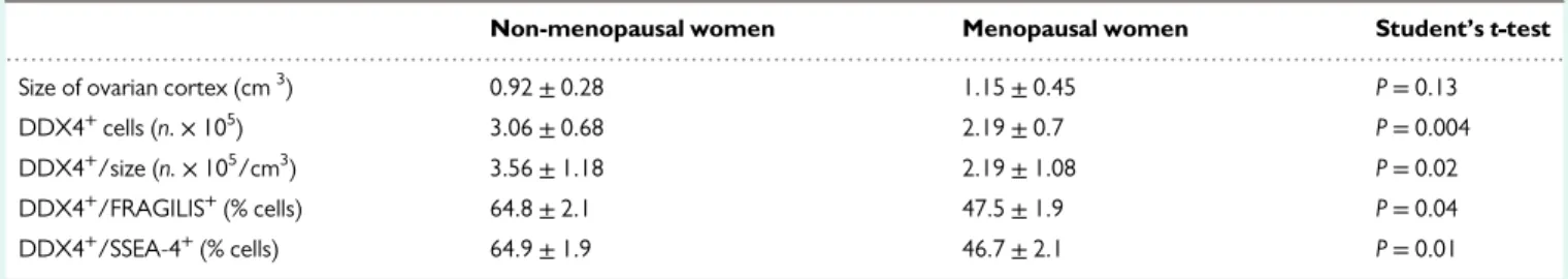
The mean sizes of the ovarian cortex samples from non-menopausal and menopausal women were similar (P= 0.13), whereas the total number of DDX4-positive cells and the ratios of the cell number ver-sus fragment size were significantly higher in the non-menopausal group (P< 0.05 in all instances).
This analysis suggested that ovarian cortex from non-menopausal women contains higher amounts of DDX4-positive cells as compared to menopausal women, and that early ontogenetic markers are preva-lent in non-menopausal women, with mean values of FRAGILIS- and SSEA-4-positive cells up to 65%. On the other hand, although at lower levels, menopausal women still maintain a considerable reserve of these cells, with a mean level<48% for both FRAGILIS- and SSEA-4-positive cell subsets. A comparison between age and the ratio of DDX4-positive cells/cm3of ovarian cortex, as an indicator of OSC reserve, showed a significant inverse correlation (Supplementary Fig. S1) (P< 0.05).
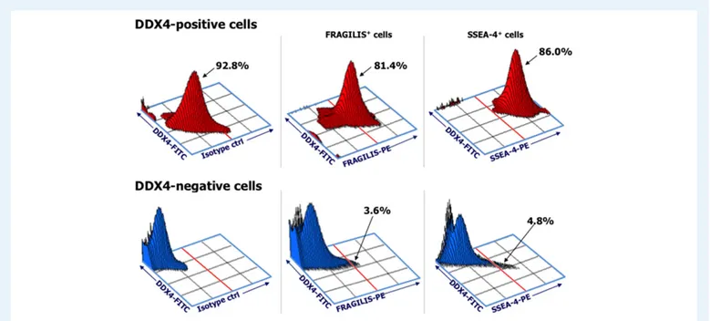
Figure 1 representatively depicts FRAGILIS and SSEA-4 protein expression in the DDX4-positive and DDX4-negative OSCs from a non-menopausal woman (#10). The FRAGILIS and SSEA-4 markers were expressed by 81.4% and 86%, respectively, of the DDX4-positive cells but by<5% (both markers) of the DDX4-negative cells. Similar patterns of antigen expression were obtained in the meno-pausal group (Supplementary Table SI). These results demonstrated
that DDX4-positive cell populations from non-menopausal and meno-pausal women are enriched in cells expressing ontogenetic markers of immature oogonial cells.
DDX4-positive cells differentiate to
oocyte-like cells in culture
DDX4-positive and DDX4-negative cells were cultured separately. On Day 1, the DDX4-positive cells were mostly uniform, small, round cells with a diameter of ~4μm (Fig.2A) whereas the DDX4-negative fractions were characterized by cells of variable size and differing morphologies includingfibroblasts.
The DDX4-positive cells in culture became progressively larger over time. By 1 week of culture, some had a diameter of ~20μm, including several that were apparently strictly adjacent tofibroblasts (Fig.2B). On Day 21 of culture, larger cells, typical of oocyte-like structures, were observed; they had a diameter up to ~80–90 μm, prominent nuclei, and a peri-nuclear accumulation of organelles (Fig.2C). These enlarged cells were presumed to be mature OSCs because their size was similar to that of oocytes, whereas the small cells were presumably immature OSCs, having maintained their original size despite minimal contamination by the DDX4-negative population. These assumptions were tested by evaluating DPPA3 gene expression by ddPCR. Based on previous studies defining mature oocytes as cells of 35–90 μm diameter (Virant-Klun et al., 2008;White et al., 2012;Woods and Tilly, 2013;
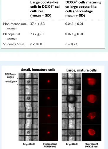
Parte et al., 2014), the number of these cells in cultures from both groups of women were counted. TableIIshows the mean values/well of the large, mature oocyte-like cells detected in single cultures. Cultures from non-menopausal women contained a higher number of these cells although the percentages of mature oocytes with respect to the original DDX4-positive cell population did not differ significantly between the two groups (P= 0.22).
The large, mature oocyte-like cells grew in the absence of other ovarian components, such as the zona pellucida or follicle-like struc-tures, that were not detected in cultures. Thus, these cells were sub-jected to further molecular analysis.
Isolation of OSCs of different sizes by
DEPArray
Cells from the 21-day-old cultures were stained with fluorescent PKH26 and sorted according to fluorescence and size using the DEPArray system (Abonnenc et al., 2013). Figure3shows four single
...
Table I Results of analyses of DDX4-positive cells in ovarian cortex samples from non-menopausal (n = 13) and menopausal (n = 19) healthy women.
Non-menopausal women Menopausal women Student’s t-test
Size of ovarian cortex (cm3) 0.92± 0.28 1.15± 0.45 P= 0.13
DDX4+cells (n.× 105) 3.06± 0.68 2.19± 0.7 P= 0.004
DDX4+/size (n.× 105/cm3) 3.56± 1.18 2.19± 1.08 P= 0.02
DDX4+/FRAGILIS+(% cells) 64.8± 2.1 47.5± 1.9 P= 0.04
DDX4+/SSEA-4+(% cells) 64.9± 1.9 46.7± 2.1 P= 0.01
Data are mean (+SD) values.
cells with a diameter much less than 60μm (left) and a single, larger cell (right) with a diameter of 80–90 μm, considered to represent two distinct populations of cultured DDX4-positive cells. The identification of these small and large cells after 3 weeks of culture suggested that the latter had undergone in vitro differentiation to oocyte-like cells.
The expression of oogonial differentiation
markers is inversely related to cell size
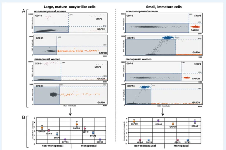
The large and small cells of the 3-week-old cultures were analyzed for their expression of GDF-9 and SYCP3, markers of advanced-stage oocyte differentiation, and DPPA3, a primordial germline marker (Parte et al., 2014). The two-dimensional dot plots in Fig. 4A show the fluorescence-based detection of individual droplets. Orange and purple dots represent droplets positive for GAPDH and GDF-9, respect-ively, blue dots positivity for SYCP3, and black dots are negative, DNA-free droplets. In addition, DPPA3 expression in each cell subset (blue dots) was analyzed in a duplex TaqMan assay. GDF-9 and SYCP3 RNAs were detected only in the large oocyte-like cells of non-menopausal and menopausal women whereas droplets of neither marker were present in the small or immature cells from the two groups of women. Figure4B shows the number of mRNA transcript copies of each gene/ μl RNA. For example, in the mature oocyte-like cells of sample #9 (non-menopausal), there were 2 and 1.2 copies/μl of GDF-9 and SYCP3, respectively, compared to 2.6 and 1.5 copies/μl in sample #19 (menopausal). By contrast, in the small immature cells from these sam-ples, only DPPA3 was detected, with 5.1 copies/μl in non-menopausal and 5.0 copies/μl in menopausal samples. No signal for this gene was detected in the mature oocyte-like cells. These results sug-gested that OSCs from non-menopausal and menopausal women
similarly differentiated in vitro to large, mature oocyte-like cells expres-sing variable levels of terminal oocyte differentiation genes. By contrast, small immature cells maintained their undifferentiated state, as evi-denced by DPPA3 expression.
Oocyte-like cells maturing from OSCs
initiate meiosis
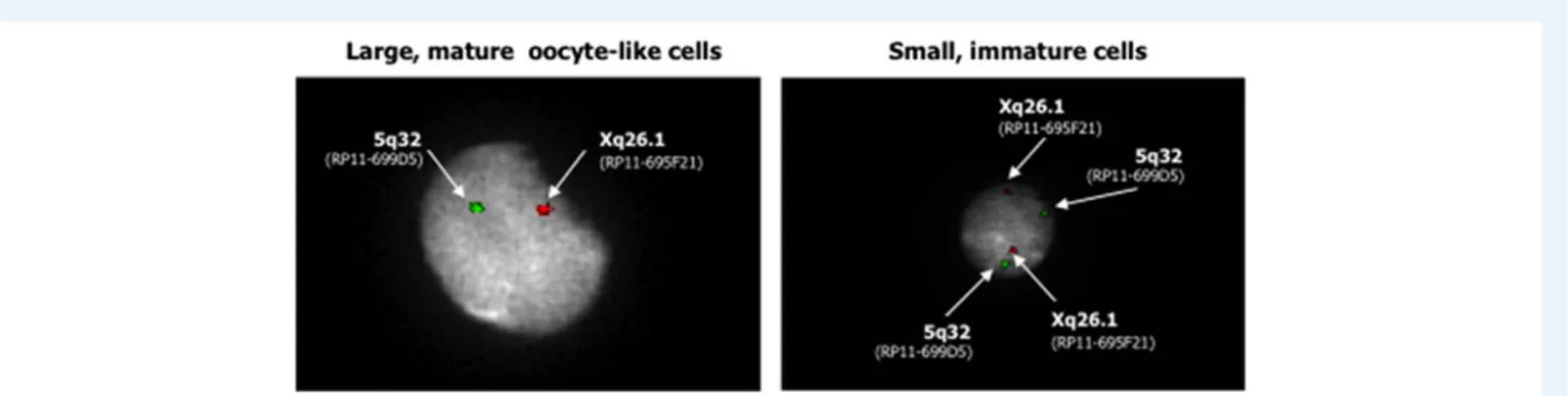
To verify the full maturation of the OSCs to oocyte-like cells, we per-formed FISH to investigate their haploid chromosomal content, using probes for chromosome X (green) and, as the control, chromosome 5 (red). The results obtained with the eight samples from the non-menopausal group and the 10 samples from the non-menopausal group are reported in Supplementary Table II. In the representative image of mature oocyte-like cells from a menopausal woman (#11) shown in Fig.5(left), the presence of a single signal for both chromosomes con-firmed the occurrence of haploidy; this was the case for the majority of oocyte-like cell preparations. Conversely, in the small immature cells, two distinct signals for each chromosome evidenced their diploid state (Fig.5, right). Thesefindings indicated the ability of the large cells isolated by DEPArray and maintained in vitro to acquire oocyte-like characteristics and undergo meiosis.
Discussion
The present study, an extension of our previous observation (Silvestris et al., 2015) of the occurrence of OSCs in the ovaries of adult women, demonstrated that OSCs can be collected from fresh ovarian cortical fragments by anti-DDX4 antibody cell sorting. In particular, using
Figure 1 Phenotypic analysis of DDX4-positive and -negative cell populations recovered by immuno-magnetic cell sorting. Dot plots of a represen-tative cell suspension from a non-menopausal woman (#10); both FRAGILIS and stage-specific embryonic antigen (SSEA)-4 primordial oogonial mar-kers are present. A high percentage of DEAD box polypeptide 4 (DDX4)-positive cells expressed both antigens whereas only a small percentage of DDX4-negative fractions expressed both FRAGILIS and SSEA-4. The plots on the left show the total DDX4-positive (top) and negative (bottom) populations.
DEPArray technology, single immature OSCs and mature oocyte-like cells could be isolated.
With increasing knowledge of ovarian physiology, the detection of OSCs in postnatal mammalian ovaries, and the putative role of these cells in female fertility, the long-accepted paradigm of afixed number of germ cells constituting the ovarian reserve during life is being chal-lenged. Studies by Tilly’s group (White et al., 2012;Imudia et al., 2013;
Woods and Tilly, 2013) demonstrated the existence of OSCs and sug-gested their potential utilization to replace the decreased or exhausted ovarian reserve associated with aging, and, especially, to treat infertility in young women with multifactorial ovarian failure (Woods and Tilly, 2012). Subsequent work by other investigators provided further
Figure 2 In vitro differentiation to oocyte-like cells. Immuno-magnetically selected DDX4-positive human cells were cultured for up to 3 weeks. On Day 1, the cells were small and round with a diameter of 4μm (A). One week later, several cells had increased in size, to ~20μm in diameter, and some were adherent to the fibroblasts used as a feeder layer (B). After 21 days, the cells took on defined oocyte-like features, reaching 75–80 μm in size, with prominent nuclei and a perinuclear accumulation of organelles (C). Their morphology was similar to that of mature oocytes. Images are representative of the culture from a single individual (#5, non-menopausal group); arrows point to the DDX4-positive cells at different stages of culture in the presence offibroblasts (magnifica-tion 60×).
...
Table II Absolute number and percentage of DDX4+ cells that matured in culture to large oocyte-like structures (cells of≥35 μm of diameter).
Large oocyte-like cells in DDX4+cell cultures (mean± SD) DDX4+cells maturing to large oocyte-like cells (percentage mean± SD) Non-menopausal women 37.4± 8.3 0.062± 0.01 Menopausal women 23.7± 6.1 0.027± 0.01 Student’s t-test P< 0.001 P= 0.22
Figure 3 DEPArray single-cell selection and isolation. Representative image of single, captured small (left) or large (right) cells from the same 3-week culture of DDX4-positive cells (menopausal woman sample #6). After their entrapment within the cartridge cages, single cells selected based on their PKH26 redfluorescence and size could be recovered individually when subjected to a dielectricfield. These two distinct populations occurred in most OSC cultures from non-menopausal as well as menopausal women and, based on their size, could be divided into small immature (left) and large mature (right) cells.
evidence of OSCs in pre- and post-menopausal women, while in mur-ine models these cells were shown to be fertilizable and capable of generating embryos (Stimpfel et al., 2013; Parte et al., 2014;
Hernandez et al., 2015).
OSCs have been isolated and cultured using different methods and protocols. Woods and Tilly (2013)used a FACS-based method to detect a cell-surface variant of DDX4 and obtain a homogeneous cell population free of both contaminating oocytes and cells of non-germline lineage. An alternative method consisted of scraping the ovarian epithelium to separate OSCs from oocytes and follicles (Virant-Klun et al., 2008; Parte et al., 2014). Once recovered, the OSCs were enriched by gradient centrifugation and then cultured, which resulted in the formation of cells within structures similar to
embryonic bodies and whose gene expression profile was that of mature oocytes, including positivity for OCT-4A, OCT-4B, C-KIT, VASA and ZP2 markers (Virant-Klun et al., 2008). Using autoMACS, these cells could be isolated based on the binding of the SSEA-4 surface anti-gen. Moreover, the number of OSCs obtained was sufficient to allow their further characterization (Parte et al., 2014). However, these methodologies and the markers used in cell separation raised several issues concerning the heterogeneity of the obtained OSC populations, their unequal sizes, and differences in their maturation (Evron and Blumenfeld, 2013;Albertini and Gleicher, 2015;Grieve et al., 2015).
Wefirst enriched the DDX4-positive population by autoMACS sep-aration and then verified the cellular expression of the primordial germline markers SSEA-4 and FRAGILIS. Based on the high-level
Figure 4 Differential expression of oogenesis-related genes in large mature and small immature cells. Growth differentiation factor (GDF-9) and syn-aptonemal complex protein (SYCP3), expressed during terminal oogonial differentiation, and developmental pluripotency-associated protein 3 (DPPA3), a primordial germline marker, were investigated by droplet digital PCR in cultured cells individually isolated by DEPArray from cultures of OSCs obtained from non-menopausal and menopausal women. GDF-9 and SYCP3 mRNAs were expressed exclusively by large (left) oocyte-like cells from non-menopausal and menopausal samples; DPPA3 mRNA was expressed only by small (right) immature cells. (A) Representative two-dimensional dot plots (non-menopausal #6; and menopausal #11) showingfluorescent individual droplets as orange and purple points, indicative of GAPDH- and GDF-9-positive droplets, respectively. Blue points are droplets positive for SYCP3, and black points, included in rectangles defined by FAM and HEX reference levels, are negative droplets. A duplex TaqMan assay was used to analyze DPPA3 expression in each cell subset (blue dots). (B) Quantitative assessment (copies/μl) of gene expression in large oocyte-like cells (left) and small immature cells (right) (QuantaSoft Software). In large oocyte-like cells from the non-menopausal and menopausal samples, 2 and 2.6 copies/μl of GDF-9 and 1.2 and 1.5 copies/μl of SYCP3, respectively, were detected. By contrast, neither GDF-9 nor SYCP3 was expressed in the small immature cells from non-menopausal women and menopausal sam-ples, whereas 5.1 and 5.0 copies/μl of the DPPA3 gene were detected in samples from the two groups. Expression was undetectable in the large oocyte-like cells.
expression of both, these putative OSCs seemed to be in the earliest stage of differentiation along the oogonial lineage. In fertile women, 65% of the OSC populations was SSEA-4- and FRAGILIS-positive but, interestingly, in menopausal women the percentage was as high as 45%. This finding is in agreement with studies from other groups (Woods and Tilly, 2013;Parte et al., 2014) and suggests that the ovar-ian reserve in menopausal women may include cells belonging to the oogonial germline but, due to the inactivity of the ovulatory cycle, are probably unable to differentiate.
In DDX4-positive cells cultured for 3 weeks, large oocyte-like cells up to 80–90 μm diameter were observed. Other authors have reported that primary OSCs from the ovarian cortex differentiate in culture to advanced stages of maturation (Woods and Tilly, 2013;
Parte et al., 2014). In the present work, we selectively purified small and large cells and then subjected them to a specific dielectric field that allowed the isolation of single cells. We subsequently showed that the larger, mature cells expressed high levels of GDF-9 and SYCP3 mRNAs, indicative of oocyte terminal differentiation. The expression of these mRNAs was not detected in the small immature cells isolated from non-menopausal and menopausal women; rather these cells expressed the primordial germ cell marker DPPA3. Nonetheless, the greater OSC reserve of non-menopausal women was demonstrated by the detection within the DDX4-enriched OSCs of a significantly lar-ger number of FRAGILIS- and SSEA-4-positive cells than in meno-pausal women, and by the higher number of large oocyte-like cells that matured in the 3-week cultures.
DEPArray technology enables the selection and purification of single cells based on theirfluorescence and size (Abonnenc et al., 2013). We were thus able to separate OSCs purified by immuno-magnetic separ-ation into populsepar-ations of small or large cells and to follow their unequal differentiation to oocyte-like cells. Why some cultured OSCs remain small, immature but viable cells while others undergo progressive enlargement and differentiate to form oocyte-like cells is presently unclear. However, the detection of a higher percentage of SSEA-4-and FRAGILIS-positive cells in non-menopausal versus menopausal women suggests that the difference is related to the occurrence of primordial cells of oogonial ontogenetic derivation. Yet, the involve-ment of other factors, including the activity of as-yet-undefined genes
that contribute to primary germline cell survival and differentiation, cannot be excluded.
Mature oocytes are haploid cells, therefore we investigated the ploi-dy of the purified large, oocyte-like cells by FISH. The single signals obtained for chromosomes X and 5 indicated that these cells were haploid. However, the haploidy of large oocyte-like cells derived from OSCs has been disputed (Oatley and Hunt, 2012), as these cells have also been shown to spontaneously express meiosis markers (Zou et al., 2009;Tedesco et al., 2011;Ghazal, 2013). FISH data in combination with the demonstrated cytoplasmic localization of SYCP3, a meiosis-related protein, and GDF-9, as a post-meiotic marker, may reflect the meiotic state of cultured OSCs (Parte et al., 2014). In the single large oocyte-like cells obtained in this study, FISH-determined haploidy correlated with the presence of both GDF-9 and SYCP3 mRNAs, suggesting the meiotic state of these cells. Although the DNA content and other‘gold stan-dards’ of meiosis were not evaluated in this study, our results suggest that the cells were in the secondary gametocyte stage that follows the first meiotic division, when nuclei are 2 C/1 N, and thus formally haploid (Handel et al., 2014). In addition, despite the absence of evidence of an entirely concluded meiosis by the oocyte-like cells, our observations are consistent with their mature, pre-meiotic state. This would imply that small DDX4-positive cells are able to initiate an in vitro developmental program of oogenesis and are thus suitable for investigations of the molecular alterations driving oocyte reserve exhaustion in infertile women. Moreover, it has been reported that, in the absence of sperm penetration, oocytes can spontaneously maintain a chromosomal haploid content allowing meiotic divisions in vitro (Woods et al., 2013).
Mature oocytes derived in vitro from the OSCs of menopausal women have been demonstrated in previous studies (Stimpfel et al., 2013). We identified a larger relative fraction of OSCs in fertile than in menopausal women, and the greater tendency of those cells to develop in vitro into large oocyte-like cells. This difference may be related to the stem cell niche, as this microenvironment is thought to stimulate the differentiation of OSCs to oocytes. In menopausal women, the stem cell niche may be defective such that fewer OSCs are supported (Niikura et al., 2009;Massasa et al., 2010;Joo et al., 2014). Nonetheless, we observed that, although at lower frequency, oocyte-like cells could also be obtained in vitro from the OSCs of
Figure 5 Haploidy of large, mature oocyte-like cells. FISH analysis of individual large, mature OSCs confirmed their meiotic entry based on the pres-ence of a single signal for chromosome X (green) and for chromosome 5 (red), as internal control, in nuclei counterstained with DAPI. This result sug-gested the presence of a haploid set of chromosomes, typical of reproductive cells (left). A FISH assay of the small DDX4-positive cells revealed their chromosomal diploidy (right) and suggested that large cells isolated by DEPArray were terminally differentiated as oocyte-like cells.
menopausal women and they expressed the same oocyte terminal dif-ferentiation genes, namely GDF-9 and SYCP3, as cells from pre-menopausal women. This observation may reflect differences in the activation state within ovarian somatic niches in vivo and/or a diverse capability to exit biological dormancy in response to the soluble factors present in culture medium, such as N2-supplement, epidermal growth factor, andfibroblast growth factor (Woods and Tilly, 2013).
In conclusion, our work provides further evidence of the occurrence of OSCs in the ovarian cortex of non-menopausal and menopausal women, and the ability of these cells to differentiate to form mature oocyte-like cells in vitro. Using single cells isolated on the basis of their size, we were able to demonstrate that, in culture, the larger cells expressed markers indicating theirfinal differentiation and underwent meiosis. Thus, oocyte-like cells can be generated in vitro even from the OSCs of menopausal women. These insights into ovarian neo-oogenesis may contribute to the development of a more effective approach to treat infertility in women.
Supplementary data
Supplementary data are available at Human Reproduction online.
Acknowledgements
The authors are grateful to Nicoletta Coccaro for FISH analysis.
Authors
’ roles
E.S. planned the study design, performed the experiments, collected the data and wrote the manuscript. P.C. and S.D’O. performed the in vitro studies and collected the data. C.F. conducted the single-cell separation as well as the digital PCR, with the related data. F.S. devel-oped the rationale of the study and wrote and revised the manuscript. G.L. contributed to the study design and to obtaining the biological samples. All authors read and approved thefinal version.
Funding
The work was funded by a grant from the Italian Association for Cancer Research (IG grant 17536), and from the Apulia Region (‘Oncogenomic Project’ and ‘Jonico-Salentino Project’).
Con
flict of interest
All authors declare no conflict of interest.
References
Abonnenc M, Manaresi N, Borgatti M, Medoro G, Fabbri E, Romani A, Altomare L, Tartagni M, Rizzo R, Baricordi O et al. Programmable inter-actions of functionalized single bioparticles in a dielectrophoresis-based microarray chip. Anal Chem 2013;85:8219–8224.
Albertini DF, Gleicher N. A detour in the quest for oogonial stem cells: methods matter. Nat Med 2015;21:1126–1127.
Dunlop CE, Telfer EE, Anderson RA. Ovarian stem cells—potential roles in infertility treatment and fertility preservation. Maturitas 2013;76: 279–283.
Evron A, Blumenfeld Z. Ovarian stem cells-the pros and cons. Clin Med Insights Reprod Health 2013;7:43–47.
Ghazal S. Oogonial stem cells: do they exist and may they have an impact on future fertility treatment? Curr Opin Obstet Gynecol 2013;25:223–228. Gheorghisan-Galateanu AA, Hinescu ME, Enciu AM. Ovarian adult stem
cells: hope or pitfall? J Ovarian Res 2014;7:71.
Grieve KM, McLaughlin M, Dunlop CE, Telfer EE, Anderson RA. The con-troversial existence and functional potential of oogonial stem cells. Maturitas 2015;82:278–281.
Handel MA, Eppig JJ, Schimenti JC. Applying‘gold standards’ to in-vitro-derived germ cells. Cell 2014;157:1257–1261.
Hernandez SF, Vahidi NA, Park S, Weitzel RP, Tisdale J, Rueda BR, Wolff EF. Characterization of extracellular DDX4- or Ddx4-positive ovarian cells. Nat Med 2015;21:1114–1116.
Imudia AN, Wang N, Tanaka Y, White YA, Woods DC, Tilly JL. Comparative gene expression profiling of adult mouse ovary-derived oogonial stem cells supports a distinct cellular identity. Fertil Steril 2013; 100:1451–1458.
Johnson J, Canning J, Kaneko T, Pru JK, Tilly JL. Germline stem cells and follicu-lar renewal in the postnatal mammalian ovary. Nature 2004;428:145–150. Joo BS, Jung IK, Park MJ, Joo JK, Kim KH, Lee KS. Differential expression of
pluripotent and germ cell markers in ovarian surface epithelium accord-ing to age in female mice. Reprod Biol Endocrinol 2014;12:113.
Lee YM, Kim TH, Lee JH, Lee WJ, Jeon RH, Jang SJ, Ock S, Lee SL, Park BW, Rho GJ. Overexpression of Oct4 in porcine ovarian stem/stromal cells enhances differentiation of oocyte-like cells in vitro and ovarian fol-licular formation in vivo. J Ovarian Res 2016;9:24.
Lichter P, Tang Chang CJ, Call K, Hermanson G, Evans GA, Housman D, Ward DC. High resolution mapping of human chromosomes 11 by in situ hybridization with cosmid clones. Science 1990;247:64–69. Massasa E, Costa XS, Taylor HS. Failure of the stem cell niche rather than
loss of oocyte stem cells in the aging ovary. Aging 2010;2:1–2.
Niikura Y, Niikura T, Tilly JL. Aged mouse ovaries possess rare premeiotic germ cells that can generate oocytes following transplantation into a young host environment. Aging 2009;1:971–978.
Oatley J, Hunt PA. Of mice and (wo)men: purified oogonial stem cells from mouse and human ovaries. Biol Reprod 2012;86:196.
Parte S, Bhartiya D, Patel H, Daithankar V, Chauhan A, Zaveri K, Hinduja I. Dynamics associated with spontaneous differentiation of ovarian stem cells in vitro. J Ovarian Res 2014;7:25.
Parte S, Bhartiya D, Telang J, Daithankar V, Salvi V, Zaveri K, Hinduja I. Detection, characterization, and spontaneous differentiation in vitro of very small embryonic-like putative stem cells in adult mammalian ovary. Stem Cells Dev 2011;20:1451–1464.
Silvestris E, D’Oronzo S, Cafforio P, D’Amato G, Loverro G. Perspective in infertility: the ovarian stem cells. J Ovarian Res 2015;8:55.
Stimpfel M, Skutella T, Cvjeticanin B, Meznaric M, Dovc P, Novakovic S, Cerkovnik P, Vrtacnik-Bokal E, Virant-Klun I. Isolation, characterization and differentiation of cells expressing pluripotent/multipotent markers from adult human ovaries. Cell Tissue Res 2013;354:593–607.
Tedesco M, Farini D, De Felici M. Impaired meiotic competence in putative primordial germ cells produced from mouse embryonic stem cells. Int J Dev Biol 2011;55:215–222.
Virant‐Klun I, Rozman P, Cvjeticanin B, Vrtacnik‐Bokal E, Novakovic S, Rülicke T, Dovc P, Meden‐Vrtovec H. Parthenogenetic embryo‐like structures in the human ovarian surface epithelium cell culture in post-menopausal women with no naturally present follicles and oocytes. Stem Cells Dev 2009;18:137–150.
Virant-Klun I, Zech N, Rozman P, Vogler A, Cvjeticanin B, Klemenc P, Malicev E, Meden-Vrtovec H. Putative stem cells with an embryonic char-acter isolated from the ovarian surface epithelium of women with no nat-urally present follicles and oocytes. Differentiation 2008;76:843–856.
Wallace PK, Tario JD Jr, Fisher JL, Wallace SS, Ernstoff MS, Muirhead KA. Tracking antigen-driven responses byflow cytometry: monitoring prolif-eration by dye dilution. Cytometry A 2008;73:1019–1034.
White YA, Woods DC, Takai Y, Ishihara O, Seki H, Tilly JL. Oocyte form-ation by mitotically active germ cells purified from ovaries of reproduct-ive age women. Nat Med 2012;18:413–421.
Woods DC, Tilly JL. Isolation, characterization and propagation of mitoti-cally active germ cells from adult mouse and human ovaries. Nat Protoc 2013;8:966–988.
Woods DC, Tilly JL. The next (re)generation of ovarian biology and fertility in women: is current science tomorrow’s practice? Fertil Steril 2012;98: 3–10.
Woods DC, Tilly JL. Woods and Tilly reply. Nat Med 2015;21:1118–1121. Woods DC, White YA, Tilly JL. Purification of oogonial stem cells from adult mouse and human ovaries: an assessment of the literature and a view toward the future. Reprod Sci 2013;20:7–15.
Zarate-Garcia L, Lane SI, Merriman JA, Jones KT. FACS-sorted putative oogonial stem cells from the ovary are neither DDX4-positive nor germ cells. Sci Rep 2016;6:27991.
Zhang H, Panula S, Petropoulos S, Edsgärd D, Busayavalasa K, Liu L, Li X, Risal S, Shen Y, Shao J et al. Adult human and mouse ovaries lack DDX4-expressing functional oogonial stem cells. Nat Med 2015;21: 1116–1118.
Zhang H, Zheng W, Shen Y, Adhikari D, Ueno H, Liu K. Experimental evi-dence showing that no mitotically active female germline progenitors exist in postnatal mouse ovaries. Proc Natl Acad Sci USA 2012;109: 12580–12585.
Zou K, Yuan Z, Yang Z, Luo H, Sun K, Zhou L, Xiang J, Shi L, Yu Q, Zhang Y et al. Production of offspring from a germline stem cell line derived from neonatal ovaries. Nat Cell Biol 2009;11:631–636.
Zuckerman S. The number of oocytes in the mature ovary. Rec Prog Horm Res 1951;6:63–108.