Dorsal tongue porphyrin autofluorescence
and Candida saprophytism: A prospective
observational study
Massimo PetruzziID1☯*, Fedora della VellaID1☯, Andrea Cassandro1, Adriana Mosca1, Mariasevera Di Comite2, Maria Contaldo3, Felice Roberto Grassi2, Dorina Lauritano4
1 Interdisciplinary Department of Medicine, University “Aldo Moro” of Bari, Bari, Italy, 2 Department of Basic
Medical Sciences, Neurosciences and Sensory Organs, University of Bari “Aldo Moro”, Bari, Italy,
3 Multidisciplinary Department of Medical-Surgical and Odontostomatological Specialties, University of
Campania “Luigi Vanvitelli”, Naples, Italy, 4 Department of Medicine and Surgery, University Milano-Bicocca, Monza, Italy
☯These authors contributed equally to this work.
Abstract
Aim
To investigate the correlation between the dorsal tongue porphyrin autofluorescence, revealed using VELscope, and Candida saprophytism.
Material and methods
Consecutive patients underwent an autofluorescence examination by the VELscope device to establish the presence or absence of porphyrin fluorescence. A tongue swab was col-lected for the Candida cultural test. Sensitivity, specificity, accuracy, negative predictive value and positive predictive value were calculated considering the oral swab as the gold standard. The degree of agreement between the two tests was calculated using Cohen’s K coefficient.
Results
One hundred twenty-six patients were enrolled. Porphyrin fluorescence method showed a sensitivity of 78%, specificity of 76% and an accuracy of 78%. Negative predictive value and positive predictive value were respectively 90% and 59%. The strength of agreement between the two methods resulted to be moderate (k = 0.551).
Conclusions
Off-label use of tongue autofluorescence examination to detect the presence of Candida species is characterized by a loss of porphyrin fluorescence. The high negative predictive value of porphyrin fluorescence loss suggests its use in preliminary selection of Candida carriers, in order to plan preventive and therapeutic strategies.
a1111111111 a1111111111 a1111111111 a1111111111 a1111111111 OPEN ACCESS
Citation: Petruzzi M, della Vella F, Cassandro A,
Mosca A, Di Comite M, Contaldo M, et al. (2019) Dorsal tongue porphyrin autofluorescence and
Candida saprophytism: A prospective observational
study. PLoS ONE 14(9): e0223072.https://doi.org/ 10.1371/journal.pone.0223072
Editor: Michael R. Hamblin, Massachusetts
General Hospital, UNITED STATES
Received: May 24, 2019 Accepted: September 12, 2019 Published: September 26, 2019
Copyright:© 2019 Petruzzi et al. This is an open access article distributed under the terms of the
Creative Commons Attribution License, which permits unrestricted use, distribution, and reproduction in any medium, provided the original author and source are credited.
Data Availability Statement: According to the
Italian Law n.169/2003 (“Italian Personal Data Protection Code”) data on patients’ health are considered “sensitive”. The ethics committee asked the research responsible (prof. Petruzzi) to sign a document where he is committed to protect the data and to relate to the ethics committee for any request on the matter. Local Ethics Committee addresses are: Comitato etico del Policlinico di Bari, Piazza Giulio Cesare 11 – 70124 Bari – ITALY.
Introduction
Autofluorescence is a biochemical-physical characteristic of the tissues that excited with an appropriate blue light source (400-460-nm), emit light with a higher wavelength [1]. Endoge-nous fluorophores as keratin, collagen, elastin, NADH and porphyrins are the molecules responsible of this phenomenon. On the other hand, haemoglobin and melanin tends to absorb the blue incident light, reducing the tissue autofluorescence [2]. Tissue modifications induced by neoplastic, potentially malignant, inflammatory and infective processes can affect the normal tissue autofluorescence pattern; actually, on this principle is based the use of auto-fluorescence for the early detection of upper aero-digestive tract cancers and related potentially malignant disorders [3–5]. McAlpine et al. classified four different patterns of autofluores-cence according to their appearance during the inspection. In particular, normal healthy tissue emits pale green autofluorescence, differently, loss of fluorescence (LOF), characterized by a dark brown to black mucosal area, is typical of dysplastic and/or neoplastic tissues. Gained fluorescence (GF) is usually observed in hyperkeratotic lesions (leukoplakia ormorsicatio buc-carum), while a porphyrin fluorescence (PF) is characterized by a red/orange light emission,
often observed on the dorsal tongue surface. The PF is characterized by a 636 nm peaks of emission caused by porphyrins produced by micro-organisms colonizing the dorsal tongue surface [6]. The soft and hard palate can also show a PF, due to their contact with the tongue [7].
The microscopic analysis conducted by Hamad et al. revealed high numbers of bacteria such asStreptococcus salivarius, Actinomyces spp. and lactobacilli on the mucosal sites emitting
a PF, confuting the hypothesis that the PF found in oral squamous cell carcinomas originates from cellular enzymatic synthesis defects [8].
To date, it has not yet been proved ifCandida species are somehow involved in oral PF,
sinceCandida species are usually commensal micro-organisms found in the oral cavity in the
40% of healthy population [9]. The frequency of isolation ofC. albicans and its mucosal density
per unit area, as measured by imprint culture, is highest on the dorsum of the tongue, particu-larly the posterior half. For this reason, tongue is considered the primary oral reservoir of the fungus, from which the rest of the oral mucosa, plaque-coated surfaces of the teeth and the saliva may become secondarily colonized [10].
Candida is currently detectable in the oral cavity by swab, smear or oral rinses, followed by
a specific microbial culture. Its colonization of the oral tissues can alter the normal microbiota balance, eventually resulting in autofluorescence properties modification.
Aim of this study was to test the efficacy of the off-label autofluorescence tongue examina-tion forCandida colonization detection.
Material and methods
Study design
This single-centre, prospective, single-arm, observational study carried out at Oral Medicine Section of Dental Clinic of University of Bari (Bari, Italy) from January 2018 to December 2018, was conducted in accordance with the Declaration of Helsinki and independently approved and reviewed by the Institution’s Ethical Committee (Comitato Etico del Policlinico di Bari, prot. n. 46815/C.E.).
Patients
Consecutive healthy subjects aged between 9 and 80 years, referred to the Dental Clinic of Uni-versity “Aldo Moro” of Bari, were enrolled. The sample size calculation was done referring to
Funding: The authors received no specific funding
for this work.
Competing interests: The authors have declared
an estimate population of Italians aged between 9 to 80 years, 40% ofCandida saprophytism
prevalence, using n = Nx/[(N-1)E2+x], with 96% confidence level and 9% margin of error. Written informed consent was obtained from each subject or /and from the parents/guard-ians of underaged patients attending the study. Individuals were excluded if assumed antimy-cotic and/or antibiotic drugs in the past 30 days, daily brushed their tongue or refused to give the consent to the study. For each subject demographic data were collected.
Autofluorescence examination
The dorsal tongue autofluorescence investigation was performed in all subjects using the med-ical device VELscope Vx (Mectron s.p.a—Carasco, Genova). The procedure was performed in a shaded room, keeping the device 5 cm distant from the tongue, pulled out by a second opera-tor with a gauze. The excitation blue light (excitation wavelength between 400 and 460 nm) was projected on the dorsal tongue mucosa by a dichroic mirror. Through the back of the handpiece, tissue autofluorescence was evaluated and classified as positive to PF if its aspect was red or orange, on the contrary it was classified as negative to PF (Fig 1). Four experienced experts in oral medicine, and routinely users of VELscope device, previously calibrated, rated as PF positive or negative each single case: only subjects showing an evident and generalized PF were considered positive. The inter-rater reliability was guaranteed setting a percent agree-ment goal of 95%.
Each case was photographed in autofluorescence, using Canon Powershot Elph 130 IS (Canon Inc, Tokyo, Japan) with a dedicated adapter provided by the manufacturer.
Oral Swab
Every subject underwent to an oral swab of the dorsal tongue after the VELscope exam. The swab was performed using a sterilized cotton swab (2150/SG, Nuova APTACA S.r.l., Canelli -AT-, Italy), rubbed and rotated on the whole surface of the dorsal tongue. In case of PF posi-tivity, the swab was performed on the red/orange revealed area, in order to avoid inter-areas contamination. The collected specimens were put in a sterile propylene cup and then sown on
Fig 1. Autofluorescence pictures. (A) Tongue presenting with porphyrin autofluorescence. (B) Tongue not presenting with porphyrin autofluorescence.
a solid ground, the Sabouraud Agar Destrosio agar. The culture test revealed the eventual pres-ence ofCandida and the species involved.
Data analysis and statistics
Theχ2test (significance set at p < 0.05) was used to test oral swab and PF outcomes (positive or negative) in relation to the sample gender and age. Global validation of the PF test results was established by calculating the sensitivity, specificity, accuracy and both the positive and negative predictive values from contingency tables.
The Cohen’s kappa coefficient was used to evaluate the interrater agreement between oral swab and PF. Concordance was evaluated according to Landis and Koch who defined values
<0 as indicating no agreement, 0 to 0.20 as slight, 0.21 to 0.40 as fair, 0.41 to 0.60 as moderate,
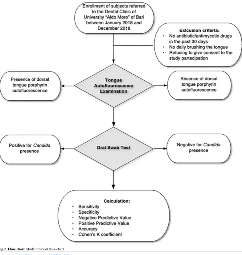
0.61 to 0.80 as substantial, and 0.81 to 1 as perfect agreement [11]. The study flow-chart is resumed inFig 2.
Results
One hundred twenty-six subjects were enrolled, including 51 males and 75 females. The mean age was 55± 17 years (mean±SD). Seventy-seven (61%) showed PF at the autofluorescence evaluation, and 37 (29%) participants resulted with a positive culture test for fungi ofCandida
species.
Out of the 49 subjects without PF, 29 were affected byCandida colonization, while out of
the 77 subjects with PF, only 8 resulted positive to the culture test. The inter-raters’ agreement was 100%.
PF and swab outcomes were not statistically influenced by age and sex (p>0.05). PF method showed a sensitivity of 78% and specificity of 76% in detectingCandida
presence.
PPV and NPV were respectively 59% and 90%, with an autofluorescence accuracy rate of 78%. The two methods showed a moderate level of agreement, according to Cohen’s k coeffi-cient (k = 0.551).
The above-mentioned data are resumed inTable 1.
Discussion
In this study the correlation betweenCandida presence on the dorsal tongue and PF elicited
by an off-label use of VELscope was investigated. The method displayed moderate values of sensitivity and specificity (78% and 76%) compared to swab specimens culture test, which remains the gold standard to detectCandida species and to perform an antimycogram to start
an effective therapy in case of candidiasis. Nevertheless, the high negative predictive value of PF (90%) make it a useful and immediate chair side, non-invasive screening tool, that permit to select the patients needing further microbial investigations.
Conventionally, autofluorescence is an ancillary tool for oral cancer and dysplastic lesions examination, with sensitivity and specificity values of 76% and 66.29% respectively, and NPV and PPV of 95.08% and 24.36% [12] but some off-label uses have also been suggested, like the valuation of the bone vitality status after surgical treatment of Medication Related Osteonecro-sis of the Jaws [13].
Fungal optical fluorescence properties have been deeply studied, even if especially in vitro: Rao S. et al. reported about the use of autofluorescence techniques as a rapid screening method for identification of fungi on routinely prepared hematoxylin-eosin-stained tissue sections slides. They found sensitivity and specificity of 97.8% and 100% respectively, concluding that autofluorescence does not require any other specialized staining procedure for fungi detection
[14]. The validity of fluorescence microscopy to diagnoseCandida infection is also
well-estab-lished [15], providing a sensitive and specific screening tool, compared to the current gold standard, i.e. periodic acid Schiff (PAS) stain [16]. Gabrielli et al. compared in mice the biolu-minescence in vivo imaging technique with colony forming units (CFU) measurement for the
Candida detection. They concluded that the bioluminescence technique was more reliable
Fig 2. Flow-chart. Study protocol flow-chart.
than CFU counts in detecting mouse’s early oral candidiasis [17]. The typical fluorescence aspect of hyphae is a yellowish-green signal, due to their flavin content [16].
Red autofluorescence, instead, is a common find of the oral cavity, generally localized on the dorsal tongue, and has been historically ascribed to multiple etiopathological theories.
Initial data reported in Literature, attributed PF to dysplastic and/or neoplastic oral tissues derailment [18] but successive assessments defined the correlation between LOF and neoplas-tic changes [1].
Otherwise, other authors considered the dorsal tongue PF as the physiological status, since it was commonly recorded using the Wood’s lamp in apparently healthy population and assumed that a reduction of PF involved nutritional deficiency and pathological conditions, such as tropical sprue, iron deficiency and pernicious anaemia [19,20].
Carrie, in 1934, argued that the normal fluorescence of the tongue observed with Wood’s lamp was caused by specific Gram-positive bacilli. The same author tried to cultivate these bacilli but did not obtain satisfactory results [21].
Tomaszewski [22] studied the effects of penicillin on PF. After four to five days of oral or inhaled treatment, the PF began to disappear, starting from the anterior area and proceeding towards the back of the base of the tongue. At the end of the therapy, the red fluorescence gradually reappeared, starting from the back of the tongue. Intramuscular penicillin adminis-tration did not produce loss of PF.
The hypothesis of a bacterial origin of PF is supported by different studies assessing the emission of red autofluorescence by oral biofilm [23,24]. Also, halitosis, known to be mainly caused by bacteria, has been linked to porphyrin fluorescence as demonstrated by Lee et al. [25] and Hitz Lindenmu¨ller et al. [26].
Nevertheless, the pathway of porphyrin production is still unknown, if directly synthesized by the bacteria or haemoglobin disruption product. PF has also been suggested as real time detection method for oropharynx bacterial infection [27].
PF was also found in the nasal vestibule, the labial commissure and the vaginal mucosa, especially in the menstrual phase, probably because of the porphyrin produced from the bacte-rial decomposition of the blood [28]. All the above-mentioned anatomical areas own a typical
Table 1. Demographic and statistical data of the study.
Total (%) Swab + (%) Swab—(%) p value PF+ (%) PF- (%) p value
Men 51 (40) 17 (33) 34 (67) p>0.05 28(55) 23 (45) p>0.05
Women 75 (60) 20 (27) 55 (73) 50 (70) 26 (30)
Aged<55 yrs 59 (46) 18 (31) 41 (69) p>0.05 32 (54) 27 (56) p>0.05
Aged>55 yrs 67 (54) 19 (28) 48 (72) 45 (67) 22 (33)
AUTOFLUORESCENCE ORAL SWAB
Positive for Candida Negative for Candida
Absence of porphyrin fluorescence 29 (True Positive) 20 (False Positive)
Presence of porphyrin fluorescence 8 (False Negative) 69 (True Negative)
Value 95% C.I.
Sensitivity 78% 61.9% to 90.17%
Specificity 76% 67.45% to 85.70%
Negative Predictive Value 90% 82.22% to 94.15%
Positive Predictive Value 59% 48.75% to 68.85%
Accuracy 78% 69.51% to 84.70%
Cohen’s K coefficient 0.551 0.356 to 0.665
Strength of agreement Moderate
microbiota, whom alterations could produce a loss of PF.Candida colonization, not
uncom-mon on these mucosal sites, is one of the possible causes of this phenomenon.
Detection of PF can have several implications in clinical practice. As demonstrated by Lodi et al. [29], it is possible to avoid a prophylactic antifungal treatment in those patients under-gone a local corticosteroid therapy, provided that they demonstrate a negativeCandida
car-riage: on the basis of the negative predictive value of autofluorescence that we found, the PF finding could allow the identification of patients eligible to receive a preventive antifungal treatment. Moreover, the topographic distribution of PF can permit a selective swab in specific oral mucosal sites, representing a fast, non-invasive and relatively inexpensive method for an initial chair-side screening for theCandida detection. A future development of this pioneering
study could consist in the enrolment of high-risk groups (e.g. diabetics, immunosuppressed, removable oral prosthesis wearers) or patients with a confirmed diagnosis of oral candidiasis or subjects with co-existing red/orange and green areas on the tongue.
Further investigations are needed to understand how the presence of PF is related to the absence ofCandida. Our hypothesis is that specific cluster of bacteria responsible for PF
antag-onize establishment and growth ofCandida species. This can be owed to an interspecies
com-petition for the colonization of the dorsal tongue ecological niches: a similar mechanism has already been demonstrated betweenCandida and Pseudomonas aeruginosa [30]. Another pos-sible explanation is an antifungal activity of porphyrin itself [31,32].
Further studies could better identify these porphyrin bacteria species and their metabolic products. The close link between PF and oral bacteria has been recently proven by Liu et al., that demonstrated the efficacy of red fluorescence imaging as an objective and promising method for dental plaque detection and quantification [33].
However, PF detection thorough VELscope does have limitations: it is not possible to define theCandida species involved in the colonization, hence PF should be considered only as an
initial and complementary screening aid. Moreover, in the present study we did not perform a culture test to identify the bacteria species associated to PF, representing an implication for future researches. This method could also be implemented with a computerized image analy-sis, using dedicated software for standardized colours identification and evaluation.
Further randomized controlled trials are needed to test the effectiveness of presence/ absence of PF inCandida detection. The "off-label" use of autofluorescence proposed in this
study has evidenced the potential of light technologies as screening support for oral diseases.
Acknowledgments
The authors are grateful to Giovanna D’Ostuni for data collection and study support.
Author Contributions
Conceptualization: Massimo Petruzzi.
Data curation: Massimo Petruzzi, Fedora della Vella, Andrea Cassandro. Formal analysis: Massimo Petruzzi, Fedora della Vella.
Investigation: Massimo Petruzzi, Fedora della Vella, Andrea Cassandro, Adriana Mosca. Methodology: Massimo Petruzzi, Fedora della Vella, Andrea Cassandro, Adriana Mosca. Project administration: Massimo Petruzzi, Dorina Lauritano.
Supervision: Massimo Petruzzi, Mariasevera Di Comite, Felice Roberto Grassi, Dorina Lauritano.
Validation: Massimo Petruzzi, Adriana Mosca, Mariasevera Di Comite, Maria Contaldo, Felice Roberto Grassi, Dorina Lauritano.
Visualization: Massimo Petruzzi, Fedora della Vella, Maria Contaldo, Felice Roberto Grassi, Dorina Lauritano.
Writing – original draft: Massimo Petruzzi.
Writing – review & editing: Massimo Petruzzi, Fedora della Vella, Mariasevera Di Comite, Maria Contaldo, Felice Roberto Grassi, Dorina Lauritano.
References
1. Lane PM, Gilhuly T, Whitehead P, Zeng H, Poh CF, Ng S, et al. Simple device for the direct visualization of oral-cavity tissue fluorescence. J Biomed Opt. 2006 Mar-Apr; 11(2):024006.https://doi.org/10.1117/ 1.2193157PMID:16674196
2. Na R, Stender IM, Henriksen M, Wulf HC. Autofluorescence of human skin is age-related after correc-tion for skin pigmentacorrec-tion and redness. J Invest Dermatol. 2001 116(4), 536–40.https://doi.org/10. 1046/j.1523-1747.2001.01285.xPMID:11286620
3. Waterhouse DJ, Joseph J, Neves AA, di Pietro M, Brindle KM, Fitzgerald RC, et al. Design and valida-tion of a near-infrared fluorescence endoscope for detecvalida-tion of early esophageal malignancy. J Biomed Opt. 2016 Aug 1; 21(8):84001.https://doi.org/10.1117/1.JBO.21.8.084001PMID:27490221
4. Jo JA, Cheng S, Cuenca-Martinez R, Duran-Sierra E, Malik B, Ahmed B et al. Endogenous Fluores-cence Lifetime Imaging (FLIM) endoscopy for early detection of oral cancer and dysplasia. Conf Proc IEEE Eng Med Biol Soc. 2018; 3009–3012.https://doi.org/10.1109/EMBC.2018.8513027PMID:
30441030
5. Petruzzi M, Lucchese A, Nardi GM, Lauritano D, Favia G, Serpico R, Grassi FR. Evaluation of autofluor-escence and toluidine blue in the differentiation of oral dysplastic and neoplastic lesions from non-dys-plastic and neonon-dys-plastic lesions: a cross-sectional study. J Biomed Opt. 2014; 19(7):76003.https://doi. org/10.1117/1.JBO.19.7.076003PMID:24996662
6. McAlpine JN, El Hallani S, Lam SF, Kalloger SE, Luk M, Huntsman DG et al. Autofluorescence imaging can identify preinvasive or clinically occult lesions in fallopian tube epithelium: a promising step towards screening and early detection. Gynecol Oncol. 2011; 120(3) 385–92.https://doi.org/10.1016/j.ygyno. 2010.12.333PMID:21237503
7. de Veld DC, Skurichina M, Witjes MJ, Duin RP, Sterenborg DJ, Star WM, Roodenburg JL. Autofluores-cence characteristics of healthy oral mucosa at different anatomical sites. Lasers Surg Med. 2003; 32 (5), 367–76.https://doi.org/10.1002/lsm.10185PMID:12766959
8. Hamad LO, Vervoorts A, Henning T, Bayer R. Ex vivo photodynamic diagnosis to detect malignant cells in oral brush biopsies. Lasers Med Sci. 2010; 25(2), 293–301. https://doi.org/10.1007/s10103-009-0712-1PMID:19662485
9. Akpan A, Morgan E. Oral candidiasis. Postgrad Med J, 2002. 78(922), 455–459.https://doi.org/10. 1136/pmj.78.922.455PMID:12185216
10. Cannon RD, Chaffin WL. Oral colonization by Candida albicans. Crit Rev Oral Biol Med. 1999; 10(3), 359–83. PMID:10759414
11. Landis JR, Koch GG. The measurement of observer agreement for categorical data. Biometrics. 1977, 33(1), 159–174. PMID:843571
12. Ganga RS, Gundre D, Bansal S, Shirsat PM, Prasad P, Desai RS. Evaluation of the diagnostic efficacy and spectrum of autofluorescence of benign, dysplastic and malignant lesions of the oral cavity using VELscope. Oral Oncol. 2017; 75, 67–74.https://doi.org/10.1016/j.oraloncology.2017.10.023PMID:
29224826
13. Giovannacci I, Meleti M, Corradi D, Vescovi P. Clinical Differences in Autofluorescence Between Viable and Nonvital Bone: A Case Report with Histopathologic Evaluation Performed on Medication-Related Osteonecrosis of the Jaws. J Oral Maxillofac Surg. 2017; 75(6),1216–1222.https://doi.org/10.1016/j. joms.2016.12.011PMID:28061356
14. Rao S, Rajkumar A, Ehtesham M, Prathiba D. Autofluorescence: a screening test for mycotic infection in tissues. Indian J Pathol Microbiol. 2008; 51(2), 215–7.https://doi.org/10.4103/0377-4929.41690
PMID:18603685
15. Idriss MH, Khalil A, Elston D. The diagnostic value of fungal fluorescence in onychomycosis. J Cutan Pathol. 2013; 40(4), 385–90.https://doi.org/10.1111/cup.12086PMID:23398499
16. Kumaraswamy Naik LR, Shetty P, Krishna Prasad MS, Karnaker VK, Shroff SE, Madathil LP. Fluores-cence of Candida in diagnosis of oral candidiasis. Indian J Dent Res. 2016; 27(6), 618–622.https://doi. org/10.4103/0970-9290.199592PMID:28169259
17. Gabrielli E, Roselletti E, Luciano E, Sabbatini S, Mosci P, Pericolini E. Comparison between biolumines-cence imaging technique and CFU count for the study of oropharyngeal candidiasis in mice. Cytometry A. 2015; 87(5), 428–36.https://doi.org/10.1002/cyto.a.22666PMID:25820122
18. Onizawa K, Okamura N, Saginoya H, Yusa H, Yanagawa T, Yoshida T. Analysis of fluorescence in oral squamous cell carcinoma. Oral Oncol. 2002; 38, 343–348. PMID:12076697
19. Hagerman G, Hirschfeld R. On the red fluorescent coating of the normal tongue and its connection with vitamin B metabolism. Acta Derm Venereol. 1947; 27, 369.
20. Costello MJ, Luttenberger LV. Fluorescence with the Wood filter as an aid in dermatologic diagnosis. N. Y. State J. Med. 1994; 44, 1778.
21. CarrièC. Die Ursache der Porphyrin-fluoreszenz in der Mundho¨hle und auf der Haut. Dermatol Z. 1935; 70, 189–193 (1935).
22. Tomaszewski W, Poznan MD. The fluorescence phenomenon of the tongue. Brit Med J. 1951; 1 (4698), 117–20.https://doi.org/10.1136/bmj.1.4698.117PMID:14812123
23. van der Veen MH, Volgenant CM, Keijser B, Ten Cate JB, Crielaard W. Dynamics of red fluorescent dental plaque during experimental gingivitis—A cohort study. J Dent. 2016; 48,71–6.https://doi.org/10. 1016/j.jdent.2016.02.010PMID:26921667
24. Volgenant CM, Hoogenkamp MA, Buijs MJ, Zaura E, Ten Cate JM, van der Veen MH. Red fluorescent biofilm: the thick, the old, and the cariogenic. J Oral Microbiol. 2016; 8, 30346.https://doi.org/10.3402/ jom.v8.30346PMID:27060056
25. Lee ES, Yim HK, Lee HS, Choi JH, Lee JH, Kim BI. Clinical assessment of oral malodor using autofluor-escence of tongue coating. Photodiagnosis Photodyn Ther. 2016; 13, 323–329.https://doi.org/10. 1016/j.pdpdt.2015.09.001PMID:26369605
26. Hitz Lindenmu¨ller I, Weiss P, Volken M, Filippi A. Diagnostics of tongue coating using autofluorescence. Swiss Dent J. 2015; 125(10), 1074. PMID:26472652
27. Blanco KC, Inada NM, Kurachi C, Bagnato VS. Fluorescence diagnosis of upper respiratory tract infec-tions. Biophotonics South America. 2015; 9531.https://doi.org/10.1117/12.2180945
28. Lipson RL, Baldes EJ, Gray MJ. Hematoporphyrin derivative for detection and management of cancer. Cancer. 1967; 20(12), 2255–7.https://doi.org/10.1002/1097-0142(196712)20:12< 2255::aid-cncr2820201229>3.0.co;2-uPMID:6073903
29. Lodi G, Tarozzi M, Sardella A, Demarosi F, Canegallo L, Di Benedetto D et al. Miconazole as adjuvant therapy for oral lichen planus: a double-blind randomized controlled trial. Br J Dermatol. 2007; 156(6), 1336–41.https://doi.org/10.1111/j.1365-2133.2007.07883.xPMID:17535232
30. Trejo-Herna´ndez A, Andrade-Domı´nguez A, Herna´ndez M, Encarnacio´n S. Interspecies competition triggers virulence and mutability in Candida albicans-Pseudomonas aeruginosa mixed biofilms. ISME J. 2014; 8(10), 1974–88.https://doi.org/10.1038/ismej.2014.53PMID:24739628
31. Moghnie S, Tovmasyan A, Craik J, Batinic-Haberle I, Benov L. Cationic amphiphilic Zn-porphyrin with high antifungal photodynamic potency. Photochem Photobiol Sc. 2017. 16(11), 1709–1716.https://doi. org/10.1039/c7pp00143fPMID:29043356
32. Beirão S, Fernandes S, Coelho J, Faustino MA, Tome´ JP, Neves MG, t al. Photodynamic inactivation of bacterial and yeast biofilms with a cationic porphyrin. Photochem Photobiol. 2014; 90(6), 1387–96.
https://doi.org/10.1111/php.12331PMID:25112506
33. Liu Z, Gomez J, Khan S, Peru D, Ellwood R. Red fluorescence imaging for dental plaque detection and quantification: pilot study. J Biomed Opt. 2017; 22(9), 1–10.https://doi.org/10.1117/1.JBO.22.9. 096008PMID:28925109