Skeletal Radiol (1986) 15:474-477
Skeletal
Radiology
Case report 381
Ugo E. Pazzaglia, M.D. 1, Francesco Benazzo, M.D. 1, Claudio Castelli, M.D. 1,
Marco Boiocchi, M.D. 2, and Giampiero Beluffi, M.D. 2
1 Clinica Ortopedica e Traumatologica dell'Universita di Pavia,
2 Servizio di Radiodiagnostica, IRCCS Policlinico San Matteo P.zale Golgi, Pavia, Italy
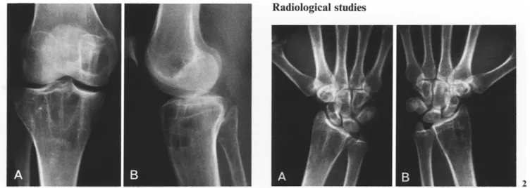
Fig. 1 A, B. Anteroposterior and lateral roentgenograms of the right knee demonstrate well defined, cystqike, lesions of the distal end of the femur and the proximal end of the tibia. The metaphyseal end of the fibula is also involved by a small lytic lesion Fig. 2A, B. Posteroanterior roentgenograms of both wrists, show similar multiple, cyst-like lesions of the carpal bones bilaterally and lesions in the distal end of each radius, similar to those in the proximal end of the right tibia. Small lytic lesions also are observed in the base of the left first metacarpal
Clinical information
This 35-year-old woman was admitted to the hospital several times because of disseminated and symmetrical skeletal lesions of a cystqike appearance. These lesions were first observed 9 years previously, following a fracture of the left fibula. One year after this incident, the left talus and navicular bones were curetted. A yellow, fatty material, filling the cyst-like lesions, was observed in the operative procedure. When examined mi- croscopically, fat tissue was noted, but a diagnosis was not made.
The family history was not significant. It was considered possible that the skeletal lesions were congenital, but this thought was negated, inasmuch as roentgenograms of the right ankle, obtained at the age of 16 years (after trauma), showed
Address reprint requests to." U.E. Pazzaglia, M.D., Department of Clinical Orthopaedic and Traumatology of the University de Pavia, Via Taramelli, 1-27100 Pavia, Italy
a normal appearance of the distal ends of the right tibia, fibula and tarsal bones.
In the following years the patient reported fractures of right and left metatarsals after a fall and of the left ankle following a vehicular accident.
The right talus and calcaneus were biopsied and curetted; material similar to that obtained in the first biopsy was again observed. A final diagnosis still was not established.
On admission to hospital at this time, a radiological survey of the skeleton showed symmetrical lesions localized in the tu- bular bones of both hands and feet and in the distal and proxi- mal ends of the long bones (both radii and ulnae, tibiae and fibulae and the lower end of the left femur) (Figs. 1 A, B, 2 A, B, 3, 4A, B). No skeletal lesions were observed in the spine, in- nominate bones or skull.
All the affected bones presented skeletal resorption, a very thin periosteum (e.g. proximal end of the tibia) with no definite periosteal reaction and no break in the cortex. The bony struc-
U.E. Pazzaglia et al. : Case report 381 475
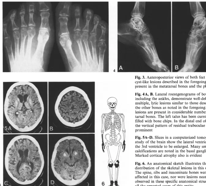
Fig. 3. Anteroposterior views of both feet show the cyst-like lesions described in the foregoing to be present in the metatarsal bones and the phalanges Fig. 4A, B. Lateral roentgenograms of both feet, including the ankles, demonstrate well-defined, multiple, lytic lesions similar to those described in the other bones as noted in the foregoing. The lesions are present in considerable number in the tarsal bones. The left talus has been curretted and filled with bone chips. In the distal end of the tibia the vertical pattern of residual trabeculae is prominent
Fig. 5A-D. Slices in a computerized tomographic study of the brain show the lateral ventricles and the 3rd ventricle to be enlarged. Many small calcifications are noted in the basal ganglia. Marked cortical atrophy also is evident Fig. 6. An anatomical sketch illustrates the distribution of the skeletal lesions in this entity. The spine, ribs and iunominate bones were not affected in this case, nor were lesions necessarily observed in these specific anatomical structures in all the reported cases of this entity
tures also consisted of course and irregular trabeculae (Fig. 2). The lytic lesions were cysMike in appearance, very well defined, differing in size and shape with thin septa, mainly longitudinal and horizontal in shape. This type of characteristic lesion was best observed in this patient at the level of the upper end of the left tibia and the distal ends of the ulnae (Figs. 1 A, B and 2 a , B).
The carpal hones and the digits of the hand showed only round, clear-cut, various sized cyst-like lesions, especially on the right. The scaphoid, lunate, capitate and hamate of the right wrists principally were affected. In the left wrist, the sca- phoid, hamate and to a lesser degree, the lunate also were af- fected (Fig. 2A, B).
Small cyst-like lesions were also detected at the distal ends of the left 3rd and 4th metacarpals, the right 3rd metacarpal and in the proximal phalanx of the right 4th finger. The proxi- mal and distal ends of the middle phalanx of the right third finger and the distal end of the middle phalanx of the right fourth finger were also involved.
The cyst-like lesions with longitudinal septa also were noted in the distal ends of the tibiae. The tarsal bones, the metatarsals (particularly in their proximal and distal ends), the fifth fingers
and the phalangies of the first toes showed spheroid, clear-cut, small, radiolucent, cyst-like defects differing in size and shape, with each lesion interspersed by thin, bony septa (Figs. 2A, B, 3, 4A, B). Each talus was affected by similar lesions, except in this instance, the lytic areas were large and divided by more sclerotic septa (calcaneus, navicular, cuboid and proximal end of the fourth left metatarsal). Furthermore, in the lateral view, the anterior edge of the left talus was irregular due to previous trauma (Fig. 4A).
All laboratory tests were within normal limits.
The patient developed mental changes which included ab- sentmindedness and loss of intellectual capacity. Because of these changes, the woman had to leave her position as a prod- uct-manager. A computerized tomographic study (Figs 5 A - D ) showed enlargement of the third ventricle and the lateral ventri- cles. The contours of the ventricles were smooth and the para- ventricular tissues showed normal density. Many small calcifi- cations were present in the basal nuclei bilaterally. Marked atrophy of the cortex of the brain also was detectable.
A biopsy of the left wrist was performed and material simi- lar to that obtained in the previous biopsies was prepared for microscopic examination.
476 U.E. Pazzaglia et al. : Case report 381
Diagnosis: Membranous lipodystrophy (MLD)
In the differential diagnosis the following entities must be considered: multiple intra-osseous lipomas,
multifocal cystic angiomatosis, polyostotic fibrous dysplasia, hyperparathyroidism, histiocytosis X and
multiple benign cysts of unknown origin. Of this group, only multiple intra-osseous lipomas and cystic
angiomatosis deserve serious consideration, since the lesions in these two entities may have an appearance
superficially resembling those noted in the skeleton in MLD. The other entities named also deserve
consideration. The radiological features, the symmetrical distribution of the skeletal lesions and the
association of the mental changes are, as a rule, sufficient to distinguish MLD from all of the previously
mentioned disorders.
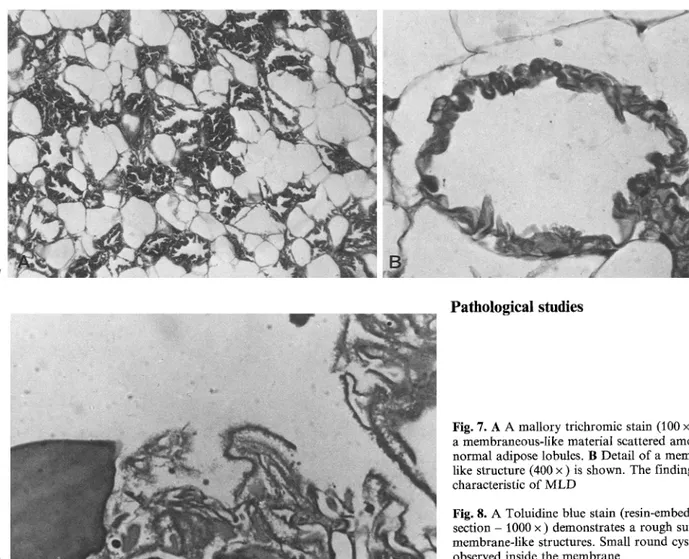
The examined specimen was entirely formed by adipose tissue. Among normal adipose lobules, amor-
phous substance, forming undulating and membranous-like structures, was observed (Fig. 7A, B), This
membrane-like materials was stained faintly by eosin and by PAS. No birefringency was observed in
polarized light. Gomori's silver impregnation and Weigert's stains for elastin were negative.
Frozen sections were made. While normal fat was fully stained by Sudan black, III and IV, the
membrane-like structures were stained only faintly by Sudan black and not by Sudan III and IV. High
resolution pictures on resin-embedded sections showed membranous structures of various thickness.
The surface was rough, with the presence of thin and short filaments, together with small sound cysts,
approximately 0.1 Ixm in size (Fig. 8).
With electron microscopy the membrane-like structures were poorly organized, with a smooth surface
on one edge and with thin projection on the other. Small lipidic droplets and disorderly arranged filaments
were noted inside the material (Fig. 9).
The histological findings were typical of membranous lipodystrophy.
Pathological studies
Fig. 7. A A mallory trichromic stain (100 x ) shows a membraneous-like material scattered a m o n g n o r m a l adipose lobules. B Detail of a membrane- like structure (400 x ) is shown. The findings are characteristic of M L D
Fig. 8. A Toluidine blue stain (resin-embedded thin s e c t i o n - 1000 x ) demonstrates a rough surface of membrane-like structures. Small r o u n d cysts are observed inside the m e m b r a n e
U.E. Pazzaglia et al. : Case report 381 477 Fig. 9. A transmission electron microscopic section (17150 • ) demonstrates the membrane-like structure to be formed by a poorly organized material with filaments and lipidic droplets within the membrane- like material. Thin projections are evident on the surface
Discussion
This disease was recognized for the first time by Jarvi in 1964 and named "Hereditary Folycystic Osteodysplasia (HPO)" [5]. In 1973, Nasu et al. [6] reported an autopsied case that pre- sented the same features as HPO and because of the histological findings, the authors of that manuscript called the entity " M e m b r a n o u s Lipodystrophy". Up to the present time only 27 cases have been reported in the world literature, exclusively in four countries - Finland, Japan, Norway and the U S A [1-7].
Mental changes and corresponding lesions of the central nervous system have been associated often with M L D [1, 3, 6], but the relationship between pathological changes in the skeleton and brain remains unclear. The etiology of the disease is unknown. Hakola [3] reported three affected members in the same family, but this observation was not confirmed in other reports.
In summary, a fascinating case of an entity called " m e m - branous lipodystrophy" has been described in a 35-year-old woman. Multiple, cyst-like disseminated, skeletal lesions were present throughout (except for the spine, innominate bones, ribs and skull). In addition, resorption of bone and a very thin periosteum were identified. Coarse and irregular trabeculae also were noted. Associated with the skeletal lesions in this instance, were hydrocephalus, cerebral cortical atrophy and multiple calcifications in the basal ganglia bilaterally.
The development of mental changes and lesions of the cen- tral nervous system as indicated are said to be associated con- sistently with MLD. The cause of the disorder is considered to be unknown.
The pathological studies were described in depth and the differential diagnosis was considered.
It is believed by the editor that this case report, describing
this entity, is a significant contribution to the literature on a disease of great interest, probably unknown to many physicians.
References
1. Akai M, Tateishi A, Cheng CH, Morii K, Abe M, Ohno T, Ben M (1977) Membranous lipodystrophy. J Bone Joint Surg 59 : 802
2. Edvardsen P, Halvorsen TB, Nesse O (1983) Lipomembran- ous osteodysplasia. A case report. Int Orthop 7 : 99
3. Hakola H P A (1972) Neuropsychiatric and genetic aspects of a new hereditary disease characterized by progressive de- mentia and lipomembranous polycystic osteodysplasia. Acta Psychiatr Scand [Suppl] 232
4. Inone K, Funayama M, Suda A, Higuchi I (1975) Intra os- seous lipomatosis. Report of a case. J Jpn Orthop Ass 49 : 223 5. Jgrvi OH, Hakola H P A (1970) A new entity of phacomato- sis; a. Bone lesions (Hereditary angionecrotic polycystic os- teodysplasia). Aeta Pathol Microbiol Scand [Suppl] 215:27 6. Nasu T, Tsukahara Y, Terayama K (1973) A lipid metabolic
disease "Membranous Lipodystrophy". An autopsy case demonstrating numerous peculiar membrane structures com- posed of compound lipid in bone and bone marrow and various adipose tissue. Acta Pathol Jpn 23:539
7. Wood C (1978) Membranous lipodystrophy of bone. Arch Pathol Lab Med 102 : 22
Dr. Howard Dorfman, New York, who reviewed the manuscript and approved it for publication also sent this editor a reprint of an excellent article by one of Dr. Dorfman's colleagues (Dr. Tsuneyoshi) on the same subject: Membranous Lipodystrophy: A Case Report (I. Matsuo, M. Suetsugu, M. Eguchi, M. Sasaki, M. Tsuneyoshi, Arch Psychiatr Nervenk, 1982, 231 : 123)