Facoltà di Scienze Matematiche, Fisiche e Naturali Dipartimento di Biologia
Scuola Dottorale in Biologia
Sezione di Biologia Applicata alla salute dell’Uomo (BASU) – XXI Ciclo
TELOMERE METABOLISM IN RESPONSE TO DNA
DAMAGE INDUCED BY LOW- AND HIGH-LET
RADIATIONS
METABOLISMO TELOMERICO IN RISPOSTA AL
DANNO AL DNA INDOTTO DA RADIAZIONI
IONIZZANTI DI DIVERSA QUALITÀ
PhD Student: Dr. Francesco Berardinelli
Supervisor: Prof. Caterina Tanzarella
ABBREVIATIONS ... 5 SUMMARY... 7 1 ITALIAN... 7 2 ENGLISH... 10 INTRODUCTION... 13 1 TELOMERES... 13
1.1 Structure and functions... 14
1.1.1 Main role of telomeres ...17
1.1.2 Regulation of cellular life span ...17
1.1.3 Additional functions of telomeres ...20
1.2 Telomere maintenance... 20
1.2.1 Telomerase ...20
1.2.2 Chromosome healing...22
1.2.3 “Telomere capture” and beyond...22
1.2.4 Alternatives to telomerase: the “ALT” mechanisms...23
1.3 NBS1 and Homologous Recombination (HR) in telomere maintenance ... 25
2 IONIZING RADIATIONS (IR) ... 27
2.1 Low-LET radiation... 27
2.2 High-LET radiation... 29
2.3 LET and Relative Biological Effectiveness (RBE)... 29
2.4 Radiation-induced DNA damage... 31
2.4.1 Isolated and clustered DNA damage ...31
2.5 Biological relevance of high-LET radiation study ... 32
3 BIOLOGICAL EFFECTS OF RADIATION ON TELOMERE METABOLISM.. 33
3.1 DNA repair and telomeres... 33
3.1.1 Non Homologous End Joining (NHEJ) ...33
3.1.2 Homologous Recombination (HR)...35
3.1.3 DNA DSBs and telomeres ...36
3.1.4 Damaged telomeres and telomerase ...37
3.2 Oxidative stress and telomere erosion... 38
3.3 Telomere length and radiosensitivity/radioresistance... 38
RATIONALE AND AIM OF THE PROJECT ... 41
RESULTS... 45
1 DNA DAMAGE INDUCED BY HIGH-LET RADIATION IS REPAIRED LESS EFFICIENTLY THAN LOW-LET ONE. ... 45
1.1 Kinetics of DSBs repair is slower in high-LET treated samples than in low-LET ones... 45 1.2 Yield of micronucleus induction is LET- dependent ... 45 2 HIGH-LET RADIATION INDUCE TELOMERE TELOMERE LENGTHENING THROUGH A RECOMBINATION MEDIATED MECHANISM SHORTLY AFTER IRRADIATION... 49
2.1 High-LET radiation induce telomere elongation in the first 24 hours after irradiation ... 49 2.2 No telomerase induction was observed in primary fibroblast exposed to low- and high-LET radiations ... 52 2.3 Increased T-SCE frequency in high-LET treated primary fibroblasts ... 53 2.4 High-LET radiations induce transient colocalization of telomere DNA and PML protein... 54 3 CELLS DEFECTIVE IN HOMOLOGOUS RECOMBINATION REPAIR DO NOT SHOW TELOMERE ELONGATION AFTER HIGH-LET IRRADIATION... 57
3.1 Neither low-LET nor high-LET radiations induce telomerase activity in lymphoblastoid cell lines... 58 4 TELOMERE LENGTH MODULATION IN HUMAN CELLS EXPOSED TO LOW- AND HIGH-LET RADIATION AND FOLLOWED UP TO 15 DAYS... 60
4.1 Different telomere length modulation dynamics in human cells exposed to low- and high-LET radiations ... 60 5 TK6 LYMPHOBLASTS WHICH SURVIVED X-RAY-IRRADIATION
DISPLAY TELOMERES LONGER THAN UNTREATED CELLS... 62 5.1 TK6 cells show telomere erosion after 3 days from X-rays exposure... 62 5.2 Determination of cellular survival in TK6 whole population.. 62 5.3 TK6 survived clones show telomere longer than untreated controls ... 64
DISCUSSION ... 67 1 DNA DAMAGE INDUCED BY HIGH-LET RADIATION IS REPAIRED LESS EFFICIENTLY THAN THAT PRODUCED BY LOW-LET ONE... 67
2 HIGH-LET RADIATION INDUCE TELOMERE LENGTHENING THROUGH A RECOMBINATION MEDIATED MECHANISM SHORTLY AFTER IRRADIATION68
3 CELLS DEFECTIVE IN HOMOLOGOUS RECOMBINATION REPAIR DO NOT SHOW TELOMERE ELONGATION AFTER HIGH-LET IRRADIATION... 70
4 TELOMERE LENGTH MODULATION IN HUMAN CELLS EXPOSED TO LOW- AND HIGH-LET RADIATION FOLLOWED UP TO 15 DAYS... 71
5 TK6 LYMPHOBLASTS WHICH SURVIVED TO X-RAY-IRRADIATION DISPLAY TELOMERES LONGER THAN UNTREATED CELLS... 72
REFERENCES... 75 REFERENCES FOR FIGURES... 103
Abbreviations
ALT Alternative Lengthening of Telomeres APBs ALT associated PML Bodies
AT Ataxia Telangiectasia BER Base Excision Repair
BFB Break Fusion Bridge
BrdU Bromo Deoxyuridine
CO-FISH Chromosome Orientated FISH DAPI 4',6-diamidino-2-phenylindole D-loop Displacement loop
DSB Double Strand Break
ECTR Extra Chromosomal Telomere Repeat FISH Fluorescence in situ Hybridization FITC Fluorescein isothiocyanate
Gy Gray
HR Homologous Recombination
IR Ionizing Radiation
LET Linear Energy Transfer LMDS Local Multiple Damaged Site
MN Micronucleus
NBS Nijmegen Breakage Syndrome NHEJ Non Homologous End Joining
PML Promyelocytic Leukaemia
Q-FISH Quantitative FISH
RBE Relative Biological Effectiveness ROS Reactive Oxygen Species RQ-TRAP Real Time Quantitative TRAP SCE Sister Chromatid Exchange SSB Single Strand Break
TIF Telomere dysfunction Induced Focus T-loop Telomeric loop
TRAP Telomerase Repeat Amplification Protocol TRF Telomere Restriction Fragment
Summary
1
Italian
Le radiazioni ionizzanti (IR) sono un noto agente genotossico, ampiamente studiato dato l’ampio spettro di possibili applicazioni in campo medico (radioterapia, diagnostica), e soprattutto per i possibili effetti sulla salute dell’uomo (esposizione occupazionale, esplorazione dello spazio). In base alla loro capacità di cedere energia alla materia le IR possono essere suddivise in radiazioni a basso- o alto- Trasferimento Lineare di Energia (LET). Nella prima categoria ricadono radiazioni in grado di cedere alla materia fino a 10 keV/m, ad esempio raggi-X e raggi-, mentre nelle seconda categoria sono incluse radiazioni che rilasciano alla materia fino 200 keV/m (protoni, ioni pesanti).
A livello cellulare, la differente capacità di cedere energia alla materia determina effetti diversi. In particolare il danno al DNA indotto da radiazioni ad alto-LET risulta più complesso e più difficile da fronteggiare rispetto a quello indotto da radiazioni a basso-LET, e tale maggiore complessità risulta in effetti cellulari più severi. Numerosi endpoint cellulari, quali mutazioni geniche, malsegregazione cromosomica, aberrazioni cromosomiche, sopravvivenza cellulare, sono stati presi in esame nello studio comparativo di radiazioni a basso- ed alto-LET. Tuttavia altri importanti aspetti come l’influenza di tali radiazioni sul metabolismo telomerico e sulla modulazione delle lunghezze telomeriche fino ad oggi, sono stati scarsamente investigati.
I telomeri sono complessi nucleoproteici localizzati alle estremità cromosomiche ed altamente conservati tra gli eucarioti. Consistono di sequenze ripetute in tandem dell’esanucleotide TAAGGG e sono strutture di fondamentale importanza per il mantenimento dell’integrità cromosomica; infatti, perdita di sequenze telomeriche o mutazioni in proteine che legano il telomero attivano una cascata di eventi che includono fusioni cromosomiche, instabilità genomica ed in ultima istanza, possono compromettere la proliferazione e/o la vitalità cellulare o condurre alla trasformazione tumorale.
Nel presente lavoro sono state utilizzate “radiazioni sparsamente ionizzanti” o a basso-LET, quali raggi-X e protoni da 62 MeV (pochi keV/m), e radiazioni “densamente ionizzanti” o ad alto-LET, quali protoni da 3 MeV e ioni carbonio (LET da 28 a 39 keV/m). Per gli esperimenti con sorgenti di
radiazione diverse dai raggi-X, ci si è avvalsi della collaborazione con ricercatori dell’INFN, e gli irraggiamenti sono stati effettuati presso i Laboratori Nazionali di Legnaro e Catania.
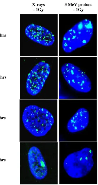
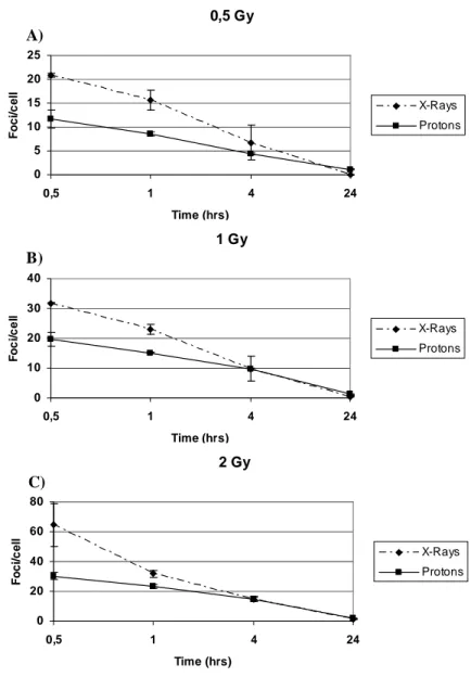
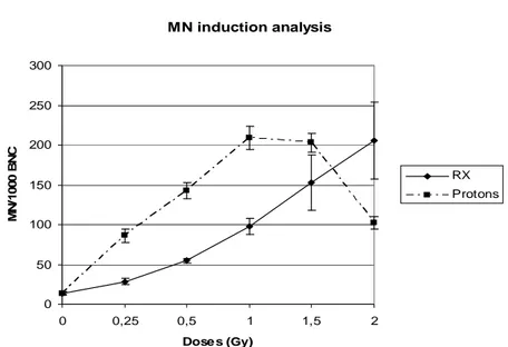
Nella prima parte del lavoro sono stati analizzati gli effetti genotossici indotti da radiazioni a basso ed alto LET in fibroblasti primari umani HFFF2. In particolare, le cellule sono state esposte a dosi crescenti (0.25-2 Gy) di raggi-X e protoni (3MeV, 28.5 keV/µm) e successivamente è stata valutata la cinetica di riparazione del danno al DNA e l’induzione di micronuclei. L’induzione di rotture alla doppia elica del DNA (DSBs) e la cinetica di riparazione di tali rotture sono state valutate seguendo la defosforilazione dell’istone H2AX nel tempo. I risultati mostrano che seppure la radiazione ad alto-LET induca inizialmente un minore numero di DSBs rispetto ai raggi-X, tali DSBs sono riparate con una cinetica più lenta nelle prime 24 ore dal trattamento. Dati ottenuti dal test del micrronucleo hanno confermato il più elevato potere clastogenico delle radiazioni ad alto-LET. L’esposizione ai protoni, infatti, è stata in grado di indurre un numero approssimativamente doppio di micronuclei rispetto a quello indotto dai raggi-X, almeno fino alla dose di 1.5 Gy. I dati ottenuti mostrano come il danno al DNA indotto da radiazioni ad alto-LET, ed in particolare da protoni a bassa energia, sia riparato in modo meno efficiente e generi danno citogenetico nel corso del primo ciclo cellulare dopo il trattamento.
Nella seconda parte del lavoro è stato valutato l’effetto del danno indotto dai protoni a 3 MeV sulla modulazione delle lunghezze telomeriche. A tale scopo fibroblasti umani HFFF2 sono stati trattati con 4 Gy di radiazioni a basso- (raggi-X, protoni da 62MeV) ed alto- (protoni da 3 MeV) LET e sono stati effettuati esperimenti di Q-FISH (Quantitative-Fluorescence In Situ Hybridization) telomerica quantitativa a 24 ore dall’esposizione. I risultati ottenuti hanno mostrato che solo la radiazione ad alto-LET è in grado di indurre un aumento significativo delle lunghezze telomeriche (22%), mentre nessuna variazione è stata riscontrata dopo trattamento con radiazioni a basso-LET. Per comprendere quale meccanismo fosse alla base del fenomeno osservato, è stata valutata l’induzione dell’enzima telomerasi e l’attivazione di meccanismi di ricombinazione telomerica. I dati ottenuti hanno escluso che il trattamento con radiazioni ionizzanti fosse in grado di indurre sia la trascrizione del gene TERT, codificante per la subunità catalitica dell’enzima, sia l’attività dell’enzima stesso, come mostrato tramite saggio TRAP (Telomerase Repeat Amplification Protocol). Risultati più incoraggianti sono invece emersi analizzando il coinvolgimento della ricombinazione telomerica nel fenomeno di allungamento osservato. Esperimenti di CO-FISH (Chromosome Orientated-FISH) infatti hanno
mostrato che i protoni da 3 MeV sono in grado di indurre un aumento della frequenza di scambi tra cromatidi fratelli nella regione telomerica. Tale evidenza ha mostrato come le radizioni ad alto-LET siano in grado di indurre, in modo transiente, un meccanismo di ricombinazione telomerica con caratteristiche simili al pathway di allungamento telomerico ALT (Alternative Lengthening of Telomeres) presente in alcune cellule tumorali. Tali dati sono stati confermati attraverso esperimenti di immuno-FISH che hanno mostrato un’induzione da parte dei protoni da 3 MeV di un altro marcatore delle cellule ALT-positive: la colocalizzazione della proteina PML (ProMyelocytic Leukaemia) e del DNA telomerico.
Recenti evidenze sperimentali hanno mostrato come la proteina NBS1 è essenziale per il fuzionamento del pathway ALT. NBS1 è una proteina coinvolta nel processo di riparazione del danno al DNA radio-indotto e in particolare nella riparazione mediata da ricombinazione omologa. Per valutare se mutazioni nel gene NBS1 influenzassero l’attivazione radio-indotta del pathway ALT, linee cellulari linfoblastoidi (LCLs) con differente genotipo per il gene NBS1 (NBS1+/+, NBS1+/-, NBS1-/-) sono state esposte a 0.5-4Gy di ioni carbonio (39 keV/µm). I risultati ottenuti hanno mostrato che solo nelle linee NBS1+/+ e NBS1+/- viene indotto un’allungamento telomerico significativo, supportando l’ipotesi del coinvolgimento del pathway ALT.
L’analisi della modulazione delle lunghezze telomeriche mediante Q-FISH è stata successivamente effettuata anche a tempi più lunghi (3/4 e 15 giorni) dall’esposizione alle diverse sorgenti di radiazioni. I dati ottenuti hanno mostrato che l’allungamento telomerico osservato a 24 ore dal trattamento con radiazioni ad alto-LET viene mantenuto nel tempo almeno fino al 15° giorno, sia in cellule primarie che immortalizzate.
Un più complesso pattern di modulazione delle lunghezze telomeriche è stato invece osservato dopo esposizione a radiazioni a basso-LET. Infatti a 3 o 4 giorni dal trattamento (3 giorni nel caso di cellule primarie HFFF2 e 4 nel caso dei LCLs) è stato riscontrato un accorciamento telomerico, seguito poi da un allungamento, come rivelato dall’analisi a 15 giorni. Risultati analoghi sono stati ottenuti su popolazioni clonali di linfoblasti umani TK6 derivati da singole cellule sopravvissute al trattamento con 4Gy di raggi-X. I dati ottenuti suggeriscono che uno dei processi implicati nella modulazione delle lunghezze telomeriche indotta da radiazioni a basso-LET sia una accresciuta radioresistenza delle cellule che nella popolazione iniziale mostravano lunghezze telomeriche medie maggiori.
2
English
Ionizing radiations are a well known genotoxic agents, widely studied for the great impact of their applications (i.e., radiotherapy and hadrontherapy) and effects (i.e., exposure risk for astronauts in space missions). Exposure to ionising radiations (IR) can result in the deposition of energy to DNA molecules, thus leading to DNA damage. IR-induced DNA damage is localized, and the level of localization is believed to increase with increasing linear energy transfer (LET) values of the radiation. Because LET is a measure of the energy released to an object along the path of the radiation, high-LET radiation can deposit more energy than low-LET one. Condensed or concentrated energy deposition results in cluster of ionization events. When the target is the DNA, the site of such lesions is termed “clustered DNA damage” or “locally multiply damaged site”, which consists in two or more lesions localized in close proximity on the DNA duplex.
In order to study the biological effects of high-LET radiations, several endpoints have been evaluated both in rodent- and in human-irradiated cells, including chromosomal aberrations, micronuclei (MN), chromosomal non-disjunction, mutations, DNA fragmentation, clonogenic survival, and cell cycle effects. However, aspects related to telomere length modulation and telomere metabolism have been so far poorly investigated both in primary and in immortalized cells exposed to low- and high-LET radiations. The aim of the first part of the study was to analyze the DNA-damage and the genotoxic effects induced by graded doses (0,25-2 Gy) of low-energy protons (high-LET radiation), and X-rays (low-LET radiation) in human primary fibroblasts. DSB induction and repair as mesured by scoring for -H2AX foci indicated that 3MeV protons, with respect to X-rays, yielded a lower number of DSBs per Gy, which showed a slower kinetics of disappearance in the first hours from irradiations. Furthermore, irrespective of dose delivered, a higher fraction of unrejoined DSBs persisted in sample harvested 24 hours from exposure to protons. The higher clastogenic effect of protons was in agreement with the extent of micronuclei (MN) induction in binucleated cells up to 1,5 Gy. Our results support the notion that DNA DNA damage produced by 28.5 keV/µm protons appears less amenable to be repaired and could be transformed in cytogenetic damage in the form of MN in the first cell cycle from irradiation .
After confirming the greater biological effectiveness of high-LET radiations compared to low-LET ones, we focused our attention on studying telomere metabolism within 24 hours from the exposure to both types of radiations.
Interestingly, data obtained showed a different kinetics of telomere length modulation in cells exposed to low- or high-LET radiations. Moreover, the phenomenon observed appeared to be conserved both in primary and in immortalized cell lines. Interestingly, exposure of human primary fibroblasts to 4Gy high-LET radiation determined a telomere elongation respect to untreated cells, whereas no telomere length modulation was observed in low-LET treated fibroblasts. In order to investigate the molecular mechanism underlying the observed elongation, the expression levels of the telomerase (i.e., hTERT) and its enzymatic activity were evaluated. Results obtained excluded the involvement of the telomerase in the observed telomere lengthening induced by high-LET radiation, thus supporting the activation of a telomerase-independent mechanism. Some mammalian cells lacking in any telomerase activity are able to maintain the length of their telomeres for many population doublings (PDs). This indicated the existence of one or more non-telomerase mechanism(s) for telomere maintenance, further termed Alternative Lengthening of Telomeres (ALT). To date, clear evidences of the existence of an ALT activity has been demonstrated only in human tumours and immortalized cell lines, and in telomerase-null mouse cell lines. To analyze whether a recombinational mechanism could be responsible for the high-LET-induced telomere lengthening observed in human primary fibroblasts, two types of experiments were performed. On one side, the incidence of recombinational events at telomeres (T-SCE) was measured, and on the other side the colocalization of telomeres and PML bodies (that are considered as an hallmark of cells with activated ALT pathway), was analyzed. Strikingly, our results indicated that the DNA damage induced by high-LET radiation is somehow able to induce telomere lengthening through the transient activation of an ALT recombinational pathway.
Recent reports demonstrated that NBS1 is essential for the correct functioning of the ALT pathway. NBS1 gene, mutated in the NBS human chromosome instability disorder, encodes for the NBS1 protein, a central player in the response to the ionizing radiation-induced DNA damage, as well as in the homologous recombination repair. In order to confirm the high-LET-induced recombinational ALT pathway, telomere length was evaluated in Lymphoblastoid Cell Lines (LCLs) heterozygous (NBS1+/-) and homozygous (NBS1-/-) for a mutation of the NBS1 gene, as well as in normal cells (NBS1+/+) exposed to 4 Gy of carbon ions (39keV/m). Remarkably, a telomere elongation was observed in NBS1+/+ and NBS1 +/-cells, but not in NBS1-/-ones. These data evidenced that the process of telomere lengthening induced by high-LET radiation is NBS1-dependent,
thus supporting the hypothesis that telomere elongation is mediated by recombinational mechanisms.
Beside the analysis performed at 24 hours, telomere length modulation was followed up to 15 days from the irradiation of both human primary fibroblasts and LCLs. Dynamics of telomere lengths modulation appeared to be different after low- and high-LET irradiation. Our data showed that the telomere lengthening observed in high-LET-treated cells seems to be maintained at 3-4 days, as well as 15 days after exposure. Interestingly, the time-course of the low-LET radiation-induced telomere length modulation appeared to be more complex than the high-LET one. In fact, after 3-4 days telomere erosion was reported, whereas after 15 days from the exposure a telomere lengthening was observed in primary as well as in immortalized cell lines.
To explain the time course of low-LET-induced telomere length modulation we have hypothesized that a direct correlation between telomere length and radioresistance/radiosensitivity could account for this phenomenon. To test our hypothesis, we decided to perform experiments in TK6 lymphoblast cells, since they represent a good and widely used radiobiological cellular model. Data obtained brought us to suggest a model: the radioresistance of cells with longer telomeres drives a selection process that led to an increased telomere length in clones survived to low-LET radiation exposure. A direct correlation between telomere length and radisensitivity/radiresistance has already been proposed in some published reports and imply that telomeres length measurement could be potentially used as a tool to predict clinical radiation response in radiotherapy.
Introduction
1
Telomeres
Telomeres or the ends of linear eukaryotic chromosomes, were first described almost 70 years ago since the pioneering studies of the geneticists Hermann Joseph Muller and Barbara McClintock in the fruit fly Drosophilia melanogaster and Zea maize, respectively [1, 2]. Muller observed that the ends of chromosomes rarely interacted with breaks that resulted from ionizing radiation (i.e., X-ray-induced chromosomal aberrations never included deletions or inversions involving the terminal regions of the chromosomes). Thus, he proposed that chromosome ends are specialized structures that he coined “telomeres”, from the Greek, telo = end, and mere = part [1]. The concept of “telomere” meant not only the physical ends of the chromosome itself but also in Muller's words “a terminal gene with a special function, that of sealing the end of the chromosome” [1]. Also by the end of 1930s, Barbara McClintock, studying chromosomal aberrations induced by X-rays in maize, found that broken chromosomes frequently fused to their sister chromatids, creating breakage-fusion-bridge (BFB) cycles, which were always accompanied by the loss of the terminal regions at the fusion site, demonstrating that broken chromosomes (i.e., without “end caps”) were subject to fusion events [2-6]. These observations lead to the idea that the ends of chromosomes or telomeres were “capped” and therefore protected from fusion reactions characteristic of ends created by chromosome breakage events. In this way, telomeres were defined as the terminal regions or physical ends of eukaryotic chromosomes, which protected them from fusion with either broken chromosomal fragments or other telomeres. Nowadays, in the light of molecular biology studies, telomeres are defined as specialized nucleoproteic complexes localized at the physical ends of linear eukaryotic chromosomes maintaining their stability and integrity [7].
Telomere biology has evolved from peripheral, albeit interesting branch of cell biology studied by a few groups to a major field involving hundreds of laboratories worldwide. Novel techniques have been developed and understanding of the structures, functions and roles of telomeres have evolved rapidly. In addition, the attention of the wider public has been stimulated through proposed links between telomeres and cancer.
1.1 Structure and functions
Telomeres are composed of both repeated DNA elements and specific DNA-binding proteins, which together form the ends of eukaryotic chromosomes [8-12]. Molecular dissection of telomeres started with the discovery of the telomeric DNA sequence of the ciliated protozoan Tetrahymena thermophila, (TTGGGG)n, by Blackburn and Gall in 1978 [13]. Telomeric DNA is characterized by being a G-rich double strand DNA composed by short fragments tandemly repeated with different sequences depending on the species considered [14, 15].
In all vertebrates, telomeres consist of tandem repeats of the hexanucleotide sequence (TTAGGG/CCCTAA)n and associated proteins [10, 16-22]. The arrays of TTAGGG repeats, first identified at human chromosome ends by Moyzis et al. in 1988 [22], are oriented 5′ → 3′ towards the end of chromosomes [19, 23] and form a 3′ single-strand G-rich overhang found at both chromosomal ends [24, 25]. The C-rich telomere strand is at the 5′ end and the G-rich telomere is at the 3′ end of each chromosomal DNA strand. These single-strand, G-rich 3′ overhangs result from both the “end replication problem”, that is the inability of DNA polymerase to replicate the very end of the telomeres, and postreplication processing [26, 27]. Thus, removal of the most distal RNA that primes lagging-strand synthesis leaves an 8- to 12-base gap at the 5′ end that, if not filled in, leads to a small loss of DNA in each round of DNA replication [28] (see paragraph 1.1.2 Regulation of cellular life spans).
The physical structure of the telomere was revealed by electron microscopy to be a large duplex loop [29-31], called t-loop, which is created when a telomere's end loops back on itself and the single-strand overhang invade an interior segment of the duplex telomeric DNA. The t-loop structure of mammalian telomeres is thought to repress the non homologous end-joining (NHEJ) DNA double-strand breaks (DSBs) repair process at chromosomal ends, thus rendering telomeres nonrecombinogenic (Fig. 1).
The length of the double strand telomeric repeat varies greatly among species [7]. For example, in the ciliate Oxytricha, it is only 20 base pairs (bp) long [64] in Saccaromyces cerevisiae, it is a few hundred bp long [32] while, in vertebrates, individual telomeres may extend to more than 100 kb, such as in some mouse cells. [33]. In normal primary human cells, the DNA at each chromosome terminus spans 5–20 kb in length [22], terminating in a 3′ single-strand overhang 100–400 nt in length [34]. In human tumor cells which use telomerase for telomere maintenance (see paragraph 1.2.1 Telomere maintenance), telomere length varies from 1 to 20 kb [35-37]. It
has been shown that in humans and mice, the length of telomere repeats at individual chromosome ends in individual cells is highly variable [38-42] and that mouse and human cell lines exhibit subpopulations of cells with different telomere lengths [43]. It was shown that, at cellular level, a stable hierarchy exists in the form of a telomere length profile of the human karyotype [44]. This rank order is conserved between different human cell types and individuals, maintained throughout a lifetime, and seems to be genetically determined [44] and [45]. The longest human telomere is found at the long arm of chromosome 4, whereas the shortest one is at the short arm of chromosome 17 [46].
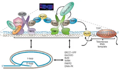
The homeostasis of mammalian telomeres is regulated by a number of telomere associated proteins (Fig. 1). Among these proteins, Telomere Repeats binding Factors, TRF1 and TRF2, directly bind double-strand telomere DNA and interact with a number of proteins to maintain telomere length and structure [47, 48]. It has been shown that the amount of telomere-bound TRF1 correlates with telomere length. Overexpression of TRF1 shortened telomeres in human cells, whereas dominant negative TRF1 led to elongated telomeres [49-51]. TRF1 may control the length of telomere repeats through multiple mechanisms. For example, TRF1 can control telomerase access through its interaction with TIN2, PTOP/PIP1, and the single-strand telomere DNA-binding protein POT1 [52-54]. TRF1 may also regulate telomerase activity through its interaction with PINX1 [55]. In comparison, TRF2 has an essential role in telomere end protection and t-loop formation [29, 47, 56]. Interference of endogenous TRF2 activity by expressing dominant negative forms of TRF2 markedly increase the rate of telomere end-to-end fusions [57]. Consistent with this role of TRF2, TRF2 forms a complex with RAP1 and associates with several proteins involved in DNA damage and repair responses, notably MRE11/RAD50/NBS1 (MRN complex), Ku86, and ERCC1/XPF [58-60]. These findings have pointed to distinct biological functions of TRF1 and TRF2. Some recent findings, however, suggest a more complex picture. In mouse embryonic stem cells, the conditional knockout of TRF1 led to significantly reduced levels of TRF2 at the telomeres, suggesting that TRF2 telomere localization may be partially regulated by TRF1 [61]. In addition, chromosome end-to-end fusion was detected in TRF1 knock-out cells, indicating that telomere end protection was compromised. Despite the wealth of information, the functional relationship between TRF1 and TRF2 in telomere maintenance remains unclear. Notably, a recent report demonstrated a direct interaction between TRF2 and the TRF1-interacting protein, TIN2 [62]. Such findings further suggest that cross-talk probably
occurs between the TRF1 and TRF2 complexes. However, whether TIN2 can simultaneously associate with both TRF1 and TRF2 in the same complex remains to be demonstrated. In addition to TRF1, several other telomeric proteins have been shown to be regulators of telomere length [47, 48, 53, 54, 63-66].
Fig. 1 The mammalian telomeric complex. The fluorescence image shows the location of a telomere within a chromosome. Mammalian telomeres consist of TTAGGG repeats with a single stranded 3' overhang of the G-rich strand. Specific protein complexes bind to the double-and single- stranded telomeric DNA. The components of the shelterin complex are shown in bold text. The single-stranded overhang can invade the double-stranded portion of the telomere, forming protective loops — such as t-loops with displacement loops (D-loops) — at the invasion site. The telomerase complex (which contains the telomerase RNA template and the reverse transcriptase TERT) interacts with the overhang and is regulated by shelterin and other telomeric proteins. Other factors that can interact with telomeres are listed. Bidirectional arrows indicate interactions. From [I].
Both inhibition of endogenous RAP1, TIN2, POT1, or PTOP expression through RNA interference and expression of dominant negative forms of these four proteins resulted in elongated telomeres in cultured cells [49,
52-54, 60, 67]. These observations suggest that RAP1, TIN2, POT1, and PTOP may function in the same pathway. All four proteins, RAP1, TIN2, POT1, and PTOP, directly or indirectly associate with TRF1 or TRF2 [53, 54, 64], pointing to a possible functional connection among these six telomeric proteins.
1.1.1 Main role of telomeres
Recently, the term ‘telomere capping’ emerged to describe the protective role of telomeres [8, 9, 68, 69], since telomeres provide a protective “cap” for the ends of chromosomic DNA. The main role of telomeres is to preserve the integrity of the chromosomes, protecting them from degradation, recombination or fusion [47] by preventing the ends of linear chromosomes from being recognized as DSB by the DNA repair machinery, i.e., they distinguish natural DNA ends from DNA ends resulting from breakage events [1, 2]. Thus, telomeres. Thus, “capping” refers to the ability of telomeres to protect chromosome ends from DNA damage responses, prevent inappropriate repair and recombination between internal DNA breaks and native chromosomal or the ligation of chromosomal ends. Accordingly, when telomeres become dysfunctional, fusions between two telomeres and between a telomere and a DSB occur, as shown by recent studies using the chromosome orientation, CO-FISH, technique [70, 71]. In mice, one of the consequences of impaired telomere function is the formation of Robertsonian-like chromosome fusions [72-74]. Therefore, the maintenance of telomere function is crucial for genomic stability and cell viability. Cells respond to dysfunctional telomeres by undergoing senescence, cell death, or genomic instability [74-86]. Cellular response to dysfunctional telomeres is governed by proteins that also control the DNA damage response [10, 15, 17, 87, 88]. The domain of telomere-associated DNA damage factors has been termed telomere dysfunction-induced focus or “TIF” by Takai et al. [87] who showed that DNA damage foci form at telomeres uncapped by TRF2 inhibition, and that uncapping of telomeres occurs in late S/G2.
1.1.2 Regulation of cellular life span
In 1972, James Watson wrote, “While 5’ to 3’ oriented growth should proceed smoothly to the end of its template, I see no simple way for 3’ to 5’ growth to reach the 3’ end of its template” [26]. Thus, he correctly predicted that the lagging strand of linear chromosomes copied by the semi-conservative replication machinery would not be fully replicated 26]. In 1973, A. M. Olovnikov proposed the ‘marginotomy theory of ageing’,
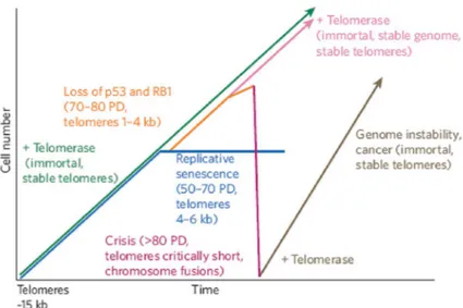
suggesting that ‘telogenes’ located at opposite ends of DNA molecules carry no genetic information and fullfil a buffer function. He stated that these telogenes are stochastically shortened during each mitotic cycle, providing a mechanism for ageing [27] (Fig. 2). Observations made by L. Hayflick in 1961 suggested that human cells derived from embryonic tissues can only divide about 50 times, and this became known as the Hayflick limit [89]. Since then, the assumption that the Hayflick limit is determined by the initial length of the telomeres and the rate of telomere shortening, as laid out in the mathematical approach of A. M. Olovnikov [27], has been proved experimentally [90, 91]. It is well established that critically short telomeres cease to function as protective units and cause the cell to die or to arrest permanently. Telomeres are now known to have many more roles than simply buffering against DNA loss; however, the initial concept of replication associated telomere shortening was correct. At present, we refer to the inability of conventional DNA polymerases to replicate linear molecules fully as the ‘end-replication problem’. This is caused by the deletion of the RNA primer of the most distal Okazaki fragment and results in the loss of about five bases of terminal genetic material per population doubling [90-94]. However, the sequence loss that is predicted to occur as a result of the end-replication problem is considerably less than that which has been observed in primary human cells, which lose about 100–200 bases of TTAGGG repeats per cell division [95-97]. Consequently, replication-associated terminal sequence loss is caused by a combination of the end-replication problem and the processing that must occur to create the G overhang on the telomeres generated by leading- and lagging-strand synthesis. When telomeres become critically short, they are detected by the cellular DNA-damage repair machinery [47]. As demonstrated in S. cerevisiae, chromosomes that lose a telomere are often eliminated, despite checkpoint and DNA damage repair machineries [98]. In human cells, p53- and RB1- (retinoblastoma 1) dependent pathways are responsible for monitoring telomere function (Fig. 2), whereas p53 seems to be the main sensor in mouse cells [80]. The minimal functional telomere length, and whether this length varies among cell types, has not been clearly defined. But even in senescent human cells, telomeric double-stranded repeats are readily detectable, suggesting that several kilobases of TTAGGG repeats are required at all times. Replicative senescence can be viewed as a mechanism to limit the potential number of population doublings a cell can undergo, hypothetically rendering it a powerful tumour-suppressor mechanism [99]. Each time a cell divides, telomeres shorten as a result of the end-replication problem and end processing. After
telomeres have become critically short, they are detected by the DNA-damage repair machinery, and the cell dies or enters senescence. At present, senescence in human cells is regarded as an irreversibly arrested state, effectively inhibiting the generation of immortal cells and therefore cancer formation. As a result, major tumour suppressive mechanisms need to be deactivated before a cell can overcome this block to immortality.
Fig. 2 Telomere shortening, senescence and cancer. Primary cells divide exponentially, and telomeres shorten from ~15 kilobases (kb) until they reach a critical length, 4–6 kb. Irreversible cell-cycle arrest then occurs (blue). Activation of telomerase before senescence allows cells to divide indefinitely and maintain a stable genome (green). If, instead, the p53 and RB1 pathways are suppressed, cells continue dividing (orange) until end protection is completely lost, resulting in telomeric crisis, cell death and massive genomic instability (dark pink). If telomerase is activated before erosion is complete, this rescues the genome from instability by re-establishing telomere maintenance (light pink). Activation of telomerase after the accumulation of mutations results in an unstable genome, allowing clones that carry multiple mutations to escape cell death (that is, to become immortal). Such cells are predisposed to oncogenic transformation (brown). PD, population doublings. From [I].
1.1.3 Additional functions of telomeres
Besides the above-mentioned functions, telomeres contribute to maintenance of chromosome topology in the cell nucleus and play a fundamental role in the proper alignment of chromosomes for recombination during the first meiotic prophase [7, 24, 75, 100-103]. An outstanding feature of telomeres is that they silence genes flanking the telomere repeat sequence [104-106]. This phenomenon, called the “telomere position effect”, is thought to be modulated by telomere length and local heterochromatin structure [107-109]. It has been shown that mutations in the encoding genes for Ku proteins lead to disruption of nuclear organization of telomeres and loss of telomeric silencing [110, 111]. Moreover, in mammalian cells, loss of Ku leads to aberrant telomere– telomere fusions [112, 113].
1.2 Telomere maintenance
1.2.1 Telomerase
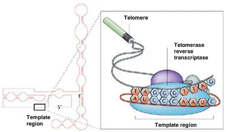
The loss of telomeric repeats is usually prevented by telomerase, a specialized reverse transcriptase-like enzyme, containing a RNA subunit (TR) and a catalytic protein subunit called telomerase reverse transcriptase (TERT) which is the rate-limiting factor for the enzyme activity (Fig. 3). This enzyme, first discovered by Greider and Blackburn in 1985 in Tetrahymena [114], works via an RNA template – using exclusively single-strand 3′ telomeric overhangs as primers [9, 11, 18, 24, 115, 116] – by adding TTAGGG repeats to the telomere. Although repressed in the majority of normal somatic cells (with the exception of a transient S phase activity thought to maintain the single-stranded overhang [117], telomerase is present in immortal cell lines, germline cells, stem cells, activated lymphocytes, and most of the tumor cells analyzed so far [35- 37, 118-120]. Telomerase activity favours 3′ overhangs over blunt DNA ends for addition of telomere sequence, at least in vitro [121, 122]. Recent observations indicate that telomerase exists as a complex tetramer composed of two RNA subunits and two catalytic subunits [123-125]. These subunits act in concert to elongate telomeres by reading from the RNA template sequence carried by the RNA subunit and synthesizing a complementary DNA strand. Mutations in conserved reverse-transcriptase catalytic residues found in telomerase eliminate the enzymatic activity of telomerase [126-129].
Fig. 3 Human telomerase is a cellular reverse transcriptase. It is composed of two essential components: telomerase reverse transcriptase catalytic subunit (hTERT) and functional telomerase RNA (hTR), which serves as a template for the addition of telomeric repeats (left side). From [II].
Other telomerase-specific motifs are also required for catalytic activity [123, 130-132]. In addition to these core components, several other proteins associate with the telomerase holoenzyme, including TEP1, p23, and Hsp90 [133, 134]; however, the physiologic function of these other proteins remains undefined because both biochemical and genetic experiments indicate that these other proteins are dispensable for telomerase activity [128, 135]. Telomere length is controlled by a mechanism involving telomerase and the telomere-binding proteins [18, 51]. Loss of telomerase enzymatic function leads to progressive telomere shortening over time, eventually resulting in the disappearance of detectable telomeric DNA and the formation of end-to-end chromosome fusions, followed by growth arrest or cell death [11, 136].
A telomere is a repeating sequence of double-stranded DNA located at the ends of chromosomes. Greater telomere length is associated with immortalized cell lines such as embryonic stem cells and cancer cells. As cells divide and differentiate throughout the lifespan of an organism or cell line, the telomeres become progressively shortened and lose the ability to
Telomerase reverse transcriptase Telomere Template region Template region
maintain their length. Telomerase is an enzyme that lengthens telomeres by adding on repeating sequences of DNA. Telomerase binds to the ends of the telomere via an RNA template that is used for the attachment of a new strand of DNA. Telomerase adds several repeated DNA sequences then releases and a second enzyme, DNA polymerase, attaches the opposite or complementary strand of DNA completing the double stranded extension of the chromosome ends. High levels of telomerase activity are detected in embryonic stem cells and cancer cells, whereas little or no telomerase activity is present in most mature, differentiated cell types. The functions of telomeres and telomerase appear to be important in cell division, normal development, and aging.
1.2.2 Chromosome healing
Besides maintaining pre-existing telomeres, telomerase can catalyze the addition of telomeric sequences directly on to non-telomeric DNA [137]. This process of direct addition of telomeric repeats to the ends of broken chromosomes by telomerase is called “chromosome healing” (a term coined by Barbara McClintock in 1941 to describe the phenomenon that halted the BFB cycles in the embryo of plants [2]), and has been observed in protozoans, yeast, plants, insects, and mammals [138-145]. Repair function of telomerase would lead to uncontrolled chromosomal fragmentation and karyotypic instability, because chromosome healing prevents repair of broken ends. Therefore, telomerase must be prevented from accessing internal DSBs. Slijepcevic and Al-Wahiby [146] proposed that Ku, a DSB protein which has a high affinity for DNA ends, acts to prevent telomerase from accessing internal DSBs. This model is supported by the fact that the efficiency of chromosome healing is extremely low, about 1% [147]. In fact, no evidence of “chromosome healing” was found in normal human lymphocytes [148] or in Ataxia-Telangiectasia (AT) cells [149], which display high radiosensitivity, exposed to ionizing radiation. The failure to recruit and/or activate telomerase at sites of DSB may contribute to the paucity of chromosome healing events.
1.2.3 “Telomere capture” and beyond
Lost telomeres in broken chromosomes can also be acquired by “telomere capture” and break-induced replication. Telomere capture is a process which involves the addition of telomeres at the site of DSB by subtelomeric cryptic translocations, undetectable by classical cytogenetic techniques [144-146, 150, 151]. In telomere capture, broken chromosomes are stabilized by the transfer of telomeres from normal chromosomes. This
phenomenon was first reported in human malignant melanoma cells [151], and has been recently observed in the leukocytes of chronic lymphocytic leukemia and chronic myeloid leukemia patients [152]. Telomere capture is essentially a non-reciprocal process, producing a chromosome with only one telomere (donor chromosome) and another one with a new telomere (recipient chromosome) plus an acentric fragment (a terminal fragment or terminal deletion, as seen by telomeric FISH, if the chromosome break occurs at G0/G1/early S phase, or a chromatid-type fragment, if the break
takes place in late S/G2). However, the donor chromosome may be involved
in secondary recombination events [145, [151, 153] by the initiation of BFB cycles in this chromosome. Slijepcevic et al. [153] showed that only a small percentage of radiation-induced chromosome/chromatid breaks may be modified by “telomere capture”.
In break-induced replication [154] and [155], the broken end of a chromosome invades a region of homology and initiates replication, thereby duplicating the end of that chromosome. Although not involving telomere restoration, the formation of ring or dicentric chromosomes can also compensate for telomere loss [156-158].
1.2.4 Alternatives to telomerase: the “ALT” mechanisms
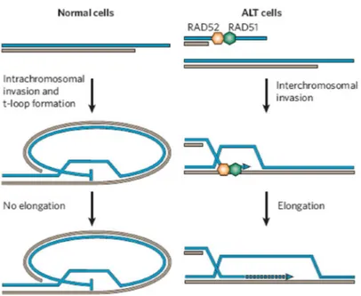
An alternative method of telomere elongation in the absence of telomerase has been described in several tumor cells and immortalized cell lines and named “ALT” (for “alternative lengthening of telomeres”) [159-166], although some tumors were found to posses neither telomerase activation nor ALT mechanisms for telomere length maintenance [167, 168]. Evidence hints that Homologous Recombination (HR) plays a primary role in most of the mammalian ALT pathways (Fig. 4). Several characteristics can be used to identify human cells using the recombination-based pathway of telomere length maintenance. First, they have no detectable telomerase activity and lack expression of the catalytic protein component, hTERT, or in some cases, they lack both hTERT and the integral RNA component, hTR [169]. Second, cells using the recombination-based pathway to maintain telomeres have very long and heterogeneous telomeres ranging in length from less than 2 kb to 50 kb [170]. Third, they contain extrachromosomal telomere repeats (ECTRs) that may be linear double-stranded fragments of telomeric DNA [171]. Fourth, cells using the recombination-based pathway to maintain telomeres have a novel type of promyelocytic leukaemia (PML) nuclear bodies called ALT-associated PML bodies (APBs) that contain telomeric DNA, telomere-associated proteins (i.e., the telomere-binding proteins TRF1 and TRF2), and
recombination-associated proteins (i.e., RAD50, RAD51, RAD52, MRE11, NBS1, BLM and WRN) [159]. The evidences that APBs appear at exactly the same time as the activation of the ALT mechanism during cell immortalization and contain HR associated proteins, suggest that PML may play a role in recombination mechanism in ALT [172-174]. Telomere exchange between sister chromatids has been suggested as a possible mechanism of ALT. It was recently found [175] that in ALT-negative cells the rates of sister chromatid exchanges in telomeres (T-SCE) are 10-fold higher compared to other DNA sequences. T-SCE rates are higher in ALT cells compared to normal cells [170, 171, 176] and has been suggested they play an important role in determining the proliferative potential of telomerase-negative cells [170, 171, 175, 176].
Fig. 4. ALT cells show an increased rate of sister chromatid exchange, suggesting that the homologous-recombination pathway is involved. From [I]
The increased telomere recombination, however, does not reflect a global increase in recombination frequencies because it was found that there was no increase in the rate of recombination at other genomic locations in ALT cells as compared to non-ALT controls [176-178]. However, while telomere recombination events are commonly called telomere sister chromatid exchanges, it is possible that the observed post replicative exchanges in ALT cells occurred with non-sister chromatids, or with the extrachromosomal telomeric repeat elements present in ALT cells. Many of these exchanges in ALT cells appear to be unequal, causing a reciprocal gain or loss of telomere sequence on the chromosome ends involved.
1.3 NBS1 and Homologous Recombination (HR) in telomere
maintenance
MRN complex is required for the maintenance of telomere length in mammals, plants and yeast [179-181]. In yeast, the XRS2 complex (homologue in yeast of human MRN complex) plays a role in at least two pathways in telomere maintenance [181-184]. The first one is telomerase-dependent and consists in the generation of 3’ ssDNA at the telomere for the recruitment and subsequent action of telomerase. In the second pathway, MRN takes part in the homologous recombination mediated telomeres elongation, which involves recombination between tracts of telomere repeats and is not dependent on telomerase function. In human, blood mononuclear cells from NBS patients analyzed for telomeres length, show shorter telomeres in comparison to cell from unaffected individuals. Primary fibroblasts isolated from NBS patients show an accelerated telomere shortening during in vitro colture [185].
In 2001 Ranganathan and co-workers demonstrated that only co-expression of NBS1 and TERT, the catalytic subunit of telomerase, leads to a significantly increase in telomere length whereas neither the introduction of NBS1 nor TERT, alone, has the same restoring effect [185]. These results suggest that the MRN complex may facilitate telomerase mediated telomere elongation by modifying telomere DNA ends or opening the T-loop [182, 186]. In 2003, Bai and Murnane elucidated another important aspect of NBS1 role in telomere maintenance. Cells expressing inducible NBS1 protein mutated at Ser278 and Ser343, showed an increased rate of telomere loss. Absence of detectable changes in the average telomere length suggest that this process is probably due to stochastic events, like complete telomere loss or loss of telomeric capping [187, 188]. These results have led to the
proposal that the MRN complex is involved in either establishment of the single stranded tail or in t-loop formation after telomeres replication. NBS1 seems to play a key role also in ALT pathway, in yeast as well in mammalian cells. NBS1 was found to be co-localized with PML, as well as to be associated with a nuclear PML-binding protein, SP100, by the BRCT-domain at its C-terminus [189]. NBS1 also functions in recruiting other recombination proteins, including RAD50, MRE11, and BRCA1 to PML nuclear bodies. Moreover, recent reports demonstrated that NBS1 protein is essential for the correct functioning of the ALT pathway, which results impaired by depleting one of the three components of MRN complex [190].
2
Ionizing Radiations (IR)
Ionizing radiation consists of subatomic particles or electromagnetic waves that are energetic enough to eject one or more orbital electrons from the atom or molecule, this process is called ionization.
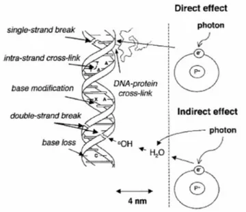
IR normally interacts with materials indirectly, via the formation of radical species. These species are mainly produced during radio-induced dissociation of the water surrounding the ultimate substrate. This process is termed “water radiolysis”. Although this indirect action can be modulated by the use of scavengers [191], in contrast to endogenous stress, these reactive species are not produced homogeneously, but are closely localised in spaces termed “clusters of ionisations”, which exist on a nanometre scale [192, 193]. In addition to water radiolysis, radiation interacts with materials in a direct way, which consists of the interaction between the radiation and the substrate itself (Fig. 5). The direct energy transfer from the radiation to the substrate can lead to either its excitation or ionisation. The relative importance of direct and indirect effects depends on the nature of the radiation, on DNA organisation and on DNA hydration, together with the scavenging properties of the environment. In order to explain the differing contribution of direct and indirect effects and the diverse biological effects observed between different ionizing radiation, the concept of linear energy transfer (LET) was introduced. LET is a macroscopic approach used to study the spatial distribution of ionisation and excitations produced along a linear path. It assumes that the energy is progressively and continuously deposited by the radiation through the matter and is expressed as the ratio between the energy loss and the corresponding path length in units of keV/µm. The LET delivered depends on several parameters such as the atomic number of the target and the velocity of the particle. It plays an important role in the understanding of the biological effects of radiation and thus allows a distinction between low-LET and high-LET radiations. For the International System of Units (SI) the unit of absorbed dose of IR is the Gray (Gy), 1 Gy is defined as 1 J kg-1.
2.1 Low-LET radiation
Low-LET radiations include electrons and electromagnetic waves such as X- and γ-rays. X-rays are produced in electrical device that accelerate electrons to high energy and then stops them abruptly in a target, usually made of tungsten or gold. On the other hand -rays are emitted by
radioactive isotopes (32P, 33P, 14C, etc.); they represent excess energy that is given off as the unstable nucleus breaks up and decays in its effort to reach a stable form. Their energy can range from 10 keV to 30 MeV. The interactions of low-LET radiation with matter depend on their nature (particles or photons) and their energy. Photons mainly interact with their biological target via the Compton process if their energy is between 20 keV and several MeV. For the lowest energetic photons, such as ultra-soft X-rays possessing energy ranging from 0.2 to 5 keV, photoelectric absorption is the preponderant process involved. Both modes of interaction result in excited and ionised matter. The trajectory of incident photons is deflected after each interaction, and dislodged electrons are emitted with a defined scattering angle and energy. Electrons mainly interact with biological material by electronic repulsion and, according to their energy, can either excite or ionise the matter. In the latter case, the dislodged electrons (termed δ electrons) can, in turn, further ionise or excite matter with deflection of the trajectory of the incident electron after each electronic interaction.
Fig. 5 - Schematic representation direct and indirect radio-induced DNA lesions. From [III].
Thus, the interaction of photons and electrons with biological material sets other electrons in motion, which in turn interact with matter, until their energy falls below 10 eV. As a consequence of their mode of interaction, these radiations are defined indirectly ionizing radiations and sparsely ionizing radiations. In fact that they do not produce chemical and biological damage themselves, but when they are absorbed in the material through which they pass they give up their energy to produce fast-moving charged particles. Moreover the ionisations and excitations produced by photons and electrons are sparsely produced in a large targeted volume (Fig. 6), and over a wide range. The LET of such radiation is low, with a range of values from <0.5 keV/µm (for 60Co γ-rays) to a few keV/µm for X-rays.
2.2 High-LET radiation
High-LET radiations include helium nuclei (-particles), neutrons, protons and heavy charged ions (198Au, 56Fe, 40Ar, 12C, 20Ne, etc.) generated by accelerators. High-LET radiations interacting with materials slow down progressively until their complete arrest. They interact by coulombic repulsion with the electrons present in the atoms of the target, leading to either excitation or ionisation of matter. As the weight of such particles is very high compared to the weight of an electron, it is not deflected and the track of the particle is almost linear and the LET is high ranging from 20 keV/µm to several hundreds of keV/µm. This radiation are also defined directly ionizing radiation and densely ionizing radiations since they have sufficient kinetic energy to disrupt the atomic structure of the absorber in which they pass through and generate a denser ionization track compared with low-LET radiation induced one (Fig. 6).
2.3 LET and Relative Biological Effectiveness (RBE)
Already early in the 20th century it was demonstrated that densely ionizing (high-LET) radiations could have a greater biological effectiveness than sparsely ionizing (low-LET) X-rays or -rays. For a given biological effect, Relative Biological Effectiveness, (RBE) is the ratio between the dose of standard X-rays (250 kV) and the dose of the type of radiation of interest required to produce this biological effect. For human cells, the RBE of high-LET radiations has direct practical implications in therapy applications and in assessing risks from environmental and occupational exposures, and it also provides analytic information on the underlying mechanisms of
radiation biology. The numerical values of RBE for a given LET can vary by large amounts (even orders of magnitude) depending on other physical and biological conditions.
Fig. 6 - Schematic representation of track structure of low and high-LET radiations. From [III].
Values of less than unity up to a few hundred have been deduced from experimental data. Values of less than unity up to a few hundred have been deduced from experimental data. Common general tendencies in mammalian systems are [192]: for mutation RBEs to be greater than cell inactivation RBEs; for lighter ions (such as protons, or a-particles) to reach
their peak at lower LETs than faster ions (such as a-particles, or carbon-ions, respectively); for RBEs to be larger at lower doses and dose-rates; and for radiosensitive cells to show lower RBEs than radioresistant cells. There are, however, many exceptions to such generalities. Therefore, we should conclude that there are a number of competing mechanisms and diverse factors that determine the effectiveness of low- and high-LET radiations. Despite these variability in effectiveness, essentially all the biological differences between low- and high-LET radiations must arise from the track structures of the ionizing charged particles that are set in motion in the cells.
2.4 Radiation-induced DNA damage
2.4.1 Isolated and clustered DNA damage
Among all the biomolecules which make up a living organism, it is widely accepted that DNA, which carries the cells vital genetic information, is the primary target of ionizing radiation (IR). The acceptance of the fact that DNA is the target of IR has been guided by much direct and indirect evidence derived from the result of extensive research in radiation biology conducted over a long period of time [194]. Considering the spatial structure of energy transfer resulting from exposure to IR, discrete DNA lesions can be generated on DNA strands along the track of IR (i.e. isolated damage). In addition, multiple lesions can be produced on DNA strands when a single track of ionizing radiation hits the DNA directly or passes close by the DNA strand. This type of lesion is referred as clustered DNA damage, or as a locally multiply damaged site (LMDS) [195].
It has been suggested that clustered DNA damage is involved in the adverse biological effects of ionizing radiation. For instance, a double strand breaks (DSB) comprised of two closely opposed single strand breaks (SSB) is a typical clustered damage, and cells deficient in DNA repair are indeed hypersensitive to ionizing radiation [196]. Another class of clustered DNA damage consists of clustered base damage comprising closely spaced lesions of different types, such as oxidized base damage, abasic sites and SSB [197, 198]. Previous studies have shown that the abortive repair of clustered base lesions on opposing DNA strands results in DSBs and can lead to adverse biological consequences, indicating a crucial role for clustered base damage togheter with DSBs. [199-202].
However, high-LET radiation produces more severe biological consequences than low-LET one. In mammalian cells, the RBE as measured by cell killing rises with increasing LET values up to 100-200 keV/micron, and then decreases due to overkill effect. Considering that high-LET
radiations generates a denser ionization track than low-LET radiation, it is possible that high-LET radiation directly or indirectly produces detrimental clustered DNA damage more efficiently than low-LET radiation, thereby resulting in severe biological consequences. Furthermore, dense ionization events associated with high-LET radiation can increase the frequency of a DNA lesion within a damage cluster and increase the structural complexity of clustered DNA damage. Thus, the quantity and complexity of clustered DNA damage are two important but relatively poor understood parameters which determine the severity of the effects of ionizing radiation [203].
2.5 Biological relevance of high-LET radiation study
Hadrontherapy is a collective word and describes the many different techniques of oncological radiotherapy which make use of fast non elementary particles such as protons, neutrons and light nuclei (hadrons) used to locally control many types of tumours minimizing the irradiation of the surrounding tissues and avoiding intercepting vital organs. In particular depending on their physical properties beam of protons, or light ions such carbon ions, allows highly conformal treatment of deep-seated tumours with millimetre accuracy, giving minimal doses to the surrounding tissues. Late effects of high-LET radiation are arguably an health risk not only for increasing number of cancer patients treated by hadrontherapy, including young adults and children, but also for the human space exploration. Space travel encompasses exposure to a broad spectrum of radiation ranging from the infrared to galactic cosmic rays. The major component of galactic cosmic rays is the highly charged energetic particles ranging from energetic protons to iron nuclei with energies upwards to 1 GeV/nucleon.
Research in the field of biological effects of high-LET radiations is needed for both hadrontherapy and protection from the exposure to galactic cosmic radiation in long-term manned space missions. Although the exposure conditions (e.g. high- vs. low-dose rate) and relevant endpoints (e.g., cell killing vs. neoplastic transformation) are different in the two fields, it is clear that a substantial overlap exists in several research topics.
However, an accurate risk calculation is required and in this respect a detailed investigation of both the physical aspects (patterns of energy deposition at the molecular/cellular level) and the biological response to high LET particles is necessary.
3
Biological effects of radiation on telomere metabolism
3.1 DNA repair and telomeres
The main function of telomeres is to protect the chromosome ends and to prevent activation of DNA damage response. Defined as the caps of linear chromosomes, they serve to distinguish normal ends from DSBs. As reported above, many proteins, involved in DNA repair and checkpoints, are also required for telomere maintenance. Therefore we could wonder how cells are able to discriminate normal from abnormal telomeres. Through the interaction between telomere maintenance and DNA repair, cells develop a sophisticated strategy to detect eroded or dysfunctional telomeres. Damaged telomere and proper repair failure might result in telomere dysfunction as shown in Fig. 7. It has been reported recently that telomere attrition or dysfunction results in the formation of the hallmark of DNA damage response [204]. Hence, uncapping or senescence elicits the formation of foci including several DNA repair proteins such as 53BP1, H2AX, ATM, MRN complex, Chk1/2. Correlation between accelerated shortening and hypersensitivity to IR in DSB repair deficiency syndromes argue in favour of a link between telomere maintenance and DNA repair [205].
3.1.1 Non Homologous End Joining (NHEJ)
NHEJ is one of most important pathway in the recognition and processing of DSBs in several organisms. In mammalian cells, NHEJ ensure alignment of DNA ends and ligation by end-joining involving several proteins and does not necessarily require sequence homology. After DSB formation, the complex Ku/DNA-PKcs (DNAdependent protein kinase catalytic subunit) is involved in initial recognition. Ku binds to DNA ends and recruits DNA-PKcs which can phosphorylate several targets [206]. This is followed by the removal of several base pairs and end-to-end ligation performed by DNA Ligase IV, XRCC4 and XLF [113, 207, 208]. Several kinds of IR-induced damage form complex DSBs which would be processed before ligation. Studies in mammalian cells revealed the role of NHEJ in the protection of chromosome ends. Several works demonstrated the implication of proteins of NHEJ in telomere maintenance.
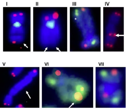
Fig. 7. Telomere dysfunctions in human cells. Metaphase spread with telomeric DNA detected by FISH (red); DNA stained with DAPI (blue). Examples of the chromosomal aberrations: (I) Loss of single telomere. (II) Loss of both telomeres. (III) Telomeres split/duplication. (IV, V) Dicentrics chromosomes with (IV) or without (V) TTAGGGq sequences at fusions junction. (VI, VII) CoFISH staining with C-rich probe (red) and G-rich (green) probe with (VI) or without telomere-SCE (T-SCE) (VII). From [IV].
For instance cells deficient in Ku86 and DNA-PKcs exhibited premature senescence, high proportions of chromosomal aberrations [209, 210], and an increase of telomere end-to-end fusions with telomeric sequences at the fusion point [112, 211-213]. In parallel deletion of Ku70 leads to and an increase of Telomere Sister chromatid Exchange (TSCE) (hallmark of HR events at telomeres) indicating that Ku70 prevents inappropriate recombination at telomeres [214]. Similarly, DNA-PKcs deficiency resulted in telomere uncapping in MEFs. Other components of NHEJ have been
![Fig. 6 - Schematic representation of track structure of low and high-LET radiations. From [III]](https://thumb-eu.123doks.com/thumbv2/123dokorg/2845860.5604/31.630.127.481.205.624/fig-schematic-representation-track-structure-high-let-radiations.webp)