University of Sassari
PhD Course in Life Sciences and Biotechnologies
Coordinator: Prof. Leonardo Antonio Sechi
Parkinson’s disease: Immune System, Infections and
Alpha-synuclein protein
Supervisor PhD student
Prof. Leonardo Antonio Sechi Dott.ssa Elisa Caggiu
Foreword
This thesis is based on several manuscripts that were published during my PhD.
The work of this PhD thesis has been completed during my enrolment as PhD student at the Department of Biomedical Sciences, Section of Microbiology and Virology, University of Sassari, Italy, in the period from November 2015 to October 2018 under the supervision of Professor Leonardo Sechi. The studies of the thesis were also conducted for a period of five months at the University of Edinburgh, from September 2017 to January 2018, under the supervision of Professors Jürgen Haas and Richard Lathe. All the subjects enrolled in the study were kindly enrolled thanks to the collaboration of Dr Kai Paulus, Dr Giannina Arru and Professor GianPietro Sechi belonging to the Neurology Clinic of the University Hospital, Department of clinical and experimental medicine, University of Sassari, Italy.
Abbreviations
AD: Alzheimer’s disease AMPs: Antimicrobial peptides Aβ: Amyloid beta
BBB: Blood-Brain Barrier
BDNF: Brain-derived neurotrophic factor CFS: Cerebrospinal Fluid
CNS: Central Nervous System COMT: Catecol-O-methyltransferase DBS: Deep Brain Stimulation
ELISA: Enzyme-Linked Immunosorbent Assay FCS: Fetal bovine serum
GFP: Green Fluorescent Protein GSH: Glutathione
HCs: Healthy Controls
HSV1: Herpes simplex virus type 1 IgG: Immunoglobulin G
IL: Interleukin
Ldopa: Levodopa
MAO-B: monoaminoxidase B
MHC I-II: Major histocompability complex I-II MPTP: 1-metil 4-fenil 1,2,3,6-tetraidro-piridina MSA: Atrophy multisystemic
OND: Other neurologic disease PBS-T: PBS-Tween 20
PD: Parkinson’s disease
PET: Positron Emission Tomography
PMBCs: Peripheral Blood Mononuclear Cells ROC: Receiver operator characteristic
SD: Standard deviation
SNpc: Substantia Nigra Pars Compacta SNPs: Single nucleotide polymorphism SSRIs: Reuptake inhibitor Serotoninergic TNF: Tumor necrosis factor
UPS: Ubiquitin-proteasome system α-syn: Alpha-synuclein
Abstract
Parkinson's disease (PD) is a neurodegenerative disorder and its etiology is unknown, but environmental factors are implicated in the development of this disease. In this project we wanted to analyze different roles played by α-syn, HSV1 and Immune System in PD. We have investigated autoimmunity in PD by ELISA test and a specific immune-stimulation using homologous peptides of HSV-1 and α-syn in PD patients versus HCs. Moreover, we have investigated the potential role of α-syn as an antimicrobial peptide and how this may contribute to α-syn aggregation, neuroinflammation, and widespread dopaminergic neuron death. Lastly, we have analyzed selected circulating miRNAs as noninvasive diagnostic candidate biomarkers of PD patients and neuroinflammation. The results obtained are in line with the hypothesis of a possible involvement of the immune system, in particular autoimmunity, in the pathogenesis of PD, and that HSV1 infections may lead to a progression of the disease. Concerning the role of α-syn as a potential antimicrobial peptides further studies are needed in order to clarify the complexity of the functions of this protein. Regarding identification of specific miRNA in PD, we have highlighted different levels of expression of some miRNA, 155 and 146a, between PD patients and HCs.
Riassunto
La malattia di Parkinson (MP) è una patologia neurodegenerativa e la sua esatta eziologia è ad oggi ancora sconosciuta, ma è noto che essendo una patologia multifattoriale i fattori ambientali possano avere un ruolo importante nella patogenesi di questa malattia. Visti questi presupposti lo scopo di questo progetto è stato quello di analizzare il ruolo svolto da diversi fattori come: la proteina α-syn, HSV1 ed il sistema immunitario nella patogenesi della MP. Abbiamo studiato il ruolo della risposta umorale, analizzando l’omologia molecolare tra HSV1 e α-syn umana, col presupposto che questa interazione possa condurre ad una più rapida progressione della MP, ed inoltre abbiamo analizzato il ruolo dell’immunità cellulo-mediata attraverso una specifica immuno-stimolazione, in vitro, con peptidi del HSV1 e gli omologhi umani dell’ α-syn attraverso la metodica ICC, analisi delle citochine intracellulari, sui pazienti con MP e controlli sani. Inoltre, attraverso degli studi in vitro, abbiamo analizzato il potenziale ruolo antimicrobico dell’α-syn, che potrebbe contribuire sia all’aggregazione di se stessa che portare a fenomeni di neuroinfiammazione e alla morte diffusa dei neuroni dopaminergici. Infine abbiamo analizzato il potenziale ruolo dei miRNA circolanti come dei possibili biomarcatori non invasivi sia di diagnosi che di neuroinfiammazione nei pazienti con MP. I risultati ottenuti indicano il mimetismo molecolare come meccanismo molecolare di autoimmunità, in particolare correlato alla cross-reattività tra peptidi HSV1 e suoi omologhi dell α-syn, nelle membrane dei neuroni dopaminergici della SNpc. Inoltre, i risultati hanno mostrato, per la prima volta, una risposta cellulare specifica per le popolazioni di CD8, CD4 e NK secernenti TNF-α dopo stimolazione nei pazienti con MP. Quindi i nostri dati sono in linea con l'ipotesi di un possibile coinvolgimento del sistema immunitario, in particolare dell'autoimmunità, nella patogenesi della MP, e che le infezioni da HSV1 possano portare a una progressione della malattia. Per quanto riguarda α-syn come un potenziale peptide antimicrobico sono necessari ulteriori studi per chiarire la complessità delle funzioni di questa proteina. Rispetto all'identificazione del miRNA come potenziali biomarcatori dell'infiammazione nella PD abbiamo evidenziato diversi livelli di espressione di alcuni miRNA, 155 e 146a, tra pazienti con MP e controlli sani. Il miRNA 155 potrebbe non solo essere un obiettivo interessante per la terapia anti-infiammatoria nella MP, ma anche la sua valutazione putrebbe aiutare la diagnosi sugli stadi della malattia.
Index Foreword………...………...………i Abbreviations……….……….ii Abstract.………...……….….iv Riassunto.………...……….v Chapter 1 : Introduction……….……..……….……..1
1.1 Parkinson’s disease: General hallmarks……..………..…1
1.2 Alpha-synuclein……..………..9
1.2.1 Structure and function of alpha-synuclein………..………...9
1.2.2 Degradation of alpha-synuclein and pathologic correlation………..13
1.2.3 Interaction between alpha-synuclein and biological membranes..………15
1.2.4 Interactions between alpha-synuclein and cytoskeleton proteins………..………....18
1.2.5 Alpha-synuclein and synaptic transmission………...……….…19
1.2.6 Secretion and propagation of alpha-synuclein……...………...……….20
1.2.7 Alpha-synuclein as an antimicrobial peptide………...………..…25
1.3 Inflammation in Parkinson's disease…...………...……….26
1.3.1 Activation of Microglia…………...………..….28
1.3.2 The role of T cells………..………29
1.3.3 Humoral immunity………...………..30
1.3.4 Proinflammatory cytokines………...………...30
1.4 miRNA, neuroinflammation and Parkinson’s disease…………..……...………...31
1.5 Herpes simplex virus type 1: outline……...………33
Chapter 2: Aims of the project…...……….…..35
Chapter 3: Materials and Methods…….……..………40
3.1 Analysis of the humoral response………..……….………....40
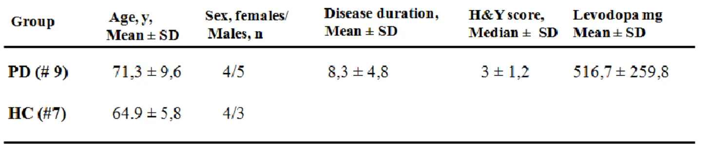
3.1.1 Subjects for immunoenzymatic assay………...………...….………...…40
3.1.3 Enzyme-Linked Immunosorbent Assay (ELISA)…...……..………...41
3.1.4 Competitive assay.……...………42
3.1.5 Statistical analysis……...………...42
3.2 Analysis of the cell-mediated response………...………....42
3.2.1 Samples for flow cytometry analysis…...………42
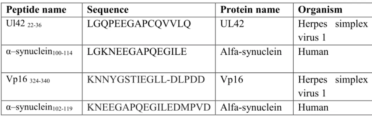
3.2.2 Antigens………..……….43
3.2.3 Cytokines and phenotyping antibodies……..………..43
3.2.4 Cell preparation and antigenic stimulation………..………44
3.2.5 Intracellular cytokine staining and phenotyping analysis…...……….44
3.2.6 Statistical analysis………..……….45
3.3 Alpha-synuclein as a antimicrobial peptide………....…..………..45
3.3.1 Effect of alpha-synuclein on HSV-1 replication (first protocol)…………..…………...45
3.3.2 Effect of alpha-synuclein on HSV-1 replication (second protocol)………..….…..46
3.3.3 Effect of alpha-synuclein on HSV-1 replication (third protocol)……..………..47
3.3.4 Transient over-expression of α-synuclein……...……….48
3.3.5 Cell viability assay………...………49
3.3.6 Statistical analysis….………..……….49
3.4 Expression of circulating miRNA analysis…...…….………...………..49
3.4.1 Samples for miRNA analysis………...………49
3.4.2 miRNAs cDNA synthesis and real-time PCR………...……...50
3.4.3 Heat Maps………..………..50
3.4.4 Statistical analysis.………...………51
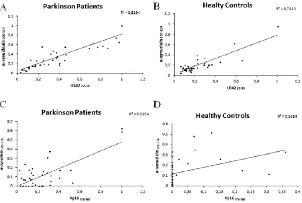
Chapter 4: Analysis of the humoral response against alpha-synuclein peptides homologous to Herpes simplex virus 1 proteins in PD patients VS healthy controls………...………..52
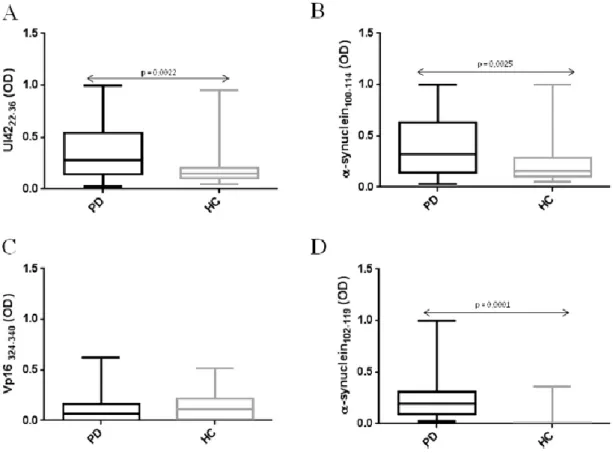
4.1 Results………...………..53
4.1.1 ELISA………..……….……….…..53
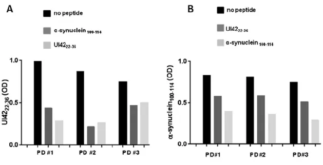
4.1.2 Competitive assay.……….……….……….………56
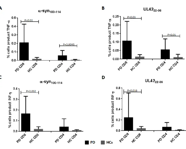
Chapter 5: Study of cell-mediated response, following stimulation with homologous alpha-synuclein and Herpes simplex virus 1 peptides, in PD patients VS healthy controls
through intracellular cytokine method………...……….……….60
5.1 Results.…...……….………61
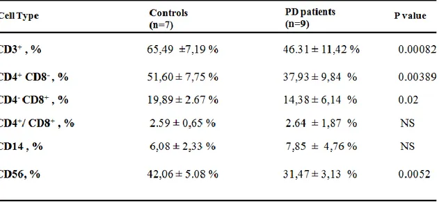
5.1.1 Population of peripheral T lymphocytes…………...………..……….61
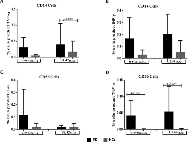
5.1.2 Activation of CD8 and CD4………...………..62
5.1.3 Stimulation of CD14 and CD56………..……….64
5.2 Discussion………..………...………..66
Chapter 6: Alpha-synuclein as a potential Antimicrobial peptide……….……70
6.1 Results………...………..………71
6.1.1 α-syn inhibition of HSV-1 infection in Hela cells ……….……….72
6.1.2 HSV-1 infection on transient over-expression of α-syn………...…..………….75
6.2 Discussion………..……….78
Chapter 7: Analysis of the expression of different miRNAs in PD patients compared to healthy controls………….……….…….81
7.1 Results………...………..………82
7.1.1 miRNA expression in patients with PD and their matched controls………82
7.1.2 miRNA expression in PD patients with different Levodopa dosages….….………83
7.2 Discussion………..………...…………..84
List of References………...……….88
List of papers done during the PhD (01-11-2015 / 31-10-2018)…………...…………107
Dr Elisa Caggiu – Parkinson’s disease: Immune System, Infections and Alpha-synuclein protein – International PhD School in Life Science and Biotechnologies – University of
Sassary, Italy
1
Chapter 1: Introduction
1.1 Parkinson's disease: General hallmarks
Parkinson's disease (PD) is a complex and progressive neurodegenerative disease whose crucial physiopathogenic moment is represented by the loss of dopaminergic neurons localized in the Substantia Nigra Pars Compacta (SNpc), in particular in its ventrolateral portion. This leads to a deficit in the dopaminergic transmission involved in complex subcortical neuronal circuits exercise the Gangli of the base, which is a group of grey substance nuclei located at the base of both the cerebral hemispheres and densely interconnected with the cerebral cortex, the thalamus and the brain stem. The basal ganglia become part of the extra-pyramidal movement control and the correct functioning of these and their connections is of crucial importance to ensure the normal execution of the motor act. In fact one of the alteration is responsible for extra-pyramidal diseases that can extrinsecate influencing speed, fluidity and quality of movements in the hypercinetic sense, such as in Huntington's disease, both in the hypokinetic sense, such as in PD. In fact, the neuronal deficit found in SNpc, leads to a reduction of neurons that send dopaminergic projections to putamen. This, in turn, determines a hyperactivity of inhibiting signals that from the Gangli Base lead to the thalamus and, in consequence, a reduction of facilitator impulses from thalamus lead pre-motor cortex (areas 6-8 Brodmann); the result is an inhibition of movement. It should be recalled that in PD, in addition to the Nigro-striatal circuit just described, other dopaminergic pathways are also involved: the mesolymbic pathway, the mesocortical pathway and the Via Tubero-Infundibolare.
Dr Elisa Caggiu – Parkinson’s disease: Immune System, Infections and Alpha-synuclein protein – International PhD School in Life Science and Biotechnologies – University of
Sassary, Italy
2
Dopaminergic depletion in SNpc neurons and the consequent alteration of the subcortical circuits that preside the execution of the movement, are responsible for the onset of the pivotal motor symptoms of PD:1. muscular rigidity of the plastic type, initially unilateral, frequently associated with trochlea phenomenon;
2. resting tremor at medium-low frequency (4-6 Hz) which reduces with movement and sleep and is accentuated during emotional states. Initially unilateral, with onset in the distal part of the upper limb, it occurs characteristically in the act of "making pills" or "counting coins";
3. bradykinesia with alteration of the velocity, amplitude and rhythm of the movement which is considerably slowed down, frequently accompanied by akinesia and therefore difficulties in initiation the motor act. Manifestations related to akinesia are: facial hypomimia, fixity of the gaze with reduction of blinking, the monotony of speech, the sialorrhea caused by the reduced frequency of swallowing, the loss of the pendular movement of the upper limbs during walking, poverty of gestural language, walking with small steps and bending attitude of the bust, micrography and so on;
4. Postural instability with late appearance and generally crippling. It is the result of akinesia, stiffness, loss of postural reflexes and straightening [1]. These are generally associated with a variety of non-motor symptoms that can sometimes also precede the above motor manifestations [2]. In fact, they would seem to be also involved in the pathology other dopaminergic pathways placed at the plexus myoenteric, intestinal level and the olfactory system.
Dr Elisa Caggiu – Parkinson’s disease: Immune System, Infections and Alpha-synuclein protein – International PhD School in Life Science and Biotechnologies – University of
Sassary, Italy
3
Givenits heterogeneity regarding the modalities of onset, the clinical
Figure 1. Shows the alteration, consequent of dopaminerg neurons dead, tipical of Parkinson’s
Dr Elisa Caggiu – Parkinson’s disease: Immune System, Infections and Alpha-synuclein protein – International PhD School in Life Science and Biotechnologies – University of
Sassary, Italy
4
appearance, the symptoms and the speed of progression, we can distinguish mainly two forms: i) A form tremorigenic essentially characterized by tremor at rest, poor bradykinesia, generally deambulation well controlled, speed of progression of slow and disabled functional mild disease; ii) a form rigid-akinetic, in which the tremor does not represent the characterizing symptom and that is frequently associated with pictures more complex phenotypics such as rigidity-akinesia, early postural instability and disorders debilitating cognitive [3].As far as non-motor symptoms are concerned, let us remember: hyposmia or olfactory dysfunctions, cognitive decay (from a dysexecutive syndrome to forms of real dementia), psychiatric symptoms such as depression (for alteration of the serotoninergic pathways, dopaminergic and noradrenergic), REM sleep disorders, constipation and dysfunction autonomic, pain, indefatigability, apathy (for degeneration of the corticosubcortical circuits that mediate the reward system). As already mentioned, some of these symptoms may precedes motor manifestations, thus representing podromic symptoms or of alarm for the development of PD and could become elements of consideration for an early diagnosis of the disease. Also, since symptoms as hyposmia or REM sleep disorders may precedes even 13-15 years the appearance of the motor symptoms, it would be identified a useful therapeutic window for prevent further damage and further loss of dopaminergic neurons, which causes of the onset of motor symptoms [4]. It is noted, in fact, that motor dysfunctions appear with the loss of at least 60% of the dopaminergic neurons of the SNpc [5]. The disease is progressive and to date, unstoppable. The only available therapeutic options are based on a symptomatic treatment of the disease which, so, can it be adequately monitored in its initial clinical manifestations, but not completely resolved. The progression of the disease implies the appearance of drug-resistant symptoms and complications determined by the long-term
Dr Elisa Caggiu – Parkinson’s disease: Immune System, Infections and Alpha-synuclein protein – International PhD School in Life Science and Biotechnologies – University of
Sassary, Italy
5
symptomatic therapy: fluctuations motor and non-motor, dyskinesias and psychosis (visual hallucinations and less commonly tactile, auditory or olfactory) [6]. In the last stages of the disease the clinical picture gets complicated with the appearance of freezing, instability postural and frequent falls, cognitive alterations, dysphagia and dysarthria. Moreover, autonomic dysfunctions such as: urinary incontinence, constipation and orthostatic hypotension are frequent [6;7]. PD has a very important social and economic-health impact, both for its great prevalence and for the functional disable to which it inevitably leads. This disease represents the second neurodegenerative disease by frequency, second only to Alzheimer's disease (AD) [8], with a prevalence of 1-3% in the population over 50 [9] and a male ratio: females of 3:2 [10]. Although the first detailed description of PD was made more than two centuries ago (James Parkinson, "An essay on the Shaking Palsy”, 1817), the pathogenesis of this disease has not been yet completely clarified [11]. The first piece of its understanding is been placed in 1912, when Lewy described of intraneuronal cytoplasmic inclusions which, subsequently, were denominated in his honor "Lewy Bodies" by Tretiakoff [12]. Subsequently the alpha-synuclein (α-syn) was discovered, described as a precursor to the non-amyloid component of the highlighted senile plaques in AD [13]. The possible pathogenetic role of this protein in PD was hypothesized in 1997, following the discovery of the SNCA gene that it is mutated in some very rare forms of familial PD with autosomal dominant transmission. Later, genetic studies have shown that polymorphisms and SNCA mutations are also related to sporadic Parkinson's forms [14], which represent the vast majority of cases. This gene, located in the chromosome 4q21-Q23, coding for the α-syn which would therefore play a fundamental role of in the pathogenesis of PD [15]. To confirm this, it was demonstrated how α-syn is the main component of Lewy bodies and Lewy neuritis, both characteristic neuropathologic elements of PD, but not only that as a result, thisDr Elisa Caggiu – Parkinson’s disease: Immune System, Infections and Alpha-synuclein protein – International PhD School in Life Science and Biotechnologies – University of
Sassary, Italy
6
disease must be considered as a full-fledged synucleinopathy, meaning by this term a heterogeneous group of neurodegenerative diseases that share common neuropathological lesions: intracellular proteinaceus inclusions consisting primarily of α-syn [16]. Although genetic, biochemical, and neuropathological studies suggest a clear pathogenic role of α-syn, how this or its derivatives actually trigger the neurodegenerative process is not still totally defined [17]. Furthermore, although several risk factors have been identified, the primum movens of the disease has not yet been identified. In this regard, efforts and studies are consistent to better understand the complex pathogenetic framework of PD.The main risk factor to be considered is, of course, the age: the prevalence of PD increases with aging and has a spike after 80 years [18]. Among the environmental risk factors includes nutritional factors [19,20], exposure to some metals [21] and pesticides such as Paraquat [22] and Diquat [23]. As stated above, PD definitely has a certain genetic component. A story family of PD or tremor is therefore an additional risk factor for the development of the disease [24]. In addition to the SNCA gene coding for α-syn, numerous other genes related to PD have been identified: LRRK2, Parkin, PINK1 and others [25].Among the environmental factors it is also suggested the role of several infectious agents such as viruses, including Herpes Simplex virus, influenza viruses, HIV and several hepatotrophic viruses. In fact, there are many infectious agents able to overcome the blood-brain barrier (BBB) and induces inflammatory processes of cerebral parenchyma such as encephalitis. Furthermore, it should be emphasized that in the symptomatology of patients with encephalitis, we can to detect also characteristic clinical elements of Parkinson's [26], as proof of a possible correlation between the two pathologies.
The diagnosis is purely clinical and is based on the search for the pivotal symptoms of disease: tremor, bradykinesia and rigidity, while the postural instability is generally a late
Dr Elisa Caggiu – Parkinson’s disease: Immune System, Infections and Alpha-synuclein protein – International PhD School in Life Science and Biotechnologies – University of
Sassary, Italy
7
symptom. In this regard, the criteria set out by the UK Parkinson's Disease Society Brain Bank are used [27]. The gold standard is, in fact, represented by the anatomopathological identification of the typical alterations of the disease, which the diagnosis is certainly post-mortem. At the present time there are no known biomarkers that to arrive at an early diagnosis, even if numerous studies are underway. A considerable help is given by imaging diagnostics. Techniques such as the DaTSCAN ™ (Ioflupane I 123 injection) allow you to make differential diagnoses with movement disorders not distinguished by the loss of dopaminergic neurons, such as the essential tremor. However these techniques are able to put into evidence of neuronal loss in the SNpc only when this has already significant [28], thus not being useful for an early diagnosis.The main differential diagnosis of PD is represented by Parkinsonian syndromes of other nature such as: multisystemic atrophy, supranuclear palsy progressive, corticobasal degeneration, disease from diffuse Lewy bodies. In establishing diagnosis helps the combination of Parkinsonian signs, rapidly evolving and poorly suited to replacement therapy, and other neurological signs specific to the disease. Other alternative diagnoses are: parkinsonism post-encephalitis lethargicor other forms of viral encephalitis; vascular Parkinsonism (generally characterized by focal neurological signs caused by ischemia, worsening steps for the succession of ischemic events and a neuroradiological picture of encephalopathy multinophartual, together with a poor dopamine response); rare forms of toxic parkinsonism from manganese, mercury, carbon monoxide or drugs (phenothiazines, CA-antagonists, butiferrones). In the forms of tremorigenic Parkinson's, the main diagnoses differentials are represented by: the essential tremor, a condition almost always familial trait with autosomal dominant transmission; the tremor associated with hyperthyroidism, high frequency and associated with other signs of thyroid dysfunction; the tremor associated with
Dr Elisa Caggiu – Parkinson’s disease: Immune System, Infections and Alpha-synuclein protein – International PhD School in Life Science and Biotechnologies – University of
Sassary, Italy
8
multiple sclerosis or chronic alcoholism. As already mentioned, the therapy of PD is exclusively symptomatic and focuses on the enhancement of dopaminergic transmission at the subcortical circuits level through drugs such as: levodopa, a natural precursor of dopamine, in the form of able to cross the BBB. Generally it is associated with drugs DOPA-decarboxylase peripheral inhibitors (Carbidopa or benserazide) in such a way as to reduce the daily intake dose. Other commonly used drugs are the MAO-B (monoaminoxidase B) and COMT (Catecol-O-methyltransferase) inhibitors that allow to reduce the dopamine metabolism at the Central Nervous System (CNS) level and increase its availability. Let us also remember the dopamino-agonists, ergolinic (Bromocriptine, Pergolide, Cabergoline) or non-ergolinic (Ropinorol and Pramipexole), which act through activation of dopaminergic postsynaptic receptors. In this class of drugs should be mentioned Apomorphine which has a pharmacological profile comparable to dopamine, and it is used in cases of PD characterized by severe motor fluctuations.All these drugs just mentioned, act by improving the symptomatology of the patient and improving the quality of life sometimes with really amazing results. The tremor, unlike the other symptoms, responds inconsistently to replacement therapy and therefore its requires the association of other drugs such as anticholinergic, although these have a limited efficacy and numerous central and peripheral side effects that allow use only in patients more young people with PD predominately tremorigenic.
The medical challenge is the management of adverse effects (nausea, daily drowsiness and impulse control disorders such as chronic gambling, hypersexuality, hyperfagia, compulsive buying syndrome) and complications of long-term therapy associated, in particular, to the use of levodopa (Ldopa). In fact, although Ldopa, represent the most drug effective for the symptomatological management of the pathology, prolonged use is accompanied by a gradual
Dr Elisa Caggiu – Parkinson’s disease: Immune System, Infections and Alpha-synuclein protein – International PhD School in Life Science and Biotechnologies – University of
Sassary, Italy
9
reduction of its effectiveness and to the onset of motor complications (dyinesias and motor fluctuations). These can be, at least in part, controlled with a better synergy between different classes of drugs, in order to optimize the concentration of dopamine at central level. The symptoms non-engines instead turn out to be more difficult to control. Talking about effects collateral it is must also mention a study that exposed a case of hyperpyrexia malignant with delirium consequent the sudden suspension of therapy with Ldopa. In this case, a rapid reduction in the concentration of dopamine, may have had an important role in the determination of the syndrome [29].Other manifestations of the disease may benefit from targeted therapy: symptoms of depression can be controlled by the use of SSRIs (reuptake inhibitors Serotoninergic), the sialorrhea with the use of anticholinergic and the injection of botulinum toxin, sleep disorders improving their hygiene and controlling the nocturia, and so on. We also remember how the motor symptomatology of PD can also benefit from surgical treatments such as Deep Brain Stimulation (DBS), which is the stimulation of nuclei involved in subcortical circuits altered by the pathology, such as Sub-thalamic nucleus of Luys or on the Globus pallidus Internal [30]. In particular, DBS is an option for patients responding to levodopa, but with a quality of life deteriorated by complications of long-term therapy.
1.2 Alpha-synuclein
1.2.1 Structure and function of alpha-synuclein
α-syn is a small neuronal protein that is closely associated with the etiology of PD. The α-syn is a protein of 140 amino acid residues, being part of a family of small proteins that also includes the beta-synuclein and the gamma-synuclein. Originally described as a non-amyloid
Dr Elisa Caggiu – Parkinson’s disease: Immune System, Infections and Alpha-synuclein protein – International PhD School in Life Science and Biotechnologies – University of
Sassary, Italy
10
component of the senile plaques in AD, α-syn certainly plays a pathogenic role in the PD. It is the major component of intraneuronal inclusions called Lewy bodies and Lewy’s neurites, observable in SNpc but also in other districts such as spinal cord and peripheral nervous system (vague nerve, sympathetic ganglia, enteric plexus nervous system, salivary glands, adrenal medulla, cutaneous nerves and sciatic nerve) [31,32,33,34]. The same neuropathologic elements are also found in a group of diseases therefore called synucleinopathies. These diseases are represented by: dementia with Lewy bodies and MSA (atrophy multisystemic) and other lesser known forms [35]. The physiological role of α-syn does not completely known: it would act modulating the release of synaptic vesicles and would therefore play a role in the regulation of synaptic transmission [36]. In physiological conditions, α-syn, actually, could be found in numerous presynaptic terminals, in proximity of vesicles containing neurotransmitters. How this modulation happen is another element still to be clarified; however it is thought that its action may be carried out by stabilizing the proteins of the complex SNARE, whose assembly and disassembly is essential for the release of neurotransmitters [37]. It has been shown how that protein is naturally “unfolded", i.e in its pure form at neutral PH it is completely deployed, without structure secondary or tertiary. However, as a result of binding with acid phospholipids of membranes of synaptic vesicles, it assumes a structure folded to alpha-helical [38]. In its structure can be identified three domains: one N-terminal, one part called NAC (non-amyloid component of the senile plaques) and a domain C-terminal. Each of these gives of the protein a particular characteristic. The N-terminal domain would determine the tendency of the protein to take on a conformation to alpha-helical when linked to vesicular membranes; the central part hydrophobic NAC, it would be responsible for the conformation Beta-leaflet and the protein'sDr Elisa Caggiu – Parkinson’s disease: Immune System, Infections and Alpha-synuclein protein – International PhD School in Life Science and Biotechnologies – University of
Sassary, Italy
11
tendency to form fibrillary aggregates. The C-terminal portion, full of negative charges, which would depend on the characteristic of protein "unfolded" [39].Considering the different studies, we can therefore affirm that this protein, physiologically, exist in various unstructured and oligomeric states in equilibrium between them thanks to factors that promote and inhibit fibrillar aggregation [40], i.e. aggregation in structures in Beta-leaflet.
Figure 2: Structure of alpha-synuclein.
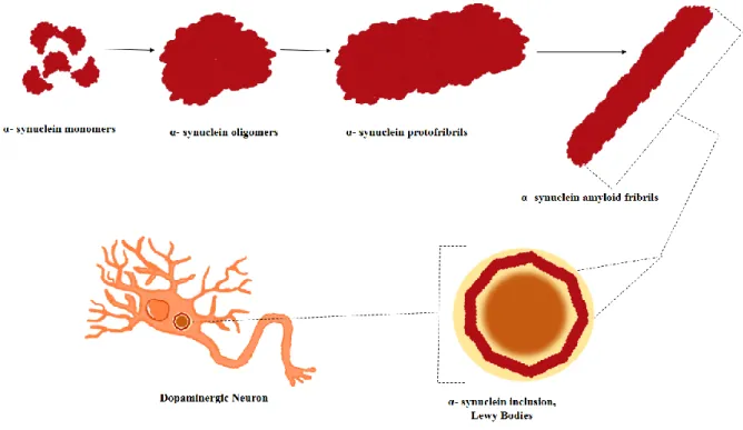
The toxic subtype of α-syn is not still identified with certainty. Some studies indicate how toxic the insoluble amyloid-like fibrils identifiable in Lewy bodies. Other studies indicated as toxic the soluble prefibrillary intermediates such as oligomers. Recently, a growing number of studies have shown how species oligomeric, rich in structures to Beta-Leaflet, are neurotoxic [41,40,42]. Then these oligomers can aggregate to form protofibrils of higher molecular weight, insoluble, whose further polymerization brings to the formation of amyloid fibrils, resembling those found in the Lewy bodies.
Dr Elisa Caggiu – Parkinson’s disease: Immune System, Infections and Alpha-synuclein protein – International PhD School in Life Science and Biotechnologies – University of
Sassary, Italy
12
Figure 3: The image shows the different unstructured forms assumed by α-syn. In
physiological conditions it is found in an inherently unfolded native form and a alpha-Helix form when it is linked to membrane phospholipids. Studies show how monomeric forms of α-syn can be aggregated into oligomeric species rich in structures in beta-leaflet and later in higher molecular weight protofibrils, insoluble, which can then further undergo polymerizing and therefore to the formation of amyloid fibrils similar to those found in Lewy bodies.
The latter could even be the result of a protective mechanism activated by neurons [43]. It would seem that dopamine and its metabolites succeed in inhibit, in vivo, the conversion of protofibrils into fibrils, thus determining accumulation of neurotoxic protofibrillar intermediates. That would explain the high sensitivity of the of dopaminergic neurons to the toxic effects of α-syn, which instead are less significant if we consider the other neuronal systems. [44]
Dr Elisa Caggiu – Parkinson’s disease: Immune System, Infections and Alpha-synuclein protein – International PhD School in Life Science and Biotechnologies – University of
Sassary, Italy
13
1.2.2 Degradation of alpha-synuclein and pathologic correlationsα-syn is encoded by the SNCA gene. Six mutations are currently known that affect this gene and that as a result cause an alteration of α-syn. These mutations are associated with a parkinsonian phenotype characterized by early onset, rapid disease progression and rapid onset of non-motor symptoms such as hallucinations, autonomic dysfunctions, dementia [15]. It should be remembered that is thanks to the study of these very rare forms of familial Parkinson's that the interest int o α-syn research was born. However, studies have shown that, if overexpressed, may cause onset of PD (as well as expression of α-syn wild type). In addition it has been confirmed that the severity of disease is directly related to the degree of overexpression of the protein. To confirm this, patients who have triplication of the SNCA gene have a more severe clinical picture and rapidly progressive than patients that present a duplication of the gene [45-46]. This indicates that the levels of the protein should be maintained into a certain physiological range, thanks to a balance between production, degradation and secretion.
In the pathogenesis of PD, alterations of the normal mechanisms to maintain proper proteostasis could be implicated, in particular: the ubiquitin-proteasome system (UPS) and the lysosome mediated autophagy system (including microautophagy, macroautophagy and autophagy chaperone-mediated according to the modalities with which the protein that it to be degraded is transported within the lysosome) [47]. Several studies have shown how the alteration of these physiological pathways can actually lead to an accumulation of α-syn, through a reduction in its clearance. For example: it is demonstrated how, total protein concentration, increases after the inhibition of lysosomal-mediated autophagy [48], while the results about the actual recognition and destruction of α-syn by the proteasome are discordant. In particular, it would seem that the wild a α-syn is selectively degraded from a
chaperone-Dr Elisa Caggiu – Parkinson’s disease: Immune System, Infections and Alpha-synuclein protein – International PhD School in Life Science and Biotechnologies – University of
Sassary, Italy
14
mediated process, whereas all forms of α-syn can be degraded by macroautophagy processes [49, 50].The inhibition of chaperone-mediated protein degradation leads to an accumulation and to the aggregation of high molecular weight intermediates and insoluble α-syn species in neurons [49], and therefore, physiologically, it would have a fundamental role in prevention of accumulation and aggregation of α-syn. So, it can be said that an alteration of the chaperone-mediated autophagy plays a pathogenetic role in the PD. Moreover, it would appear that α-syn itself determines the alteration of the lysosomal function, thus going to create a vicious circle: accumulation of α-syn→ lysosomal damage → further accumulation of protein. In this regard some studies show how the main proteins involved in chaperone-mediated autophagy (Hsc70 and Lamp2a) are reduced in the brain tissue of patients with PD [51].
The alteration of the chaperone-mediated function could occur through a compensatory activation of macroautophagy processes. In fact, several animal models of synucleinopathies have shown indicative elements of an excessive activation of macroautophagy processes (e.g. certain specific markers) or indicative, however, of an alteration of the fusion of the autophagic vacuoles with lysosomes [52].The consequences of this compensatory activation are not clear: some studies indicate how this mechanism can be protective against α-syn neurotoxicity; other studies, on the contrary, indicate how the activation of macroautophagy is deleterious. These different effects may not exclude each other and also depend on the stage of the disease.
In the early stages, the compensatory activation of macroautophagia could represent a defense mechanism that proves to be counterproductive and harmful in the advanced stages of the disease [17]. Further evidence of the involvement of the lysosomal function in PD, comes from the study of Gaucher disease. This is a storage disease, hereditary, determined by
Dr Elisa Caggiu – Parkinson’s disease: Immune System, Infections and Alpha-synuclein protein – International PhD School in Life Science and Biotechnologies – University of
Sassary, Italy
15
mutations “Loss of function”of the GBA gene coding for the glucocerebrosidase, a lysosomal enzyme. It is characterized by the accumulation of glucosilceramide in the reticulo-endothelial cells of the spleen, bone marrow and liver. The presence of this mutation also leads to an increase in the risk of developing PD or other types of diseases with Lewy bodies [53]. In fact, the mutation determines a whole series of consequences:1. A lesser affinity of the enzyme Glucocerebrosidase for α-syn compared to the enzyme Wild-Type; [54]
2. A reduction in the activity of Glucocerebrosidase and consequently an α-syn accumulation and neurotoxicity; [55]
3. Accumulation of glucosylceramide that would seem to “stabilize” species oligomeric α-syn, and increase its toxic potential. [55]
It was also demonstrated that overexpression of the Glucocerebrosidase enzyme wild-type in animal models of Gaucher disease, is able to invert the process of accumulation of α-syn and its histopathological alterations [56]. So, most likely, the accumulation of α-syn plays a role in the pathogenesis of PD.
1.2.3 Interaction between alpha-synuclein and biological membranes
The mechanisms through which α-syn or its different isoforms exert their toxic potential are different and not completely known. Some studies suggest that normal and neuropathogenic functions of α-syn may be different from the molecular point of view, contributing to neurodegeneration. The role of α-syn oligomers in binding and permeabilization of cell membranes has been also reported [57], the α-syn oligomers have the ability to bind themselves to the lipids of different biological membranes, going to increase its permeability
Dr Elisa Caggiu – Parkinson’s disease: Immune System, Infections and Alpha-synuclein protein – International PhD School in Life Science and Biotechnologies – University of
Sassary, Italy
16
at the mitochondrial level, lysosomal and of synaptic vesicles. Through this link, it could be explained both the physiological effects of proteins, as well as pathological ones. In fact it has been shown as this link can lead to an increase in the influx of calcium ions within the organelles, alterations in ionic equilibrium and finally cellular death through activation of the pathway activated by the Caspase 3 [58].It has also been shown how the α-syn molecules be subject to a series of post-translational modifications that might somehow affect its toxic potential and the ability to bind to membranes. Among these modifications we remember: phosphorylation in sites Ser87 and Ser129 of α-syn isolated from tissue from encephalic patients with PD [59]; processes of nitration and oxidation, which they should reduce the propensity of α-syn to form insoluble fibrils and they would “stabilize” the oligomers, there by increasing the toxicity [60] and many other modifications. So this protein could play a role physiopathological also through the connection with the biological membranes, going to alter the functionality of different intracellular organelles. The interactions between α-syn and mitochondria have been widely studied: in different works, the protein has been found in association with outer membranes of these organelles, closely tied to Cardiolipin [61]; its bondto the inner membrane of the same, turns out to be less certain [62]. Furthermore α-syn is able to determine a down-regulation of complex I, i.e the succinate-coenzyme Q reductase involved in the mitochondrial respiratory chain.
It has also demonstrated as both the mutated protein and the wild-type can determine alterations of mitochondrial morphology [63]. These alterations consist in: swelling, membrane distortion or crystal formation even to the fragmentation of mitochondria. These effects would seem to be mediated by the bond of the protein with the mitochondrial membrane, towards which it presents marked affinity due to its wealth in Cardiolipin [61].
Dr Elisa Caggiu – Parkinson’s disease: Immune System, Infections and Alpha-synuclein protein – International PhD School in Life Science and Biotechnologies – University of
Sassary, Italy
17
What kind of α-syn can to determine these effects, is not yet been clarified. The fragmentation of mitochondria could trigger a cascade of events that culminates with the loss of potential of mitochondrial transmembrane, respiratory chain alteration and neuronal death [61]. The same interactions could also occur with the biological membranes of other organelles like the already mentioned lysosomes, presynaptic vesicles, endoplasmic reticulum or the Golgi apparatus. Also, the effects exercised by the α-syn on the mitochondria, could determine the release of reactive oxygen species, thus triggering a vicious cycle that leads to the further accumulation and aggregation of the protein [64]. As already affirmed the presence of α-syn causes excessive activation of the macroautophagy processes. In parallel, a specific pathway appears to activate the mitophagia who, together to the already mentioned macroautophagy, leads to a depletion of mitochondria. Also the mitophagia, therefore, could represent a mode through which the process of neurodegeneration is determined.However, it is necessary to clarify how the aforementioned subcellular alterations may represent also a consequence of normal ageing, which always represents the main risk factor for the development of PD [20].
The involvement of mitochondria in the pathogenesis of PD has been widely described by several studies using mitochondrial toxins such as MPTP (1methyl-4 Phenyl-1, 2, 3.6 tetrahydropyridine) or 6-Ohda. These molecules are able to induce onset of Parkinson's symptoms without determining the appearance of Lewy bodies. For example the MPTP, found for the first time as a contaminant of a drug in a cluster of young students who have developed PD, is metabolized by the enzyme MAO-B to form the MPP + ion, which is able to inhibit complex I of the mitochondrial respiratory chain by inducing cell apoptosis.
Furthermore, mutations that alter mitochondrial function have been identified and are responsible for some cases of autosomal recessive PD. These mutations involve the genes:
Dr Elisa Caggiu – Parkinson’s disease: Immune System, Infections and Alpha-synuclein protein – International PhD School in Life Science and Biotechnologies – University of
Sassary, Italy
18
1 Parkin coding for an E3 ligase involved in mitophagy processes.2 PINK1 is a mitochondrial kinase that is always involved in the process of mitophagy.
3 Others like LRRK2, DJ-1.
1.2.4 Interactions between alpha-synuclein and cytoskeleton proteins
α-syn could exert its pathological role, as well as on organelles cytoplasmic, also on other cellular constituents. The interactions between this protein and the elements of the cytoskeleton were therefore studied, in order to highlight a possible physiopathogenetic correlation with PD. The studies in this regard are manifold and sometimes contrasting. The extracellular application of α-syn would seem to lead to a reduction in polymerization of tubulin [65]. Vice versa, studies conducted on yeast cultures, have shown how the inhibition of polymerization of tubulin may trigger the aggregation of α-syn [66]. This last statement has been questioned by other studies which, on the contrary, have shown as the oligomerization of tubulin promotes the aggregation of α-syn [67]. It has also been shown that α-syn is able to determine the hyperphosphorylation of Tau free protein, this protein stabilizes the structure of microtubules and regulates their spatial organization [68]. The effects of this phosphorylation remains to be clarified. We remember that Tau protein, like α-syn, is involved in the development of a whole series of neurodegenerative disease characterized by an alteration of its metabolism, such as the AD.
A confirmation of a possible interaction between cytoskeleton and PD comes, once again, from genetic studies. In fact, there are very rare cases of autosomal recessive hereditary Parkinson related to mutations in the gene LRRK2. This gene coding for a kinase whose activity is directly related to the functions of the cytoskeleton. In fact, similarly to α-syn,
Dr Elisa Caggiu – Parkinson’s disease: Immune System, Infections and Alpha-synuclein protein – International PhD School in Life Science and Biotechnologies – University of
Sassary, Italy
19
LRRK can determine cytoskeletal instability through the hyperphosphorylation of tau or directly through the phosphorylation of beta-tubulin.1.2.5 Alpha-synuclein and synaptic transmission
Given the physiological role of α-syn in the release of synaptic vesicles, it follows that its alteration will inevitably reflect on the synaptic transmission. It has been shown that an alteration of the α-syn leads to:
1 Loss of pre-synaptic proteins essential for the release of neurotransmitter-rich vesicles; [69]
2 Reduction of the pool of synaptic vesicles and consequently the release of neurotransmitters in synaptic space; [70]
3 Redistribution of SNARE proteins; [71] 4 Morphological alterations of synaptic vesicles; 5 Reduction of synaptic vesicle recycling.
As Calcium plays a key role in synaptic transmission, probably α-syn acts by altering the homeostasis of this ion. In fact, it would seem that α-syn determines the formation of pores at the cellular membrane level [72], significantly increasing the intracellular flow of calcium ions. This leads to an alteration of the membrane potential of the synaptic terminal and the loss of physiological pace-maker activity in dopaminergic neurons that normally is guaranteed by an optimum calcium concentration and the voltage-dependent calcium channels L [29]. Through the same mechanism, it is thought that α-syn can also bind to synaptic vesicles, forming pores with consequent loss of molecules of neurotransmitter within the cytosol. In particular, in dopaminergic neurons, an excess cytosolic dopamine can be detrimental to the cell with induction of oxidative stress and cellular death [73]. Finally, we
Dr Elisa Caggiu – Parkinson’s disease: Immune System, Infections and Alpha-synuclein protein – International PhD School in Life Science and Biotechnologies – University of
Sassary, Italy
20
remember that the name of α-syn is due to the fact that this protein has been initially highlighted both at the synaptic level but also at the nuclear level. However the levels of protein found at the nuclear level are generally inconsistent [74].1.2.6 Secretion and propagation of alpha-synuclein
Although the studies are countless and the results are sometimes promising, the triggers that lead to the accumulation of α-syn are not yet identified, and it is not known what it is exactly the way that towards which it presents marked affinity due to its wealth in Cardiolipin leads to neurodegeneration.
Figure 4: α-syn and synapses. Notes the accumulation of Ca++ in the cytoplasm of synaptic terminal.
Dr Elisa Caggiu – Parkinson’s disease: Immune System, Infections and Alpha-synuclein protein – International PhD School in Life Science and Biotechnologies – University of
Sassary, Italy
21
The understanding of these mechanisms would lead to the resolution of numerous question marks that characterize the different aspects of the PD, but it would also open the way to the understanding of other related neurodegenerative diseases and especially to new horizons and therapeutic possibilities to reduce the neurotoxicity of α-syn [75]. The new knowledge and information about it are constantly increasing.For example: α-syn has been considered, for a long time, a protein exclusively intracellular. In fact, new evidence underline how it can be found also in different extracellular fluids, such as plasma and Cerebrospinal Fluid (CFS) [76,77]. This new data also opens the way to new diagnostic opportunities. Studies have been conducted on saliva samples of PD patients, to seek a correlation between protein levels and the onset of the disease, in the hope of identifying a cut-off that may lead to an early diagnose. To that regard we quote one study where the total amount of α-syn in the saliva of PD patients was reported considerably lower than that of HCs. Also, the same study, highlighted a greater salivary levels of α-syn oligomers in PD patients versus HCs [78]. This data could be used in the future in order to develop a test for the early diagnosis of PD.
The modality of secretion of this protein has not yet been completely clarified, although we think of a mechanism of non-classical exocytosis through exosomes, of the endocytic vesicles derived from multivesicular bodies and released with a Ca-mediated mechanism.
This process could represent a real language of communication between one cell and the other, but it could also represent a way of propagation of α-syn. This hypothesis has been processed as a result of the following discovery: fetal dopaminergic transplants in human striatum develop Lewy bodies disease years after the transplant, suggesting a possible transmission "Host-Graft" of syn. However it has not been possible to ascertain that the α-syn included in the transplant were derived from the host and were not primarily formed in
Dr Elisa Caggiu – Parkinson’s disease: Immune System, Infections and Alpha-synuclein protein – International PhD School in Life Science and Biotechnologies – University of
Sassary, Italy
22
the tissue transplanted [79]. This ability to propagate α-syn, would depend on the capacity of the cells to capture the protein. Actually, studies, show how α-syn oligomers are particularly prone to being captured by the cells and therefore they would have a role in the dissemination of pathology. [80] The transmission cell-to-cell of α-syn has been demonstrated in vitro [81], but also in vivo in transgenic mice [82] [83].These notions suggest the hypothesis of a possible prion-like transmission of α-syn, which could also explain the advancement of the pathology in PD and explained in the Braak staging. This in fact divide the pathology into different stages based on the location of the neuropathological alterations. Initially there is an exclusive involvement of the ganglia autonomic, the anterior olfactory nucleus and of the dorsal motor nucleus of the vagus. After the disease progresses in an upward way, involving extensive areas of the CNS and in particular the SNpc, determining the appearance of the motor manifestations of PD: tremor, bradykinesia and rigidity. As the disease progresses, the anatomopathological alterations also spread at the cortical level, associated to cognitive decline and psychiatric disorders [84]. According to prion-like theory, α-syn misfolded could have a pathogenetic behavior similar to PRPSC prion protein and, like this, it would propagate from cell to cell inducing formation of further α-syn misfolded, thus contributing to the progression of the disease.
We recall that prion diseases includes different forms of transmissible spongiforms encephalopathies, where the pathogen agent is not a common microorganism, but it is represented by an abnormal protein (PRPSC) which, once formed, is able not only to spread in the individual, but is also endowed with infectious properties.
These diseases, such as Creutzfeldt-Jacob disease and its variants, Gerstmann-Straussler-Scheinker disease, Kuru and fatal familial insomnia, can be transmitted from individual to individual.
Dr Elisa Caggiu – Parkinson’s disease: Immune System, Infections and Alpha-synuclein protein – International PhD School in Life Science and Biotechnologies – University of
Sassary, Italy
23
We talk about prion-like transmission of α-syn, because this protein could have the same transmission capacity cell and the ability to induces the formation of a further protein misfolded, namely the ability to induce alteration of endogenous α-syn normally conformed. However, the infectious capacity of the protein has not been demonstrated, and in fact no reports of cases of inter-human transmission of synucleinopathies are so far reported.According to the Braak’s theory of and his collaborators and called “dual-hit” hypothesis, a pathogen agent with prion-like properties, such as α-syn, would initially be localized at the olfactory epithelium level, probably in response to the inhalation of a neurotropic agent not better specified. Then it could be also localized at the level of the intestinal epithelium following the ingestion of nasal secretions together with saliva. Again according to Braak's theory, this hypothetical agent, it would be propagated from the olfactory epithelium to the temporal lobe and in via retrograde from the intestinal epithelium to the CNS, through the fibers of the vagus nerve [85,86]. Actually Lewy bodies were found both along the structures of the olfactory via (anterior olfactory nucleus, olfactory tubercle and olfactory cortex)[87] which in nerve cells of the enteric plexus of patients with PD [88].
It is unlikely that α-syn may penetrate into the organism directly through the olfactory via. It's more likely to be present in these locations, as a result of contact with substances present in the environment, such as viral agents or pesticides, which actually represent a risk factor for the development of the disease. This theory was very successful, as the filaments of the olfactory nerve are the only nerves that are directly in contact with elements from the environment. To confirm a possible propagation of the α-syn, a study has put in evidence also the correlation between the activity of this protein and mitochondrial activity. Indeed the intragastric administration of Rotenone, an inhibitor of the mitochondrial complex I, leads to the appearance of α-syn included in the intestinal epithelium, which gradually also affect the
Dr Elisa Caggiu – Parkinson’s disease: Immune System, Infections and Alpha-synuclein protein – International PhD School in Life Science and Biotechnologies – University of
Sassary, Italy
24
CNS, including dopaminergic neurons. This study, therefore, indicates how α-syn can propagate independently from the point of origin of its accumulation [89].Although this theory is appealing, there are still numerous unresolved questions and arguments against it. For example: there are no studies that confirm how the dopaminergic neurons, primarily affected by PD, have the ability to capture α-syn from other cells or that the transfer of the protein could induces the formation of Lewy bodies. Furthermore, it should be stressed out that the central role of α-syn in triggering neurodegenerative events is not accepted unanimously. In fact, there are numerous alternative theories that explain the etiopathogenesis of PD. Some studies, for example, have shown a decrease in the concentration of reduced Glutathione (GSH) in the SNpc of PD patients. This could uncover other mechanism involved in neurodegeneration and also possible new therapeutics target. A study has highlighted the therapeutic efficacy of the GSH in patients with PD untreated with a substitute therapy [90]. Moreover, other studies have shown that GSH deficiency is directly proportional to the severity clinical pathology, further emphasizing the potential role of this substance in the pathogenesis of PD [91]. Other studies suggest that alteration of the α-syn and its aggregation, could be an epiphenomenon caused by other processes such as, for example, neuroinflammation, which actually is detected in postmortem histologic preparations of patients with PD [92]. It's also true though that the same extracellular α-syn, once secreted, is able to induces neuroinflammation by activating glial cells. To support this theory, it has been shown how the glial cells are able to capture and degrade the α-syn aggregates in a more effective way than neurons [93]. Activation of microglial cells would cause the release of protective molecules as brain-derived neurotrophic factor (BDNF) but also pro-inflammatory cytokines, reactive oxygen and nitrogen species [94], there by definitely playing a role in the progression of neurodegenerative disease. Also, it has been
Dr Elisa Caggiu – Parkinson’s disease: Immune System, Infections and Alpha-synuclein protein – International PhD School in Life Science and Biotechnologies – University of
Sassary, Italy
25
shown as the α-syn nitrated is not recognized as a self and, therefore, causes the formation of harmful species of helper T lymphocytes that could be another cause of neuronal damage [95]. This indicates the importance of the Immune System in the pathogenesis of PD. The maintenance of a correct extracellular homeostasis of α-syn, would be another key piece to keep a correct brain function and could represent a possible therapeutic target in the near future.1.2.7 Alpha-synuclein as an antimicrobial peptide
Currently, the exact physiological function of α-syn is still not fully known, and also the exact mechanisms that lead to toxicity with subsequent neuronal death are still unclear, but the deposition of α-syn fibrils in Lewy Bodies in dopaminergic neurons is one of the main features of PD, Lewy Bodies are composed largely of beta-sheet rich α-syn amyloid fibrils [96]. Oligomerization in particular is viewed as a pathogenic pathway and α-syn oligomers are assumed to be intrinsically abnormal. Antimicrobial peptides (AMPs), an evolutionarily very old family of proteins, have the characteristic of generating oligomers and fibrils, as well as α-syn. These properties play a key role in mediating the processes of defense of innate immunity, AMPs are the first-line of defense against pathogens and act as potent broad-spectrum antibiotics and immunomodulators that target bacteria, mycobacteria, enveloped viruses, fungal, and protozoans, and in some cases, transformed or cancerous host cells [97]. AMPs are expressed in many tissues, but it has been reported that there is a notable expression in the brain [98], as well as in other tissues where the intervention of the adaptive immune system is limited. Normally the action of this class of peptides turns out to be protective, but their dysregulation can lead to toxic effects in the host cells [99,100], such as chronic inflammation [101,102,100,103] and degenerative diseases [104]. Recent studies
Dr Elisa Caggiu – Parkinson’s disease: Immune System, Infections and Alpha-synuclein protein – International PhD School in Life Science and Biotechnologies – University of
Sassary, Italy
26
reveal a possible antimicrobial action of amyloid-beta (Aβ), suggesting that Aβ deposits may be a consequence of the protective action of this peptide against infections, for example Bourgade et al showed Aβ as an antiviral activity in vitro study of HSV-1 infection of human fibroblasts, epithelial and neuronal cells [105,106,107,108, 109].Regarding the α-syn Park et al demonstrate that α-syn exhibits antibacterial activity against Escherichia coli and Staphylococcus aureus. In addition, the authors demonstrate a role for α-syn in inhibiting various pathogenic fungal strains such as Aspergillus flavus, Aspergillus fumigatus and Rhizoctonia solani. They also analyzed localizations of recombinant α-syn protein in E. coli and Candida albicans. These results suggest that in addition to α-syn's role in neurotransmitter release, it appears to be a natural AMP [110]. Beatman et al reported that the neuronal expression of α-syn is able to inhibit viral infection by West Nile Virus (WNV), in fact the authors observed an increase of α-syn expression in neurons infected with WNV. At the same time they observed that knockout mice for α-syn develop a higher viral titre respect to wild-type animals. this data suggest a possible action of α-syn as AMPs [111].
1.3 Inflammation in Parkinson's disease
Inflammation has been increasingly studied as part of the pathophysiology of neurodegenerative diseases, corroborating the hypothesis that the immune system may be the nexus between environmental and genetic factors, and the abnormal immune function can lead to disease. Since 1988, McGeer's research team suggested that inflammation could be the first mechanism for PD [112]. At the same time the role of inflammation in PD is indirectly proven by the use of non-steroidal anti-inflammatory drugs that decrease the risk for PD. Several scientific evidences documented inflammatory processes in PD patients, such as microglia activation, cytokine production and the presence of autoantibodies [113, 114, 112].
Dr Elisa Caggiu – Parkinson’s disease: Immune System, Infections and Alpha-synuclein protein – International PhD School in Life Science and Biotechnologies – University of
Sassary, Italy
27
In vitro studies showed the presence in serum of PD patients of antibodies that recognize some of the membrane proteins in a model of dopaminergic neurons. McGeer throughimmoistological assay reported the presence of microglia in the striatum of PD patients with a consequent production of pro-inflammatory cytokines[115]. Nagatsu et al documented in striatum autopsy findings and in CSF of PD patients high levels of cytokines and also high levels of proapototic proteins, a clear sign that inflammation is a constant element in the disease [116]. However, it remains to be explained whether inflammation represents the primary cause of neurodegeneration or it is the results of damage processes and cell
degeneration. Inflammation appears to be a constant feature in this disease, and at the same time neurons death would seem to support the inflammatory process in the CNS [117]. It is known that the neurotoxin MPTP is able to cause neuronal damage and parkinsonian syndromes and a study conducted by Langston et al showed that people who had been
exposed to this toxin presented an activation of microglia that could be found post-death up to 16 years after death in the autopsy findings [118]. This is a further evidence that neuronal damage is associated to a neuro-inflammation process. Moreover all these data are supported by the numerous studies conducted on Parkinson's animal models.
Several studies conducted in animal models showed that the MPTP [119] rotenone (Gao et al., 2002a; Sherer et al., 2002) and 6-hydroxydopamine (6-OHDA) are able to activate microglia. In the same way it has been observed that microglial activation LPS-inducted cause dopaminergic neurodegeneration in vitro and in vivo studies [122, 121, 122, 123, 124, 125].
Dr Elisa Caggiu – Parkinson’s disease: Immune System, Infections and Alpha-synuclein protein – International PhD School in Life Science and Biotechnologies – University of
Sassary, Italy
28
1.3.1 Activation of Microglia
The microglia cells protect and repair neurons in the CNS [126]. Different kind of insults can activate the microglia, and they can be external or internal signals such as neuronal
dysfunction, trauma or some toxin. Furthermore a wide range of substances such as viral or bacterial proteins, α-syn, cytokines and antibodies can induce the activation of microglia [127]. Usually after that microglia produces different molecular mediators that have a chemotactic and immunomodulatory function, such as reactive oxygen species, prostanoids and cytokines. In PD Tumor necrosis Factor (TNF) has an important role: it would seem to be able to modulate synaptic plasticity [128, 129, 130]. In the state of chronic neuroinflammation it has been observed the expression of MHC-II molecules in microglial cells while it has not been observed in the CNS of healthy people [131]. Different authors reported in PD brains the presence of HLA-DR+ microglia, and observed that the microglia and macrophage activation marker CD68 had a positive correlation with disease duration and with the deposit of α-syn. Moreover studies conducted by PET confirmed the activation of microglia in PD [112, 132, 133]. People with single nucleotide polymorphism (SNPs) in MCH-II locus showed an increased risk to develope PD, it is an indirect proof of the importance of adaptive immunity in PD [134]. Furthermore the expression of MHC-II in neurons can modulate the
immunoresponse and the neuroinflammation in the CNS. There are numerous factors that can induce microglia activation leading to neuroinflammation and destruction of dopaminergic neurons. Activation factors can be proteins such as: α-syn aggregates, Neuromelanin, MMP-3, Fibrinogen; or environmental toxins such as: LPS, MPTP, Rotenone, Paraquat, Pesticides, Proteasome, Heavy metals[135].
Dr Elisa Caggiu – Parkinson’s disease: Immune System, Infections and Alpha-synuclein protein – International PhD School in Life Science and Biotechnologies – University of
Sassary, Italy
29
1.3.2 The role of T cells
Naïve and memory T cells perform homeostatic surveillance in the CNS [136, 137], for this reason they may be involved in initiating and propagating PD pathogenesis. T-cell infiltration has been observed in postmortem brain sections of PD patients [138]. Several studies have analyzed the composition of T-cell subsets in the peripheral blood of PD patients showing that immune response is altered in these patients. The overall numbers of lymphocytes was
decreased but not the frequency [139, 140]. Besides an increased number of memory T cells but a decreased number of naïve T cells was observed in PD in comparisonto other neurologic diseases (OND) [141]. Memory T cells responded faster and with greater magnitude than activated naïve T cells [142]. In PD patients a decreased CD4+:CD8+ ratios and a shift to more IFNγ− versus IL-4-producing T cells was observed suggesting a the cytotoxic T-cell response [143, 140, 144]. To date, nothing is known about the identity of CNS antigens to which activated and memory T cells are responding in PD patients.
CD8+ T cells in these kind of patients have a lower frequency of Vβ8 expressing cells [145].
Some candidate proteins such as β-fibrinogen and transaldolase have been identified within T cells and investigated as possible biomarkers [146]. PD patients presented some pathogenic changes in PBL. Lymphocytes displayed an increased incidence of micronuclei, single-strand DNA breaks, and oxidized purine bases [147]. Interestingly, DNA damage seemed to shrink after levodopa treatment [148]. Moreover in lymphocytes of PD patients were observed increased level of apoptosis, caspase-3 activation, and Cu/ Zn superoxide dismutase activity [149]. T cells contribution to PD-like pathology has been assessed mainly in animal models using neurotoxins. In mouse model of PD the overexpression of intranigral AAV-human-α-synuclein, caused a B- and T-lymphocyte infiltration that persisted in the CNS after peak of