Phan Thang - Molecular approach to early diagnosis of colonizing or invasive Candida in critically ill ventilated patients - Doctorate Thesis of PhD School in Biomolecular and Biotechnological Sciences,
University of Sassari 1
University of Sassari
Department of Biomedical Sciences
INTERNATIONAL PHD SCHOOL IN BIOMOLECULAR AND BIOTECHNOLOGICAL SCIENCES
XXXI Cycle
MOLECULAR APPROACH TO EARLY DIAGNOSIS OF
COLONIZING OR INVASIVE CANDIDA IN CRITICALLY ILL
VENTILATED PATIENTS
Director: Prof. LEONARDO A. SECHI
Tutor: Prof. SALVATORE RUBINO Co-tutor: Dr. TON NU PHUONG ANH Dr. ANTONELLA SANTONA
PhD thesis of Dott. PHAN THANG
Phan Thang - Molecular approach to early diagnosis of colonizing or invasive Candida in critically ill ventilated patients - Doctorate Thesis of PhD School in Biomolecular and Biotechnological Sciences,
University of Sassari 2
ATTESTATION OF AUTHORSHIP
I hereby declare that this submission is my own work to the best of my knowledge and belief. It contains no material previously published or written by another person except for what appears in the citations and acknowledgements.
Phan Thang - Molecular approach to early diagnosis of colonizing or invasive Candida in critically ill ventilated patients - Doctorate Thesis of PhD School in Biomolecular and Biotechnological Sciences,
University of Sassari 3 TABLES OF CONTENTS LIST OF ABBREVIATION ... 7 LIST OF TABLES ... 9 LIST OF FIGURES ... 10 LIST OF IMAGES ... 11 ABSTRACT ... 12 1. INTRODUCTION ... 14 1.1. Candida species ... 14 1.1.1. History... 14 1.1.2. Taxonomy ... 14 1.1.3. Cell biology ... 14 1.1.4. Morphogenesis ... 15 1.1.5. Hyphal-specific proteins ... 17
1.1.6. A first cytolytic peptide toxin in a human fungal pathogen “Candidalysin” ... 18
1.2. Candida species in ventilator-associated pneumonia patients ... 21
1.3. Invasive Candidiasis diagnostic test and treatment ... 24
2. RESEARCH OBJECTIVES ... 28
3. MATERIALS AND METHODS ... 30
3.1. Study site ... 30
3.2. Study population ... 30
3.2.1. Inclusion criteria ... 30
3.2.2. Exclusion criteria ... 30
3.2.3. VAP diagnosis according to CDC 2013 ... 30
3.2.4. Protocol of broncho-alveolar lavage sampling ... 31
3.2.5. Protocol of blood sampling ... 33
3.2.6. Bacteria species identification and antimicrobial susceptibility ... 33
3.2.7. Candida species identification ... 35
3.2.8. Antifungal susceptibility testing ... 36
3.2.9. Indirect ELISA protocol ... 37
3.3. Ethical issue ... 38
Phan Thang - Molecular approach to early diagnosis of colonizing or invasive Candida in critically ill ventilated patients - Doctorate Thesis of PhD School in Biomolecular and Biotechnological Sciences,
University of Sassari 4
4.1. General clinical and laboratory characteristics ... 40
4.1.1. Clinical symptoms ... 40
4.1.2. Inflammatory symptoms of infection... 40
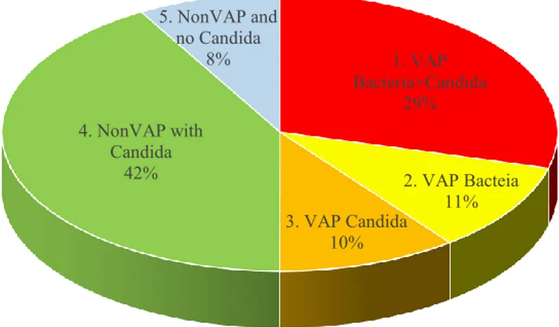
4.1.3. Group classification based on VAP definition and BAL culture results ... 41
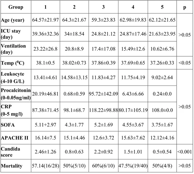
4.2. Clinical and laboratory data among five groups ... 41
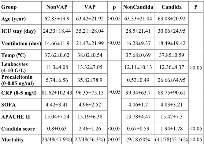
4.3. Clinical and laboratory data between VAP and nonVAP group ... 42
4.4. Microbiology results ... 43
4.4.1. Frequencies of bacteria species isolation from BAL ... 43
4.4.2. Frequencies bacteria species isolation between two hospitals ... 44
4.4.3. Antimicrobial susceptibility of A. baumannii ... 44
4.4.4. Antimicrobial susceptibility of K. pneumoniae ... 45
4.4.5. Antimicrobial susceptibility of S. aureus... 46
4.4.6. Fungal isolation from BAL ... 46
4.4.7. Antifungal susceptibility of Candida species ... 47
4.4.8. Antifungal susceptibility of C. albicans ... 47
4.4.9. Antifungal susceptibility of C. tropicalis... 48
4.4.10. Antifungal susceptibility of C. glabrata ... 49
4.5. Treatment ... 50
4.5.1. Firstline antibiotics prescription ... 50
4.5.2. Suitable of firstline antibiotics treatment ... 51
4.6.2. Indirect ELISA results in 5 groups ... 51
4.6.3. Indirect ELISA results between VAP group and nonVAP group... 52
4.6.4. Mean of ECE1, HWP1 optical density in serum and BAL of 5 groups ... 53
4.6.5. Mean of ECE1, HWP1 optical density of VAP group and nonVAP group ... 53
4.6.6. Sensitivity and specificity of indirect ELISA test... 54
4.6.7. Association between indirect ELISA results and clinical data ... 54
4.6.8. Correlation between indirect ELISA results and clinical data ... 54
5.1. Clinical characteristics of ICU ventilated patients ... 56
5.2. Bacteria species characteristics from ICU ventilated patients ... 57
5.3. Candida species characteristics from ICU ventilated patients ... 57
5.4. IC diagnosis by indirect ELISA results ... 60
6. CONCLUSION ... 62
Phan Thang - Molecular approach to early diagnosis of colonizing or invasive Candida in critically ill ventilated patients - Doctorate Thesis of PhD School in Biomolecular and Biotechnological Sciences,
University of Sassari 5
ACKNOWLEDGEMENTS
“Nothing can succeed without the help of the teacher” My teacher, a word that I have been calling all people who have contributed to the completion of my PhD study during the last three years. I gratefully acknowledge all my teachers in Italy and Vietnam for supporting me with the ideas or suggestions, equipments, and materials during the research process.
Firstly, I would like to express my great gratitude to my tutor, Prof. Salvatore Rubino, for the continuing support for my research process. I am especially thankful for my co-tutor, Prof. Ton Nu Phuong Anh and Dr. Antonella Santona, a believable and reliable tutor, who trained and let me primary experience the research of microbiology techniques, encouraged and supported me in processing my research. I will never forget a generous teacher who made my ideas come into reality with their enthusiasm in medical research. I would like to thank Prof. Piero Cappuccinelli, teacher of the teachers, whose ideas have been the most inspiring so that we could hurdle all the obstacles in completing my study.
Secondly, I also would like to send best wishes and good success to Dr. Maura Fiamma, Dr. Valentine Margarita, Dr. Anna Rita Cocco, Prof. Pierluigi Fiori, Prof. Nguyen Văn Minh, Dr. Tran Xuan Thinh, Prof. Tran Van Huy, Mrs. Nguyen Thi Y Nhi, Mr. Bui Manh Hung, Dr. Ngo Thi Minh Chau, Prof. Nguyen Gia Binh, Prof. Dao Xuan Co, Dr. Le Van An and Dr. Nguyen Hoang Bach for whose honorable supports are essential in my research. A special thanks to Prof. Leonardo A. Sechi, Rector of international PhD School and the staff of the Microbiology Department, Sassari University, who gave me a good environment to study and helped me to interact in Italian culture. I also would like to thank to Dr. Giovanni Sini, a humorus man, who gave me strong supports to finishing all documents relating my PhD course.
Thirdly, I sincerely express my profound gratitude to the leader of Hue University of Medicine and Pharmacy, Prof. Cao Ngoc Thanh and Prof. Nguyen Vu Quoc Huy, who provided me an opportunity to enroll this PhD programme and helped me to complete this study.
Phan Thang - Molecular approach to early diagnosis of colonizing or invasive Candida in critically ill ventilated patients - Doctorate Thesis of PhD School in Biomolecular and Biotechnological Sciences,
University of Sassari 6
unit, microbiology, hematology, genetic department of Hue University of Medicine and Pharmacy Hospital and the intensive care unit, microbiology department of Hue Central Hospital for their contributions during the time when I collected the samples. I express my warm thanks to all the technicians at the Parasitology department, especially M.D. Do Thi Bich Thao and MD. Ha Ngoc Thuy who spent a lot of time to help me do my research.
With my profound gratitude I also sincerely thank all the patients and their kinships, without whom I could not complete this thesis. One patient was one of my clinical teachers. I will never forget them.
Finally, and most importantly, I would like to thank my family for their love and encouragement. From the bottom of my heart, I’d love to send all my love and special thanks to my better half - my wife, who is the inspiration for me to complete all the works in my life. My deepest love and special thanks are for my son. He is my motivation for me to finish my study as soon as possible.
Words can’t express the gratitude. Again, sincerely thank you! Phan Thang
Phan Thang - Molecular approach to early diagnosis of colonizing or invasive Candida in critically ill ventilated patients - Doctorate Thesis of PhD School in Biomolecular and Biotechnological Sciences,
University of Sassari 7
LIST OF ABBREVIATION
A. baumannii Acinetobacter baumannii Als Agglutinin-like sequence
APACHE II Acute Physiology and Chronic Health Evaluation II BAL Broncho-alveolar lavage
BDG β-D-Glucan C. albicans Candida albicans C. nonalbicans Candida nonalbicans
CDC Centers for Disease Control and Prevention CRP C-reactive protein
ECE1 Extent of cell elongation 1
ELISA Enzyme-linked immunosorbent assay
FiO2 Fraction of inspired oxygen
HIV Human Immunodeficiency Virus HCH Hue Central hospital
HUMP Hue University of Medicine and Pharmacy HWP1 Hyphae wall protein 1
HYR1 Hyphally regulated gene 1 IC Invasive Candidiasis ICU Intensive Care Unit IV Intravenous
K. pneumoniae Klebsiella pneumoniae
MALDI - TOF Matrix assisted laser desortion ionization time-of-flight Neg Negative
PCR Polymerase chain reaction
PEEP Possitive end - expiratory pressure
P. aeruginosa Pseudomonas aeruginosa
Pos Positive
Phan Thang - Molecular approach to early diagnosis of colonizing or invasive Candida in critically ill ventilated patients - Doctorate Thesis of PhD School in Biomolecular and Biotechnological Sciences,
University of Sassari 8
SDA Sabouraud dextrose agar
SOFA Sequential Organ Failure Assessment S. aureus Staphylococcus aureus
VAP Ventilator-associated pneumonia
Phan Thang - Molecular approach to early diagnosis of colonizing or invasive Candida in critically ill ventilated patients - Doctorate Thesis of PhD School in Biomolecular and Biotechnological Sciences,
University of Sassari 9
LIST OF TABLES
Table 1.1. Bias of β-D-Glucan results for IC diagnosis ... 26
Table 3.1. Distribution of samples on study ... 33
Table 3.2. Zone diameter interpretive instructions of antimicrobial susceptibility ... 34
Table 3.3. Zone diameter interpretive instructions of antifungal susceptibility ... 37
Table 3.4. Amino acidic sequences of synthetic peptides in this study ... 37
Table 4.1. Clinical symptoms of patients ... 40
Table 4.2. Inflammatory symptoms of patients ... 41
Table 4.3. Clinical data among 5 groups... 42
Table 4.4. Clinical symptoms between VAP group and nonVAP group ... 43
Table 4.5. Cutoff value of indirect ELISA results ... 51
Table 4.6. Indirect ELISA results among 5 groups ... 52
Table 4.7. Indirect ELISA results between VAP group and nonVAP group ... 52
Table 4.8. Association between clinical data and indirect ELISA results ... 54
Phan Thang - Molecular approach to early diagnosis of colonizing or invasive Candida in critically ill ventilated patients - Doctorate Thesis of PhD School in Biomolecular and Biotechnological Sciences,
University of Sassari 10
LIST OF FIGURES
Figure 1.1. Structure of the C. albicans cell wall ... 15
Figure 1.2. Candida species morphology ... 16
Figure 1.3. Model of Candidalysin-induced pore-formation ... 20
Figure 1.4. Schematic of the role of C. albicans ECE1-III in another stage infection of epithelial cells... 20
Figure 1.5. The steps of C. albicans tissue invasion ... 22
Figure 3.1. Scheme of study ... 39
Figure 4.1. Primary admission diagnosis of patients ... 40
Figure 4.2. Group classification of all patients ... 41
Figure 4.3. Bacteria species isolation from BAL ... 43
Figure 4.4. Bacteria species isolation from BAL in the two hospitals ... 44
Figure 4.5. Antimicrobial susceptibility of A. baumannii ... 44
Figure 4.6. Antimicrobial susceptibility of A. baumannii in the two hospitals ... 45
Figure 4.7. Antimicrobial susceptibility of K. pneumoniae ... 45
Figure 4.8. Antimicrobial susceptibility of S. aureus ... 46
Figure 4.9. Fungal isolation from BAL ... 46
Figure 4.10. Antifungal susceptibility of Candida species ... 47
Figure 4.11. Antifungal susceptibility of C. albicans ... 47
Figure 4.12. Antifungal susceptibility of C. albicans between two hospitals... 48
Figure 4.13. Antifungal susceptibility of C. tropicalis ... 48
Figure 4.14. Antifungal susceptibility of C. tropicalis between two hospitals ... 49
Figure 4.15. Antifungal susceptibility of C. glabrata ... 49
Figure 4.16. Frequencies of firstline antibiotics indication... 50
Figure 4.17. Characteristic of firstline antibiotic therapy ... 50
Figure 4.18. Suitable of firstline antibiotics treatment ... 51
Figure 4.19. Mean of ECE1, HWP1 optical density of 5 groups ... 53
Phan Thang - Molecular approach to early diagnosis of colonizing or invasive Candida in critically ill ventilated patients - Doctorate Thesis of PhD School in Biomolecular and Biotechnological Sciences,
University of Sassari 11
LIST OF IMAGE
Image 3.1. Bronchoendoscopy to the wedge of bronchus ... 31 Image 3.2. Antifungal susceptibility of C. albicans to fluconazole ... 37
Phan Thang - Molecular approach to early diagnosis of colonizing or invasive Candida in critically ill ventilated patients - Doctorate Thesis of PhD School in Biomolecular and Biotechnological Sciences,
University of Sassari 12
ABSTRACT
Candida colonization is a frequent event in respiratory tract of non-immunocompromised intensive care unit (ICU) ventilated patients. From 5 to 30% of Candida colonization patients will develop Invasive Candidiasis (IC), which is usually a late-onset ICU acquired infection. Until now, a lot of data highlight the necessity for new IC noninvasive diagnostic in high risk patients. IC is a serious complication in the ICU patients, around 35% mortality and up to 90% in patients with septic shock. How to diagnosis IC early and give appropriate antifungal therapy are the key for a remarkable reduction in mortality. The overall objective of this study was to identify the etiology of Candida and bacteria species in lower respiratory tract in the central of Vietnam, and to discriminate invasive or colonizing Candida by indirect ELISA (Enzyme-linked immunosorbent assay).
Ninety six critically ill ventilated patients from 2 hospital in Hue (central Vietnam)
were followed in this study. The 3 main isolated fungal pathogens were C. albicans (42%), C. tropicalis (37%) and C. glabrata (16%). The fluconazole resistance of Candida species was 21.11% and caspofungin was 4.44%. C. tropicalis, that is becoming a predominant opportunistic in nosocomial fungal infections of ICU in developing country, showed highest fluconazole resistance (34.29%) and caspofungin resistance (5.71%). In ICU, 3 main bacteria resulted in ventilator-associated pneumonia (VAP) were A. baumannii (43.2%), K. pneumoniae (28.4%) and S. aureus (14.8%), with high levels of antimicrobial resistance. A. baumannii showed resistance to all cephalosporin 2, 3, 4 generation (100%) and carbapenem (94%). A 50% of K. pneumoniae was carbapenem-resistant while 100% S. aureus was resistant to methicillin.
To discriminate invasive or colonizing Candida, we chose 2 proteins, ECE1, present in C. albicans and C. dubliniensis, and HWP1, present in almost Candida species, selecting specific epitopes to develop indirect ELISA. ELISA results showed that 47.4% of patients with C. albicans had IC and 28.9% had invasive C. albicans pneumonia. In 19.23% of patients with Candida species had IC and 2.56% had invasive Candida species pneumonia. The sensitivity and specificity of ECE1 and HWP1 antibody detecting were 80% and 96% and 60% and 77% respectively, indicating the selected ECE1 epitope as a
Phan Thang - Molecular approach to early diagnosis of colonizing or invasive Candida in critically ill ventilated patients - Doctorate Thesis of PhD School in Biomolecular and Biotechnological Sciences,
University of Sassari 13
good marker for IC due to C. albicans and C. dubliniensis. A correlation between the ELISA results and 4 clinical parameters (Candida score, procalcitonin, length of ICU stay, ventilation day) was also investigated, that should help physicians to decide early antifungal therapy waiting for a new IC test that include all Candida species.
Phan Thang - Molecular approach to early diagnosis of colonizing or invasive Candida in critically ill ventilated patients - Doctorate Thesis of PhD School in Biomolecular and Biotechnological Sciences,
University of Sassari 14
1. INTRODUCTION 1.1. Candida species
1.1.1. History
The original name of Candida comes from the Latin term “candidus”. Candida albicans (C. albicans) was identified in the nineteenth century from three independent sources. First, in 1841, Fredrick Berg, a medical practitioner, discovered that thrush was caused by fungus with filaments that dispersed into epithelial cells. In 1842, David Gruby, a medical practitioner, fully described the cells of thrush fungus and compared to that causing tinea. Thrush fungus was later named in 1853 as Oidium albicans by Charles Phillipe Robin. In 1923, Christine Berkhout, a mycologist, changed the name to C. albicans till now [1].
1.1.2. Taxonomy
The taxonomy of the genus Candida is increasing overtime because of the reclassification of certain species and the discovery of new species such as Candida dubliniensis (C. dubliniensis), Candida orthopsilosis (C. orthopsilosis), and Candida metapsilosis (C. metapsilosis). C. orthopsilosis and C. metapsilosis were previously classified as part of the Candida parapsilosis (C. parapsilosis) complex. More than 200 species of Candida have been described, most of which exist as saprophytes organisms. Approximately 20 species can infect humans and C. albicans is the most prevalent species [2]. C. albicans, Candida glabrata (C. glabrata), C. parapsilosis, Candida tropicalis (C. tropicalis), and Candida krusei (C. krusei) were results in 90 - 92% of all cases of candidiasis [3, 4]. These species are able to cause both superficial infections of the skin and mucosa as well as systemic infections. The dissemination of the fungus through the blood stream and subsequent organ colonization is life-threatening with high mortality rates.
1.1.3. Cell biology
Cell biology characteristics of Candida species are the same to those of eukaryotes and especially similar to Saccharomyces cerevisiae [5]. Polysaccharides are an indispensalbe compound in the cell walls of Candida species [5, 6]. Candida cell walls consist of mannans, glucans and a few of chitin (Figure 1.1) [7, 8]. These components are closely
Phan Thang - Molecular approach to early diagnosis of colonizing or invasive Candida in critically ill ventilated patients - Doctorate Thesis of PhD School in Biomolecular and Biotechnological Sciences,
University of Sassari 15
bound to polypeptides and proteins found on the cell membrane [7]. Three types of adhesion molecules consist of glycoproteins, the protein moiety of glycoproteins, and the polysaccharide portion of a mannoprotein [5]. Moreover, the mannan polysaccharides structures found on the walls of Candida play an important role in its pathogenicity [5, 9]. C. albicans mannan is required for disruption of host processes that function to inactivate pathogens, leading to survival and escape of this fungal pathogen from host phagocytes [10].
Figure 1.1. Structure of the C. albicans cell wall [9]
Phospholipids and sterols are dominant in lipid structure of Candida species. Ergosterol is the major membrane sterol. These lipids provide the site of action for the synthesis of enzymes involved in cell wall morphogenesis and antifungal action. Lipid alterations can occur during a yeast to mycelium transition [11].
1.1.4. Morphogenesis
Genus Candida constitutes a heterogeneous group of eukaryotic, dimorphic, or polymorphic organisms [5, 12, 13]. Candida species can grow in widely pH from below 2.0 to nearly 10.0 under microaerophilic and even anaerobic conditions as well as the more normal aerobic atmospheres of incubation [5, 14]. Normally, in the gut microbiota of humans or animals blastoconidia of Candida species exits in round and oval shape. Candida species grow as yeast cells or blastoconidia. Yeast cells are approximately 2 - 10
Phan Thang - Molecular approach to early diagnosis of colonizing or invasive Candida in critically ill ventilated patients - Doctorate Thesis of PhD School in Biomolecular and Biotechnological Sciences,
University of Sassari 16
µm in the largest dimension, round to oval and reproduce by budding. They multiply principally by the blastoconidia production. However, Candida species can have different morphogenesis depending on the species and environmental triggers (Figure 1.2) [5, 15]. When the blastoconidia are produced from one another in a linear fashion without separating, a structure termed a pseudohypha is formed. Under certain circumstances, some yeast may produce true hyphae. True hyphae differ from pseudohyphae in forming long narrow filaments with parallel sides and no constrictions at the sites of septation, whereas pseudohyphae are generally shorter and wider with obvious constrictions showing at the septation sites [16]. Further morphological forms are opaque yeast cells and chlamydospores [17, 18]. The switch from yeast cells to opaque cells plays an important role in the mating process, whereas chlamydospores can only be observed in vitro when grown on nutrient-poor media and may therefore present a dormant growth form developing under harsh environmental conditions [18, 19].
Figure 1.2. Candida species morphology [16, 20, 21]
The presence of budding yeasts, pseudohyphae, opaque cells, chlamydospores, and hyphae in the infected tissue are usually indicative of candidiasis [16, 22]. The
Yeast
Opaque cells Pseudohyphal cells
Gut cell
Chlamydospores
Hyphal cells
Phan Thang - Molecular approach to early diagnosis of colonizing or invasive Candida in critically ill ventilated patients - Doctorate Thesis of PhD School in Biomolecular and Biotechnological Sciences,
University of Sassari 17
morphological flexibility in Candida pathogen play an important role in allowing C. albicans to penetrate and proliferate in a wide variety of host tissues [23].
1.1.5. Hyphal-specific proteins
In C. albicans and C. nonalbicans species, hypha formation is characterised by the expression of specific hypha-associated proteins, the most important hydrolytic enzymes are proteases and phospholipases [24] including the secreted aspartic proteases (SAP), agglutinin-like sequence (ALS), hyphally regulated gene (HYR1), hyphal wall protein 1 (HWP1), and extent of cell elongation 1 (ECEl) [16, 25].
Several studies have demonstrated a correlation between an increase in the synthesis and the activity of hydrolytic enzymes and an increase in clinical symptoms of severe candidiasis [26]. Ten SAP isoenzymes are responsible for the proteinase activity, SAP produce by C. albicans, C. parapsilosis, C. tropicalis, C. dubliniensis, C. guilliermondii, C. kefyr, C. lusitaniae, and C. krusei [27, 28]. These enzymes produce non-specific proteolysis of host proteins related the defence against the infection. Different kinds of SAP are associated with different locations within the yeasts and different pathogenicity [28].
C. albicans, C. dubliniensis, C. tropicalis, C. glabrata, C. krusei, C. lusitaniae and C. parapsilosis also produce phospholipases [5, 29]. These enzymes take in controlling of yeast growth, remodeling of fungal cell membranes and spreading in host tissues through the hydrolysis of phospholipids [29]. Seven phospholipase genes have been characterized. However, the role of the enzymes encoded by these genes remains unclear [30]. PLB1p is a glycoprotein present at hyphal tips during the tissue invasion and has hydrolase and lysophospholipase-transacylase activity [31]. The growth of hyphae, a virulence mechanism, plays an important function in the tissue invasion and the resistance to phagocytosis [32].
HYR1 is a Candida germ tube specific cell wall glycoprotein with a glycosylphosphatidylinositol motive. HYR1 play a structural role in the Candida cell wall architecture. HYR1 protein shares significant structural homology to A. baumannii cell surface proteins, and becomes the receptor for A. baumannii binding to the fungus [33].
Phan Thang - Molecular approach to early diagnosis of colonizing or invasive Candida in critically ill ventilated patients - Doctorate Thesis of PhD School in Biomolecular and Biotechnological Sciences,
University of Sassari 18
to host surfaces. Eight genes in the Als family have been discovered presently [34]. The expression of these proteins is correlated with Candida species infection. Among the Als family, Als3 play main important role in epithelial adhesion [35, 36].
Hwp1 gene encodes for a fungal cell wall protein. HWP1 protein consists of 634 amino acids sequence. It’s specific for C. albicans, C. tropicalis, C. dubliniensis, C. africana and play an important role in adhesin that is required for mating, hyphal development and biofilm formation [37]. HWP1 also promotes the binding of Candida to epithelial cells relating to oroesophageal candidiasis in mice [38].
Hypha formation of Candida species is essential for host tissue damage and immune activation and the extent of cell elongation 1 protein (ECE1), present in C. albicans and C. dubliniensis is one of the most early and abundantly expressed proteins during this process. ECE1 expression was not detected when C. albicans grew as a budding yeast cell but it was observed within 30 min after cells had been induced to the form of hyphae. Birse et al were able to show that ECE1 expression correlated with the extent of cell elongation, but the function of ECE1 remained unknown for a long time [39]. The characterization of this protein was determined since 1993 but only in 2016 a portion of ECE1 protein, called “Candidalysin”, was indicated as the first cytolytic peptide toxin in a human fungal pathogen.
1.1.6. A first cytolytic peptide toxin in a human fungal pathogen “Candidalysin”
Cytolytic proteins and peptide toxins are virulence factors of bacterial pathogens which disrupt epithelial barrier function, damage cells and stimulate host immune responses [40, 41]. However, cytolytic peptide toxins in fungi pathogenic had not been identified for long time, until the discovery of the ECE1 toxin.
ECE1 is a protein consist of 271 amino acids, including a signal peptide for secretion (recognized by the signal peptidase) and seven dibasic lysine-arginine (KR) motifs which are recognized by the Golgi complex-associated endoproteinase Kex1p and Kex2p [42, 43]. These subtilisin or kexin-like proteases have been implicated in the activation of various bacterial toxins [44]. C. albicans Kex2p is a member of a family of eukaryotic proprotein protease enzymes including proprotein convertase 1, proprotein convertase 2 and furin, which possess catalytic domains homologous to the degradative serine proteases
Phan Thang - Molecular approach to early diagnosis of colonizing or invasive Candida in critically ill ventilated patients - Doctorate Thesis of PhD School in Biomolecular and Biotechnological Sciences,
University of Sassari 19
of the subtilisin family [45]. C. albicans Kex1p is a protease with a carboxypeptidase B-like function involved in releasing the C-terminal processing of the lysine and arginine residues from protein precursors. Recent studies proved that ECE1 was cleavaged completely by Kex2 resulting in a signal peptide and eight other peptides, with seven of these peptides ending in KR. After Kex2p processed, these peptides were subsequently cleaved by Kex1p for removing the C-terminal R [46-48].
C. albicans ECE1 amino acid sequence
MKFSKIACATVFALSSQAAIIHHAPEFNMKRDVAPAAPAAPADQAPTVPAPQEFN
TAITKRSIIGIIMGILGNIPQVIQIIMSIVKAFKGNKREDIDSVVAGIIADMPFVVRAV
DTAMTSVASTKRDGANDDVANAVVRLPEIVARVATGVQQSIENAKRDGVPDVG LNLVANAPRLISNVFDGVSETVQQAKRDGLEDFLDELLQRLPQLITRSAESALKDS QPVKRDAGSVALSNLIKKSIETVGIENAAQIVSERDISSLIEEYFGA SP ECE1- I1-31 ECE1- II32-61 ECE1- III62-93 ECE1-IV94-126 ECE1-V127-160 ECE1-VI161-194 ECE1-VII195-228 ECE1- VIII229-271 + ECE1-I1-31: MKFSKIACATVFALSSQAAIIHHAPEFNMKR + ECE1-II32-61: DVAPAAPAAPADQAPTVPAPQEFNTAITKR + ECE1-III62-93: SIIGIIMGILGNIPQVIQIIMSIVKAFKGNKR + ECE1-IV94-126: EDIDSVVAGIIADMPFVVRAVDTAMTSVASTKR + ECE1-V127-160: DGANDDVANAVVRLPEIVARVATGVQQSIENAKR + ECE1-VI161-194: DGVPDVGLNLVANAPRLISNVFDGVSETVQQAKR + ECE1-VII195-228: DGLEDFLDELLQRLPQLITRSAESALKDSQPVKR + ECE1-VIII229-271: DAGSVALSNLIKKSIETVGIENAAQIVSERDISSLIEEYFGA
Moyes et al discovered that among these eight peptides, only ECE1-III peptide was found in the presence of epithelial cells, indicating that the fungus secretes this toxin during mucosal infection [47]. In early infection progress, ECE1-III62-92K
(SIIGIIMGILGNIPQVIQIIMSIVKAFKGNK) levels accumulate into a membrane-bound ‘invasion pocket’ [49, 50] which affect direct tissue damage and stimulate the release of lactate dehydrogenase from the host epithelium (Figure 1.3).
During stages of infection, concentrations of ECE1-III62-92K induce epithelial immunity
by activating the ‘danger response’ pathway (MAPK, p-MKP1/c-Fos) resulting in the production of immune regulatory cytokines and alerting the host to the transition from
Phan Thang - Molecular approach to early diagnosis of colonizing or invasive Candida in critically ill ventilated patients - Doctorate Thesis of PhD School in Biomolecular and Biotechnological Sciences,
University of Sassari 20
colonizing yeast to invasive, toxin-producing hyphae (Figure 1.4). As a result, C. albicans ECE1-III62-92K is proved as the first cytolytic peptide toxin in a human fungal pathogen and
reveals the molecular mechanisms of epithelial damage and the host recognition of this clinically important fungus [46-48].
Figure 1.3. Model of Candidalysin-induced pore-formation [48]
Figure 1.4. Schematic of the role of C. albicans ECE1-III in another stage infection of epithelial cells [47]
Phan Thang - Molecular approach to early diagnosis of colonizing or invasive Candida in critically ill ventilated patients - Doctorate Thesis of PhD School in Biomolecular and Biotechnological Sciences,
University of Sassari 21
1.2. Candida species in ventilator-associated pneumonia patients
Nowadays, pneumonia is a leading cause of death worldwide. Ventilator-associated respiratory infection (VARI), which consists of ventilator-associated tracheobronchitis (VAT) and ventilator-associated pneumonia (VAP), is the commonest hospital-acquired infection in ICU [51]. According to the Centers for Disease Control and Prevention (CDC) definition, VAP is a pneumonia where the patient is on mechanical ventilation for > 2 calendar days on the date of event [52]. VAP is a significant problem in a resource-restricted ICU which remains important cause of morbidity (9% - 35%) and mortality (30% - 40%) despite the advances in prevention strategies and antimicrobial therapy [53, 54].
Previously study showed that the main microorganism pathogens causing VAP in United State of America are Staphylococcus aureus (S. aureus) 27.9%, Pseudomonas species 16.3%, Klebsiella species 13.3%, and Candida species 6.3% [54]. The incidence of VARI and VAP in Vietnam were 24.6% and 9.9% respectively, mainly caused by Gram-negative organisms Acinetobacter baumannii (A. baumannii) 43.8%, Klebsiella pneumoniae (K. pneumoniae) 35.6%, and Pseudomonas aeruginosa (P. aeruginosa) 32.9% [53]. In other studies, VAP with Candida species isolation was 8.8% - 19.5% [55, 56].
Nowadays, Candida species become more and more a predominant microrganism in healthcare-associated infections. CDC estimated that 46000 healthcare-associated Candida infections occur among hospitalized patients each year. Candida species is the fourth most common causes of healthcare-associated bloodstream infections in the United States [2]. Candida species are human commensals which commonly appear on the mucosal surfaces of gastrointestinal, respiratory tracts, urinary tracts, skin and under fingernails [5, 57]. Moreover, Candida species are also isolated from hospital sources, such as the floor, water, soil, food, medical equipments, medical staff, etc [10]. The prevalence of Candida colonization varies depending on site, population sampled, sampling equipment and sampling method. It’s estimated that between 25 - 40 % of people are colonized by C. albicans [11], this rate is approximately 47% (13 - 76%) in hospitalized patients [5]. Almost 100% of humans carry one or more Candida species from the mouth to the colon. The numbers of yeasts carried at any point in the gut can critically increase in ill patients.
Phan Thang - Molecular approach to early diagnosis of colonizing or invasive Candida in critically ill ventilated patients - Doctorate Thesis of PhD School in Biomolecular and Biotechnological Sciences,
University of Sassari 22
At ICU, the critically ill ventilated patients commonly have a lot of risk factors such as total parenteral nutrition, invasive procedures, a long time broad spectrum antibiotics, immunotherapy and hemodialysis, that contribute to Candida species infections increasingly [58]. Furthermore, the automatic protecting reflection of respiratory tract was reduced by the feeding tube, endotube and the main disease during mechanical ventilation. By this condition, Candida species have opportunity to penetrate and break the epithelial barriers and enter the blood stream (Figure 1.5). Therefore, infections can become life-threatening.
(1) Adhesion and colonization (2) Hyphal penetration and invasion
(3) Vascular dissemination
(4) Endothelial colonization and penetration
Figure 1.5. The steps of C. albicans tissue invasion [9]
The ventilated ICU patients can have Candida species infections due to haematogenous spread or pulmonary aspiration of the contents of colonies of oropharyngeal or gastric origin [24]. As a result, the colonization or even invasion to the respiratory tract by Candida species is common in patients receiving mechanical ventilation for a long time. However, distinguishing invasive Candida from colonizing Candida in respiratory tract has been a challenge until now. In the clinical practice guidelines for the management of candidiasis of Infectious Disease Society of America (IDSA) and European Society for Clinical Microbiology and Infectious Diseases (ESCMID) showed that Candida species from respiratory secretions usually indicate colonization and rarely requires any treatment
Phan Thang - Molecular approach to early diagnosis of colonizing or invasive Candida in critically ill ventilated patients - Doctorate Thesis of PhD School in Biomolecular and Biotechnological Sciences,
University of Sassari 23
with the antifungal therapy [59, 60]. However, in a multiple Candida colonization patient with signs and symptoms of infection, it might prompt antifungal treatment [61]. Moreover, the isolation of Candida species from respiratory tract samples in a patient who is severely immunosuppressed should trigger a search for evidence of IC and requires antifungal treatment. Until now, some studies supported that Candida pneumonia and Candida lung abscess are very uncommon [62], only rarely after the aspiration of oropharyngeal material has primary Candida pneumonia or abscess [63, 64]. Because of the rarity of Candida pneumonia, the common Candida colonization in the respiratory tract and the lack of sensitivity and specificity of diagnostic test [65], a decision to initiate antifungal therapy should not be based only on the respiratory tract culture results for avoiding the overuse of antifungal therapy [59].
Recently, some studies indicated that the infection symptoms, the duration of mechanical ventilation and ICU stay as well as the mortality correlate to Candida species in the respiratory tract. Azoulay et al found an association between Candida colonization of the respiratory tract secretions and a prolonged period of mechanical ventilation, longer ICU stay, hospital stay and an increased risk for Pseudomonas VAP [66]. In other studies concluded that the colonization of the airway with Candida species is associated with the development of bacterial colonization and pneumonia. Candida airway colonization was also associated with worse clinical outcomes and higher mortality [67-70]. Candida species colonization not only affected the pulmonary epithelial, but also promoted the antibiotic-resistant and the biofilm formation with the bacteria in suspected VAP patients [66, 70]. Candida species become more and more microinvasive in this ventilated ICU patients who had a lot of risk factors of IC [71, 72] such as:
+ Hospitalisation in ICU
+ Acute or chronic organ dysfunction requiring intensive care or invasive procedures (e.g. mechanical ventilation, vasoactive drugs, renal substitution, extracorporeal circulation systems, high-volume fluid or haemocomponents infusions, tracheostomy) + Solid organ transplantation
+ Onco-haematological diseases and stem cell transplantation, especially with graft-versus-host disease (GVHD)
Phan Thang - Molecular approach to early diagnosis of colonizing or invasive Candida in critically ill ventilated patients - Doctorate Thesis of PhD School in Biomolecular and Biotechnological Sciences,
University of Sassari 24
+ Surgery (especially abdominal surgery and surgical revision), trauma and burn patients
+ Pediatric and neonatal intensive care units
+ Multiple underlying medical conditions (e.g. elderly patients in medical wards) + Immunosuppressive therapy
+ Renal failure requiring haemodialysis or haemofiltration + Neutropaenia
+ APACHE II score > 20 + Multiple site colonisations + Duration of hospital stay
+ Previous history of Candida infection
+ Disruption of physiological barriers in the digestive tract + Total parenteral nutrition and use of indwelling catheters + Diabetes mellitus
+ Previous prolonged antibiotic therapy
1.3. Invasive Candidiasis diagnostic test and treatment
IC ranges from 5 to 10 cases per 1000 ICU admissions and represents 5% to 10% of all ICU-acquired infections [73, 74]. Candida species are the fourth most common cause of candidemia in United State of America and the drugs resistant of Candida infections results in significant increasingly healthcare expenditures each year [2]. In Europe, the incidence of Candidemia ranges from 2 - 3% of the blood stream isolates, but Candidiasis stays among the top ten blood stream pathogens [75]. Although C. albicans is the most common cause of invasive fungal infections, the increasing number of infections from C. nonalbicans species is recently reported as a major source of infection [76, 77]. Pfaller et al determined that C. albicans was the most common cause of IC (63 - 70%) all worldwide, followed by C. glabrata (44%), C. tropicalis (6%), and C. parapsilosis (5%) [77]. However, the species distribution of C. non albicans is different. While in North America and Europe, C. glabrata is the second species, in South America C. tropicalis is the most commonly after C. albicans [78]. 5 - 30% of Candida colonization patients will develop IC, which is usually a late-onset ICU acquired infections [73, 79]. The increasing
Phan Thang - Molecular approach to early diagnosis of colonizing or invasive Candida in critically ill ventilated patients - Doctorate Thesis of PhD School in Biomolecular and Biotechnological Sciences,
University of Sassari 25
of IC is related to the use of a broad spectrum of antibiotics, total parenteral nutrition, surgical procedures, indwelling invasive devices, intensive care support, hemodialysis, cytotoxic therapy and immune suppressive therapy [58, 74, 80], usually related to high rates of mortality, approximately 30%, as well as increases in cost and length of hospital stay [2].
Distinguishing invasive Candida from Candida colonizing is very difficult till now and may guide to deciding a proper antifungal therapy. Clinical symptoms suggestiveness of IC did not differ from those of other nosocomial infections. Candida isolation from blood cultures has low sensitivity. Cultures turn round times of several days and turn positive late in the course of disease [81] whereas antifungal therapy delayed after blood sampling had been associated with an increase of hospital mortality [82, 83]. An important factor contributing to the outcome of an IC also lies in a timely diagnosis. Quickly IC diagnostic test help to decide early proper antifungal therapy leading to reducing the mortality in IC patients. The development of nonculture assays is critical to providing the occasion for earlier IC diagnosis. Untill now, the available IC diagnostic tests are:
- Role of the mannan antigen or antimannan antibody test: The mannan or antimannan detection test may be useful for the diagnosis of IC. Several standard serological tests detecting antibodies against Candida mannan have been invented. In a meta-analysis of 14 studies, the sensitivity/specificity of mannan and anti-mannan IgG were 58%/93% and 59%/83% [84]. However, the specific is low because anti-mannan antibodies are ubiquitous in human sera and the sensitiveness was extremely poor in severely immunosuppressed patients [85]. Consequently, the separate detection of either mannan or antimannan was not recommended on the guideline [71].
- Beta-D-glucan (BDG) test: BDG is a cell wall constituent of Candida species and others fungi, but not on mammalian and bacterial cells [86]. Therefore, BGD detection in blood or other bodily specimens may represent a marker of a fungal disease. The sensitivity and specificity of serum BDG testing for diagnosing IC have ranged from 57% to 97% and 56% to 93% [87]. The BDG test are recommended on guideline as a diagnostic test in a patient with signs and symptoms of IC infection [71]. However, the
Phan Thang - Molecular approach to early diagnosis of colonizing or invasive Candida in critically ill ventilated patients - Doctorate Thesis of PhD School in Biomolecular and Biotechnological Sciences,
University of Sassari 26
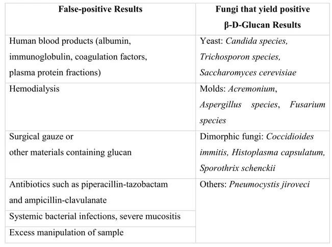
inaccuracy for BDG detection is in specificity and false-positivity value. False-positive results are commonly in patients with gram-positive and gram-negative bacteremia and ICU residents [88]. True-positive results are not specific for IC and several other causes of false-positivity have been identified [81], such as:
False-positive Results Fungi that yield positive β-D-Glucan Results
Human blood products (albumin, immunoglobulin, coagulation factors, plasma protein fractions)
Yeast: Candida species, Trichosporon species, Saccharomyces cerevisiae
Hemodialysis Molds: Acremonium,
Aspergillus species, Fusarium species
Surgical gauze or
other materials containing glucan
Dimorphic fungi: Coccidioides immitis, Histoplasma capsulatum, Sporothrix schenckii
Antibiotics such as piperacillin-tazobactam and ampicillin-clavulanate
Others: Pneumocystis jiroveci Systemic bacterial infections, severe mucositis
Excess manipulation of sample
Table 1.1. Bias of β-D-Glucan results for IC diagnosis
- Nucleic acid-based diagnostic techniques: Molecular-based diagnostic tests may potentially be sensitive in detecting an invasive fungal infection and can provide results more rapidly than culture, therefore enabling the possibility for earlier diagnosis and more timely initiation of antifungal therapy [89-91]. Nevertheless, molecular-based diagnostic techniques are not yet recommended on the guideline, because of the heterogeneity of the available results, the lack of reliable reference standards and differences in techniques [71]. -D-glucan, mannan antigen or antimannan antibody and molecular-based diagnostic assay of blood samples are recommended as adjuncts to cultures for the diagnosis of IC. However, these assays do not provide high sensitive and specific data for IC diagnosis.
Phan Thang - Molecular approach to early diagnosis of colonizing or invasive Candida in critically ill ventilated patients - Doctorate Thesis of PhD School in Biomolecular and Biotechnological Sciences,
University of Sassari 27
Currently, for management of IC infections and an early proper antifungal therapy decision, a physician should usually consider the combination of clinical symptoms such as individual risk factor of IC [71, 72], Candida colonization index [92, 93], Candida score [94], Ostrosky-Zeichner prediction rule [95, 96] and laboratory test result for deciding anearly proper antifungal therapy for their patients.
Phan Thang - Molecular approach to early diagnosis of colonizing or invasive Candida in critically ill ventilated patients - Doctorate Thesis of PhD School in Biomolecular and Biotechnological Sciences,
University of Sassari 28
2. RESEARCH OBJECTIVES
Ventilator-associated pneumonia (VAP) becomes the most frequent ICU-acquired infection nowadays with significant mortality (35%) despite the advances in the understanding of contributing causes and prevention. Candida species is the most common opportunistic mycosis at intensive care unit and Candida colonization is a frequent microrganism in respiratory tract of mechanically ventilated non-immunocompromised ICU patients [70, 94]. CDC estimates that each case of Candida species infection causes 3 - 13 days of additional hospitalization and a total of $6,000 - $29,000 in direct healthcare costs per patient [2]. Therefore, a better knowledge of the epidemiologic features of Candida in ventilated patients will be critically, enable physicians to provide appropriate prevention and treatment strategy [58].
A 5 - 30% of Candida colonization patients will develop IC [79]. IC is a serious complication in the ICU patients, around 35% mortality [2, 59, 60] and up to 90% in patients with septic shock [97]. Early diagnosis IC still remains a major challenge especially in respiratory tract now. Blood cultures, which were considering the gold standard for diagnosis are positive in a minority (50%) of cases and often late in the course of infection [71, 81]. Deep-seated tissue sampling usually requires extremely invasive procedures at high risk of complications and has a low specific especially in ICU patients who have received empirical therapy, whereas the antifungal therapy delayed beyond 12 hours after the sampling of blood has been associated with an increase of in-hospital mortality from under 20% to 40% [82, 83]. Early appropriate antifungal therapy is the key for a remarkable reduction in mortality [59, 98]. Using antifungal therapy early for Candida colonization in ICU patients with fever despite broad-spectrum antibiotics was recommended in the guideline. However, 70% of critically ill patients are receiving systemic antifungal therapy although they have no documented invasive fungal infection, suggesting an urgent need for antifungal stewardship strategies [59, 98]. The lack of rapid and sensitive IC diagnostic tests has led to overuse of antifungal therapy resulting in increased costs, drugs resistance and drugs toxicity. Until now, all available data highlight the essential for new noninvasive diagnostic tools for IC in high risk patients. In Vietnam, the total price of one intravenous antifungal therapy is from 250€ with fluconazole to
Phan Thang - Molecular approach to early diagnosis of colonizing or invasive Candida in critically ill ventilated patients - Doctorate Thesis of PhD School in Biomolecular and Biotechnological Sciences,
University of Sassari 29
3800€ with caspofungin, while GDP was 1950€ (2017). Furthermore, insurance did not accept intravenous antifungal payments in almost hospitals. “Treatment or not?” is still a big question to a physician and the relationship of patients in Vietnam. Therefore, this research has been carried out with the following objectives:
1. To identify the etiology of Candida and Bacteria species in lower respiratory tract infections in the central of Vietnam.
2. To discriminate between invasive or colonizing Candida by indirect ELISA test by selecting specific epitopes from invasive ECE1, HWP1 proteins.
Phan Thang - Molecular approach to early diagnosis of colonizing or invasive Candida in critically ill ventilated patients - Doctorate Thesis of PhD School in Biomolecular and Biotechnological Sciences,
University of Sassari 30
3. MATERIALS AND METHODS 3.1. Study site
This study was conducted in the Hue University of Medicine and Pharmacy and University of Sassari from September 2016 to August 2018, in the following departments: - ICU of Hue University of Medicine and Pharmacy (HUMP) Hospital
- ICU of Hue Central Hospital (HCH)
- Department of Parasitology, Hue University of Medicine and Pharmacy Hospital - Carlo Ubani Center, Department of Microbiology, Hue University of Medicine and Pharmacy
- Microbiology laboratory, Department of Biomedical Sciences, University of Sassari.
3.2. Study population
Patients, admitted to the ICU of HUMP hospital and HCH, were included in the study following specific criteria:
3.2.1. Inclusion criteria
- Patients over 18 years old
- Admission intensive care unit within the last 48 hours - Mechanical ventilation over 48 hours
3.2.2. Exclusion criteria
- VAP or suspected VAP before ICU admission
- End stage Human Immunodeficiency Virus (HIV) patients, neutropenic patients - Refused permission to join on this study given by patient's relatives
3.2.3. VAP diagnosis according to CDC 2013
Deterioration in ventilation following a period of stability
PEEP: ≥ 2 days of stable or decreasing daily minimum PEEP followed by a rise in daily minimum PEEP of ≥ 2.5 cmH2O, sustained for ≥ 2 calendar days
Or
FiO2: ≥ 2 days of stable or decreasing daily minimum FiO2 followed by a rise in daily
minimum FiO2 of ≥ 0.15 points, sustained for ≥ 2 calendar days And systemic signs
Phan Thang - Molecular approach to early diagnosis of colonizing or invasive Candida in critically ill ventilated patients - Doctorate Thesis of PhD School in Biomolecular and Biotechnological Sciences,
University of Sassari 31
Or
WBC > 12 x 109/L or < 4 x 10⁹/L And
Chest radiography: New and persistent infiltrate, consolidation, or cavitation as read by
two study physicians
Or
Decision to replace new antibiotics, physician starts antibiotics within a window period of 2 days before the deterioration in ventilation to 2 days after.
And pulmonary secretions
Increased/new purulent tracheobronchial secretions Or
≥ 25 neutrophils per low power field (10 objectives) on Gram stain of endotracheal aspirate [52, 99].
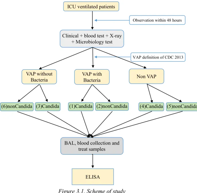
All ventilated patients were divided 5 groups based on VAP definition and the Broncho-alveolar lavage (BAL) culture results:
- Group 1: VAP with Bacteria and Candida species on BAL - Group 2: VAP with only Bacteria species on BAL
- Group 3: VAP with only Candida species on BAL - Group 4: NonVAP with Candida species on BAL - Group 5: NonVAP without any agents on BAL
Baseline demographic, pertinent clinical data and medications were recorded on admission to the study. Necessary variables were recorded in order to calculate the Acute Physiological and Chronic Health Assessment II (APACHE II) and Sequential Organ Failure Assessment (SOFA) scores on the sampling day. The microbiology results, the length of mechanical ventilation, ICU stay and hospital outcome were recorded completely.
3.2.4. Protocol of broncho-alveolar lavage sampling A. Preparation
A.1. Medical staff
Phan Thang - Molecular approach to early diagnosis of colonizing or invasive Candida in critically ill ventilated patients - Doctorate Thesis of PhD School in Biomolecular and Biotechnological Sciences,
University of Sassari 32
- 01 nurse who had training in bronchoscopy
- Patients: The relatives of patient were explained about the procedure, the benefit and
the side effect during the procedure. The procedure was performed when the relatives of patients accepted and signed on medical record. Patients had not been feeding for 4 hours before the procedure.
A.2. Instruments and drugs
- Instrument: Bronchoscopy tube (Pentax, Japan), light system (XD 320, Germany), suction machine (High-vacuum, Taiwan), sterile bottle sample, sterile gloves, sterile gauze, surgery clothes, ambu, oxygen, monitor, ventilation machine were used.
- Drugs utilized were atropin 0.25 mg, ephedrin 30 mg, lidocain 2%, propofol 200 mg, fentanyl 0.5 mg, methylprednisolon 40 mg, adrenalin 1 mg, salbutamol 0,5mg, midazolam 5 mg, natriclorua 0,9%.
- Medical records: X-ray, blood count cells, coagulation, HIV negative, blood gas and
electrocardiography had already done. A.3. Procedure
Propofol 2 - 3 µg/kg IV plus fentanyl 2 - 3 µg/kg IV was injected in patients before 5 minutes the procedure or midazolam 150 - 350 µg/kg IV was injected in unstable hemodynamic patients.
Endoscopy tube was slipped into endotube from mouth to tracheal. The lidocain 2% was used for local anesthesial before moving to bronchus. After connecting a steril bottle sample to endoscopy tube, the endoscopy tube was put at wedge broncho position which intends to clean. Normal saline was slowly instilled through the bronchoscope, the total volume was 150 - 200 ml and divided into three to four aliquots. By keeping the endoscopy tube and slightly suctionning (80 mmHg), the lavage fluid was flowing inside a sterile bottle sample. When the BAL was enough, the sterile bottle sample and endoscopy tube were taken out the patients. Patients were observed carefully during the procedure and 15 minutes after this procedure. It was 2 hours after the procedure that patients could eat.
Phan Thang - Molecular approach to early diagnosis of colonizing or invasive Candida in critically ill ventilated patients - Doctorate Thesis of PhD School in Biomolecular and Biotechnological Sciences,
University of Sassari 33
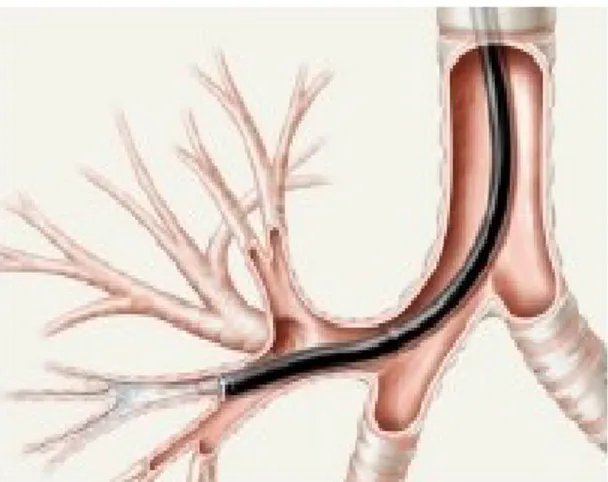
Image 3.1. Bronchoendoscopy to the wedge of bronchus A.4. Handling of the BAL fluid
BAL fluids were put on ice after sampling and quickly transportated to laboratory within 30 minutes. In the laboratory, BAL fluid was centrifuged at 15000 rpm for 10 min at 40C. The supernatant was separated from the pellet. The supernatant was stores at -200C
for detecting antibody by ELISA, the cellpellet was use for microbiological culture. 3.2.5. Protocol of blood sampling
2 ml nonheparinized blood of the patient was collected in sterilized vials. The blood was centrifuged at 3000 rpm for 2 minutes to collect serum. The sera were store at -200C
until the the ELISA test was performed.
Samples
Place BAL Blood
BAL control Blood control
Negative Positive Negative Positive
HUMP hospital 30 30 5 0 30 0
HCH 66 66 10 0 0 0
Bach Mai hosppital # # # # # 9
Viet Duc hospital # # # # # 3
74 central hospital # # # # # 2
Da Nang hospital # # # # # 1
Nouro hospital # # # # # 1
Table 3.1. Distribution of samples on study 3.2.6. Bacteria species identification and antimicrobial susceptibility
Phan Thang - Molecular approach to early diagnosis of colonizing or invasive Candida in critically ill ventilated patients - Doctorate Thesis of PhD School in Biomolecular and Biotechnological Sciences,
University of Sassari 34
blood culture or from BAL culture. Microbiological techniques are varied by site in line with routine clinical microbiological work of HCH and HUMP hospital. BAL samples were subjected to Gram staining prior to incubation on rabbit blood in blood agar base, brain heart infusion, drigalski lactose agar, and chocolate blood agar.
Antimicrobial susceptibility test of bacteria strains was performed by Kirby-Bauer disk diffusion method according to CLSI 2015 guideline [100] and manufacturer's instructions. The standard medium used for disk diffusion test was Mueller-Hinton agar, tested antibiotics were cefotaxime, cotrimoxazol, ciprofloxacin, levofloxacin, gentamicin, ceftazidime, imipenem, meropenem, amikacin, augmentin, colistin. Antimicrobial agents were used at the concentrations indicated in table 3.2.
Drugs Code Potency Zone in diameter (mm)
S I R Cefotaxime CTX 30 µg ≥ 23 - ≤ 14 Coxtrimoxazol SXT 25 µg ≥ 16 - ≤ 10 Ciprofloxacin CIP 5 µg ≥ 21 15 - 16 ≤ 15 Levofloxacin LEV 5 µg ≥ 17 15 - 16 ≤ 13 Gentamicin GM 10 µg > 15 7 - 9 < 12 Ceftazidime CAZ 30 µg ≥ 18 14 - 22 ≤ 14 Imipenem IMP 10 µg ≥ 16 15 - 18 ≤ 13 Meropenem MEM 10 µg ≥ 19 16 - 20 ≤ 15 Amikacin AMC 30 µg ≥ 18 13 - 17 ≤ 12 Augmentin AN 30 µg Colistin COL 10 µg ≥ 11 < 10
(S: Susceptible, I: Intermediate, R: Resistant)
Table 3.2. Zone diameter interpretive instructions of antimicrobial susceptibility The colonies were suspended in 5 ml of sterile 0.85% saline, and the turbidity was adjusted to yield 1×10⁸ cells/ml (0.5 McFarland standard). Next, a sterile cotton swab was
dipped into the suspension and rotated several times. Any excess fluid from the swab was removed by pressing firmly against the inside wall above the fluid level before dispensing suspension inoculated on the plate surface. Streaked the swab all over the surface of the
Phan Thang - Molecular approach to early diagnosis of colonizing or invasive Candida in critically ill ventilated patients - Doctorate Thesis of PhD School in Biomolecular and Biotechnological Sciences,
University of Sassari 35
medium three times, rotating the plate through an angle of 60o after each application then
passed the swab round the edge of the agar surface. Left the inoculum to dry for a few minutes at room temperature with the lid closed. Antimicrobial disks were placed on the inoculated agar with a forceps, and the plates were incubated at 37oC. A maximum of
seven discs can be placed on a 9 - 10 cm plate. Six discs may be spaced evenly, approximately 15 mm from the edge of the plate, and 1 disc placed in the center of the plate. Each disc was gently pressed down to ensure even contact with the medium. The plates were incubated at 37°C. The zone of inhibition was recorded after 24 hours and 48 hours.The diameter of each zone (including the diameter of the disc) was made with a ruler on the under-surface of the plate without opening the lid.
The results of culture and antimicrobial susceptibility were usually available after 3 days.
3.2.7. Candida species identification A. Microbiological media
- Sabouraud dextrose agar (SDA) medium and Brilliance Candida agar medium were used.
- 5 ml of BAL was centrifuged 15000 rpm for 10 minutes at 40C. The cellpellet was
dispensed on the plate surface containing Sabouraud dextrose agar (SDA) medium and chloramphenicol to incubate for 24 - 48 hours at 370C. All Candida strains isolated from
colony in SDA medium were sub-cultured in Brilliance Candida agar medium and incubated aerobically at 37°C. The plates were checked at 24, 48 and 72 hours. Brilliance Candida agar was used to identify the Candida species following the guide line of the manufacturer. The species of Candida were also selected basing on different colour of colonies. This media contains two chromogens (5-bromo-4-chloro-3-indolyl N acetyl ß-D-glucosaminide and 5 bromo-6-chloro-3-indolyl phosphate p-toluidine salt), which help identify the presence of the two target enzymes, the hexosaminidase and the alkaline phosphatase. The presense of either enzymes allows the differentiation of C. albicans and C. tropicalis from other species of Candida within 48 hours. The green colour of C. albicans comes by the chromogenic reaction as well as the dark blue color of C. tropicalis. Other species were very difficult to differentiate based on the colour. The isolated Candida strains were stored for matrix assisted laser desortion ionization time-of-flight
Phan Thang - Molecular approach to early diagnosis of colonizing or invasive Candida in critically ill ventilated patients - Doctorate Thesis of PhD School in Biomolecular and Biotechnological Sciences,
University of Sassari 36
(MALDI-TOF) identification. B. MALDI-TOF MS
Candida species was subcultured in SDA plates. After 24 hour incubation at 370C,
single colony was transferred directly and spotted in duplicate into the MALDI target. 1μl pure ethanol was added to each well to fix the sample. Next, 1µl of 70% formic acid was added and mixed gently. When the liquid medium was evaporated completely, each spot was overlaid with 1 μl of HCCA matrix solution and dried at room temperature. The loaded plate was analyzed by MALDI Biotyper CA System. The spectrum obtained was compared with the Maldi database. Identification was provided with accompanying scores as the manufacture schemes:
- Score < 1.7: No reliable identity
- Score from 1.7 - < 2.0: Identity at genus level - Score from 2.0 to upper: Identity at species level
In our study, one hundread fungal strains were isolated and identified from ninety six BAL samples.
3.2.8. Antifungal susceptibility testing
- Mueller-Hinton medium supplemented with 2% dextrose and 0.5 μg/ml methylene blue (Liofilchem Laboratories, Italy) and antifungal disk (Liofilchem Laboratories, Italy) were used.
- Candida species were suspended in 5 ml of sterile 0.9% normal saline, shaked 15 seconds and the turbidity was adjusted to yield 1 × 10 5 - 1 × 10⁶ cells/ml (0.5 McFarland
standard). A sterile cotton swab was soaked into the suspension and pressed firmly against the inside wall above the fluid level. The Candida species was spread on media by moving the swab 3 times arround the plates. After that, the plates were dried at room temperature for 3 - 5 min. Antifungal disks were put on the inoculated agar with forceps, and the plates were incubated at 37°C. The antifungal tested included fluconazole 10 μg/disk, intraconazole 8 μg/disk, amphotericin B 20 μg/disk, nystatin 100 unit/disk, flucytosine 10 μg/disk, caspofungin 5 μg/disk. The zone of inhibition was recorded after 24 hours and 48 hours. The C. albicans strain ATCC 90028, C. parapsilosis ATCC 22019 and C. krusei ATCC 6258 were used as standard strains. Zone diameter interpretive standards for Antifungal Disk Diffusion Susceptibility Testing of Candida species following
Phan Thang - Molecular approach to early diagnosis of colonizing or invasive Candida in critically ill ventilated patients - Doctorate Thesis of PhD School in Biomolecular and Biotechnological Sciences,
University of Sassari 37
manufacture instructions.
Sensitive of C. albicans Resistant of C. albicans Image 3.2. Antifungal susceptibility of C. albicans to fluconazole
Drugs Code Potency Zone in diameter (mm)
S I R Fluconazole FLU 25 µg ≥ 19 15 - 18 (DD) ≤ 14 Intraconazole ITC 8 µg > 16 10 - 15 (DD) < 9 Amphotericin B AMB 10 µg ≥ 15 10 - 14 < 10 Flucytosine AFY 1 µg ≥ 20 12 - 19 ≤ 11 Nystatin NY 100 UI ≥ 15 10 - 14 < 9 Caspofungin CAS 5 µg ≥ 16 13 - 15 ≤ 12
(S: Susceptible, I: intermediate, R: resistant, DD: Dose dependent) Table 3.3. Zone diameter interpretive instructions of antifungal susceptibility 3.2.9. Indirect ELISA protocol
A. Epitopes selection
ECE1, HWP1 proteins were chosen as antigen. ECE1-III62-93K Candidalysin sequence
[46, 47] and HWP1 protein sequence (CP017626.1) were used to select two epitopes by using BepiPred 2.0 software. Peptides from 14 to 16 amino acids were produced with a C-terminal cysteine residue to allow cross-linking with maleimide activated carrier proteins
Peptide Sequence Molecular mass (Da)
ECE1 H-CIQIIMSIVKAFKGNK-OH 1793.26
HWP1 H-CDNPPQPDQPDDNP-OH 1551.56
![Figure 1.2. Candida species morphology [16, 20, 21]](https://thumb-eu.123doks.com/thumbv2/123dokorg/8366217.134986/16.892.126.788.547.917/figure-candida-species-morphology.webp)
![Figure 1.4. Schematic of the role of C. albicans ECE1-III in another stage infection of epithelial cells [47]](https://thumb-eu.123doks.com/thumbv2/123dokorg/8366217.134986/20.892.146.775.740.1011/figure-schematic-role-albicans-stage-infection-epithelial-cells.webp)
![Figure 1.5. The steps of C. albicans tissue invasion [9]](https://thumb-eu.123doks.com/thumbv2/123dokorg/8366217.134986/22.892.149.714.378.749/figure-steps-c-albicans-tissue-invasion.webp)
![Nella sua versione più semplice, un sistema Radar si presenta come in Figura I.1 [Ber 05]; in essa si distinguono i blocchi di trasmissione e di ricezione:](data:image/gif;base64,R0lGODlhAQABAIAAAP///wAAACH5BAEAAAAALAAAAAABAAEAAAICRAEAOw==)