Development and in Vitro Evaluation of a Microbicide Gel
Formulation for a Novel Non-Nucleoside Reverse Transcriptase
Inhibitor Belonging to the
N‑Dihydroalkyloxybenzyloxopyrimidines
(N-DABOs) Family
Cristina Tintori,
†Annalaura Brai,
†Maria Chiara Dasso Lang,
†Davide Deodato,
†Antonia Michela Greco,
†Bruno Mattia Bizzarri,
†Lorena Cascone,
†Alexandru Casian,
†Claudio Zamperini,
†Elena Dreassi,
†Emmanuele Crespan,
‡Giovanni Maga,
‡Guido Vanham,
§Elisa Ceresola,
⊥Filippo Canducci,
⊥Kevin K. Ariën,
§and Maurizio Botta
*
,†,∥†
Dipartimento Biotecnologie, Chimica e Farmacia, Universita
̀ degli Studi di Siena, Via A. De Gasperi 2, I-53100 Siena, Italy
‡Istituto di Genetica Molecolare, IGM-CNR, Via Abbiategrasso 207, I-27100 Pavia, Italy
§
Virology Unit, Institute of Tropical Medicine, Nationalestraat 155, B-2000 Antwerpen, Belgium
∥
Biotechnology College of Science and Technology, Temple University, Biolife Science Building, Suite 333, 1900 N 12th Street,
Philadelphia, Pennsylvania 19122, United States
⊥
Department of Biotechnology and Life Sciences, University of Insubria, Dunant 3, 21100, Varese, Italy
*
S Supporting InformationABSTRACT:
Preventing HIV transmission by the use of a
vaginal microbicide is a topic of considerable interest in the
fight against AIDS. Both a potent anti-HIV agent and an
e
fficient formulation are required to develop a successful
microbicide. In this regard, molecules able to inhibit the HIV
replication before the integration of the viral DNA into the
genetic material of the host cells, such as entry inhibitors or
reverse transcriptase inhibitors (RTIs), are ideal candidates for
prevention purpose. Among RTIs, S- and
N-dihydro-alkyloxybenzyloxopyrimidines (S-DABOs and N-DABOs) are
interesting compounds active at nanomolar concentration
against wild type of RT and with a very interesting activity
against RT mutations. Herein, novel N-DABOs were synthesized and tested as anti-HIV agents. Furthermore, their mode of
binding was studied by molecular modeling. At the same time, a vaginal microbicide gel formulation was developed and tested for
one of the most promising candidates.
■
INTRODUCTION
Since its discovery in 1981, human immunode
ficiency virus
(HIV) infection continues to be a major health threat
worldwide. Combination antiretroviral therapy (cART) can
signi
ficantly reduce the viral load and prolong patients’ life
expectancy, but it is not able to totally eradicate the virus.
1Furthermore, the emergence of resistant strains continues to
limit the e
fficacy of current drugs.
2Accordingly, a great variety
of chemotherapy agents were developed against HIV-1 in the
past 20 years. Most of the anti-HIV studied small-molecules
target the viral enzyme reverse transcriptase (RT) by binding
the polymerase active site (nucleoside RT inhibitors) or an
allosteric pocket (non-nucleoside RT inhibitors or NNRTIs).
3,4More than 30 di
fferent molecular structures of NNRTIs were
developed, comprising nevirapine, delavirdine, efavirenz,
rilpivirine, and etravirine FDA-approved drugs. Among
NNRTIs, S- and N-dihydroalkylthiobenzyloxopyrimidines
(S-DABOs and N-(S-DABOs) have been extensively modi
fied by our
research group with the aim of improving their inhibitory
activity toward RT wild-type and clinically relevant
mu-tants.
5−13Several compounds showed nanomolar inhibitory
potency against wild-type HIV-1 and relevant drug-resistant
mutants. Furthermore, previous studies revealed that the
conversion of the S-DABO derivatives into the corresponding
N-DABOs may lead to new derivatives characterized by
improved ADME profiles, especially aqueous solubility (
Chart
1
).
13On the other hand, HIV transmission remains high in
developing countries, and with a vaccine not yet in sight, the
development of an e
ffective prevention strategy would be
desirable. Topical microbicides are preparations able to prevent
the transmission of HIV when applied to the genital and/or
Received: December 22, 2015 Published: February 20, 2016
lower gastrointestinal graft via the rectum.
14It is generally
accepted that a microbicide should act prior to the integration
of proviral DNA into the host cell DNA. Accordingly, the two
main compound classes that could be used to such a purpose
are entry inhibitors and RT inhibitors. Examples of RT
inhibitors in preclinical and clinical evaluation as microbicides
include tenofovir, dapivirine, MIV150, UC781, UAMC01398,
and DABO.
15−20Microbicide candidates must have a good
selectivity index (SI), inhibit virus replication at low, nontoxic
concentrations in vitro, have a good resistance pro
file, be stable,
and have the potential for reasonable pricing.
21−24In this
context, the purpose of this work was to initiate a study on
N-DABO NNRTIs in order to investigate their potential for the
development of new microbicides. To this aim, a novel series of
N-DABO derivatives was synthesized. Biological evaluation
showed that these compounds are able to inhibit HIV-1
replication at nanomolar concentration. Remarkably, the most
promising derivative in terms of activity against mutant strains,
25e, was also characterized by a high selectivity index, 5-fold
higher than dapivirine, a good cell permeability, and stability
higher than 95%. On this basis, a vaginal microbicide gel
formulation of 25e was developed and evaluated in vitro.
Moreover, in the present work we also reported the
development of a predictive QSAR model for a large set of
S-DABO/N-DABO derivatives.
■
RESULTS AND DISCUSSION
Chemistry. The new compounds were synthesized using
the classical approach for the synthesis of N-DABO, based on
the nucleophilic displacement of the methylsulfonyl group at
C2 position with the appropriate amine. According to
Scheme
1
, the key intermediates 13 and 14 were synthesized starting
from the appropriate carboxylic acids (3 and 4) that were
methylated with MeI using LDA as a base to obtain compounds
5
and 6 in good yields, while compound 12 was synthesized
directly from acid 4.
The
β-ketoesters 9−11 were prepared by activation of acids
4
−6 using 1,1′-carbonyldiimidazole (CDI), followed by
treatment with potassium monoethyl malonate 8 in the
presence of anhydrous MgCl
2and Et
3N. Then condensation
of 9
−11 with thiourea in the presence of EtONa led to
substituted thiouracils 12
−14. Subsequent alkylation with MeI
in DMF gave compounds 15
−17 in excellent yield.
C2-methylthio group of thiouracils 15
−17 was then converted in
the corresponding sulfone derivatives 18
−20, by oxidation.
Secondary amines 22b
−g were synthesized by reductive
amination of the appropriate aldehyde (21b
−g) with
methyl-amine and sodiumborohydride. Nucleophilic substitution of the
C2-methylsulfonyl group of 18
−20 with the appropriate amine
led to
final compounds 23a−f, 24a, 24c−f, and 25d−g
(
Scheme 2
). Boc deprotection of compounds 23b and 24b led
to compounds 23a and 24a (
Scheme 3
).
Aldehydes 21b
−d were not commercially available and were
synthesized by nucleophilic displacement of the 4-
fluoroben-zaldehyde with the appropriate amine (
Scheme 4
).
Biology. Antiviral Activity against HIV-1 and
Cytotox-icity. Compounds 23a−f, 24a, 24c−f, and 25d−g were
Chart 1. Hit Compounds Belonging to S-DABO and
N-DABO Families
Scheme 1
aaReagents and conditions: (a) LDA, HMPA, CH
3I, dry THF,−78 °C to rt, 12 h; (b) KOH, dry EtOH, 0 °C to rt, 12 h; (c) carbonyldiimidazole, MgCl2, TEA, dry CH3CN, rt, 12 h; (d) Na0, thiourea, dry EtOH, reflux, 12 h; (e) CH3I, KOH, EtOH, rt, 1 h; (f) m-CPBA, dry DCM, 0°C to rt, 12 h.
evaluated for their anti-HIV-1 activity and cytotoxicity in
TZM-bl cells in comparison with TMC120 (dapivirine). The results,
expressed as EC
50(50% e
ffective concentration), CC
50(50%
cytotoxic concentration), and SI (selectivity index given by the
CC
50/EC
50ratio) values are summarized in
Table 1
. Among the
novel N-DABOs, compounds 23e, 23f, 24e, 24d, 25d, 25e, and
25f
retained an activity similar to that of the lead 1 and showed
very potent inhibitory activities against the replication of the
V106A
25mutant virus which is resistant to nevirapine (
Table
2
). On the other hand, the compounds were also active at
nanomolar concentration against V90I, V106A, E138K, and
K101E mutants while modestly active against the L100I,
K103N, and Y181C
25mutants. Moderate activities against WT
RT were found for derivatives 23d, 24a, and 24c. Remarkably,
the most active compounds also showed low cytotoxicity and
resulting high selectivity indices, with the best compound, 25e,
characterized by SI higher than 10 000. This feature together
with the good activity pro
file makes this compound suitable for
microbicide formulation.
Antireverse Transcriptase Activity. Compounds 23c,
23e, 23f, and 24e were further evaluated for antireverse
transcriptase activity in comparison with nevirapine in an
enzymatic recombinant HIV-1 RT activity assay, and results
expressed as IC
50were compared with anti-HIV-1 activity.
Comparable activity values were found, con
firming that the
target of these antiviral agents is the RT and that compounds
are able to permeate through the cellular membrane, inhibit
RT, and subsequently block the HIV-1 replication.
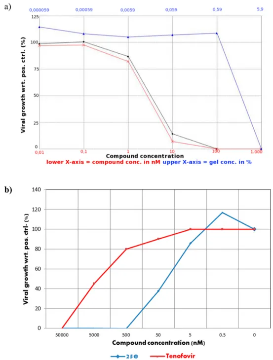
Antiviral Activity for 25e Gel. Antiviral activity testing
was performed with a gel formulation of 25e in direct
comparison with the free drug and the blank gel. As measured
in TZM-bl cells, the EC
50values of formulated and native 25e
are similar, which indicates that formulating 25e does not alter
the in vitro potency of the compound (
Figure 1
a). In addition,
no loss in antiviral activity was observed for the 25e gel
formulation after storage for 1 month at room temperature and
without light exposure.
To exclude the e
ffect of β-cyclodextrins on the observed
antiviral activity and the toxicity of these gels in vitro even at
low gel concentrations (toxicity was observed at 5% gel
concentration), gels were reformulated without
β-cyclodextrins.
Moreover, to reproduce more physiological conditions, TZM-bl
cells were seeded into Transwells and the gel was applied onto
the cell monolayer at 50%
fixed concentration with serial drug
(25e or tenofovir) dilutions. This experiment con
firmed the
high antiviral activity of 25e gel (EC
50= 30 nM) also in
comparison with the observed e
fficacy of tenofovir gel (EC
50=
3
μM) (
Figure 1
b).
In Vitro ADME Studies. Selected N-DABOs were profiled
in vitro for aqueous solubility at pH 4.2 and 7.4, liver
microsomal stability, and membrane permeability (
Table 3
).
Their aqueous solubility (
−log S ranging from −6.51 to −8.60
and from
−5.49 to −7.99 at pH 7.4 and 4.2, respectively),
although improved over the S-DABOs previously developed,
was rather low. This aspect does not prevent the e
fficacy of the
gel formulation which is composed of cyclodextrins, commonly
used to increase drug solubility.
26,27Furthermore, cyclodextrins
have been shown to determine a dose-dependent inactivation
of the virus by the removal of cholesterol from the membrane
of HIV-infected cells, thus enhancing the infection prevention
of the microbicide formulation. On the other hand, passive
membrane permeability in a PAMPA assay indicated acceptable
cell permeability values for compound 25e (5.54 and 3.78
×
10
−6cm/s, at pH 4.2 and 7.4, respectively). Moreover, stability
tests disclosed that all analogues showed excellent metabolic
stability in liver microsomes (
∼99%).
Stability of 25e Gel Formulation. 25e gel (85% of
compound was loaded that corresponded with a
final
concentration of 17
μM) at pH 4.2 (vaginal formulation) was
found to be stable at room temperature without light exposure:
the 25e recovery after 1 month of storage amounted to about
100%. In addition, the viscosity and pH of this gel remained
stable during storage.
Molecular Modeling. Docking Studies. Docking studies
have been previously used to characterize the binding mode of
S-DABOs within the non-nucleoside binding pocket of RT wild
type.
11,12A schematic representation of the main interactions
Scheme 2
aaReagents and conditions: (a) MeNH
2, NaHCO3, MeOH, reflux 4 h; (b) NaBH4, 0°C to rt, 1 h; (c) dry toluene, reflux, 48 h.
Scheme 3
aa
Reagents and conditions: (a) 10% TFA in dry DCM, rt 1 h.
Scheme 4
aaReagents and conditions: (a) di-tert-butyl dicarbonate, MeOH, microwave, 2 min; (b) 4-fluorobenzaldehyde, K2CO3, dry DMF, 130 °C, 12 h.
established by S-DABOs is reported in
Figure 2
. Two hydrogen
bonds were detected between the amide moiety of the
pyrimidinone ring and the backbone of Lys101. The C6-benzyl
group established hydrophobic interactions with the aromatic
cage formed by Tyr181, Tyr188, Phe227, Trp229, and Leu100,
while the C6
′-group interacted with Val179. Finally profitable
lipophilic interactions were found between the S side chain and
a solvent-exposed channel lined by residues Val106, Pro225,
Tyr318, Phe227, and Pro236.
Herein, molecular docking simulations were conducted on
new N-DABOs following the computational protocol
pre-viously adopted on S-DABOs. Besides Autodock,
28two more
software programs were used, namely, Gold
29and Glide,
30,31and the three computational procedures were extended to a
large set of compounds including 168
S-DABOs/N-DABOs
11−13,32,33characterized by IC
50values ranging from
0.0003 and 50
μM against RT wt (
Table S1, Supporting
Information
). In parallel, the same docking analyses were
conducted by considering a well-ordered water molecule
crystallized into the binding pocket of 1RT2 crystal structure,
which forms a triad of hydrogen bonds involving the inhibitor,
the main chain nitrogen of Lys101, and a carboxyl oxygen of
Table 1. Antienzymatic Activity, Antiviral Activity, and Cytotoxicity of the New N-DABO Derivatives
aData represent the mean of at least two experiments.bND: not determined.
Table 2. Anti-HIV Activities of Selected Compounds against Mutant Strains
acompd EC50(nM) pNL4.3 WT V90I (nM) L100I (nM) E138 K (nM) K101E (nM) K103N (nM) V106A (nM) Y181C (nM)
23d 100 ND ND 274 >1000 792 18 704 23e 15 ND ND ND ND ND 10 680 23f 8.6 13 220 35 51 367 ND 201 24c 96 ND ND 256 942 1502 15 1288 24d 29 ND ND 103 294 1039 6.3 627 24e 12 ND ND ND ND ND 10 209 25d 7.9 22 1570 42 138 2460 ND 331 25e 5 8.7 84 17 31 102 ND 80 25f 10 30 1498 65 134 518 ND 814 25g 329 687 >10000 950 >10000 >10000 ND >10000 1 93 ND ND 246 450 549 56 1608 2 3 ND ND 9.1 17 34 0.87 94
aAll these viruses are site-directed mutants. pNL4.3 replication competent viruses with single point mutations introduced in HIV RT by site-directed mutagenesis.
Glu138 in the p51 chain. Remarkably, the three software
programs converged to the same solution in both the presence
and absence of the water molecule and the poses obtained for
N-DABOs perfectly overlapped with those of S-DABOs
derivatives previously reported. As an example,
Figure 3
shows the binding mode of compound 24e aligned with the
congeneric S-DABO.
Although coincident poses were obtained through the use of
di
fferent docking procedures which made the results highly
con
fident, no correlation was found between the scoring
functions employed (Glide SP and XP, Autodock, ChemScore)
and the experimental activity values. Bad results were also
obtained through pose rescoring by X-Score
34(an empirical
Figure 1.Anti-HIV activity of 25e (black line) and 25e gel (red line) in human Tzm-Bl. (a) The EC50 values for the free drug and the gel formulation were similar, ranging from 2.3 to 3.3 nM, respectively. Each concentration was evaluated in triplicate. Toxicity was evaluated in parallel, and the blank gel showed toxicity at a concentration higher than 100 nM. (b) The EC50 was evaluated by Transwell assays and without β-cyclodextrins in the gel formulation. The usage of Transwells has allowed use of the gels at a 50%fixed concentration at all dug dilutions with no toxicity on the cells. Tenofovir was used as control.
Table 3. In Vitro ADME Pro
file of N-DADO Derivatives
compd PAMPA, pH 7.4 (10−6cm/s) PAMPA, pH 4.2 (10−6cm/s) water solubility, pH 7.4 (log S)a water solubility, pH 4.2 (log S)a metabolic stability (%)b 23c 21.9 ND −8.6 −7.80 99 23d 1.37 0.58 −6.51 −5.49 99 24d 0.91 ND −8.7 −7.85 99 23f 10.93 1.90 −7.55 −6.70 99 25e 5.54 3.78 −8.60 −7.99 99 alog S = log mol L−1.bExpressed as percentage of unmodified parent drug.
scoring function) and MM-GBSA
35(a force
field based
function) methods. This was not surprising and was also
recently found for another family of NNRTIs.
36In order to
derive a quantitavive structure
−activity relationship, the new
N-DABO derivatives together with additional N-N-DABOs and
S-DABOs previously described by us
11,13and other research
groups
32,33were submitted to a multiple linear regression
(MLR) analysis with the aim of identifying the molecular
determinants able to influence their activity against wild type
HIV-1. For this purpose, the MLR algorithm implemented in
Canvas was applied. In particular, 68 compounds with activity
data spanning over 6 orders of magnitude were used as learning
set. Several models were generated by using the cellular data
(EC
50) as the independent variable, 87 descriptors as
dependent variables, and choosing in turn an increasing
number of X variables as best subset. Besides the parameters
coming from docking studies, 2D
fingerprint descriptors and
QikProp properties were calculated. The learning set (the
first
68 compounds in
Table 1S
) was randomly divided into a
training set (80%) and a test set (20%). A good predictive
ability, both internal and external, was found with an equation
characterized by seven descriptors: evdw (van der Waals energy
calculated by Glide during the docking with water), PISA (
π,
carbon, and attached hydrogen, component of the SASA), three
prime descriptors
37,38(prime mmgbsa bind lipo, prime mmgbsa
DG bind, primesolv GB), HPScore,
34dendritic (2D
finger-print).
39Equation was assessed by both internal and external
validation procedures. Good r
2(0.76) and leave-three-out
cross-validated correlation coe
fficients q
2(0.72) were found
(
Figure 4
). We decided to stop our analysis to seven properties
because increasing the number of descriptors, even to
determine an improvement of the predictive power of the
model on the training set, led to bad q
2values on the external
test set. Remarkably, the 68 compounds belonging to the
learning set were extracted from the larger original database of
168 derivatives on the basis of a solubility criteria (QPLogS <
−6.0). Indeed, because of their scarce solubility, some
Figure 2. Binding mode of S-DABOs within the NNBP of RT wt (PDB code 1RT2) as previously predicted by docking studies through the Autodock software.
Figure 3.Binding mode of compound 24e (sky blue stick) within the NNBP of RT. For comparison purposes, the congeneric S-DABO (gray stick) is also visualized. For the sake of clarity, only a few key residues are labeled, hydrogen atoms are omitted, and hydrogen-bonding interactions are represented by black dashed lines.
Figure 4.Experimental activity (−log EC50) versus predicted activity (Pred−log(EC50)) in thefinal MLR model. Red points represent prediction for the training set, while blue points represent predictions for the test set.
derivatives tended to precipitate from medium, and their
activity values were thus underestimated. Accordingly, the
models generated including all compounds were characterized
by lower r
2values (r
2< 0.5, data not shown). Overall, this study
suggests that docking scoring functions are poor in predicting
the activity of DABO derivatives against RT. However, a
regression model combining docking results with other 2D
descriptors gives the possibility to obtain more accurate
prediction. The model generated herein could be exploited
for the design of new S-DABO/N-DABO RT inhibitors taking
into due consideration that the application domain of this
QSAR model is limited to compounds with QPLogS <
−6.0.
■
CONCLUSIONS
N-DABOs, which previously appeared to be very potent against
HIV-1 with a good activity against NNRTI-resistant viruses,
also proved to have an excellent cellular safety pro
file and to
possess pharmacokinetic characteristics that make them
promising microbicide gels to be used for HIV prevention.
This family of RT inhibitors was further explored herein by the
synthesis and biological evaluation of new derivatives. Among
them, 25e was chosen to be formulated because of its good
activity pro
file and high selectivity index (>17 000), an
important feature for the development of an e
ffective topical
microbicide. The gel formulation has been shown to be stable
and e
ffective against HIV-1 replication. Remarkably, 25e
formulated in gel form demonstrated ability in treating HIV-1
infection comparable to that of the free drug. Overall, results
reported herein support the anti-HIV microbicide potential of
N-DABO RT inhibitors.
■
EXPERIMENTAL SECTION
Methods. All commercially available chemicals were used as purchased. DCM and CH3CN were dried over sodium hydride. EtOH was dried over Mg. THF and toluene solvents were dried over Na/ benzophenone prior to use. Anhydrous DMF was used as purchased. Anhydrous reactions were run under a positive pressure of dry N2or Ar.
Instrumentation.1H NMR and13C NMR spectra were measured on a 400 and 100 MHz spectrometer, respectively. Chemical shifts for protons are reported in parts per million (δ scale) and internally referenced to the CDCl3signal atδ 7.24 ppm, to the DMSO at δ 2.50 ppm, and to the D2O signal atδ 4.79 ppm. Chemical shifts for carbon are reported in parts per million (δ scale) and referenced to the carbon resonances of the solvent (CDCl3:δ 77.76 ppm, the middle peak). Data are presented as follows: chemical shift, multiplicity (s = singlet, d = doublet, dd = double doublet, td = triple doublet, t = triplet, q = quartet, m = multiplet and/or multiplet resonances, br = broad), coupling constant in hertz (Hz), and integration. The purity of compounds was assessed by reverse phase liquid chromatography and a mass spectrometer with a UV detector at λ= 254 nm and an electrospray ionization source (ESI). All the solvents were HPLC grade. Mass spectral (MS) data were obtained using a LC/MSD VL system with a 0.4 mL/minflow rate using a binary solvent system of 95:5 methanol/water. UV detection was monitored at 254 nm. Mass spectra were acquired in positive or negative mode scanning over the mass range of 100−1500. The following ion source parameters were used: drying gasflow, 9 mL/min; nebulize pressure, 40 psig; drying gas temperature, 350°C. Microwave reactions were conducted using a CEM Discover synthesis unit (CEM Corp., Matthews, NC). The machine consists of a continuous focused microwave power delivery system with operator-selectable power output from 0 to 300 W. The temperature of the contents of the vessel was monitored using a calibrated infrared temperature control mounted under the reaction vessel. All experiments were performed using a stirring option whereby the contents of the vessel are stirred by means of a rotating magnetic
plate located below the microwave cavity and a Teflon-coated magnetic stir bar in the vessel.
Chromatographic analysis was performed using a Varian Polaris 5 C18-A column (150 mm × 4.6 mm, 5 μm particle size) at room temperature. Analysis was carried out using gradient elution of a binary solution; eluent A was ACN, while eluent B consisted of water. The analysis started at 0% A for 3 min, then rapidly increased up to 98% in 12 min, andfinally remained at 98% A until 18 min. The analysis was performed atflow rate of 0.8 mL min−1, and injection volume was 20 μL. Purity of compounds (as measured by peak area ratio) was >95%. Chemistry. General Procedure for the Preparation of 2-(2,6-Dihalophenyl)alkanoic Acids (5 and 6). Spectroscopic and analytical data for compounds are in agreement with those reported in the literature.
Example: 2-(2,6-Difluorophenyl)propanoic Acid 640. A 1.6 M
solution of n-butyllithium in hexane (8.35 mL, 13.4 mmol) was added dropwise in a round-bottomedflask charged with a magnetic stirrer and diisopropylamine (1.87 mL, 13.4 mmol), under anhydrous conditions at −78 °C. After 15 min the mixture was diluted with dry THF (16 mL) and stirred at−78 °C for further 30 min. A solution of the appropriate acid (1.00 g, 5.8 mmol) in dry THF (14 mL) and HMPA (1.52 mL, 8.7 mmol) were added dropwise, and the mixture was warmed to−10 °C and stirred for 30 min. The orange solution obtained was cooled to−78 °C, iodomethane (0.54 mL, 8.7 mmol) was added, and the colorless solution obtained was gently warmed to room temperature and stirred for 12 h. The mixture was then treated with HCl 1 N (100 mL) and extracted with EtOAc (3× 100 mL). The combined organic phases were washed with HCl 1 N and brine, dried over Na2SO4,filtered, and concentrated under reduced pressure. The crude residue was purified by flash chromatography on silica gel, eluting with 25% DCM/EtOAc to give pure compound 6 (isolated yield 92%).1H NMR (400 MHz, CDCl 3)δ ppm 11.10 (bs, 1H) 7.32− 7.12 (m, 1H), 6.98−6.80 (m, 2H), 4.15 (q, J = 7.3 Hz, 2H), 1.50 (t, J = 16.1 Hz, 3H). LRMS (ESI) m/z = 184 [M− H]−. 2-(2,6-Dichlorophenyl)propanoic Acid 5.1H NMR (400 MHz, CDCl3)δ ppm 11.73 (bs, 1H) 7.32−7.28 (m, 1H), 7.13 (t, J = 8.0 Hz, 2H), 4.58 (q, J = 7.2 Hz, 1H), 1.54 (d, J = 7.2 Hz, 3H). LRMS (ESI) m/z = 218 [M− H]−.40
Potassium 3-Ethoxy-2-methyl-3-oxopropanoate 8. A stirred solution of diethyl methylmalonate (2.00 g, 11.5 mmol) in dry EtOH (7 mL) under Ar atmosphere was cooled to 0°C, and a solution of KOH (0.64 g, 11.5 mmol) in dry EtOH (7 mL) was added dropwise. The mixture was stirred at 0 °C for 30 min and then at room temperature for 12 h. Solvent was evaporated, and the residue was triturated with DCM. The white solid obtained wasfiltered, washed with hexane, and used in the next step without further purification (isolated yield 56%).1H NMR (400 MHz D
2O)δ ppm 4.11 (q, J = 7.1 Hz, 2H), 3.29 (q, J = 7.2 Hz, 1H), 1.24 (d, J = 7.2 Hz, 3H), 1.19 (t, J = 7.1 Hz, 3H).
General Procedure for the Preparation of Ethyl 4-(2,6-Dihalophenyl)-2-methyl-3-oxoalkanoates (9−11). Example: 4-(2,6-Difluorophenyl)-2-methyl-3-oxopentanoate 11. To stirred solution of 8 (1.59 g, 8.6 mmol) in dry CH3CN (20 mL) under argon atmosphere triethylamine (1.66 mL, 12.0 mmol) and magnesium chloride (0.85 g, 8.9 mmol) were added. The mixture was stirred at room temperature for 1 h, and then a solution of 6 (0.51 g, 2.8 mmol) and carbonyldiimidazole (0.50 g, 3.0 mmol) in dry CH3CN (15 mL) was added dropwise. The mixture was stirred at room temperature for 12 h, then refluxed for 2 h. After cooling, the mixture was gently treated with HCl 3 N (20 mL) and the layers were separated. The organic phase was washed with brine and a saturated solution of NaHCO3(3× 20 mL). The organic solution was dried over Na2SO4, filtered, and concentrated under reduce pressure. The crude residue was purified by flash chromatography on silica gel, eluting with 10% EtOAc/Hex to give pure compound 11 (isolated yield 61%).1H NMR (400 MHz, CDCl3)δ ppm 7.22−7.19 (m, 1H), 6.90−6.84 (m, 2H), 4.15 (dq, J = 7.2 Hz, 2H), 4.08−4.05 (m, 1H), 3.47−3.38 (m, 1H) 1.38 (t, J = 17.6 Hz, 3H), 1.29−1.22 (m, 6H).13C NMR (100 MHz, CDCl3)δ ppm 203.67, 170.13, 162.24, 129.21, 11.80, 111.69, 111.55,
111.43, 61.16, 49.94, 46.04, 41.40, 14.76, 13.61, 12.7. LRMS (ESI) m/z = 293 [M + H]+. Ethyl 4-(2,6-Dichlorophenyl)-2-methyl-3-oxobutanoate 9. 1H NMR (400 MHz, CDCl 3) δ ppm 7.29 (d, J = 8.0 Hz, 2H), 7.11−7.09(m, 1H), 4.30 (s, 2H), 4.20 (q, J = 7.2 Hz, 2H), 3.68 (q, J = 7.4 Hz, 1H), 1.41(d, J = 7.1 Hz, 3H), 1.29 (t, 3H). LRMS (ESI) m/z = 290 [M + H]+. Ethyl 4-(2,6-Dichlorophenyl)-2-methyl-3-oxopentanoate 10. 1H NMR (400 MHz, CDCl 3)δ ppm 7.33(d, J = 8.0 Hz, 2H), 7.20− 7.15 (m, 2H), 4.61 (q, J = 7.2 Hz, 1H), 4.09 (q, J = 6.2 Hz, 2H), 3.45 (q, J = 5.2 Hz, 1H), 1.45 (d, J = 7.2 Hz, 3H), 1.29 (d, J = 5.2 Hz, 3H), 1.22 (t, J = 6.2 Hz, 3H). LRMS (ESI) m/z = 304 [M + H]+, 325 [M + Na]+.
General Procedure for the Preparation of 6-[1-(2,6-Dihalophenyl)ethyl]-2-thioxo-2,3-dihydropyrimidin-4(1 H)-ones (12, 13, and 14). Example: 6-(1-(2,6-Di fluorophenyl)-ethyl)-5-methyl-2-thioxo-2,3-dihydropyrimidin-4(1H)-one 14. To a stirred solution of sodium metal (0.06 g, 2.7 mmol) in dry EtOH (5 mL) under Ar atmosphere thiourea (0.15 g, 1.9 mmol) was added, and the mixture was stirred at room temperature until complete dissolution. A solution of 11 (0.40 g, 1.3 mmol) in dry EtOH (15 mL) was added dropwise, and the mixture was stirred at reflux for 12 h. Solvent was evaporated and the residue treated with H2O and neutralized with AcOH 0.5 N. The aqueous phase was extracted with EtOAc (3× 30 mL), dried over Na2SO4,filtered, and concentrated under reduced pressure. The crude residue was recrystallized from EtOH to give compound 14 as a white solid (isolated yield 65%).1H NMR (400 MHz DMSO)δ ppm 11.59 (s, 1H), 7.46−7.29 (m, 1H), 7.08 (t, J = 8.6 Hz, 2H), 4.47 (q, J = 7.3 Hz, 1H), 1.61 (d, J = 7.3 Hz, 3H), 1.55 (s, 3H). LRMS (ESI) m/z = 305 [M + Na]+, 283 [M + H]+. 6-(1-(2,6-Dichlorophenyl)ethyl)-2-mercaptopyrimidin-4(3 H)-one 12.1H NMR (400 MHz DMSO)δ ppm 9.44 (bs, 1H), 8.40 (bs, 1H), 7.45−7.25 (m, 3H), 4.18 (s, 1H), 2.09 (s, 3H). LRMS (ESI) m/z = 302 [M + H]+. 6-(1-(2,6-Dichlorophenyl)ethyl)-2-mercapto-5-methylpyri-midin-4(3H)-one 13.1H NMR (400 MHz DMSO)δ ppm 12.50 (s, 1H), 10.99 (s, 1H), 7.48−7.43 (m, 2H), 7.31 (t, J = 8.0, Hz, 1H), 4.35−4.76 (m, 1H), 1.56 (d, J = 7.6 Hz, 3H), 1.36 (s, 3H). LRMS (ESI) m/z = 315 [M + H]+.
General Procedure for the Preparation of 6-[1-(2,6-Dihalophenyl)ethyl]-2-(methylthio)pyrimidin-4(3H)-ones (14− 17). Example: 6-(1-(2,6-Di fluorophenyl)ethyl)-5-methyl-2-(methylthio)pyrimidin-4(3H)-one 17. To a stirred solution of KOH (0.01 g, 0.16 mmol) in EtOH (2 mL) 14 (0.05 g, 0.16 mmol) was added, and the mixture was stirred at room temperature until complete dissolution. Iodomethane (0.01 mL, 0.16 mmol) was added dropwise, and the mixture was stirred at room temperature for 1 h. Solvent was evaporated and the residue dissolved in EtOAc (20 mL) and washed with H2O (2× 15 mL). The organic phase was dried over Na2SO4,filtered, and concentrated under reduced pressure. The crude residue was purified by flash chromatography on silica gel, eluting with 40% EtOAc/Hex to give pure compound 17 (isolated yield 95%).1H NMR (400 MHz DMSO)δ ppm 7.36−7.23 (m, 1H), 7.00 (m, 2H), 4.52 (q, J = 7.1 Hz, 1H), 2.36 (s, 3H), 1.82 (s, 3H), 1.56 (d, J = 7.1 Hz, 3H). LRMS (ESI) m/z = 319 [M + Na]+, 297 [M + H]+.
6-(1-(2,6-Dichlorophenyl)ethyl)-5-methyl-2-(methylthio)-pyrimidin-4(3H)-one 15. Yield 94%, white solid. 1H NMR (400 MHz, CDCl3)δ ppm 7.24 (d, J = 8.0 Hz, 2H), 7.07 (t, J = 8.0 Hz, 1H), 4.92−4.87 (m, 1H), 2.59 (s, 3H), 1.65 (s, 3H), 1.63 (s, 3H). LRMS (ESI) m/z = 329 [M + H]+.
6-(2,6-Dichlorobenzyl)-5-methyl-2-(methylthio)pyrimidin-4(3H)-one 16. Yield 98%, white solid.1H NMR (400 MHz, CDCl
3)δ ppm 7.30−7.10 (m, 3H), 4.22 (s, 2H), 2.17 (s, 3H), 2.15 (s, 3H). LRMS (ESI) m/z = 316 [M + H]+.
General Procedure for the Preparation of 6-(1-(2,6- Dihalophenyl)ethyl)-5-methyl-2-(methylsulfonyl)pyrimidin-4(3H)-ones (18−20). Example: 6-(1-(2,6-Difluorophenyl)ethyl)-5-methyl-2-(methylsulfonyl)pyrimidin-4(3H)-one 20. To a stirred solution of 17 (0.03 g, 0.10 mmol) in dry DCM (5 mL) at 0 °C, 3-chloroperbenzoic acid (0.05 g, 0.25 mmol) was added, and the mixture was stirred at 0°C for 1 h and then at room temperature for
12 h. The reaction was quenched with a saturated solution of NaHCO3(5 mL) and extracted with EtOAc (3× 10 mL). The organic phase was dried over Na2SO4, filtered, and concentrated under reduced pressure. The crude residue was purified by flash chromatography on silica gel, eluting with 2% MeOH/DCM to give pure compound 20 (isolated yield 63%).1H NMR (400 MHz, CDCl
3) δ ppm 7.23−7.10 (m, 1H), 6.83 (t, J = 8.4 Hz, 2H), 4.68 (q, J = 7.1 Hz, 1H), 3.29 (s, 3H), 2.09 (s, 3H), 1.69 (d, J = 7.1 Hz, 3H). LRMS (ESI) m/z = 351 [M + Na]+, 329 [M + H]+.
6-(2,6-Dichlorobenzyl)-5-methyl-2-(methylsulfonyl)-pyrimidin-4(3H)-one 18. Yield 63%, white solid. 1H NMR (400 MHz, CDCl3)δ ppm 7.30−7.10 (m, 3H), 4.22 (s, 2H), 2.17 (s, 3H), 2.15 (s, 3H). LRMS (ESI) m/z = 349 [M + H]+.
6 ( 1 ( 2 , 6 D i c h l o r o p h e n y l ) e t h y l ) 5 m e t h y l 2 -(methylsulfonyl)pyrimidin-4(3H)-one 19. Yield 65%, white solid. 1H NMR (400 MHz, CDCl
3)δ ppm 7.25(d, J = 8.0 Hz, 2H), 7.10 (t, J = 8.0 Hz, 1H), 5.10 (q, J = 6.8 Hz, 1H), 3.30 (s, 3H), 1.73 (s, 3H), 1.69 (d, J = 6.8 Hz, 3H). LRMS (ESI) m/z = 362 [M + H]+.
tert-Butyl Piperazine-1-carboxylate (26b). Piperazine (100 mg, 1.61 mmol) and di-tert-butyl dicarbonate [(Boc)2O] (386 mg, 1.71 mmol) were suspended in MeOH (2 mL) in a 10 mL glass vial equipped with a small magnetic stirring bar. The mixture was irradiated for 2 min at 100°C using an irradiation power of 300 W. The reaction was monitored using TLC. After that time, cold water was added (10 mL), and the reaction mixture was extracted with EtOAc (3× 5 mL). The organic phase was washed with brine, dried over Na2SO4,filtered, and concentrated under reduced pressure. The resulting residue was purified by flash chromatography (MeOH/ EtOAc 1:5). Yield 86%, white solid.1H NMR (400 MHz, CDCl3) δ (ppm) 3.35−3.33 (m, 4H), 2.77−2.75 (m, 4H), 2.03 (bs, 1H), 1.41 (s, 9H). LRMS (ESI) m/z: 187 [M + H]+.
General Procedure for the Preparation of Aminobenzalde-hydes (21b−d). Example: tert-Butyl 4-(4-Formylphenyl)-piperazine-1-carboxylate (21b). 4-Fluorobenzaldehyde (266 mg, 2.14 mmol), tert-butyl 4-(4-formylphenyl)piperazine-1-carboxylate (400 mg, 2.14 mmol, K2CO3, 296 mg, 4.28 mmol) were stirred in dry DMF at 130 °C under nitrogen atmosphere. After 12 h, the reaction was quenched by addition of water. The resulting mixture was extracted with EtOAc (3× 25 mL), washed with brine, and dried over anhydrous Na2SO4. The resulting residue was purified by flash chromatography (PE/EtOAc 4:1). Yield 65%, white solid.1H NMR (400 MHz, CDCl3)δ (ppm) 9.75 (s, 1H), 7.73 (d, J = 8.1 Hz, 2H), 6.87 (d, J = 8.1 Hz, 2H), 3.57−3.55 (m, 4H), 3.37−3.34 (m, 4H), 1.46 (s, 9H). LRMS (ESI) m/z: 291 [M + H]+.
4-Morpholinobenzaldehyde (21c). Yield 99%, yellow solid.1H NMR (400 MHz, CDCl3)δ (ppm) 9.79 (s, 1H), 7.76 (d, J = 8.0 Hz, 2H), 6.91 (d, J = 8.0 Hz, 2H), 3.84−3.82 (m, 4H), 3.33−3.31 (m, 4H). LRMS (ESI) m/z: 192 [M + H]+. 4-(4-Methylpiperazin-1-yl)benzaldehyde (21d). Yield 95%, yellow solid. 1H NMR (400 MHz, CDCl 3) δ (ppm) 9.76 (s, 1H), 7.72 (d, J = 8.0 Hz, 2H), 6.90 (d, J = 8.0 Hz, 2H), 3.41−3.39 (m, 4H), 2.55−2.53 (m, 4H), 2.34 (s, 3H). LRMS (ESI) m/z: 205 [M + H]+. General Procedure for the Preparation of 1-(Aralkyl)- N-methylmethanamines (22b−g). Example: 1-(1H-Indazol-3-yl)-N-methylmethanamine 22g. A 40% aqueous solution of methyl-amine (0.16 mL, 2.05 mmol) was diluted with MeOH (8 mL). 1H-Indazole-3-carboxaldehyde (0.10 g, 0.69 mmol) and NaHCO3(0.03 g, 0.82 mmol) were added. The mixture was stirred at reflux for 4 h. Then it was cooled to 0°C and NaBH4 (0.12 g, 1.37 mmol) was added and the mixture was stirred at room temperature for 1 h. The reaction was quenched with H2O (10 mL) and extracted with EtOAc (3× 5 mL). The organic phase was dried over Na2SO4,filtered, and concentrated under reduced pressure. The crude residue was purified byflash chromatography on silica gel, eluting with a 0.5/1/9 mixture of TEA/MeOH/DCM to give pure compound 22g (isolated yield 52%).1H NMR (400 MHz, CDCl
3)δ ppm 7.68 (d, J = 8.0 Hz, 1H), 7.33 (d, J = 8.3 Hz, 1H), 7.24 (t, J = 7.4 Hz, 1H), 7.04 (t, J = 7.3 Hz, 1H), 4.15 (s, 2H), 2.47 (s, 3H). LRMS (ESI) m/z = 162 [M + H]+. tert-Butyl 4-(4-((Methylamino)methyl)phenyl)piperazine-1-carboxylate (22b). Yield 90%, foam.1H NMR (400 MHz, CDCl
δ (ppm) 7.23 (d, J = 8.0 Hz, 2H), 6.88 (d, J = 8.0 Hz, 2H), 4.80 (s, 1H), 3.73 (s, 2H), 3.56−3.54 (m, 4H), 3.11−3.09 (m, 4H), 2.42 (s, 3H), 1.47 (s, 9H). LRMS (ESI) m/z: 306 [M + H]+.
N-Methyl-1-(4-morpholinophenyl)methanamine (22c). Yield 95%, light yellow oil.1H NMR (400 MHz, CDCl
3)δ (ppm) 9.79 (s, 1H), 7.11 (d, J = 8.3 Hz, 2H), 6.75 (d, J = 8.3 Hz, 2H), 3.86−3.84 (m, 4H), 3.55 (s, 2H), 3.14−3.12 (m, 4H), 2.29 (s, 3H). LRMS (ESI) m/ z: 207 [M + H]+.
N-Methyl-1-(4-(4-methylpiperazin-1-yl)phenyl)-methanamine (22d). Yield 70%, yellow solid.1H NMR (400 MHz, CDCl3)δ (ppm) 7.10 (d, J = 8.0 Hz, 2H), 6.78 (d, J = 8.0 Hz, 2H), 3.54 (s, 2H), 3.09−3.07 (m, 4H), 2.46−2.44 (m, 4H), 2.34 (s, 3H). LRMS (ESI) m/z: 220 [M + H]+.
(4-Methoxyphenyl)-N-methylmethanamine (22e). Yield 97%, colorless oil.1H NMR (400 MHz, CDCl 3)δ (ppm) 7.13−7.11 (d, 2H, J = 8.3 Hz), 6.77−6.75 (d, 2H, J = 8.3 Hz), 3.67 (s, 3H), 3.57 (s, 2H), 2.32 (s, 3H). LRMS (ESI) m/z: 152 [M + H]+. N-Methyl-1-(4-nitrophenyl)methanamine (22f). Yield 95%, orange solid. 1H NMR (400 MHz, CDCl3) δ (ppm) 8.17 (d, J = 8.6 Hz, 2H,), 7.49 (d, J = 8.6 Hz, 2H), 3.85 (s, 2H), 2.46 (s, 3H). LRMS (ESI) m/z: 167 [M + H]+.
General Procedure for the Preparation of 2-(((1 H-Indazol-3-yl)methyl)(methyl)amino)-6-(1-(2,6-di fluorophenyl)ethyl)-5-methylpyrimidin-4(3H)-ones. Example: 2-(((1H-Indazol-3-yl)-methyl)(methyl)amino)-6-(1-(2,6-di fluorophenyl)ethyl)-5-methylpyrimidin-4(3H)-one 25g. To stirred solution of 19 (0.03 g, 0.17 mmol) in dry toluene (5 mL) 22g (0.02 g, 0.06 mmol) was added, and the mixture was stirred at reflux for 48 h. After cooling, the mixture was diluted with MeOH and evaporated under reduced pressure. The crude residue was purified by flash chromatography on silica gel, eluting with 5% MeOH/DCM to give pure compound 25g (isolated yield 60%).1H NMR (400 MHz, CDCl 3)δ ppm 7.47 (dd, J = 7.8, 5.8 Hz, 2H), 7.41−7.30 (m, 1H), 7.12−6.96 (m, 2H), 6.75 (t, J = 8.3 Hz, 2H), 5.21 (d, J = 15.3 Hz, 1H CH-N), 4.96 (d, J = 15.3 Hz, 1H CH-N), 4.58 (q, J = 7.1 Hz, 1H), 3.05 (s, 3H), 1.93 (s, 3H), 1.65 (d, J = 7.1 Hz, 3H).13C NMR (100 MHz, CDCl 3)δ ppm 165.97, 165.52, 151.45, 142.43, 127.69, 126.97, 121.37, 120.88, 120.32, 111.37, 111.13, 109.95, 106.16, 45.13, 34.28, 17.60, 9.35, 5.45. LRMS (ESI) m/z = 432 [M + Na]+, 410 [M + H]+. 6-(2,6-Dichlorobenzyl)-2-((4-methoxybenzyl)(methyl)-amino)-5-methylpyrimidin-4(3H)-one (23e). The residue was crystallized from EtOH. Yield 57%. White solid. 1H NMR (400 MHz, CDCl3)δ ppm 9.48 (s, 1H), 7.25 (d, J = 8.0 Hz, 2H), 7.04 (t, J =7.8 Hz, 1H), 6.88 (d, J = 8.0 Hz, 2H), 6.72−6.70 (m, 2H), 4.35 (s, 2H), 4.17 (s, 2H), 3.77 (s, 3H), 2.86 (s, 3H), 2.08 (s, 3H).13C NMR (100 MHz, CDCl3) δ ppm 158.72, 135.90, 135.73, 129.91, 129.67, 128.98, 128.19, 113.84, 55.32, 50.99, 35.89, 34.24, 16.21, 10.08. LRMS (ESI) m/z = 419 [M + H]+. 6-(1-(2,6-Dichlorophenyl)ethyl)-2-((4-methoxybenzyl)-(methyl)amino)-5-methylpyrimidin-4(3H)-one (24e). The resi-due was purified by flash chromatography (DCM/MeOH 99:1). Yield 57%. White solid.1H NMR (400 MHz, CDCl3)δ ppm 11.48 (s, 1H), 7.24 (d, J = 8.0 Hz, 2H), 7.19 (d, J = 8.0 Hz, 2H), 7.08−7.05 (m, 1H), 6.83 (d, J = 8.0 Hz, 2H), 6.92 (q, J = 7.3 Hz, 1H), 4.81 (m, 1H), 4.63 (m, 1H), 3.78 (s, 1H), 3.06 (s, 3H), 1.58 (d, J = 7.2 Hz, 3H), 3.06 (s, 3H), 1.58 (s, 3H), 1.51 (s, 3H). 13C NMR (100 MHz, CDCl 3) δ ppm158.72, 135.90, 135.73, 129.91, 129.67, 128.98, 113.84, 55.32, 50.99, 35.89, 34.24, 16.21, 15.68, 10.08. LRMS (ESI) m/z = 433 [M + H]+. 6-(1-(2,6-Dichlorophenyl)ethyl)-5-methyl-2-(methyl(4-morpholinobenzyl)amino)pyrimidin-4(3H)-one (24c). The resi-due was purified by flash chromatography (DCM/MeOH 95:5). Yield 62%, white solid.1H NMR (400 MHz, CDCl 3)δ ppm 7.27−7.02 (m 3H), 7.20−7.18 (d, J = 8.0 Hz, 2H), 6.98−6.96 (J = 8.0 Hz, 2H), 4.84−4.78 (q, J = 8.0 Hz, 1H), 4.94−4.59 (m, 2H), 3.85 (t, J = 4.0 Hz, 4H), 3.14 (t, J = 4.0 Hz, 4H), 3.06 (s, 3H), 1.55 (d, J = 7.1 Hz, 3H), 1.51 (s, 3H).13C NMR (100 MHz, CDCl3)δ ppm 161.5, 152.3, 150.4, 147.9, 141.0, 134.4, 128.8, 127.7, 126.8, 125.9, 122.7, 112.7, 66.3, 53.3, 52.3, 33.4, 18.1, 9.7. LRMS (ESI) m/z = 509 [M + Na]+, 487 [M + H]+. 6-(1-(2,6-Dichlorophenyl)ethyl)-5-methyl-2-(methyl(4-morpholinobenzyl)amino)pyrimidin-4(3H)-one (23c). The resi-due was purified by flash chromatography (DCM/MeOH 99:1). Yield 65%. White solid.1H NMR (400 MHz, CDCl 3)δ ppm 10.59 (s, 1H), 7.23 (d, J = 8.0 Hz, 2H), 7.03 (t, J = 7.9 Hz, 1H), 6.88 (d, J = 8.0 Hz, 2H), 6.73 (d, J = 8.0 Hz, 2H), 4.34 (s, 2H), 4.16 (s, 2H), 3.84 (t, J = 4.0 Hz, 4H), 3.11 (t, J = 4.0 Hz, 4H), 2.89 (s, 3H), 2.04 (s, 3H).13C NMR (100 MHz, CDCl3) δ ppm 158.72, 145.90, 135.73, 129.91, 129.67, 128.98, 113.84, 71.45, 58.91, 55.32, 50.99, 35.89, 34.24, 16.21, 10.08. LRMS (ESI) m/z = 474 [M + H]+. 6-(2,6-Dichlorobenzyl)-5-methyl-2-(methyl(4-(4-methylpi-perazin-1-yl)benzyl)amino)pyrimidin-4(3H)-one (23d). The res-idue was purified by flash chromatography (DCM/MeOH 99:1). Yield 85%. White solid.1H NMR (400 MHz, CDCl 3)δ ppm 10.50 (bs 1H), 7.25(d, J = 8.0 Hz, 2H), 7.03(d, J = 8.0 Hz, 2H), 7.22−6.73 (m, 3H), 4.34 (s, 2H), 4.23 (s, 2H), 3.19−3.17 (t, J = 4.0 Hz, 4H), 2.83 (s, 3H), 2.58 (t, J = 4.0 Hz, 4H), 2.31 (s, 3H), 1.98 (s, 3H).13C NMR (100 MHz, CDCl3) δ ppm 161.5, 152.3, 147.9, 145.8, 138.5, 135.7, 128.8, 127.5, 126.8, 125.9, 123.5, 57.2, 52.3, 52.2, 52.0, 46.6, 33.4, 20.1, 9.4. LRMS (ESI) m/z = 486 [M + H]+, 508 [M + Na]+. 6-(1-(2,6-Dichlorophenyl)ethyl)-5-methyl-2-(methyl(4-(4-methylpiperazin-1-yl)benzyl)amino)pyrimidin-4(3H)-one (24d). The residue was purified by flash chromatography (DCM/ MeOH 95:5). Yield 60%. White solid.1H NMR (400 MHz, CDCl
3)δ ppm 7.24−7.02 (m, 3H), 7.16 (d, J = 8.0 Hz, 2H), 6.84 (d, J = 8.0 Hz, 2H), 4.91−4.57 (m, 2H), 4.83−4.80 (q, J =4.0 Hz, 1H), 3.21 (t, J = 4.0 Hz, 4H), 3.04 (s, 3H), 2.60 (t, J = 4.0 Hz, 4H), 2.36 (s, 3H), 1.58 (t, J = 8.0 Hz, 3H), 1.51 (s, 3H).13C NMR (100 MHz, CDCl3)δ ppm 166.0, 165.4, 151.3, 150.4, 139.9, 135.2, 129.2, 116.2, 115.8, 115.5, 106.4, 55.0, 49.3, 48.9, 48.6, 46.0, 34.4, 16.0, 9.3. LRMS (ESI) m/z = 500 [M + H]+, 522 [M + Na]+. tert-Butyl 4-(4-(((4-(2,6-Dichlorobenzyl)-5-methyl-6-oxo-1,6- dihydropyrimidin-2-yl)(methyl)amino)methyl)phenyl)-piperazine-1-carboxylate (23b). The residue was purified by flash chromatography (DCM/MeOH 95:5). Yield 65%. White solid. 1H NMR (400 MHz, CDCl3)δ ppm 10.56 (bs, 1H), 7.25−7.02 (m, 3H), 6.99−6.73 (m, 4H), 4.34 (s, 2H), 3.56−3.54 (m, 4H), 3.08−3.06 (m, 4H), 2.89 (s, 3H), 2.04 (s, 3H), 1.47 (s, 9H).13C NMR (100 MHz, CDCl3)δ ppm 165.5, 161.6, 154.7, 152.3, 151.5, 150.6, 136.4, 135.9, 129.2, 127.6, 116.4, 112.7, 106.2, 79.9, 51.8, 49.4, 36.1, 34.1, 8.4, 9.7, 9.4. LRMS (ESI) m/z = 573 [M + H]+. 6-(2,6-Dichlorobenzyl)-5-methyl-2-(methyl(4-(piperazin-1-yl)benzyl)amino)pyrimidin-4(3H)-one 2,2,2-Trifluoroacetate (23a). The residue was crystallized from ethanol. Yield 95%. White solid.1H NMR (400 MHz, CDCl 3)δ ppm 10.42 (bs, 1H), 7.24−7.09 (m, 3H), 6.89−6.62 (m, 4H), 4.77 (s, 2H), 4.27 (s, 2H), 3.52−3.49 (m, 4H), 2.88−2.95 (m, 4H), 2.89 (s, 3H), 1.94 (s, 3H).13C NMR (100 MHz, CDCl3)δ ppm 165.0, 165.4, 150.9, 150.2, 140.2, 136.1, 128.6, 115.7, 115.5, 106.4, 55.0, 49.3, 48.9, 48.6, 46.4, 9.1. LRMS (ESI) m/z = 472 [M + H]+, 495 [M + Na]+. tert-Butyl 4-(4-(((4-(1-(2,6-Dichlorophenyl)ethyl)-5-methyl- 6-oxo-1,6-dihydropyrimidin-2-yl)(methyl)amino)methyl)-phenyl)piperazine-1-carboxylate (24b). The residue was purified by flash chromatography (DCM/MeOH 95:5). Yield 65%. White solid.1H NMR (400 MHz, CDCl 3)δ ppm 7.36−7.31 (m, 3H), 7.15 (d, J = 8.0 Hz, 2H), 6.92 (d, J = 8.0 Hz, 2H),), 5.07 (q, J = 8.0 Hz, 1H), 4.96 (d, J = 15.1 Hz, 1H N), 4.59 (d, J = 15.1 Hz, 1H CH-N), 3.52−3.50 (m, 4H), 3.04−3.02 (m, 4H), 2.89 (s, 3H), 1.89 (s, 3H), 1.56 (d, J = 8.0 Hz, 3H), 1.47 (s, 9H) ppm.13C NMR (100 MHz, CDCl3)δ ppm 165.5, 161.6, 154.7, 152.3, 151.5, 150.6, 136.4, 135.9, 129.2, 127.6, 116.4, 112.7, 106.2, 79.9, 51.8, 49.4, 36.1, 34.1, 8.4, 9.7, 9.4. 164.8, 160.6, 153.1, 152.7, 151.9, 151.6, 137.4, 135.9, 129.2, 128.6, 117.3, 113.7, 107.1, 80.0, 51.6, 48.1, 36.0, 34.3, 18.2, 8.6, 9.9, 9.3. LRMS (ESI) m/z = 586 [M + H]+, 608 [M + Na]+. 6-(1-(2,6-Dichlorophenyl)ethyl)-5-methyl-2-(methyl(4-(pi-perazin-1-yl)benzyl)amino)pyrimidin-4(3H)-one 2,2,2-Trifluor-oacetate (24a). The residue was crystallized from ethanol. Yield 95%. White solid. 1H NMR (400 MHz, CDCl
3) δ ppm 1H NMR (400 MHz, CDCl3)δ ppm 7.23−7.01 (m, 3H), 7.15 (d, J = 8.0 Hz, 2H), 6.82 (d, J = 8.0 Hz, 2H),), 4.91−4.57 (m, 2H), 4.82 (q, J = 8.0 Hz, 1H), 3.52−3.50 (m, 4H), 3.04−3.02 (m, 4H), 2.89 (s, 3H), 1.89 (s,
3H), 1.56 (d, J = 8.0 Hz, 3H) ppm.13C NMR (100 MHz, CDCl 3)δ ppm 165.5, 161.6, 161.2, 152.3, 151.5, 150.6, 136.4, 135.9, 129.2, 127.6, 116.4, 112.7, 106.2, 51.8, 49.4, 36.1, 34.1, 8.4, 9.7, 9.4. 164.8, 160.6, 153.1, 152.7, 151.9, 151.6, 137.4, 135.9, 129.2, 128.6, 117.3, 115.9, 113.7, 107.1, 80.0, 51.6, 48.1, 36.0, 18.2, 8.6, 9.9, 9.3. LRMS (ESI) m/z = 486 [M + H]+, 508 [M + Na]+. 6-(1-(2,6-Dichlorophenyl)ethyl)-5-methyl-2-(methyl(4-nitrobenzyl)amino)pyrimidin-4(3H)-one (24f). The residue was purified by flash chromatography (DCM/MeOH 95:5). Yield 40%. White solid.1H NMR (400 MHz, CDCl 3)δ ppm 8.16 (d, J = 8.0 Hz, 2H), 7.40 (d, J = 8.0 Hz, 2H), 7.24−7.20 (m, 2H), 7.04 (t, J = 8.0 Hz, 1H), 5.12−4.78 (m, 1 H), 3.43 (s, 2H), 3.14 (s, 3H), 1.55 (s, 3H), 1.51−1.48 (m, 3H). 13C NMR (100 MHz, CDCl 3) δ ppm 162.3, 156.6, 152.1, 146.2, 142.5, 141.1, 135.5, 129.0, 127.9, 127.0, 124.2, 123.0, 55.1, 36.5, 35.0, 21.1, 14.2. LRMS (ESI) m/z = 448 [M + H]+. 6-(1-(2,6-Di fluorophenyl)ethyl)-5-methyl-2-(methyl(4-nitrobenzyl)amino)pyrimidin-4(3H)-one (25f). The residue was purified by flash chromatography (DCM/MeOH 95:5). Yield 64%. White solid.1H NMR (400 MHz, CDCl 3)δ ppm 8.10 (d, J =8.0 Hz, 2H), 7.31−7.26 (m, 2H), 7.09−7.03 (m, 1H), 6.73 (t, J = 8.0 Hz, 1H), 5.09 (d, J = 15.3 Hz, 1H CH-N), 4.62 (d, J = 15.3 Hz, 1H CH-N), 4.53 (q, J = 8.0 Hz, 3H), 3.09 (s, 3H), 1.89 (s, 3H), 1.56 (d, J = 7.1 Hz, 3H).13C NMR (100 MHz, CDCl 3)δ ppm 163.38, 163.38, 161.49, 155.86, 147.59, 144.57, 130.19, 128.29, 128.29, 123.78, 123.78, 122.27, 113.52, 112.06, 112.06, 57.42, 38.24, 38.01, 21.95, 13.66. LRMS (ESI) m/z = 400 [M + H]+. 6-(1-(2,6-Di fluorophenyl)ethyl)-2-((4-methoxybenzyl)-(methyl)amino)-5-methylpyrimidin-4(3H)-one (25e). The resi-due was purified by flash chromatography (DCM/MeOH 95:5). Yield 60%. White solid.1H NMR (400 MHz, CDCl3)δ ppm 7.10−7.08 (m, 3H), 6.79−6.75 (m, 4H), 6.82 (d, J = 8.0 Hz, 2H), 4.86 (d, J = 15.3 Hz, 1H CH-N), 4.55−4−52 (m, 3H), 2,76 (s, 3H), 3.00 (s, 3H), 1.86 (s, 3H), 1.62 (d, J = 7.1 Hz, 3H).13C NMR (100 MHz, CDCl3) δ ppm 163.38, 163.38, 161.49, 159.18, 155.86, 130.19, 128.91, 128.91, 128.00, 122.27, 113.54, 113.54, 113.52, 112.06, 112.06, 57.42, 56.04, 38.24, 38.01, 21.95, 13.66. LRMS (ESI) m/z = 400 [M + H]+.
Biology. Cells. The JC53-BL cell line, also known as the TZM-bl cell line (NIH AIDS Research and Reference Reagent Program, Germantown, USA), was used for the evaluation of anti-HIV-1 activity of the N-DABOs. TZM-bl cells were cultured in Dulbecco’s minimum essential medium (DMEM) (Lonza) containing 10% heat-inactivated FBS and 50μg of gentamycin/mL at 37 °C in a humidified 5% CO2, 95% air environment. Twice a week the cells were treated with 0.25% trypsin−1 mM EDTA (Lonza) for 10 min. The resulting cell suspension was washed with an equivalent amount of TZM-bl medium and subsequently seeded in a T75 culture flask (Greiner Bio-One, Germany) at 106cells in 20 mL of medium.
Antiviral Activity Assay. The antiviral activity of the newly designed compounds was measured by preincubating 10 000 TZM-bl cells (at 105cells/mL in culture medium supplemented with 30μg/mL DEAE dextran) in a 96-well plates for 30 min at 37 °C, 5% CO2 in the presence or absence of serial dilutions of the respective compound. Subsequently, 200 TCID50of HIV-1 WT or mutant virus was added to each well and cultures were incubated for 48 h before quantifying luciferase activity, using a TriStar LB941 luminometer (Berthold Technologies GmbH & Co., KG, Bad Wildbad, Germany). Each condition was evaluated in triplicate wells and in at least two independent experiments. The antiviral activity of the compound was expressed as the percentage of viral inhibition compared to the untreated controls and subsequently plotted against the compound concentration. Nonlinear regression analysis was used to calculate the 50% effective concentration (EC50) based on at least two independent measurements and using GraphPad Prism, version 5.03, for Windows (GraphPad Software, San Diego, CA, USA). The antiviral activity of the 25e gel formulation was tested by the same procedure and in comparison with free 25e in solution and the gel without compound (blank).
Antiviral Activity by Using a Transwell Assay. To avoid the antiviral effect of β-cyclodextrins and the toxic effect on monolayers of highly concentrated gels (toxicity was observed with the original
preparation at 5% concentration), 25e gel was reformulated without these additives and antiviral activity was investigated with a Transwell experiment. TZM-bl cells were plated at 4× 10⁴cells/well into each Transwell apical chamber of a 24-well plate (pore size 3 μm and diameter 6.5 mm, Corning Costar). The insert was placed in the bottom plate, which contains cell culture medium and cultured overnight. NL4.3 HIV virus was titrated as follows: medium was removed from all wells, and 100μL of blank gel was added to all the apical chambers. A serial dilution of NL4.3 stock virus was made and 100 μL was added in triplicate to the top chamber, while culture medium was added in the bottom compartment. After 72 h luciferase activity was quantified as above.
For efficacy testing, 60 TCID50of virus was added to each apical well in the presence of 100μL of 25e gel, tenofovir gel, or blank gel at thefinal drug concentrations of 5000, 500, 50, 5, and 0.5 nM. In all apical wells, gels were used at 50% concentration directly on monolayers, but cells were exposed to the culture medium present in the bottom plate. Inhibition was determined as previously based on deviations from blank gel.
Reverse Transcriptase Activity Assay. Chemicals. [3H]dTTP (40 Ci/mmol) was from PerkinElmer, and unlabeled dTTP was from Promega. PerkinElmer was the supplier of the GF/Cfilters. All other reagents were of analytical grade and purchased from Merck or Sigma-Aldrich.
Polymerase Assay. The homopolymer poly(rA) (Pharmacia) was mixed at weight ratios in nucleotides of 10:1 to the oligomer oligo(dT)12−18 (Pharmacia) in 20 mM Tris-HCl (pH 8.0) containing 20 mM KCl and 1 mM EDTA, heated at 65 °C for 5 min, and then slowly cooled at room temperature. The coexpression vectors pUC12N/p66(His)/p51 with the wild-type or mutant forms of HIV-1 RT p66 were kindly provided by Dr. S. H. Hughes (NCI-Frederick Cancer Research and Development Center). Proteins were expressed in Escherichia coli and purified as described.40
RNA-dependent DNA polymerase activity was assayed as follows: afinal volume of 25μL contained reaction buffer (50 mM Tris-HCl, pH 7.5, 1 mM DTT, 0.2 mg/mL BSA, 4% glycerol), 10 mM MgCl2, 0.5μg of poly(rA)/oligo(dT)10:1 (0.3μM 3′-OH ends), 10 μM [3H]dTTP (1 Ci/mmol), and 2−4 nM RT. Reactions were incubated at 37 °C for 20 min. Ten microliter aliquots were then spotted on glassfiber filters GF/C, which were immediately immersed in 5% ice-cold TCA. Filters were washed twice in 5% ice-cold TCA and once in ethanol for 5 min and dried. Acid-precipitable radioactivity was quantitated by scintillation counting (Trilux Perkinelmer).
Inhibition Assays. Inhibition assays were performed under the conditions described for the HIV-1 RT RNA-dependent DNA polymerase activity assay. Incorporation of radioactive dTTP into poly(rA)/oligo(dT) at different substrate (nucleic acid or dTTP) concentrations was monitored in the presence of increasing fixed amounts of inhibitor. Data were then plotted according to Line-weaver−Burke and Dixon. For inhibition constant (Ki) determination, an interval of inhibitor concentration between 0.2Kiand 5Kiwas used. WST-1 Cytotoxicity Assay. The water-soluble tetrazolium-1 (WST-1) cell proliferation assay is a colorimetric assay for the measurement of cell proliferation and viability. The assay is based on the cleavage of the tetrazolium salt WST-1 (4-[3-(4-iodophenyl)-2-(4-nitrophenyl)-2H-5-tetrazolio]-1,3-benzene disulfonate) to a formazan dye by a complex cellular mechanism. This bioreduction is largely dependent on the glycolytic production of NAD(P)H in viable cells. Therefore, the amount of formazan dye formed correlates directly to the number of viable cells in the culture and can be quantified by measuring the absorbance at 450 nm in a multiwell plate reader. The greater is the number of viable cells, the greater is the amount of formazan dye produced following the addition of WST-1. Cytotoxicity of each compound was evaluated using this WST-1 viability assay, according to the manufacturer’s instructions (Roche, Vilvoorde, Belgium). Briefly, 10 000 TZM-bl cells were seeded in a 96-well plates and cultured for 2 days in the presence of a serial dilution of compound. After this 48 h exposure, cell proliferation reagent, WST-1, was added and absorbance at 450 nm was quantified after 90 min using a microplate reader (BioRad, Tokio, Japan). Each compound was tested in three replicate
wells and in at least two independent experiments. The percentage cell viability, compared to untreated controls, was plotted against the compound concentration, and nonlinear regression analysis was performed using GraphPad Prism, version 5.02, for Windows (GraphPad Software, San Diego, CA, USA) to calculate the 50% cytotoxic concentration (CC50).
ADME Assay. Chemicals and Excipients. All solvents, L -α-phosphatidylcholine, 2-hydroxypropyl-β-cyclodextrin (2-HP-βCD), hydroxyethylcellulose (HEC), and propionic acid were from Sigma-Aldrich Srl (Milan, Italy). Dodecane was purchased from Fluka (Milan, Italy). Milli-Q quality water (Millipore, Milford, MA, USA) was used. Hydrophobic filter plates (MultiScreen-IP, Clear Plates, 0.45 μm diameter pore size), 96-well microplates, and 96-well UV-transparent microplates were obtained from Millipore (Bedford, MA, USA).
Parallel Artificial Membrane Permeability Assay (PAMPA). Donor solution (0.5 mM) was prepared by diluting 1 mM dimethylsulfoxide (DMSO) compound stock solution using two buffers: phosphate buffer (25 mM, pH 7.4) and acetate buffer (50 mM, pH 4.2). Filters were coated with 5 μL of a 1% (w/v) dodecane solution of phosphatidylcholine. Donor solution (150μL) was added to each well of thefilter plate. To each well of the acceptor plate was added an amount of 300μL of solution (50% DMSO in phosphate buffer). All compounds were tested in three different plates on different days. The sandwich was incubated for 5 h at room temperature under gentle shaking. After the incubation time, the plates were separated, and samples were taken from both receiver and donor sides and analyzed using LC with UV detection at 280 nm.
LC analysis was performed with a Varian Prostar HPLC system (Varian Analytical Instruments, USA) equipped with a binary pump with a manual injection valve and model Prostar 325 UV−vis detector. Chromatographic separation was conducted using a Phenomenex Kinetex C18-100A column (150 mm× 4.6 mm, 5 μm particle size) at a flow rate of 0.6 mL/min with a mobile phase composed of 60% ACN/40% formic acid (0.1%) or 35% ACN/65% formic acid (0.1%) for compounds 23f, 25e, 23c, 24c, 23d, 24d, respectively.
Permeability (Pe) was calculated according to the following equation, from Reis et al.,42in order to obtain effective permeability values in cm/s, = − − + ⎡ ⎣⎢ ⎤⎦⎥
(
)
A t Pe ln 1 C tC V V ( ) 1 1 A eq D Awhere Ceqis the equilibrium concentration reported by the following formula: = + + C C t V C t V V V ( ) ( ) eq D D A A D A
VAis the volume in the acceptor well, VDis the volume in the donor well (cm3), A is the“effective area” of the membrane (cm2), CA(t) is the compound concentration in acceptor well at time t, CD(t) is the compound concentration in donor well at time t, and t is the incubation time (s).
Water Solubility Assay. Each solid compound (1 mg) was added to 1 mL of phosphate buffer (25 mM, pH 7.4) or acetate buffer (50 mM, pH 4.2). The samples were shaken in a shaker bath at room temperature for 24−36 h. Each suspension was filtered through a 0.45 μm nylon filter (Acrodisc), and the solubilized compound was determined by LC−MS−MS assay. For each compound the determination was performed in triplicate. For the quantification the following was used: an LC−MS system consisting of a Varian apparatus (Varian Inc.) including a vacuum solvent degassing unit, two pumps (212-LC), a triple quadrupole MSD (model 320-LC) mass spectrometer with ES interface, and Varian MS Workstation System Control, version 6.9 software. Chromatographic separation was obtained using a Pursuit C18 column (50 mm× 2.0 mm) (Varian) with 3μm particle size and gradient elution, with eluent A being ACN and eluent B consisting of an aqueous solution of formic acid (0.1%). The analysis started with 0% of eluent A, which was linearly increased
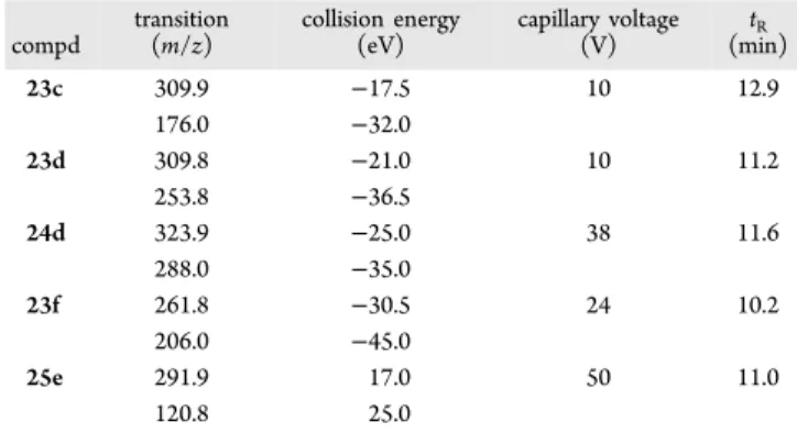
up to 70% in 10 min, then slowly increased up to 98% up to 15 min. Theflow rate was 0.3 mL/min, and injection volume was 5 μL. The instrument operated in positive mode, and parameters were the following: detector 1850 V, drying gas pressure 25.0 psi, desolvation temperature 300.0°C, nebulizing gas 40.0 psi, needle 5000 V, and shield 600 V. Nitrogen was used as nebulizer gas and drying gas. Collision induced dissociation was performed using argon as the collision gas at a pressure of 1.8 mTorr in the collision cell. The transitions as well as the capillary voltage and the collision energy used for compound are summarized inTable 4.
Quantification of the single compound was made by comparison with apposite calibration curves realized with standard solutions in methanol.
Solubility Assessment. The solubilizing capacity of formulation media was measured in the presence of 50% w/w of 2-HP-β-CD. Stock solution of compound 25e (DMSO) was added to obtain a concentration of 20μM in a final volume of 1 mL (DMSO did not exceed 2%). After sonication, the samples were shaken in a shaker bath at room temperature for 24 h to reach equilibrium conditions. Each pregel solution was analyzed before and afterfiltration by a 0.45 μm nylonfilter (Acrodisc). The loaded compound was determined using LC−UV−MS method already described for the permeability.
Molecular Modeling. Protein Preparation. The reverse tran-scriptase three-dimensional coordinates were selected from the Protein Data Bank (PDB entry code 1RT2)41and was prepared by means of Protein Preparation Wizard workflow implemented in the Maestro suite.42 In particular, the inhibitor and the water molecules were deleted, hydrogen atoms were added, and bond orders and charges were assigned; the orientation of hydroxyl groups on Ser, Thr, and Tyr, the side chains of Asn and Gln residues, and the protonation state of His residues were optimized. Steric clashes were relieved by performing a small number of minimization steps, not intended to minimize the system completely. In our study, the minimization (OPLS forcefield) was stopped when the rmsd of the non-hydrogen atoms reached 0.30 Å. The same procedure was applied to prepare the RT protein in complex with the water molecule making a bridge between the crystallized ligand TNK-651 and the amino acids Lys101 of chain A and Glu138 of chain B. In this case, the water molecule 1023 was kept in the system during the Protein Preparation Wizard workflow. To prepare the input structure for Autodock calculations, the RT structure was further manipulated by removing nonpolar hydrogen atoms, while Kollman united-atom partial charges and solvent parameter were added. Autogrid was then used to generate grid maps.
Ligand Preparation. 168 S-DABOs/N-DABOs were collected from previous work and were built with the Schrodinger Maestro 9.2 graphical interface.42 Compounds were then processed with the Schrodinger LigPrep tool to generate separate files for all possible enantiomers and protonation states at phisiological pH. OPLS_2005 was used as forcefield. Ligands for Autodock were further refined by
Table 4. Chromatographic and MS Parameters (Monitored
Transition, Collision Energy, Capillary Voltage, and
Retention Time
t
R) of the Selected Compounds
compd transition (m/z) collision energy (eV) capillary voltage (V) tR (min) 23c 309.9 −17.5 10 12.9 176.0 −32.0 23d 309.8 −21.0 10 11.2 253.8 −36.5 24d 323.9 −25.0 38 11.6 288.0 −35.0 23f 261.8 −30.5 24 10.2 206.0 −45.0 25e 291.9 17.0 50 11.0 120.8 25.0
deleting the nonpolar hydrogen atoms and adding Gasteiger atomic charges.
Docking Simulation. Docking studies were performed within the NNBP of RT using three software packages: Autodock,28Gold,29and
Glide.30,31The reliability of the docking protocols wasfirst checked by simulating the binding mode of known ligands for which the poses have been experimentally determined (TNK-651, MKC-442, and HEPT). The three docking algorithms were able to correctly predict the binding modes of the reference compounds. With regard to the Gold program, the ChemScore scoring function was found to be the best one in terms of pose prediction (in comparison with GoldScore, CHEMPLP, and ASP) and was thus used in the following calculations. The genetic algorithm parameter settings were employed using the search efficiency set at 100%, and 100 runs were carried out for each ligand. Finally, results differing less than 1 Å in ligand-all atom rmsd were clustered together. For each inhibitor, thefirst ranked solution was selected for further analysis. The Lamarckian genetic algorithm (LGA) was used in Autodock to explore the possible orientations/ conformations of the inhibitors in the binding site. For each compound, the following protocol was applied: 50 independent LGA runs, a population size of 150 individuals, and a maximum number of 2 500 000 energy evaluations. Moving to the Glide software, compounds were docked and scored using the Glide Standard Precision (SP) mode.
Multiple Linear Regression. QSAR calculations were performed using Canvas implemented in Maestro (version 9.5).42The conformer of each inhibitor was extracted from the complex with RT and submitted to the calculation of both QikProp and 2D fingerprint descriptors. These calculated properties, together with the parameters previously derived from docking studies with three different software as well as with the rescoring methods employed (MM-GBSA, XSCORE), were used as dependent variables to generate multiple linear regression (MLR) models, while enzymatic or cellular data were in turn used as the independent variable (expressed as−log IC50 or −log EC50). In the case of chiral compounds, the 3D energetic parameters coming from docking studies were calculated as the medium of the values obtained with all the possible diastereomeric forms. Molecules together with the calculated descriptors were imported in the cheminformatics package Canvas, and a MLR was carried out. The Simulated Annealing algorithm of Canvas was applied to descriptors to efficiently search the wide solution space and to identify the best subsets of descriptors to build robust QSAR models. The number of Monte Carlo steps was set to 1000.
■
ASSOCIATED CONTENT
*
S Supporting InformationThe Supporting Information is available free of charge on the
ACS Publications website
at DOI:
10.1021/acs.jmed-chem.5b01979
.
2D structures of S-DABO/N-DABO used for molecular
modeling analysis (
)
Molecular formula strings (
CSV
)
■
AUTHOR INFORMATION
Corresponding Author
*Phone: +39 0577 234306. Fax: +39 0577 234306.
Notes
The authors declare no competing
financial interest.
■
ACKNOWLEDGMENTS
This work was supported by the European Union collaborative
project
“CHAARM” (Grant HEALTH-F3-2009-242135) and
by the Italian Ministero dell
’Istruzione, dell’Università e della
Ricerca, Prin 2010 research project (Grant 2010W2KM5L).
Tenofovir was obtained through the NIH AIDS Reagent
Program, Division of AIDS, NIAID, NIH.
■
ABBREVIATIONS USED
cART, combination antiretroviral therapy; RTI, reverse
tran-scriptase inhibitor; S-DABO,
S-dihydroalkyloxybenzyl-oxopyrimidine; N-DABO,
N-dihydroalkyloxybenzyl-oxopyrimidine; WST-1,
4-[3-(4-iodophenyl)-2-(4-nitrophen-yl)-2H-5-tetrazolio]-1,3-benzene disulfonate; CDI, 1,1
′-carbonyldiimidazole; LDA, lithium diisopropylamide; HEC,
hydroxyethylcellulose
■
REFERENCES
(1) Looney, D.; Ma, A.; Johns, S. HIV therapy-the state of art. Curr. Top. Microbiol. Immunol. 2015, 389, 1−29.
(2) Franzetti, M.; Violin, M.; Antinori, A.; De Luca, A.; Ceccherini-Silberstein, F.; Gianotti, N.; Torti, C.; Bonora, S.; Zazzi, M.; Balotta, C. Trends and correlates of HIV-1 resistance among subjects failing an antiretroviral treatment over the 2003−2012 decade in Italy. BMC Infect. Dis. 2014, 14, 398.
(3) Zhan, P.; Liu, X.; Li, Z. Recent advances in the discovery and development of novel HIV-1 NNRTI platforms: 2006−2008 update. Curr. Med. Chem. 2009, 16, 2876−2889.
(4) Song, Y.; Fang, Z.; Zhan, P.; Liu, X. Recent advances in the discovery and development of novel HIV-1 NNRTI platforms (Part II): 2009−2013 update. Curr. Med. Chem. 2014, 21, 329−355.
(5) Manetti, F.; Esté, J. A.; Clotet-Codina, I.; Armand-Ugón, M.; Maga, G.; Crespan, E.; Cancio, R.; Mugnaini, C.; Bernardini, C.; Togninelli, A.; Carmi, C.; Alongi, M.; Petricci, E.; Massa, S.; Corelli, F.; Botta, M. Parallel solution-phase and microwave-assisted synthesis of new S-DABO derivatives endowed with subnanomolar anti-HIV-1 activity. J. Med. Chem. 2005, 48, 8000−8008.
(6) Mugnaini, C.; Manetti, F.; Esté, J. A.; Clotet-Codina, I.; Maga, G.; Cancio, R.; Botta, M.; Corelli, F. Synthesis and biological investigation of S-aryl-S-DABO derivatives as HIV-1 inhibitors. Bioorg. Med. Chem. Lett. 2006, 16, 3541−3544.
(7) Radi, M.; Contemori, L.; Castagnolo, D.; Spinosa, R.; Esté, J. A.; Massa, S.; Botta, M. A versatile route to C-6 arylmethyl-functionalized S-DABO and related analogues. Org. Lett. 2007, 9, 3157−3160.
(8) Mugnaini, C.; Alongi, M.; Togninelli, A.; Gevariya, H.; Brizzi, A.; Manetti, F.; Bernardini, C.; Angeli, L.; Tafi, A.; Bellucci, L.; Corelli, F.; Massa, S.; Maga, G.; Samuele, A.; Facchini, M.; Clotet-Codina, I.; Armand-Ugón, M.; Esté, J. A.; Botta, M. Dihydro-alkylthio-benzyl-oxopyrimidines as inhibitors of reverse transcriptase: synthesis and rationalization of the biological data on both wild-type enzyme and relevant clinical mutants. J. Med. Chem. 2007, 50, 6580−6595.
(9) Radi, M.; Falciani, C.; Contemori, L.; Petricci, E.; Maga, G.; Samuele, A.; Zanoli, S.; Terrazas, M.; Castria, M.; Togninelli, A.; Esté, J. A.; Clotet-Codina, I.; Armand-Ugón, M.; Botta, M. A multi-disciplinary approach for the identification of novel HIV-1 non-nucleoside reverse transcriptase inhibitors: S-DABOCs and DAVPs. ChemMedChem 2008, 3, 573−593.
(10) Botta, M.; Corelli, F.; Petricci, E.; Radi, M.; Maga, G.; Estè, J. A.; Mai, A. WO/2007/043094, 2007.
(11) Radi, M.; Angeli, L.; Franchi, L.; Contemori, L.; Maga, G.; Samuele, A.; Zanoli, S.; Armand-Ugon, M.; Gonzalez, E.; Llano, A.; Esté, J. A.; Botta, M. Towards novel S-DABOC inhibitors: synthesis, biological investigation, and molecular modeling studies. Bioorg. Med. Chem. Lett. 2008, 18, 5777−5780.
(12) Radi, M.; Maga, G.; Alongi, M.; Angeli, L.; Samuele, A.; Zanoli, S.; Bellucci, L.; Tafi, A.; Casaluce, G.; Giorgi, G.; Armand-Ugon, M.; Gonzalez, E.; Esté, J. A.; Baltzinger, M.; Bec, G.; Dumas, P.; Ennifar, E.; Botta, M. Discovery of chiral cyclopropyl dihydro-alkylthio-benzyl-oxopyrimidine (S-DABO) derivatives as potent HIV-1 reverse transcriptase inhibitors with high activity against clinically relevant mutants. J. Med. Chem. 2009, 52, 840−851.
(13) Radi, M.; Pagano, M.; Franchi, L.; Castagnolo, D.; Schenone, S.; Casaluce, G.; Zamperini, C.; Dreassi, E.; Maga, G.; Samuele, A.; Gonzalo, E.; Clotet, B.; Esté, J. A.; Botta, M. Synthesis, biological activity, and ADME properties of novel S-DABOs/N-DABOs as HIV reverse transcriptase inhibitors. ChemMedChem 2012, 7, 883−896.