British Medical Bulletin, 2017, 122:123–133 doi: 10.1093/bmb/ldx008 Advance Access Publication Date: 24 March 2017
Invited Review
Apoptosis and rotator cuff tears: scienti
fic
evidence from basic science to clinical
findings
Leonardo Osti
†, Matteo Buda
‡, Angelo Del Buono
§, Raffaella Osti
‡,
Leo Massari
‡, and Nicola Maffulli**
,††,*
†
Unit of Arthroscopy and Sports Medicine, Hesperia Hospital, Via Arqua
’, Modena, Italy,
‡Department of
Trauma and Orthopaedic Surgery, University of Ferrara, S.Anna Hospital, Via Aldo Moro, Ferrara, Italy,
§Department of Orthopedic and Trauma Surgery, Fidenza Hospital, Via Tincati, Fidenza, Italy,
**Department of Trauma and Orthopaedic Surgery, University of Salerno, Italy, and
††Centre for Sports
and Exercise Medicine Queen Mary University of London Barts and The London School of Medicine and
Dentistry, Mile End Hospital London, UK
*Correspondence address. Department of Trauma and Orthopaedic Surgery, University of Salerno, Italy. E-mail: [email protected]
Editorial Decision 28 February 2017; Accepted 8 March 2017
Abstract
Introduction: Excessive apoptosis has been hypothesized as possible cause
of tendinopathy and tear in the tendons of the rotator cuff (RC). Different
mechanisms and molecules play a key role in cell regulation. Biological
interventions can affect the process of apoptosis to control the
tendinopa-thy process, and may be useful to design new treatments.
Source of data: We identi
fied basic science, in vitro and in vivo preclinical
and clinical studies listed in the Pubmed Google Scholar, CINAHL,
Cochrane Central and Embase Biomedical databases in English, Spanish,
Italian and French concerning the effects of apoptosis on RC tendons.
Areas of agreement: The homeostasis between the apoptotic and in
flam-matory processes is dynamic and controlled by pro- and anti-apoptotic
mechanisms and signals, with variable balance in different areas of the RC
tendons in human specimens.
Areas of controversy: Apoptosis can be identi
fied along the whole tendon,
not only in the area of the lesion. Therefore, it is not necessary to undertake
wide debridement of the torn edges of the tendon when undertaking a
repair.
© The Author 2017. Published by Oxford University Press. All rights reserved. For permissions, please e-mail: [email protected]
Growing points: The identi
fication of the various factors that control
apop-tosis and its mechanisms can help to design new treatments and exert
positive effects in the recovery from tendon tears.
Areas timely for developing research: Further studies are needed to
pro-duce clear guidelines to determine how to balance the apoptosis process to
reduce the failed healing response found in non-traumatic RC tears.
Key words: rotator cuff, tendons, apoptosis, shoulder, tenocytes, tendinopathy
Introduction
Apoptosis, or programmed cell death, is an import-ant component in the control of cells proliferation by regulating embryogenesis, organogenesis and tis-sue morphogenesis, and maintaining homeostasis in many adult tissues.1The pathogenesis of atraumatic
rotator cuff (RC) tendon tears, in which the disease is the result of a combination of intrinsic tendon and extrinsic factors, is still controversial.2
Of the intrinsic factors, apoptosis seems to may play a key role in the regulation and development of the lesions.
Yuan et al.3in 2002 postulated excessive apop-tosis as a primary cause of tendinopathy and tears within the supraspinatus tendon, rather than tears being a secondary effect of degeneration, identifying apoptotic cells in supraspinatus tendon tears com-pared with control samples. Furthermore, excessive apoptosis seems to involve the entire RC in the presence of a supraspinatus tear.4
Apoptosis is triggered by exogenous and endogen-ous stimuli that initiate two different highly complex and sophisticated pathways: intrinsic and extrinsic.
The mitochondrial or intrinsic pathway is trig-gered by different factors (i.e. oxidative stress, genetic damage, high concentration of cytosolic calcium ions) within the cell that increase mitochondrial membrane permeability. Bax protein, migrating to the surface of the mitochondrion and inhibiting the protective effect of anti-apoptotic Bcl-2 protein, inserts itself into the outer mitochondrial membrane, perforating it. This results in an opening of the mitochondrial permeabil-ity transition pore and release of two main groups of normally sequestered pro-apoptotic proteins from the
intermembrane space into the cytosol: cytochrome c and the serine protease.
Cytochrome c binds to the protein Apaf-1 (apop-totic protease activating factor-1) and together form apoptosomes which bind to and activate caspase-9. Caspase-9 cleaves and, in so doing, activates other caspases (caspase-3 and -7).
The activation of these ‘executioner’ caspases creates an expanding cascade of proteolytic activ-ity (similar to what happens in blood clotting and complement activation) which leads to digestion of structural proteins in the cytoplasm, degradation of chromosomal DNA and phagocytosis of the cell.
The death-receptor mediated or extrinsic pathway is activated by transmembrane receptor-mediator ligand involving receptors which are members of the tumor necrosis factor (TNF) gene. Fas Ligand (FasL), binding the TNF receptor, induces apoptosis activating caspase 8 and starting a cascade of caspase activation leading to phagocytosis of the cell. Nitric oxide induces apoptosis by dissipating the membrane potential of mitochondria, and therefore makes the mitochondrial wall more permeable.5
The mechanism responsible for the modulation of apoptosis may be a combination of different factors such as withdrawal of positive signals (i.e. growth factors for neurons or Interleukin-2) or receipt of negative signals (i.e. increased levels of oxidants within the cell, damage to DNA by oxi-dants, death activators as TNF-α, TNF-β and FasL). Furthermore, hypoxia, extracellular matrix (ECM), cytokines, genetic factor and other pro-teins interact in the apoptotic pathway.6
Basic science studies focus on both physiological and pathological processes regulating apoptosis, and external factors which can decrease or balance the apoptosis.
This review analyzes the relationship between the factors which have been identified as possible regulators of the apoptotic process in RC tendons, and evaluates how they can influence apoptosis.
Methods
Search strategy
In August 2016, a systematic search was conducted in the online PubMed, Google Scholar, CINAHL, Cochrane Central and Embase Biomedical data-bases using various combinations of the following key terms and MesH terms: RC, tendons, tenocyte, tendinopathy, apoptosis and shoulder, with no lim-its regarding the year of publication and following the Preferred Reporting Items for Systematic Reviews and Meta-Analyses (PRISMA) guidelines.7
Our PRISMA checklist is presented in Figure 1. Two authors (M.B. and L.O.) screened all the arti-cles identified for title, abstract and full text con-cerning the effects of apoptosis in RC. All in vitro, in vivo preclinical and clinical studies in English, Spanish, Italian and French were evaluated and analyzed in this review.
Results
Genetic in
fluence
Genetic factors play a role in the development of full thickness tears of the RC.8,9Siblings of patients with RC tear had more than twice the relative risk to develop a RC lesion, and nearly five times the risk of experiencing symptoms.10 Tashjian et al.11 identified two single-nucleotide polymorphism asso-ciated with RC tears. They were located within two genes SAP30BP (on chromosome 17) and SASH1 (on chromosome 6); both these genes play a marked role in apoptosis, regulating tendon cell apoptosis and predisposing individuals to RC tears.
Hypoxia
Tenocytes are capable of withstanding acute and repetitive reductions of blood perfusion, and therefore are likely able to tolerate transient hypoxia. Hypoxia promotes the expression of pro-inflammatory cyto-kines, which are key apoptotic mediators, and drives matrix component synthesis towards a collagen type III profile by human tenocytes.12,13
Millar et al.14 noted that hypoxia could start and regulate early tendinopathy by showing in apoptotic cells an increase of hypoxia-inducible fac-tor 1α (HIF1α), a transcription factor that regulates several genes to promote survival in low oxygen,
Fig. 1 PRISMA 2009flow diagram.
125 Apoptosis and rotator cuff tears, 2017, Vol. 122
Bcl-2, a regulator protein and clusterin, a molecular chaperone and a member of the small heat shock protein (HSP) family. Hypoxia reduces the ERK (extracellular signal regulated kinase) and p38 pro-duction, increasing collagen III synthesis compared with type I. The final result is a decrease of the resistance of tendons to tensile forces.
Benson et al.15 showed that the expression of HIF-1α was greatest in mild impingement and in association with different stages of RC tendinopa-thy (partial, small, medium and large RC tears). Bcl-2 Nineteen KiloDalton interacting protein (BNip3) expression, a pro-apoptotic member of the Bcl-2 family with a role in hypoxia-induced death in many cell types, increased significantly in RC tears, but was reduced in massive tears. This is pos-sibly a consequence of the reduction of BNip3-positive cells from apoptosis. Excessive apoptosis was found within full thickness tears of the RC, with almost a threefold increase compared with the control or mild RC tear groups. Furthermore, there was a statistically significant association between the proportion of apoptotic cells and the age of the patients. This is contrary to Yuan et al.3study, in which there was no association between the propor-tion of apoptotic cells and age, durapropor-tion of symp-toms or size of tear. This is a likely consequence of the relatively small number of patients studied and the limited age range (50–70 years).
Nuclear factor-kB (NF-kB) also plays a role in the pathogenesis of RC tears. Evaluating the tear margins of 63 patients with non-traumatic RC tears (mean age 65 years, range: 52–74), it was shown that NF-kB, activated by hypoxia, was increased on the margins of the tendon tear with increasing size of the lesion. NF-kB seems to lessen the progression of the damage, supporting the healing response, and lessening the rate of tendon cells death.16
Cytokines and caspases
Cytokines are important molecular messengers in the response of soft tissue from injury to wound healing, and play an important role in oxidative-stress induced cellular apoptosis.17
The caspases are a family of cysteine proteases for aspartic acid residues, which cleave specific intracellular substrates and regulate cell death induced by apoptosis.18
Millar et al.19 confirmed the presence of the
cytokines IL-18, IL-15, IL-6 and macrophage inhibitory factor (MIF) in both rat and human mod-els of tendinopathy. They identified significantly increased levels of key mediators, caspases 3 and 8 and of Fas-Ligand apoptosis in the margins of full thickness tears of the RC compared with matched subscapularis tendon and normal control tendon. In another study, Millar et al.20showed an increase of
IL-17A in early RC tendinopathy. This cytokine seems to regulate and modulate thefirst phases of tendon degeneration by regulating inflammatory, apoptotic pathways and collagen matrix synthesis, as it increases collagen III and decreases collagen I synthesis.
Voloshin et al.21 showed an elevated expression of IL-1, IL-6 and TNF-α in the subacromial bursa in 10 patients with full thickness tears the RC comparing them to eight patients (control group) (P< 0.001).
Blain et al.22 found an increase of IL-1, IL-6,
TNF, COX-1, COX-2, MMP-1 and MMP-9 in 14 patients with subacromial bursitis undergoing mini-open RC repair compared to four patients undergo-ing surgery for shoulder instability.
Lundgreen et al.23comparing biopsies from nine patients (five men and four women; mean age 54) with partially torn supraspinatus tendons and the matched intact subscapularis tendons, and 10 refer-ence subscapularis tendons (five women and five men, mean age 43.9, undergoing an arthroscopic labral repair and who demonstrated no RC path-ology), showed a significant increase of apoptotic tenocytes, and cells expressing p53 and caspase c in both the supraspinatus and subscapularis tendons of patients presenting partial-thickness tears. In this study, however, the control group was significantly younger than the patient group, and two different types of tendons were compared.
Fabis et al.,24,25 in nine patients, noted a reduc-tion of caspases 3, 8, 9 and Bax (pro-apoptotic molecules) along a 1 cm section taken from the edge
of a torn supraspinatus tendon (P < 0.05). TNF-α decreases in a distal to proximal direction within 1 cm of the edge of a torn supraspinatus tendon, contrary to an increase of the anti-apoptotic mole-cules Bcl-2 and expression of the anti-inflammatory cytokine IL-10, which showed an opposite trend (P< 0.05).
Furthermore, the expression of type I collagen, IL-4 and IL-13 significantly increased, and IL-1b and IFN-γ decreased from the distal to the proximal parts of the tendon edge (P< 0.05). The homeosta-sis between the apoptotic and inflammatory pro-cesses can be dynamic and is highly dependent on the distance from the edge of the tear. The authors recommended careful resection of the torn edges of the RC tear, as resection 4–7 mm from the edge of the torn supraspinatus tendon may enhance the healing process.
Lee et al.26 quantified the extrinsic and intrinsic
apoptosis pathways by examing the activities of caspase 3/7, 8 and 9 and cytochrome c in tendon biopsies collected from 14 patients undergoing arthroscopic RC repair for RC tears. When these biopsies were compared with tendon biopsies from seven patients undergoing surgery for proximal humeral fractures, the activity of caspase 9 and cytochrome c was more than double that of other proteins. The increase of caspase 9 and cytochrome c activity suggests that the intrinsic (mitochondrial) pathway might be more important in the apoptosis mechanism of RC tears. They noted a significant difference in the apoptotic activities harvesting the samples from three different sites of the tendons. This suggests that apoptosis is a generalized phe-nomenon throughout the entire tendon. They hypothesized that apoptosis could start from the lateral aspect of the RC, and then propagate in all directions.
Yuan et al.27 showed that oxidative stress,
exposing to hydrogen peroxide (H2O2) human
tenocytes from patients undergoing surgery for RC repair, induced apoptosis most likely through releasing cytochrome c from mitochondria to cyto-sol, and activating caspase-3 in a time and dose-dependent manner. Lower concentration of H2O2
(50μM) led to tenocytes apoptosis, while there was
a significant increase in necrotic cell death with higher concentrations of H2O2.
Scott et al.28examined apoptosis using antibodies
against fragmented DNA in 32 male Sprague-Dawley rats randomly divided into controls (stand-ard cage care) and runners (stand(stand-ard cage care plus treadmill running which induced supraspinatus ten-dinopathy). They did not find consistent results regarding apoptosis in the primary stages of tendino-pathy, concluding that apoptosis might play a sec-ondary role in the more advanced stages of injury such asfibrosis or after frank tendon tears.
Heat shock protein
HSPs protect cells from the cytotoxic effects of cyto-kines and apoptotic mediators, and play a protect-ive role as molecular chaperones in cells by facilitating the folding, intracellular transport, assembly and disassembly of other proteins.29
Millar et al.30using 24 rat and 17 torn supraspi-natus tendons collected from patients with RC tears undergoing shoulder surgery (mean age 57 years but with wide range, 39–75 years) suggested that HSP plays a central role in caspase dependent apoptotic cell signaling and in the cascade of failed healing response found in tendinopathy. Over-expression of HSP 27 is essential in preventing cells from undergo-ing apoptosis, inhibitundergo-ing specifically the cytochrome C and ATP-triggered activity of caspase 9 on the apoptotic pathway. Furthermore, HSP 27 indirectly interferes with cell death because of its ability to modulate intracellular glutathione, while HSP 70 interacts with Apaf-1, thereby preventing its inter-action with the caspases, preventing apoptosis and protecting cells from heat stress, the cytotoxic effects of TNFα and from nitric oxide.
Extracellular matrix
The ECM of tendons is composed largely of type I collagen, which provides structural integrity and accounts for mechanical strength. Disruption of the collagen matrix is characterized by loss of its nor-mal longitudinal alignment. Fragmentation of colla-gen bundles decreases the mechanical property of the tendon, and may result in tendon rupture.31
127 Apoptosis and rotator cuff tears, 2017, Vol. 122
Wu et al.32 demonstrated that the percentage of
cells undergoing apoptosis increased gradually with the degree of ECM breakdown. The increase of apop-totic cells changes severely the tendon through the loss offiber structure, hyalinization and loss of paral-lel arrangement, with no discernible orientation of the fiber. A reduced cell population might affect the rate of collagen synthesis and capacity of healing after injury by contributing to matrix deterioration and promoting further cell death and cell differentiation.
The increase of type III collagen is expression of tendinopathy and hypoxia changes, and alters colla-gen synthesis by tenocytes, with a disproportionate concentration of type III collagen compared with type I, with detrimental changes in the ECM.14
Exogenous substance and hormones
Hyaluronic acid (HA) has an anti-inflammatory and anti-adhesive activity in tendons and in synovial fibroblasts derived from RC tear without evident adverse effect.33,34 Osti et al. in in vitro studies in RC degenerative torn tendon samples demonstrated a decreased rate of apoptosis when the tendon-derived cells were exposed to HA, stimulating and reducing the synthesis of collagen type I and type III respectively. The reduction in collagen type III seems to be a protective factor for tendons. The molecular weight of HA seems not to affect this process, although the presence of sorbitol 4% increases the expression of collagen I, limiting HA degradation.35
The effectiveness of corticosteroid (CS) injections results from their analgesic and anti-inflammatory properties, but they exert profound negative effects on human tenocytes. CSs induce early tendon fibro-blasts death and inhibit their proliferation at the injection site, and decrease the biomechanical prop-erties of the injected tendons.36 High doses of CS (1.0 mg/mL of triamcinolone acetonide) cause irre-versible effects on the human RC-derived tenocytes by prolonging the suppression of their viability and promoting apoptosis. CS increase mRNA expres-sions of caspase-3, 7, 8 and 9, indicating that both the endogenous and the exogenous apoptosis path-ways are activated. Lower concentrations of CS
(0.1 mg/mL of triamcinolone acetonide) seem to have reversible effects on the tendons in 3 weeks, suggesting intervals longer than 3 weeks for safe CS re-administration.37,38
Furthermore, the use of lidocaine with the injec-tion causes cytotoxicity, decreases biomechanical properties, induces apoptosis and delays collagen organization in torn RC tendons both in vivo and in vitro increasing reactive oxygen species produc-tion regulated by extracellular signals and activa-tion of caspase-3/7.39
Recently, platelet rich plasma (PRP), a blood frac-tion containing a high concentrafrac-tion of platelets, has been used to improve healing in tendon injuries, stimulating cell proliferation and total collagen pro-duction in human tenocyte cultures. Kim et al. showed that self assembled peptide (SAP)+ PRP can be effective in healing a RC tear by enhancing the collagen arrangement and inhibiting inflammatory changes and apoptosis.40 Muto et al. confirm that the deleterious effect and apoptosis of CS injections can be prevented by adding PRP in vitro.38On the other hand, Carr et al.41in a randomized controlled
trial including 60 patients (age: 35–75 years) showed that the co-application of PRP during arthroscopic acromionplasty did not improve clinical outcomes at any time of the study (P< 0.001). In that study, PRP caused potential deleterious effects on healing ten-dons, decreasing blood vessel number and tendon cellularity and increasing the levels of p53 protein in tendon biopsies taken 12 weeks after acromion-plasty. The authors concluded that PRP treatment could actually be pro-apoptosis.
Three studies showed how oxidative stress increased autophagy and RC tears, and how antiox-idants protect against autophagic cell death.42 Anthocyanins, a natural antioxidant, inhibiting intracellular ROS production and reducing the H2O2-induced phosphorylation of ERK1/2, JNK
and p38, plays an important anti-apoptotic role in the prevention of RC tendinopaty.43,44
Nevertheless, other studies are necessary to explain better their molecular interactions and how these molecules could be used to regulate the apop-tosis process.
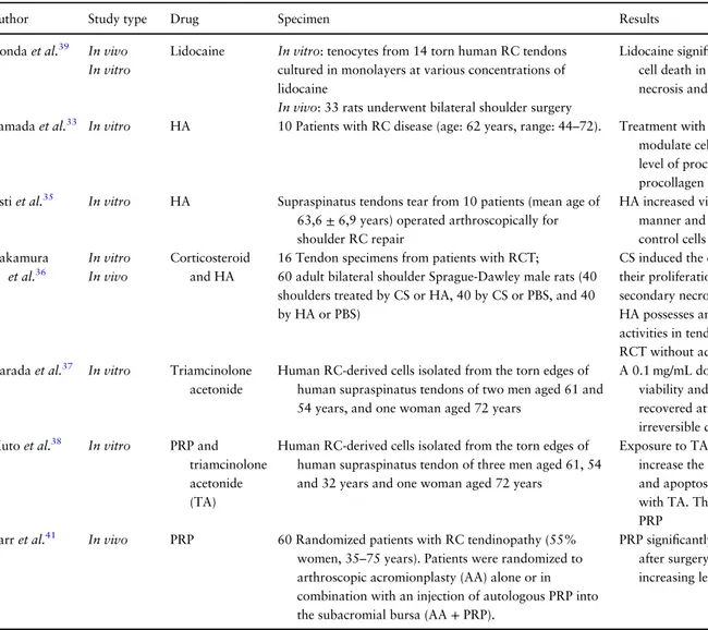
Table 1 Effects of exogenous substances
Author Study type Drug Specimen Results
Honda et al.39 In vivo In vitro
Lidocaine In vitro: tenocytes from 14 torn human RC tendons cultured in monolayers at various concentrations of lidocaine
In vivo: 33 rats underwent bilateral shoulder surgery
Lidocaine significantly inhibit cell proliferation and cause cell death in tenocytes, induce apoptosis, collagen necrosis and decrease biomechanical strength
Yamada et al.33 In vitro HA 10 Patients with RC disease (age: 62 years, range: 44–72). Treatment with various concentrations of HA significantly modulate cell proliferation decreasing the expression level of procollagen alpha1 (III) mRNA, but not that of procollagen alpha1 (I) mRNA
Osti et al.35 In vitro HA Supraspinatus tendons tear from 10 patients (mean age of 63,6± 6,9 years) operated arthroscopically for shoulder RC repair
HA increased viability and proliferation, in dose-dependent manner and reduce apoptosis at 24 h compared to control cells Nakamura et al.36 In vitro In vivo Corticosteroid and HA
16 Tendon specimens from patients with RCT;
60 adult bilateral shoulder Sprague-Dawley male rats (40 shoulders treated by CS or HA, 40 by CS or PBS, and 40 by HA or PBS)
CS induced the death of tendonfibroblasts and inhibited their proliferation with features typical of apoptosis and secondary necrosis.
HA possesses anti-inflammatory and anti-adhesive activities in tendon and synovialfibroblasts derived from RCT without adverse effects
Harada et al.37 In vitro Triamcinolone
acetonide
Human RC-derived cells isolated from the torn edges of human supraspinatus tendons of two men aged 61 and 54 years, and one woman aged 72 years
A 0.1 mg/mL dose of TA temporarily decreased cell viability and increased cell apoptosis, which was recovered at 21 days, however, 1 mg/mL of TA caused irreversible damage to cell morphology and viability Muto et al.38 In vitro PRP and
triamcinolone acetonide (TA)
Human RC-derived cells isolated from the torn edges of human supraspinatus tendon of three men aged 61, 54 and 32 years and one woman aged 72 years
Exposure to TA significantly decreased cell viability and increase the number of apoptotic cells, but cell viability and apoptosis did not decrease when PRP was added with TA. The deleterious effect of TA was prevented by PRP
Carr et al.41 In vivo PRP 60 Randomized patients with RC tendinopathy (55%
women, 35–75 years). Patients were randomized to arthroscopic acromionplasty (AA) alone or in combination with an injection of autologous PRP into the subacromial bursa (AA+ PRP).
PRP significantly alters the tissue characteristics in tendons after surgery reducing cellularity and vascularity and increasing levels of apoptosis
Continued 129 Apoptosis and rotator cuff tears, 2017, Vol. 122
Cigarette smoke has a negative effect on tendon healing, and many studies have remarked on the increase of the risk of RC tears in smookers; in par-ticular, nicotine and carbon monoxide decrease microperfusion and tissue oxygenation, leading to tissue hypoxia.45,46
Lundgreen et al.47showed that the supraspinatus
tendons in smokers have significantly more advanced degenerative changes, with increased density of apoptotic cells, reduced tenocyte density and down regulation of proliferative activity.
Endogenous substances may also regulate apop-tosis. Recent studies have investigate the role of hormones in the pathogenesis of tendinopathy, and it seems that hormonal imbalances cause negative effects on the healing and maturation process of tendon healing.48
Oliva et al. investigated the presence of thyroid hormones receptors in RC tendons, and described their possible role in the proliferation and apoptosis of human tenocyte by enhancing tenocyte growth and counteracting apoptosis in healthy tenocytes.49,50 Berardi et al. confirmed this hypothesis, in an in vitro study evaluating biopsies fromfive different healthy supraspinatus tendons, and showing how thyroid hormones modulate ECM proteins secre-tion (such as collagen I, biglycan and Cartilage Oligomeric Matrix Protein) in tenocytes.51
Maman et al.52 described the reaction of
teno-cytes to the estrogens and calciotropic stimulations: female sex steroids and vitamin D promoted tendon-derived cell proliferation via estrogen recep-torα and vitamin D receptor.
The results are summarized in Table1.
Conclusion
Apoptosis in RC tears may be induced by many stimuli, both endogenous and exogenous. In vivo and in vitro studies showed that repetitive stress and overuse in normal human tenocytes produce hypoxia, and start a typical pathway that induce apoptosis and contribute to tendon failed healing response.
In RC tears, the concentration of cytokines and caspases is increased. They play a key role in
Table 1 Continued Author Study typ e Dru g Specim en Results Kim et al. 40 In vivo PRP + self asse mbled pept ide (SAP ) 27 Sprague -Dawley rats random ly divided in four grou ps (7 SAP, 7 P R P , 7 SAP –PRP and six cont rol) SAP –PRP can be effec tive in healing a rotator cuf f tear by enha ncing the collag en arrang ement and in hibiting in fl amm atory cha nges and apop tosis Kim et al. 42 In vitro Cyanid in Huma n su prasp inatus tendo n tissues collected from thre e pat ients (two males and one femal e, ag es 50, 56 and 55 year s) Cyanidin inh ibits autopha gic cell death Nam et al. 44 In vivo Cyanid in and delph inidin Suprasp inatus tendo ns of adult male Sprague -Dawley rats Cyanidin and delphin idin exerted dos e-depe ndent ant i-apop totic effects (P < 0.001) Park et al. 43 In vitro Anthoc yanin s Suprasp inatus tendo ns of male Spra gue-D awley Anthocya nins has an in hibitory effec t o n H2O2-ind uced apop tosis by supp ressing both the intrac ellular ROS prod uction and acti vation of JNK and ERK 1/2 Lundgre en et al. 47 In vivo Smoke 10 Smoke rs vs 15 non smoke rs with full-thickness supra spinatu s tend on tears The su prasp inatus tendo ns from smoke rs hav e signi fi cantly more adv anced degenera tive chan ges with increa sed densi ty of apoptot ic cells, reduce d ten ocyte densi ty and upregu lation prolif erative activit y
cellular apoptosis and in the regulation of the homeostasis between the apoptotic and inflamma-tory processes in the RC tendon tears.
The pro-apoptotic signals increase moving away from the tear margins, and can play an important role during the surgical repairs of RC tear on the healing response of the tendon. It appears that wide debridement the edge of a tendon tear is not neces-sary to enhance the healing process.
Genetic influences increase the susceptibility to the earlier incidence of tears and progression of symptoms and tear size. Antioxidants could play a role in the development of RC tears, but other stud-ies are necessary to confirm this. Understanding the pathological cascade should lead to the develop-ment of cell-target treatdevelop-ment modalities for early RC tendinopathy.
Several drugs are injected to manage RC tear. CS seems to reduce pain, but with deleterious effects on tendons. HA reduces apoptosis acting on the produc-tion of collagen type I and reducproduc-tion of type III, but it is still unclear what are the most appropriate molecular weight and therapeutic protocol.
In conclusion, knowing which stimuli may trig-ger the cascade of RC tendinopathy and its modi fi-able mediators would allow to reduce the damage and increase the potential healing of such injuries.
Con
flict of interest statement
The authors have no potential conflicts of interest.
References
1. Thompson CB. Apoptosis in the pathogenesis and treat-ment of disease. Science 1995;267:1456–62.
2. McFarland e.g., Maffulli N, Del Buono A, et al. Impingement is not impingement: the case for calling it ‘Rotator Cuff Disease’. Muscles Ligaments Tendons J 2013;3:196–200.
3. Yuan J, Murrell GA, Wei AQ, et al. Apoptosis in rota-tor cuff tendinopathy. J Orthop Res 2002;20:1372–9. 4. Lundgreen K, Lian OB, Engebretsen L, et al. Tenocyte
apoptosis in the torn rotator cuff: a primary or secondary pathological event? Br J Sports Med 2011;45:1035–9. 5. Elmore S. Apoptosis: a review of programmed cell
death. Toxicol Pathol 2007;35:495–516.
6. Maffulli N, Longo UG, Berton A, et al. Biological fac-tors in the pathogenesis of rotator cuff tears. Sports Med Arthrosc 2011;19:194–201.
7. Moher D, Liberati A, Tetzlaff J, et al. Preferred report-ing items for systematic reviews and meta-analyses: the PRISMA statement. Ann Intern Med 2009;151:264–9. 8. Longo UG, Berton A, Papapietro N, et al.
Epidemiology, genetics and biological factors of rotator cuff tears. Med Sport Sci 2012;57:1–9.
9. Chaudhury S, Carr AJ. Lessons we can learn from gene expression patterns in rotator cuff tears and tendinopa-thies. J Shoulder Elbow Surg 2012;21:191–9.
10. Harvie P, Ostlere SJ, Teh J, et al. Genetic influences in the aetiology of tears of the rotator cuff: sibling risk of a full-thickness tear. J Bone Joint Surg 2004;86-B:696–700. 11. Tashjian RZ, Granger EK, Farnham JM, et al.
Genome-wide association study for rotator cuff tears identifies two significant single-nucleotide polymorphisms. J Shoulder Elbow Surg 2016;25:174–9.
12. Liang M, Cornell HR, Zargar Baboldashti N, et al. Regulation of hypoxia-induced cell death in human tenocytes. Adv Orthop 2012;2012:984950.
13. Petersen W, Varoga D, Zantop T, et al. Cyclic strain influences the expression of the vascular endothelial growth factor (VEGF) and the hypoxia inducible factor 1 alpha (HIF-1 alpha) in tendonfibroblasts. J Orthop Res 2004;22:847–53.
14. Millar NL, Reilly JH, Kerr SC, et al. Hypoxia: a critical regulator of early human tendinopathy. Ann Rheum Dis 2012;71:302–10.
15. Benson RT, McDonnell SM, Knowles HJ, et al. Tendinopathy and tears of the rotator cuff are asso-ciated with hypoxia and apoptosis. J Bone Joint Surg Br 2010;92:448–53.
16. Gumina S, Natalizi S, Melaragni F, et al. The possible role of the transcription factor nuclear factor-κB on evolu-tion of rotator cuff tear and on mechanisms of cuff ten-don healing. J Shoulder Elbow Surg 2013;22:673–80. 17. Nguyen MT, Lue H, Kleemann R, et al. The cytokine
macrophage migration inhibitory factor reduces pro-oxidative stress-induced apoptosis. J Immunol 2003; 170:3337–47.
18. McIlwain DR, Berger T, Mak TW. Caspase functions in cell death and disease. Cold Spring Harb Perspect Biol 2015;5:a008656.
19. Millar NL, Wei AQ, Molloy TJ, et al. Cytokines and apoptosis in supraspinatus tendinopathy. J Bone Joint Surg Br 2009;91:417–24.
20. Millar NL, Akbar M, Campbell AL, et al. IL-17A med-iates inflammatory and tissue remodeling events in early human tendinopathy. Sci Rep 2016;6:27149.
131 Apoptosis and rotator cuff tears, 2017, Vol. 122
21. Voloshin I, Gelinas J, Maloney MD, et al. Proinflammatory cytokines and metalloproteases are expressed in the sub-acromial bursa in patients with rotator cuff disease. Arthroscopy 2005;21:1076.e1–1076.e9.
22. Blaine TA, Kim YS, Voloshin I, et al. The molecular pathophysiology of subacromial bursitis in rotator cuff disease. J Shoulder Elbow Surg 2005;14:84S–9S. 23. Lundgreen K, Lian Ø, Scott A, et al. Increased levels of
apoptosis and p53 in partial-thickness supraspinatus tendon tears. Knee Surg Sports Traumatol Arthrosc 2013;21:1636–41.
24. Fabis´ J, Szemraj J, Strek M, et al. Is resection of the ten-don edge necessary to enhance the healing process? An evaluation of the homeostasis of apoptotic and in flam-matory processes in the distal 1 cm of a torn supraspi-natus tendon: part I. J Shoulder Elbow Surg 2014;23: 1772–8.
25. Fabis´ J, Szemraj J, Strek M, et al. Is resection of the ten-don edge necessary to enhance the healing process? An evaluation of the expression of collagen type I, IL-1β, IFN-γ, IL-4, and IL-13 in the distal 1 cm of a torn supraspinatus tendon: part II. J Shoulder Elbow Surg 2014;23:1779–85.
26. Lee HJ, Kim YS, Ok JH, et al. Apoptosis occurs throughout the diseased rotator cuff. Am J Sports Med 2013;41:2249–55.
27. Yuan J, Murrell GA, Trickett A, et al. Involvement of cytochrome c release and caspase-3 activation in the oxidative stress-induced apoptosis in human tendon fibroblasts. Biochim Biophys Acta 2003;1641:35–41. 28. Scott A, Cook JL, Hart DA, et al. Tenocyte responses to
mechanical loading in vivo: a role for local insulin-like growth factor 1 signaling in early tendinosis in rats. Arthritis Rheum 2007;56:871–81.
29. Hartl FU, Martin J, Neupert W. Protein folding in the cell: the role of molecular chaperones Hsp70 and Hsp60. Annu Rev Biophys Biomol Struct 1992;21:293–322. 30. Millar NL, Wei AQ, Molloy TJ, et al. Heat shock
pro-tein and apoptosis in supraspinatus tendinopathy. Clin Orthop 2008;466:1569–76.
31. Del Buono A, Oliva F, Longo UG, et al. Metalloproteases and rotator cuff disease. J Shoulder Elbow Surg 2012;21:200–8.
32. Wu B, Chen J, Dela Rosa T, et al. Cellular response and extracellular matrix breakdown in rotator cuff tendon rupture. Arch Orthop Trauma Surg 2011;131:405–11. 33. Yamada T, Gotoh M, Nakama K, et al. Effect of
hya-luronan on cell proliferation and mRNA expression of procollagensɑ1(I) and ɑ1(III) in tendon-derived fibro-blasts from patients with rotator cuff disease. Am J Sports Med 2007;35:1870–6.
34. Osti L, Buda M, Buono AD, et al. Clinical evidence in the treatment of rotator cuff tears with hyaluronic acid. Muscles Ligaments Tendons J 2016;5:270–5.
35. Osti L, Berardocco M, Di Giacomo V, et al. Erratum to: hyaluronic acid increases tendon derived cell viability and collagen type I expression in vitro: comparative study of four different hyaluronic acid preparations by molecular weight. BMC Musculoskelet Disord 2015;16:334. 36. Nakamura H, Gotoh M, Kanazawa T, et al. Effects of
corticosteroids and hyaluronic acid on torn rotator cuff tendons in vitro and in rats. J Orthop Res 2015; 33:1523–30.
37. Harada Y, Kokubu T, Mifune Y, et al. Dose- and time-dependent effects of triamcinolone acetonide on human rotator c6uff-derived cells. Bone Joint Res 2014;3:328–34. 38. Muto T, Kokubu T, Mifune Y, et al. Platelet-rich plasma protects rotator cuff-derived cells from the deleterious effects of triamcinolone acetonide. J Orthop Res 2013;31:976–82. 39. Honda H, Gotoh M, Kanazawa T, et al. Effects of lidocaine
on torn rotator cuff tendons. J Orthop Res 2016;34: 1620–7.
40. Kim SJ, Lee SM, Kim JE, et al. Effect of platelet-rich plas-ma with self-assembled peptide on the rotator cuff tear model in rat. J Tissue Eng Regen Med 2017;11:77–85. 41. Carr AJ, Murphy R, Dakin SG, et al. Platelet-rich
plas-ma injection with arthroscopic acromioplasty for chronic rotator cuff tendinopathy: a randomized con-trolled trial. Am J Sports Med 2015;43:2891–7. 42. Kim RJ, Hah YS, Sung CM, et al. Do antioxidants
inhibit oxidative-stress-induced autophagy of tenofibro-blasts? J Orthop Res 2014;32:937–43.
43. Park HB, Hah YS, Yang JW, et al. Antiapoptotic effects of anthocyanins on rotator cuff tenofibroblasts. J Orthop Res 2010;28:1162–9.
44. Nam DC, Hah YS, Nam JB, et al. Cytoprotective mech-anism of cyanidin and delphinidin against oxidative stress-induced tenofibroblast death. Biomol Ther (Seoul) 2016;24:426–32.
45. Baumgarten KM, Gerlach D, Galatz LM, et al. Cigarette smoking increases the risk for rotator cuff tears. Clin Orthop Relat Res 2010;468:1534–41. 46. Carbone S, Gumina S, Arceri V, et al. The impact of
preoperative smoking habit on rotator cuff tear: cigar-ette smoking influences rotator cuff tear sizes. J Shoulder Elbow Surg 2012;21:56–60.
47. Lundgreen K, Lian OB, Scott A, et al. Rotator cuff tear degeneration and cell apoptosis in smokers versus non-smokers. Arthroscopy 2014;30:936–41.
48. Oliva F, Piccirilli E, Berardi AC, et al. Hormones and tendinopathies: the current evidence. Br Med Bull 2016; 117:39–58.
49. Oliva F, Berardi AC, Misiti S, et al. Thyroid hormones and tendon: current views and future perspectives. Concise review. Muscles Ligaments Tendons J 2013;3: 201–3.
50. Oliva F, Berardi AC, Misiti S, et al. Thyroid hormones enhance growth and counteract apoptosis in human tenocytes isolated from rotator cuff tendons. Cell Death Dis 2013;4:e705.
51. Berardi AC, Oliva F, Berardocco M, et al. Thyroid hor-mones increase collagen I and cartilage oligomeric matrix protein (COMP) expression in vitro human tenocytes. Muscles Ligaments Tendons J 2014;4:285–91.
52. Maman E, Somjen D, Maman E, et al. The response of cells derived from the supraspinatus tendon to estrogens and calciotropic hormones stimulations: in-vitro study. Connect Tissue Res 2015;57:124–30.
133 Apoptosis and rotator cuff tears, 2017, Vol. 122