Inaccuracy of Insulin-Like Growth Factor (IGF) Binding
Protein (IGFBP)-3 Assessment in the Diagnosis of
Growth Hormone (GH) Deficiency from Childhood to
Young Adulthood: Association to Low GH Dependency of
IGF-II and Presence of Circulating IGFBP-3
18-Kilodalton Fragment
Stefano Cianfarani, Alice Liguori, Sergio Boemi, Mohamad Maghnie, Lorenzo Iughetti,
Malgorzata Wasniewska, Maria E. Street, Stefano Zucchini, Gianluca Aimaretti, and Daniela Germani Department of Public Health and Cell Biology (S.C., A.L., D.G.), Tor Vergata University, 00133 Rome, Italy; Division of Nuclear Medicine (S.B.), San Eugenio Hospital, 00144 Rome, Italy; Department of Pediatrics (M.M.), Istituto di Ricovero e Cura a Carattere Scientifico Policlinico San Matteo, University of Pavia, 27100 Pavia, Italy; University of Modena and Reggio Emilia (L.I.), 41100 Modena, Italy; University of Messina (M.W.), 98100 Messina, Italy; University of Parma (M.E.S.), 43100 Parma, Italy; University of Bologna (S.Z.), 40100 Bologna, Italy; and Department of Internal Medicine (G.A.), University of Turin, 10100 Turin, Italy
Context: Poor sensitivity of IGF binding protein (IGFBP)-3 assess-ment in the work-up of GH deficiency (GHD) has been ascribed to the equal affinity of IGFBP-3 for IGF-I and IGF-II and to IGFBP-3 proteolysis.
Objective: The objective of this study was to determine the IGF-II GH dependency and IGFBP-3 proteolysis in patients with GHD from childhood to young adulthood.
Design: This study was cross-sectional.
Setting: This was a national multicenter study performed in uni-versity hospitals.
Patients: One hundred thirty-one subjects (chronological age, 1.3–25 yr), 72 patients with GHD and 59 subjects with idiopathic short stature, were studied.
Interventions: IGF-I, IGF-II, and IGFBP-3 serum concentrations were measured by immunoradiometric assay. IGFBP-3 circulating forms were assessed by Western immunoblot (WIB) analysis.
Main Outcome Measures: Main outcome measures were sensitivity and specificity of IGF-I, IGF-II, and IGFBP-3 measurements. Results: Sensitivity and specificity of IGFBP-3 measurement were 27 and 100%, respectively. IGFBP-3 sensitivity was 46% in young adult-hood. Sensitivity and specificity of IGF-I were 69 and 81%, respec-tively. Sensitivity and specificity of IGF-II assessment were 23 and 97%, respectively. IGFBP-3 WIB revealed the presence of the intact form and the major 29-kDa fragment in both GHD and subjects with idiopathic short stature. In patients with GHD, WIB showed the presence of an additional smaller IGFBP-3 fragment migrating at approximately 18 kDa.
Conclusions: Our results suggest that in children and young adults with GHD, the low GH dependency of IGF-II together with IGFBP-3 proteolytic activity yielding the 18-kDa fragment concur to reduce the sensitivity of IGFBP-3 assessment, ultimately making it too inaccu-rate as a screening test in the work-up of GHD. (J Clin Endocrinol
Metab 90: 6028 – 6034, 2005)
T
HE DIAGNOSIS OF GH deficiency (GHD) is not straightforward in childhood and adolescence, requir-ing comprehensive clinical, anthropometric, biochemical, en-docrine, and neuroradiological assessment (1, 2). Although pharmacological GH stimulation tests are still considered the gold standard for GHD diagnosis, they are burdened by both poor specificity and side effects (3). The threshold value usedto define a normal GH response was chosen arbitrarily. Ini-tially, it was set at 5g/liter, thereafter gradually moved up to the cutoff value of 10g/liter, after the increased avail-ability of biosynthetic GH rather than a physiology based rationale (2, 4).
Although difficult, the diagnosis of GHD still has a number of important clinical implications. The child with GHD must be started on daily sc injections of GH that may be lifelong. Furthermore, appropriate replacement ther-apy enables the GHD child to achieve a normal adult height. Finally, once GHD is diagnosed, the child must be investigated for other pituitary deficiencies such as hy-pothyroidism and adrenal insufficiency, the latter being one of the major causes of death in patients with GHD (4, 5).
First Published Online August 9, 2005
Abbreviations: CV, Coefficient(s) of variation; GHD, GH deficiency; IGFBP, IGF binding protein; IRMA, immunoradiometric assay; ISS, id-iopathic short stature; MRI, magnetic resonance imaging; WIB, Western immunoblot.
JCEM is published monthly by The Endocrine Society (http://www. endo-society.org), the foremost professional society serving the en-docrine community.
doi: 10.1210/jc.2005-0721
The measurement of IGF-I and IGF binding protein (IGFBP)-3, due to the close GH dependency, was proposed for diagnosing GHD (6, 7). However, a number of factors such as technical problems, degree of sexual maturation, nutritional status, intestinal absorption, liver function, thy-roid hormones, and genetic determinants, may affect the measurements (8). We previously reported that although specificity of IGFBP-3 assessments is high (above 90%), sen-sitivity is no more than 50% in diagnosing childhood-onset GHD (9, 10). To explain the low sensitivity of IGFBP-3 as-sessment, two major causes have been proposed. IGFBP-3 concentrations would reflect the total IGF concentrations, IGF-I⫹ IGF-II, IGF-II being less GH dependent than IGF-I (11). In addition, the measurement of IGFBP-3 serum con-centrations is complicated by the existence of a family of proteases that fragment IGFBP-3 (12). These proteases, which include plasmin, matrix metalloproteinases, and prostate-specific antigen, degrade IGFBP-3, thereby reducing its af-finity for IGFs and eventually leading to increased IGF bio-availability. Proteolysis might affect IGFBP-3 measurement (8). Indeed, most of the available commercial kits employ anti-IGFBP-3 antibodies that recognize all the IGFBP-3 cir-culating forms, including IGFBP-3 fragments (13–16).
To date, no systematic study has been carried out to assess the IGF-II GH dependency and to evaluate IGFBP-3 prote-olysis in a large cohort of patients with GHD ranging from childhood to young adulthood, defined as the time window encompassing the 6 –7 yr after the achievement of adult height (17). The measurement of IGFBP-3 concentrations seems to be more accurate in the diagnosis of adult-onset GHD (18, 19). Our work hypothesis was that increased IGFBP-3 proteolysis in children with GHD might represent a compensatory mechanism to maximize the biological ac-tions of the reduced IGF-I concentraac-tions during linear growth. After the achievement of adult height, proteolysis might progressively vanish.
We set up a national multicenter study to assess sensitivity and specificity of the measurements of IGFBP-3, IGF-I, and IGF-II in patients with GHD from childhood to young adult-hood and children with idiopathic short stature (ISS). Fur-thermore, we investigated IGFBP-3 circulating forms in both groups of subjects.
Subjects and Methods
Seventy-two patients with GHD (47 boys and 25 girls) and 59 subjects (37 males and 22 females) with ISS were evaluated. Patients with GHD were enrolled into the national multicenter study, whereas all subjects with ISS were recruited from the Outpatient Growth Clinic of the Rina Balducci Center of Pediatric Endocrinology (Tor Vergata University, Rome, Italy). Anthropometric measurements and pubertal stage eval-uations were performed according to standard procedures (20), and data were compared with the standards of Tanner and Whitehouse (21). Patients’ heights, growth rates, and bone ages were expressed as Z scores for chronological age and sex. Z scores were calculated with the fol-lowing formula: Z score⫽ (x ⫺ average x)/sd where x is the observed measurement, average x is the mean of this measurement at the relevant age, and sd is the sd from the mean. Bone age was estimated by the method of Greulich and Pyle (22). The protocol was approved by the Institutional Review Boards of all participating centers, and informed consent was obtained from the patients or their parents or guardians. GHD diagnosis was based on fulfillment of all the following anthro-pometric, endocrine, and radiological criteria: stature less than⫺2 Z score, delayed bone age (at least 1 yr), growth rate less than 25th centile,
peak GH response to two different provocative tests less than 10g/liter (⬍19g/liter for GHRH ⫹ arginine test), brain magnetic resonance imaging (MRI) positive for hypothalamus-pituitary abnormalities such as pituitary hypoplasia, stalk agenesis, and ectopic posterior lobe (23), and catch-up growth during the first year of GH replacement therapy (growth rateⱖ 75th centile). Patients with GHD who had achieved adult height were retested after 3 months wash-out and included only if peak GH response to insulin tolerance test was less than 3g/liter. Children with GHD were studied off-therapy, pretherapy, or after at least 2 months wash-out. Patients with GHD secondary to intracranial tumors were tested at least 6 months after completion of radiotherapy and/or chemotherapy. All patients with intracranial tumors had surgical pitu-itary and/or stalk resection. No patient had surgery for at least 3 months before the enrolment.
In young adult patients, severe GHD was defined by the presence of structural hypothalamic-pituitary abnormalities and peak GH response less than 3g/liter. Among 72 patients with GHD, 54 had isolated GHD, 11 had combined GH, TSH, ACTH, and gonadotrophin deficiency, three had GHD and TSH deficiency, three had GHD and gonadotrophin deficiency, and one had GHD and diabetes insipidus.
Children referred for anthropometry resembling that of GHD but with normal peak GH responses and normal brain MRI were diagnosed as having ISS. Brain MRI was performed in those subjects with ISS referred for reduced growth rate to rule out intracranial lesions. In all subjects, IgA-endomysial and tissue transglutaminase antibody testing and free thyroxine and thyroid-stimulating hormone assessment were performed to exclude celiac disease and hypothyroidism, respectively. Karyotype was normal in all girls, and no patient had dysmorphic features, malnutrition (percentage of ideal body weight was more than 85% in all patients), or chronic diseases.
Clonidine (100g/m2body surface area, orally), arginine (0.5 g/kg
body weight up to a maximum of 30 g iv), insulin (0.10 IU/kg body weight iv), glucagon (100g/kg body weight up to a maximum of 1 mg im), and, in patients with GHD only, GHRH plus arginine (GHRH, 1 g/kg iv) stimulation tests were used to assess GH secretion. All subjects were tested in fasting conditions in each participating center. Prepu-bertal boys older than 10 yr were primed with 100 mg depot testosterone 3 d before testing. Prepubertal girls older than 9 yr were primed with 50g/d ethinyl estradiol for 3 consecutive days before testing.
Hormone assays
In Rome and Messina, serum GH was measured by immunoradio-metric assay (IRMA) (Diagnostic Systems Laboratories, Inc., Webster, TX). The intraassay coefficient of variation (CV) was 3.1– 4.4%, the in-terassay CV was 5.9 –11.5%, and the sensitivity limit was 0.01g/liter. In Pavia, serum GH was measured by fluoroimmunoassay DELFIA (Wallac Oy, Turku, Finland). The intraassay CV was 1.6 – 8.4%, the interassay CV was 2.0 –9.0%, and the sensitivity limit was 0.0038g/ liter. In Modena, serum GH was measured by chemiluminescence assay (Nichols Institute Diagnostics, San Juan Capistrano, CA). The intraassay CV was 4.9 – 6.7%, the interassay CV was 7.2–9%, and the sensitivity limit was 0.1g/liter. In Parma and Bologna, serum GH was measured by RIA (Nichols Institute Diagnostics). The intraassay CV was 2.8 – 4.2%, the interassay CV was 3.5–7.2%, and the sensitivity limit was 0.02g/ liter. In Turin, serum GH was measured by IRMA (Sorin, Saluggia, Italy). The intraassay CV was 1.5–2.9%, the interassay CV was 4.9 – 6.5%, and the sensitivity limit was 0.15g/liter.
IGF-I, IGF-II, and IGFBP-3 assessments were centralized in the lab-oratory of the Rina Balducci Centre of Pediatric Endocrinology (Rome, Italy). Blood samples were collected without anticoagulant between 0800 and 0900 h from fasting subjects, allowed to coagulate, and cen-trifuged at room temperature. Serum samples were subdivided in two aliquots (A, 0.8 –1 ml; B, 0.1– 0.2 ml) and stored at⫺20 C. Thereafter, frozen serum samples were shipped in dry ice and kept at ⫺20 C. Samples thawed just before the assays that were performed within 6 months from blood collection. IGF-I, IGF-II, and IGFBP-3 measurements were made on serum aliquot A, IGFBP-3 Western immunoblot (WIB) analysis was performed on aliquot B.
Serum IGFBP-3 was measured by IRMA (Diagnostic Systems Labo-ratories, Inc.). The intraassay CV was 1.8 –3.9%, the interassay CV was 0.5–1.9%, and the sensitivity limit was 0.5 mg/liter (17.5 nmol/liter). Serum IGF-I was measured by IRMA (Nichols Institute Diagnostics).
IGF-I was measured after extraction obtained by acidification followed by the addition of excess IGF-II to block the IGFBP binding sites. The intraassay CV was 3.3– 4.6%, the interassay CV was 9.3–15.8%, and the sensitivity limit was 6g/liter (0.8 nmol/liter). Serum IGF-II was mea-sured by IRMA (Diagnostic Systems Laboratories, Inc.). The intraassay CV was 3.4 – 6.5%, the interassay CV was 4.5– 6.3%, and the sensitivity limit was 12g/liter (1.6 nmol/liter). Both IGF-I and IGFBP-3 assays were validated in our laboratory by assessing the blood concentrations in 82 normal children and comparing the results with the normative data provided by the manufacturer’s kit (9).
IGFBP-3 WIB analysis
To visualize circulating proteolytic fragments of IGFBP-3, WIB anal-ysis was performed as previously described (24). Briefly, after addition of nonreducing sodium dodecyl sulfate sample buffer, serum samples (3l) were processed by SDS-PAGE (12% acrylamide gel). Separated proteins were electroblotted onto nitrocellulose filters in a Hoeffer semi-dry transfer unit (San Francisco, CA). Filters were blocked with 1% BSA and sequentially incubated with goat anti-IGFBP-3 antibody (Diagnostic Systems Laboratories, Inc.) overnight at 4 C and with goat antisheep IgG conjugated with horseradish peroxidase (Amersham Biosciences UK,
Ltd., Little Chalfont, Buckinghamshire, UK) for 2 h at room temperature. Filters were exposed to enhanced chemiluminescence reagents (Amer-sham Biosciences UK, Ltd.) for 1 min at 20 C and exposed to hyperfilm ECL for 1 min to 1 h at 20 C. Densitometric analysis of bands was performed using a Bio-Rad GS 700 imaging densitometer (Bio-Rad, Richmond, CA). The relative amount of each IGFBP-3 circulating frag-ment was estimated by calculating the absorbance of the fragfrag-ment band over the sum of the intact IGFBP-3 and IGFBP-3 fragments in the same lane.
Statistical analysis
Results are reported as mean⫾ sd. Differences between means were assessed using unpaired two-tailed Student’s t test. Significance was assigned for P⬍ 0.05. Sensitivity (true positive ratio) was defined as the number of true positives divided by the sum of true positives plus false negatives. Specificity (true negative ratio) was defined as the number of true negatives divided by the sum of true negatives plus false positives.
Results
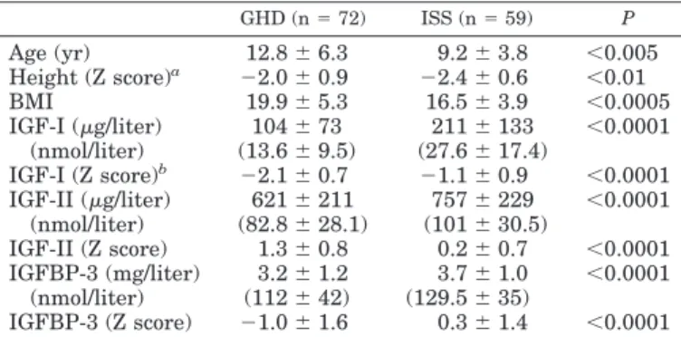
Clinical and endocrine data of patients with GHD and ISS are summarized in Table 1.
IGFBP-3 assessment
IGFBP-3 serum levels were evaluated in 52 patients with GHD (30 males and 22 females) and in 58 children with ISS (36 males and 22 females). The majority of patients with GHD had IGFBP-3 values within the normal range (Fig. 1). Only 10 of 30 males and four of 22 females had IGFBP-3 concen-trations equal or below⫺2 Z score, with an overall sensitivity of 27%.
In young adult patients with GHD (from approximately 15–25 yr, 15 males and nine females), the sensitivity of IGFBP-3 assessment was 46%.
None of the children with ISS had IGFBP-3 levels equal or below ⫺2 Z score. Therefore, the overall specificity of IGFBP-3 measurement was 100% (Fig. 1).
TABLE 1. Clinical and endocrine data of GHD and ISS subjects GHD (n⫽ 72) ISS (n⫽ 59) P Age (yr) 12.8⫾ 6.3 9.2⫾ 3.8 ⬍0.005 Height (Z score)a ⫺2.0 ⫾ 0.9 ⫺2.4 ⫾ 0.6 ⬍0.01 BMI 19.9⫾ 5.3 16.5⫾ 3.9 ⬍0.0005 IGF-I (g/liter) 104⫾ 73 211⫾ 133 ⬍0.0001 (nmol/liter) (13.6⫾ 9.5) (27.6⫾ 17.4) IGF-I (Z score)b ⫺2.1 ⫾ 0.7 ⫺1.1 ⫾ 0.9 ⬍0.0001 IGF-II (g/liter) 621⫾ 211 757⫾ 229 ⬍0.0001 (nmol/liter) (82.8⫾ 28.1) (101⫾ 30.5) IGF-II (Z score) 1.3⫾ 0.8 0.2⫾ 0.7 ⬍0.0001 IGFBP-3 (mg/liter) 3.2⫾ 1.2 3.7⫾ 1.0 ⬍0.0001 (nmol/liter) (112⫾ 42) (129.5⫾ 35) IGFBP-3 (Z score) ⫺1.0 ⫾ 1.6 0.3⫾ 1.4 ⬍0.0001
aHeight Z scores were determined at the time of blood collection
for IGF-I/IGF-II/IGFBP-3 measurements.
bAccording to reference standards in Z score reported in a different
IGF-I IRMA commercial kit (Diagnostic Systems Laboratories, Inc.).
FIG. 1. Individual data for IGFBP-3, expressed as Z score, in GHD (n⫽ 52, F) and ISS (n⫽ 58, ‚).
IGF-I assessment
IGF-I was measured in all 72 patients with GHD (47 boys and 25 girls) and 59 subjects with ISS (37 males and 22 females). The commercial kit we used provides reference values according to age in the form of centiles (5th to 95th centiles). Taking the fifth centile as threshold value, 50 of 72 patients with GHD showed IGF-I levels equal or below the cutoff value, with a sensitivity of 69% (Fig. 2, A and B). In children with ISS, IGF-I concentrations were equal or below
the fifth centile in 11 of 59 subjects, with a specificity of 81% (Fig. 2, A and B). It is noteworthy that specificity of IGF-I measurement was 91% in children younger than 11, whereas in subjects older than 11, specificity was 53%.
IGF-II assessment
IGF-II serum levels were evaluated in 52 patients with GHD (30 males and 22 females) and 40 children with ISS (20 males and 20 females). Twelve of 52 subjects with GHD
FIG. 2. A, Individual data for IGF-I, ex-pressed as centiles, in GHD (n⫽ 47, F) and ISS (n⫽ 37, ‚) males. B, Individual data for IGF-I, expressed as centiles, in GHD (n⫽ 25, F) and ISS (n ⫽ 22, ‚) females.
showed concentrations equal or below ⫺2 Z score, with a sensitivity of 23% (Fig. 3). In young adult patients with GHD (from approximately 15–25 yr, 12 males and seven females), the sensitivity of IGF-II assessment was 26%. All but one child with ISS had IGF-II levels above⫺2 Z score. The overall specificity of IGF-II measurement was 97.5% (Fig. 3).
IGFBP-3 WIB analysis
WIB analysis was performed in all 72 patients with GHD and in 40 children with ISS. In both groups, IGFBP-3 immu-noblot analysis revealed the presence of two major forms: the intact form represented by a doublet migrating at approxi-mately 42–39 kDa molecular mass and the major product of proteolysis migrating at approximately 29 kDa (Fig. 4, A and B).
Densitometry did not show any relationship between the relative amounts of the two IGFBP-3 forms and age, sex, anthropometry, peak GH response, and IGF-I and IGF-II concentrations. In all patients with GHD, WIB analysis re-vealed the presence of an additional smaller fragment mi-grating at approximately 18 kDa (Fig. 4B). Densitometric analysis did not show any relationship between the relative amount of the 18-kDa fragment and age, sex, anthropometry, peak GH response, and IGF-I and IGF-II levels. This 18-kDa fragment was absent in all children with ISS.
Discussion
IGFBP-3 is the major serum carrier of IGFs and circulates as part of a ternary complex consisting of the binding protein, an IGF peptide, and an acid-labile subunit (11). IGFBP-3 serum levels are constant throughout the day and are closely GH dependent (11). These characteristics led to propose IGFBP-3 assessment as a reliable and simple screening test in the work-up of children with short stature. Preliminary re-sults were promising (25). Moreover, IGFBP-3 measurement seemed to offer several important advantages over IGF-I determination: 1) no extraction step is required before mea-surement, improving the precision and facilitating the pro-cedure; 2) IGFBP-3 normally circulates in the serum at high concentrations, so that assay sensitivity is not problematic; 3) IGFBP-3 serum concentrations, like those of IGF-I, are
age-dependent, but the normal range varies only modestly with age and pubertal status; 4) the impact of nutritional status is not as great as it is with IGF-I; and, finally, 5) the molar concentrations of IGFBP-3 approximate the sum of the molar concentrations of IGF-I⫹ IGF-II, making it possible to esti-mate total IGF content. Several studies have addressed the issue of sensitivity and specificity of IGFBP-3 assessment in the diagnosis of GHD, yielding conflicting results (19, 26 –34). We previously reported poor sensitivity and high specificity of IGFBP-3 evaluation, suggesting that proteolysis likely af-fects the IGFBP-3 assay results (9, 10). Only three studies reported IGFBP-3 sensitivity equal or greater than 90% (19, 25, 26), whereas most previous results showed a failure of IGFBP-3 measurement to diagnose GHD, the sensitivity ranging from 22–79% (27–34). To date, however, no sys-tematic investigation has been carried out to elucidate the mechanisms underlying the poor accuracy of IGFBP-3 measurement.
The results of the present study performed in patients with GHD due to structural hypothalamic-pituitary abnormalities confirm our previous observations. IGFBP-3 assessment shows an overall sensitivity of 27% in childhood. In young adult patients with GHD, the measurement of IGFBP-3 pro-vides slightly higher sensitivity (46%), suggesting that nor-mal IGFBP-3 concentrations do not rule out GHD both in children and young adults. Because specificity was 100%, low IGFBP-3 levels are strongly suggestive of GHD.
One of the mechanisms proposed to explain the low sen-sitivity of IGFBP-3 measurement in the diagnosis of GHD is that IGFBP-3 concentrations reflect the total IGF circulating concentrations, IGF-I⫹ IGF-II, but IGF-II is less GH depen-dent than IGF-I (11, 35). Consistent with this, our results show poor GH dependency of IGF-II circulating levels, the overall sensitivity of IGF-II assessment being 23 and 26% in young adulthood. Therefore, normal IGF-II values may be found in more than 70% of patients with GHD. The speci-ficity of IGF-II assay was 97.5%, suggesting that low con-centrations are strongly in favor of severe GHD.
The measurement of IGF-I had a sensitivity of approxi-mately 70% and specificity of 80%. It is noteworthy that the specificity of IGF-I measurement was 91% in children
FIG. 3. Individual data for IGF-II, expressed as Z score, in GHD (n⫽ 52, F) and ISS patients (n ⫽ 40, ‚).
younger than 11 and only 53% in the older ones, probably reflecting the lack of appropriate reference standards for bone age and pubertal stage. However, other intrinsic factors may affect the accuracy of IGF-I assessment in the diagnosis of GHD. The biological variability in IGF-I measurements is up to 32% in the same subject tested on different days (36). Furthermore, genetic determinants account for 40% of the variability in serum IGF-I (37).
Our results suggest that normal IGF-I concentrations do not exclude GHD in 30% of patients, whereas subnormal values strongly suggest GHD in children with age below 11 yr. During puberty and young adulthood, appropriate nor-mative data are needed (38).
We investigated IGFBP-3 circulating molecular forms in patients with GHD and children with ISS at the different age ranges to study the potential impact of proteolysis on the accuracy of the IGFBP-3 assay. In human serum, two im-munoreactive forms of IGFBP-3, a 42–39-kDa and a 29-kDa form, have been described, the former representing the intact form, the latter the major fragment of IGFBP-3 (39). Our results show that patients with GHD have IGFBP-3 proteo-lytic activity yielding an additional low-molecular mass frag-ment migrating at 18 kDa. This pattern of fragfrag-mentation was constant in all patients with GHD from childhood to young adulthood and was absent in all children with ISS. This finding strongly supports the hypothesis that IGFBP-3 pro-teolysis is increased in GHD, at least up to young adulthood, and might affect the results of IGFBP-3 measurements. How-ever, the results of IGFBP-3 WIB analysis were not controlled for the interassay variability; hence, semiquantitative com-parisons between GHD and ISS samples could not be made. A similar small IGFBP-3 fragment was described by Spagnoli
et al. (14) in the urine and serum of normal children and
patients with GHD on GH replacement therapy. The small number of subjects studied by Spagnoli et al. (14), the less
restrictive diagnostic criteria for diagnosing GHD used in that study, and the use of different antibodies may account for the different results. Lassarre et al. (15) first reported that the results of IGFBP-3 measurement may depend on the relative affinities of antibodies for the intact and proteolyzed forms of the protein. However, they studied only 10 patients with GHD in whom they observed that IGFBP-3 proteolysis (yielding exclusively the major 29-kDa fragment) coincided with a decrease in immunoassayable IGFBP-3 levels (15).
Our finding of the 100% specificity for the 18-kDa IGFBP-3 fragment might suggest it to be a specific marker of GHD. However, this fragment was previously identified in serum from healthy children (14), healthy neonates (24), infants born small for gestational age (24), and children with insulin-dependent diabetes mellitus (40).
In conclusion, our results show that the low GH depen-dency of IGF-II secretion together with IGFBP-3 proteolytic activity yielding the 18-kDa fragment concur to reduce the sensitivity of IGFBP-3 assessment, ultimately making it too inaccurate as a screening test in the work-up of short stature both in childhood and young adulthood. Normal values of IGF-I and IGF-II concentrations do not rule out GHD. The high specificity of IGF-I, IGF-II, and IGFBP-3 measurements suggests that a subnormal value of at least two of the three variables, in addition to anthropometric criteria and brain imaging, might enable the diagnosis of GHD in prepubertal children without any further test.
Acknowledgments
The authors thank the Italian Society for Pediatric Endocrinology and Diabetology and the Italian Society for Pediatric Endocrinology and Diabetology study group on pathophysiology of growth processes for supporting this work.
Received April 5, 2005. Accepted August 3, 2005. FIG. 4. A and B, Representative autoradiogram of WIB of
IGFBP-3 in serum from ISS (A) and GHD (B) subjects in prepuberty, puberty, and young adulthood. The intact form of IGFBP-3 is seen at approximately 42–39 kDa, and the major IGFBP-3 fragment is seen at about 29 kDa. Addi-tional smaller IGFBP-3 fragments are seen at approxi-mately 21 and 18 kDa in patients with GHD only (B).
Address all correspondence and requests for reprints to: Stefano Cianfarani, M.D., Rina Balducci Center of Pediatric Endocrinology, De-partment of Public Health and Cell Biology, Room E-178, Tor Vergata University, Via Montpellier 1, 00133 Rome, Italy. E-mail: stefano. [email protected].
References
1. GH Research Society 1997 Consensus guidelines for the diagnosis and treat-ment of growth hormone (GH) deficiency in childhood and adolescence. J Clin Endocrinol Metab 85:3990 –3993
2. Shalet SM, Toogood A, Rahim A, Brennan BMD 1998 The diagnosis of growth hormone deficiency in children and adults. Endocr Rev 19:203–223 3. Sizonenko PC, Clayton PE, Cohen P, Hintz RL, Tanaka T, Laron Z 2001
Diagnosis and management of growth hormone deficiency in childhood and adolescence. Growth Horm IGF Res 11:137–165
4. Badaru A, Wilson DW 2004 Alternatives to growth hormone stimulation testing in children. Trends Endocrinol Metab 15:252–258
5. Hintz RL 1996 Eternal vigilance: mortality in children with growth hormone deficiency. J Clin Endocrinol Metab 81:1691–1692
6. Moore DC, Ruvalcaba RHA, Smith EK, Kelly VC 1982 Plasma somatome-din-C as a screening test for growth hormone deficiency in children and adolescents. Horm Res 16:49 –55
7. Baxter RC, Martin JL 1986 Radioimmunoassay of growth hormone dependent insulin-like growth factor binding protein in plasma. J Clin Endocrinol Metab 78:1504 –1512
8. Rosenfeld RG, Gargosky SE 1996 Assays for insulin-like growth factors and their binding proteins: practicalities and pitfalls. J Pediatr 128:S52–S57 9. Cianfarani S, Boemi S, Spagnoli A, Cappa M, Argiro` G, Vaccaro F, Manca
Bitti ML, Boscherini B1995 Is IGF binding protein-3 assessment helpful for the diagnosis of GH deficiency? Clin Endocrinol (Oxf) 43:43– 47
10. Cianfarani S, Tondinelli T, Spadoni GL, Scire` G, Boemi S, Boscherini B 2002 Height velocity and IGF-I assessment in the diagnosis of childhood-onset GH insufficiency: do we still need a second GH stimulation test? Clin Endocrinol (Oxf) 57:161–167
11. Baxter RC 1993 Circulating binding proteins for the insulin-like growth factors. Trends Endocrinol Metab 4:91–96
12. Fowlkes JL 1997 Insulin-like growth factor-binding protein proteolysis. Trends Endocrinol Metab 8:299 –306
13. Jones JL, Clemmons DR 1995 Insulin-like growth factors and their binding proteins. Endocr Rev 16:3–34
14. Spagnoli A, Gargosky SE, Spadoni GL, MacGillivray M, Oh Y, Boscherini
B, Rosenfeld RG1995 Characterization of a low molecular mass form of insulin-like growth factor binding protein-3 (17䡠7 kDa) in urine and serum from healthy children and growth hormone deficient patients: relationship with GH therapy. J Clin Endocrinol Metab 80:3668 –3676
15. Lassarre C, Lalou C, Perin L, Binoux M 1994 Protease-induced alteration of insulin-like growth factor binding protein-3 as detected by radioimmunoassay. Agreement with ligand blotting data. Growth Regul 4:48 –55
16. Lassarre C, Binoux M 2001 Measurement of intact insulin-like growth-factor binding protein-3 in human plasma using a ligand immunofunctional assay. J Clin Endocrinol Metab 86:1260 –1266
17. Clayton PE, Cuneo RC, Juul A, Monson JP, Shalet SM, Tauber M 2005 Consensus statement on the management of the GH-treated adolescent in the transition to adult care. Eur J Endocrinol 152:165–170
18. De Boer H, Blok GJ, Popp-Snijders C, van der Veen EA 1994 Diagnosis of growth hormone deficiency in adults. Lancet 343:1645–1646
19. Boquete HR, Sobrado PGV, Fideleff HL, Sequera AM, Giaccio AV, Suarez
MG, Ruibal GF, Miras M2003 Evaluation of diagnostic accuracy of insulin-like growth factor (IGF)-I and IGF-binding protein-3 in growth hormone-deficient children and adults using ROC plot analysis. J Clin Endocrinol Metab 88:4702– 4708
20. Cameron N 1984 The measurements of human growth. Sydney: Croom-Helm 21. Tanner JM, Whitehouse RH 1976 Clinical longitudinal standards for height, weight, height velocity, weight velocity and stages of puberty. Arch Dis Child 51:170 –179
22. Greulich WW, Pyle SI 1959 Radiographic atlas of skeletal development of hand and wrist. Stanford, CA: Stanford University Press
23. Maghnie M, Triulzi F, La rizza D, Preti P, Priora C, Scotti G, Severi F 1991 Hypothalamic-pituitary dysfunction in growth hormone-deficient patients with pituitary abnormalities. J Clin Endocrinol Metab 73:79 – 83
24. Cianfarani S, Germani D, Rossi P, Rossi L, Germani A, Ossicini C, Zuppa
A, Argiro` G, Holly JMP, Branca F1998 Intrauterine growth retardation (IUGR): evidence for the activation of the IGF-related growth promoting ma-chinery and the presence of a cation-independent IGFBP-3 proteolytic activity by two months of life. Pediatr Res 44:374 –380
25. Blum WF, Ranke MB, Kietzmann K, Gauggel E, Zeisel HJ, Bierich JR 1990 A specific radioimmunoassay for the growth hormone (GH)-dependent so-matomedin binding protein: its use for diagnosis of GH deficiency. J Clin Endocrinol Metab 70:1292–1298
26. Hasegawa Y, Hasegawa T, Aso T, Kotoh S, Nose O, Ohyama Y, Araki K,
Tanaka T, Saisyo S, Yokoya S, Nishi Y, Miyamoto S, Sasaki N, Kurimoto F, Stne M, Tsuchiya Y1994 Clinical utility of insulin-like growth factor binding protein-3 in the evaluation and treatment of short children with suspected growth hormone deficiency. Eur J Endocrinol 131:27–32
27. Nunez AB, Municchi G, Barnes KM, Rose SR 1996 Insulin-like growth factor-I (IGF-I) and IGF-binding protein-3 concentrations compared to stimulated and night growth hormone in the evaluation of short children-a clinical research centre study. J Clin Endocrinol Metab 81:1927–1932
28. Juul A, Skakkebaek NE 1997 Prediction of the outcome of growth hormone provocative testing in short children by measurement of serum levels of insulin-like growth factor I and insulin-like growth factor binding protein 3. J Pediatr 130:197–204
29. Tillman V, Buckler JMH, Kibirige MS, Price DA, Shalet SM, Wales JKH,
Addison MG, Gill MS, Whatmore AJ, Clayton PE1997 Biochemical tests in the diagnosis of childhood growth hormone deficiency. J Clin Endocrinol Metab 82:531–535
30. Rikken B, van Doorn J, Ringeling A, van den Brande JL, Massa G, Wit JM 1998 Plasma levels of insulin-like growth factor (IGF)- I, II and IGF-binding protein-3 in the evaluation of childhood growth hormone deficiency. Horm Res 50:166 –176
31. Mitchell H, Dattani MT, Nanduri V, Hindmarsh PC, Preece MA, Brook CGD 1999 Failure of IGF-I and IGFBP-3 to diagnose growth hormone insufficiency. Arch Dis Child 80:443– 447
32. Weinzimer SA, Homan SA, Ferry RJ, Moshang T 1999 Serum IGF-I and IGFBP-3 concentrations do not accurately predict growth hormone deficiency in children with brain tumours. Clin Endocrinol 51:339 –345
33. Granada ML, Murillo J, Lucas A, Salinas I, Lopis MA, Castells I, Foz M,
Sanmartı` A2000 Diagnostic efficiency of serum IGFI, IGF-binding protein-3 (IGFBP-3), IGF-I/IGFBP-3 molar ratio and urinary GH measurements in the diagnosis of adult GH deficiency: importance of an appropriate reference population. Eur J Endocrinol 142:243–253
34. Das U, Whatmore AJ, Khosravi J, Wales JKH, Butler G, Kibirige MS,
Dia-mandi A, Jones J, Patel L, Hall CM2003 IGF-I and IGF-binding protein-3 measurements on filter paper blood spots in children and adolescents on GH treatment: use in monitoring and as markers of growth performance. Eur J Endocrinol 149:179 –185
35. Rosenfeld RG, Wilson DM, Lee PDK, Hintz RL 1986 Insulin-like growth factors I and II in evaluation of growth retardation. J Pediatr 109:428 – 433 36. Milani D, Carmichael JD, Welkowitz J, Ferris S, Reitz RE, Danoff A,
Klein-berg DL2004 Variability and reliability of single serum IGF-I measurements: impact on determining predictability of risk ratios in disease development. J Clin Endocrinol Metab 89:2271–2274
37. Harrela M, Koistinen H, Kaprio J, Lehtovirta M, Tuomilehto J, Eriksson J,
Toivanen L, Koskenvuo M, Leinonen P, Koistinen R, Seppala M1996 Genetic and environmental components of interindividual variation in circulating levels of IGF-I, IGF-II, IGFBP-1, and IGFBP-3. J Clin Invest 98:2612–2615 38. Maghnie M, Aimaretti G, Bellone S, Bona G, Bellone J, Baldelli R, de Sanctis
C, Gargantini L, Gastaldi R, Ghizzoni L, Secco A, Tinelli C, Ghigo E2005 Diagnosis of growth hormone deficiency in the transition phase period: ac-curacy of insulin tolerance test and insulin-like growth factor-I measurement. Eur J Endocrinol 152:1– 8
39. Hossenlopp P, Sergovia B, Lassarre C, Roghani M, Bredon M, Binoux M 1990 Evidence of enzymatic degradation of insulin-like growth factor binding pro-teins (IGFBPs) in the 150 K complex during pregnancy. J Clin Endocrinol Metab 71:797– 805
40. Cianfarani S, Bonfanti R, Manca Bitti ML, Germani D, Boemi S, Chiumello
G, Boscherini B2000 Growth and insulin-like growth factors (IGFs) in children with insulin-dependent diabetes mellitus at the onset of disease: evidence for normal growth, age dependency of the IGF system alterations, and presence of a small (approximately 18-kilodalton) IGF-binding protein-3 fragment in serum. J Clin Endocrinol Metab 85:4162– 4167
JCEM is published monthly by The Endocrine Society (http://www.endo-society.org), the foremost professional society serving the endocrine community.