A
A
l
l
m
m
a
a
M
M
a
a
t
t
e
e
r
r
S
S
t
t
u
u
d
d
i
i
o
o
r
r
u
u
m
m
–
–
U
U
n
n
i
i
v
v
e
e
r
r
s
s
i
i
t
t
à
à
d
d
i
i
B
B
o
o
l
l
o
o
g
g
n
n
a
a
DOTTORATO DI RICERCA IN
Scienze Biomediche: Progetto n.1 “Biotecnologie Mediche”
Ciclo XXV
Settore Concorsuale di afferenza: 06/A3 Settore Scientifico disciplinare: MED/07
Human papillomavirus (HPV) and associated diseases:
between applied diagnostic and basic research
Presentata da: Dott.ssa Daniela Barbieri
Coordinatore Dottorato
Relatore
Prof. Lucio Cocco
Prof.ssa Marialuisa Zerbini
Summary
1. INTRODUCTION...1
1.1. Human Papillomavirus (HPV)...1
1.1.1. Viral structure and genome organization...1
1.1.2. Classification...2
1.1.3. Viral cycle: productive infection and viral proteins...4
1.1.4. Molecular pathogenesis: transforming infection, integration and latency...6
1.2. Clinical relevance of mucosal alpha-HPVs infection...9
1.2.1. Cervical cancer and other HPV-associated diseases in the anogenital region...10
1.2.2. Non-anogenital cancers: HPV-associated head and neck squamous cell carcinoma...14
1.3. Control and prevention of cervical cancer: diagnosis, screening and vaccination...17
1.3.1. Cervical cancer screening...17
1.3.2. Techniques for HPV detection in clinical samples: HPV-DNA test...18
1.3.3. The prophylactic vaccine...20
1.4. HPV and immune response: antigens presentation, immunoproteasome and interferons...20
1.4.1. IFN- 1.4.2. Type I IFNs in cervical cancer therapy...25
2. AIMS OF THE THESIS...27
3. MATHERIALS AND METHODS...29
PART 1: COMPARISON OF HPV SIGN GENOTYPING TEST WITH INNO-LIPA HPV GENOTYPING EXTRA ASSAY ON HISTOLOGIC AND CYTOLOGIC CERVICAL SPECIMENS (Barbieri et al, 2012)………...29
3.1. Clinical specimens………...29
3.2. DNA isolation………...29
3.3. INNO-LiPA HPV Genotyping Extra assay (Innogenetics, Ghent, Belgium)………...29
3.4. HPV sign® Genotyping Test (Qiagen, Hilden, Germany)………...30
3.5. Genotype-specific quantitative real time-PCRs………...31
3.6. Statistical analysis………...32
PART 2: VIROLOGICAL MARKERS IN HPV-ASSOCIATED CERVICAL ADENOCARCINOMA AND OROPHARYNGEAL CARCINOMA………...33
3.7. Populations...33
3.7.1. Cervical Adenocarcinoma (AdCa)...33
3.7.2. Oral and Oropharyngeal Squamous Cell Carcinoma (OSCCs and OPSCCs)...34
3.8. Cell lines, samples and nucleic acids extraction...34
3.9. HPV Genotyping and mRNA detection...35
3.10. HPV 16 and 18 viral load and physical state...35
3.11. CpG methylation analysis of HPV 16 LCR and 3’L1 by pyrosequencing...37
3.11.1. Assay design...37
3.11.2. DNA conversion by bisulfite treatment...38
3.11.3. Amplification and pyrosequencing...38
3.11.4. Specificity and sensitivity...40
3.12. Statistical analysis...42
PART 3: ANALYSIS OF THE RESPONSE TO IFN- TRANSFECTION IN CERVICAL CANCER AND HPV-POSITIVE HNSCC CELL LINES...42
3.13. Cell Lines...42
3.14. IFN- plasmid...42
3.15. IFN- transfections and IFN-/IFN- treatments...43
3.16. mRNA extraction and RT-qPCR analysis...43
3.17. Proteins extraction and Western-blot analysis...45
3.18. Antiviral activity assay (cytopathic effect reduction assay)...45
3.19. Cell proliferation assay...46
3.20. Statistical analysis...46
4. RESULTS...47
PART 1: COMPARISON OF HPV SIGN GENOTYPING TEST WITH INNO-LIPA HPV GENOTYPING EXTRA ASSAY ON HISTOLOGIC AND CYTOLOGIC CERVICAL SPECIMENS (Barbieri et al, 2012)………...47
4.1. HPV-detection concordance between HPV sign and INNO-LiPA tests...47
4.2. HPV genotyping concordance between HPV sign and INNO-LiPA tests...47
4.3. Analysis of discordant samples...48
PART 2: VIROLOGICAL MARKERS IN HPV-ASSOCIATED CERVICAL ADENOCARCINOMA AND OROPHARYNGEAL CARCINOMA………...51
4.4. HPV prevalence and mRNA in cervical AdCa...51
4.5. HPV 16 and 18 viral load and physical state in cervical AdCa...52
4.6. HPV 16 DNA methylation...52
4.6.1. Specificity and sensitivity of the HPV 16 DNA methylation analysis...52
4.6.2. HPV 16 DNA methylation pattern in cervical AdCa: a general overview...53
4.6.3. Methylation frequency of E2BSs in cervical AdCa...56
4.6.4. HPV 16 E6 promoter methylation and in cervical AdCa...56
4.7. HPV prevalence in OPSCCs and OSCCs...58
4.8. HPV 16 viral load and physical state in OPSCCs...60
4.9. HPV 16 DNA methylation pattern in OPSCCs...61
4.10. HPV 16 methylation frequency in the E2BSs in OPSCCs...63
4.11. HPV 16 E6 promoter methylation frequency and clinical/virological data in OPSCC63 4.12. Viral physical state and methylation as potential prognostic markers in OPSCCs...65
PART 3: ANALYSIS OF THE RESPONSE TO IFN- TRANSFECTION IN CERVICAL CANCER AND HPV-POSITIVE HNSCC CELL LINES...66
4.13. Basal IFN- and type I IFNRs expression...66
4.14. Effects of recombinant IFN- transfection and IFN- IFN- treatment...66
4.14.1. MHC class I proteins and immunoproteasome...67
4.14.2. Antiviral Response...70
4.14.3. IFN- expression...72
4.14.4. NLRC5 expression...74
4.15. Effects of long-term IFN- transfection: proliferation and IFN- expression...74
5. DISCUSSION...77
PART 1: COMPARISON OF HPV SIGN GENOTYPING TEST WITH INNO-LIPA HPV GENOTYPING EXTRA ASSAY ON HISTOLOGIC AND CYTOLOGIC CERVICAL SPECIMENS (Barbieri et al, 2012)………...77
PART 2: VIROLOGICAL MARKERS IN HPV-ASSOCIATED CERVICAL ADENOCARCINOMA AND OROPHARYNGEAL CARCINOMA………...80
5.1.1. HPV genotyping as primary virological marker for risk of cervical AdCa development...80 5.1.2. Viral load and physical state seems not to be suitable diagnostic marker...81 5.1.3. Methylation frequency of the early promoter as suitable marker of invasion in cervical AdCa...82 5.2. Oral and Oropharyngeal SCCs...83 5.2.1. HPV 16 is confirmed the most prevalent genotype in OPSCCs...83 5.2.2. Preferentially high viral load and mixture of episomal and integrated forms of the virus
is present in OPSCCs………84 5.2.3. There are differences in methylation frequency of the LCR among HPV-driven OPSCCs...85 5.2.4. Methylation frequency of the early promoter as suitable prognostic marker in OPSCCs...86 5.3. A mechanistic suggestion for HPV-mediated carcinogenesis alternative/synergistic to integration………87 PART 3: ANALYSIS OF THE RESPONSE TO IFN- TRANSFECTION IN CERVICAL CANCER AND HPV-POSITIVE HNSCC CELL LINES...90
1
1. Introduction
In 2008, Prof. Harald zur Hausen (German Cancer Research Center, Heidelberg, Germany) won the Nobel Prize in Medicine “for his discovery of Human Papillomaviruses causing
cervical cancer” at the beginning of the ‘70s (zur Hausen et al., 1975), giving the necessary
scientific grounds to the Italian physician Rigoni-Stern’s observations in 1842 about the possible sexual transmission of cervical cancer. This event definitely underlined the global relevance of the research on HPV and associated diseases.
1.1 Human Papillomavirus (HPV)
1.1.1 Viral structure and genome organization
HPVs are small (55 nm of diameter), non-enveloped viruses with an icosahedral proteic capsid, belonging to the family of Papillomaviridae. The genome is a circular double stranded molecule of DNA (dsDNA) about 8000 bp long, which can be divided in three major regions separated by two polyadenilation sites (AE and AL) (Fig. 1) (Zheng & Baker, 2006). The early region contains six open reading frames (ORFs) expressed during the early phase of the viral cycle, which codify for functional proteins involved in cell cycle deregulation (E5, E6 and E7), control of viral genome replication and transcription (E1 and E2) and interactions with the cytoskeleton (E4). In the late region there are only two genes for the production of the major and minor structural proteins of the capsid (L1 and L2, respectively). These two regions are under the tight control of two different promoters, the early and the late promoters (P97 and P670 in HPV 16), whose activation is strictly dependent on cell differentiation status.
The third region is the long control region or upper regulatory region (LCR or URR), a ~850bp long non-codifying region which contains sequences involved in the control of viral genes expression and replication (TATA box, enhancer and origin of replication). Here there are the four sequences specific for the binding of the viral protein E2 (E2 binding sites, E2BSs, 5’-ACCN6GGT-3’) which allow the transcriptional regulation of E6 and E7
(Androphy et al, 1987). When the concentration of E2 is low, it binds preferably to E2BS1 far from the early promoter, leading to E6/E7 expression. As E2 concentration increases, the other E2BSs will also be bound by E2, resulting in E6/E7 repression (Hegde, 2002). The interaction of E2 with E2BS1 was found to be the most stable interaction compared to binding
2
of E2 to the other binding sites, with E2BS2 in the origin of replication being the least stable. In addition to E1 and E2, other cellular transcription factors, such as TFIID, Sp1, Ap1, YY1 and NF1, can bind to this region influencing E2 binding and viral transcription (Lewis et al, 1999).
The viral genome is very compact and can encode multiple proteins by transcribing polycistronic transcripts which undergo alternative RNA splicing to express different proteins. For example, HPV 16 E6 and E7 pre-mRNAs are transcribed from the same P97
early promoter as bicistronic E6/E7 pre-mRNAs, and have an intron in the E6 coding region with one 5′ splice site and three alternative 3′ splice sites. Splicing of E6/E7 pre-mRNAs by alternative utilization of these three 3′ splice sites produce E6^E7, E6*I and E6*II mRNAs. If this intron remains unspliced, the resulting E6/E7 mRNA expresses the oncogenic full length E6 (Zheng & Baker, 2006).
Figure 1 – Predicted structure and genomic organization of HPV 16: function of the codified proteins and cellular transcription factors which bind to the LCR (Lazarczyk et al, 2009).
1.1.2 Classification
Actually, more than 120 Papillomaviruses (PV) have been described based on the isolation of complete genome but more are probably to exit. The L1 ORF is the most conserved gene within the genome and has been used for the identification of new PV types over the past 20 years. A new PV isolate is recognized as such if the complete genome has been cloned and
3
the DNA sequence of the L1 ORF differs by more than 10% from the closest known PV type. Differences between 2% and 10% homology define a subtype and less than 2% a variant (de Villiers et al, 2004). The designation, HPV (number), is only given after isolation and characterization of the complete genome. This full-length genome is deposited at the Reference Centre for Papillomaviruses (in Heidelberg, Germany) where the genome organization and sequence is verified as a new PV type.
The alpha-, beta- and gamma-genus include the HPV types which have different genomic and biological properties (Fig. 2). In fact, HPVs can be divided in two major groups: the cutaneous and the mucosal HPV, depending on the type of epithelium which can preferentially infect. The alpha-genus include both mucosal and cutaneous HPVs with the E5 ORF, the beta- and gamma-genus only cutaneous HPVs without E5 ORF.
Figure 2 – Phylogenetic tree containing the sequences of 118 Papillomavirus types (human and non-human), based on the differences/similarities in the L1 ORFs (de Villiers et al, 2004). Red circles underline the genus which include the mucosal and cutaneous HPV types.
4
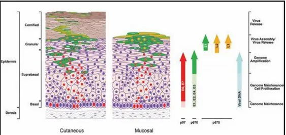
1.1.3 Viral cycle: productive infection and viral proteins
As previously mentioned, HPV viral cycle is strictly dependent on the differentiation status of the infected host cells and for this reason epithelial raft cultures has been developed to better investigate it in vitro (Andrei et al, 2010).
The normal productive infection starts when the virus reaches the dividing basal cells of the epithelium through small lesions, entering in the cytoplasm via clathrin- or caveolin-dependent endocytic mechanisms (Fig. 3) (Day et al, 2003). The major capsid protein L1 interacts with heparan sulfate proteoglycans on the cell surface with the involvement of a secondary receptor and a possible role for the minor capsid protein, L2. Particles disassemble in late endosomes and/or lysosomes, with the transfer of viral DNA to the nucleus being facilitated by L2 (Doorbar, 2006). In experimental systems, viral transcripts can be detected as early as 12 h post-infection, with mRNA levels increasing over the course of several days. In this early part of the infection, the viral genome is a stable episome (not integrated into the host genome) and acquires euchromatic structure (association with histones) allowing nuclear transcription factors to regulate the viral expression during the productive infection. The proteins E1 and E2 are required for its maintenance, replication with the cellular genome during the S-phase and segregation (Table 1). Moreover, E2 regulates transcriptional levels of E6 and E7 in a dose-dependent manner: these proteins are necessary to drive to and block the cell in the S-phase via interactions with p53 and pRb. Upon infection of basal cells, HPV genomes are replicated up to 50–100 copies per cell.
Suprabasal cells normally exit the cell cycle and begin the process of terminal differentiation in order to produce the protective barrier that is normally provided by the skin. At the same time, HPVs need to amplify their genome and produce new viral particles. What triggers the onset of late events is not yet fully understood, but appears to depend in part on changes in the cellular environment as the infected cell moves towards the epithelial surface, leading to the increase of viral proteins involved in replication (i.e. E1, E2, E4, E5). As E2 increases in abundance, occupancy of the remaining sites leads to the displacement of basal transcription factors, such as Sp1 and TBP (TATA-box-binding protein), that are necessary for promoter activation. It appears that the increase in E2 expression leads eventually to the down-regulation of E6/E7 expression and to the eventual loss of the replicative environment necessary for viral DNA synthesis. E5 is a transmembrane protein which resides predominantly in the endoplasmic reticulum, but it can associate with the vacuolar proton ATPase delaying the process of endosomal acidification. It is thought that this affects the
5
recycling of growth factor receptors on the cell surface, leading to an increase in epidermal growth factor (EGF)-mediated receptor signalling and the maintenance of a replication competent environment in the upper epithelial layers. On the other hand, E4 causes cell-cycle arrest in G2 phase and antagonizes E7-mediated cell proliferation.
The events that link genome amplification to the synthesis of the capsid proteins are not yet fully understood, but are dependent on changes in mRNA splicing and on the generation of transcripts that terminate at the late (rather than the early) polyadenylation site. The assembly of infectious virions in the upper epithelial layers is thought to require E2 in addition to the capsid proteins L1 and L2, and it has been suggested that E2 may improve the efficiency of genome encapsidation during natural infection. L2 localizes to the nucleus by virtue of nuclear localization signals located at its N- and C termini and, once there, it associates with PML (promyelocytic leukaemia) bodies. Although some PV L2 proteins can associate directly with DNA, the specific recruitment of viral genomes to PML bodies is thought to require E2, which can associate with viral DNA through its specific recognition sites. L1 assembles into capsomeres in the cytoplasm (360 copies of L1 organized into 72 capsomeres with 1 copy of L2 in the center) prior to nuclear relocation and is recruited into PML bodies only after L2 has bound and has displaced the PML component sp100.
In the end, virus release requires efficient escape from the cornified envelope at the cell surface, which may be facilitated by the E4 protein by disrupting the keratin network and affecting the integrity of the cornified envelope. The viral infectious cycle is exclusively intraepithelial: there is no viremia and no virus-induced cytolysis or cell death, and viral replication and release are not associated with inflammation.
Figure 3 – Viral life cycle in cutaneous and mucosal epithelia and gene expression in different epithelial layers (Doorbar, 2006).
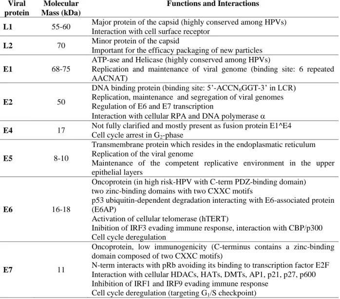
6
Table 1- Viral proteins and their functions (Doorbar, 2006; Ganguly & Parihar, 2009; Hegde, 2002; Miller et al, 2012).
Viral protein
Molecular Mass (kDa)
Functions and Interactions
L1 55-60 Major protein of the capsid (highly conserved among HPVs) Interaction with cell surface receptor
L2 70 Minor protein of the capsid
Important for the efficacy packaging of new particles
E1 68-75
ATP-ase and Helicase (highly conserved among HPVs)
Replication and maintenance of viral genome (binding site: 6 repeated AACNAT)
E2 50
DNA binding protein (binding site: 5’-ACCN6GGT-3’ in LCR) Replication, maintenance and segregation of viral genomes Regulation of E6 and E7 transcription
Interaction with cellular RPA and DNA polymerase
E4 17 Not fully clarified and mostly present as fusion protein E1^E4 Cell cycle arrest in G2-phase
E5 8-10
Transmembrane protein which resides in the endoplasmatic reticulum Replication of the viral genome
Maintenance of the competent replicative environment in the upper epithelial layers
E6 16-18
Oncoprotein (in high risk-HPV with C-term PDZ-binding domain) two zinc-binding domains with two CXXC motifs
p53 ubiquitin-dependent degradation interacting with E6-associated protein (E6AP)
Activation of cellular telomerase (hTERT)
Inibition of IRF3 evading immune response, interaction with CBP/p300 Cell cycle deregulation
E7 11
Oncoprotein, low immunogenicity (C-terminus contains a zinc-binding domain composed of two CXXC motifs)
N-term interacts with pRb avoiding its binding to transcription factor E2F Interaction with cellular HDACs, HATs, DMTs, AP1, p21, p27, p600 Inhibition of IRF1 and IRF9 evading immune response
Cell cycle deregulation (targeting G1/S checkpoint)
1.1.4 Molecular pathogenesis: transforming infection, integration and latency
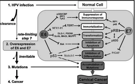
In the previous paragraph, we described the productive infection of HPV, which does not cause a symptomatic effect nor a high immune response. However, the majority of HPV infections (90%) are cleared in 2 years without any complication for the host (Gravitt, 2011). But what happens if the infection persists? As we have seen, the key molecules during the viral replication are the oncoproteins E6 and E7, which interact with a great number of cellular proteins (Tab 1 and Fig 4). In experimental systems these interactions have been shown to induce proliferation and eventually immortalization and malignant transformation of cells, but the mechanisms which determine the “switch” from productive to transforming infection have not been completely understood yet. Since there are differences between E6/E7
7
proteins of different HPV types, actually we distinguish, among the mucosal group, high risk-HPVs (HR-risk-HPVs), probably HR-risk-HPVs and low risk-risk-HPVs (LR-risk-HPVs), depending on the different capacity of HPV types to establish a transforming infection. For example, HR-HPVs E6 protein contains a PDZ-binding domain targeting proteins in the cellular membrane-cytoskeleton interface, disrupting cell junctions and promoting transformation.
Figure 4 – Multi-step pathogenesis of HPV and some of the interactions of viral oncoproteins E6 and E7 with cellular proteins (Yugawa & Kiyono, 2009).
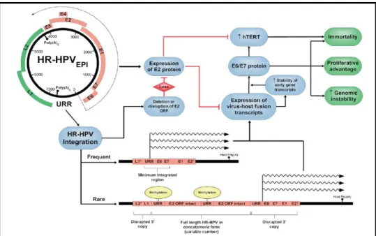
The crucial point during viral life cycle is the regulation of E6 and E7 expression. If these proteins are overexpressed, cell cycle/ mitosis deregulation and suppression of the immune response are increased as well, leading to genomic instability and accumulation of mutations at different sites (random) of the cellular chromosomes. Together, all these events are necessary for cancer development and define the malignant phenotype (multipolar mitoses are hallmark of HR-HPV-associated carcinomas) (Muñoz et al, 2006; Yugawa & Kiyono, 2009). One of the main consequences of genomic instability during persistent HPV infection is random integration of the viral genome into cellular chromosomes through double strand breaks (DSBs) (Pett & Coleman, 2007; Pett et al, 2004; Wentzensen et al, 2004). Usually, this event occurs in the viral E2 ORF, leading to the complete or partial loss of E2 protein. Since E2 is fundamental for the correct regulation of E6 and E7 transcription, viral integration contributes to the overexpression of these oncoproteins. More rarely, concatameric integrants are observed, where viral copies (including intact E2) are arranged in a head-to-tail fashion with partially deleted copies at the 5’- and 3’- ends (Fig 5).
During the last 20 years, viral integration was thought to be the key mechanism for HPV-associated cancer development, but the findings of episomal viral DNA in late stage cancers
8
suggested the presence of alternative pathways for oncoproteins overexpression in the presence of active E2 (Arias-Pulido et al, 2006; Vinokurova et al, 2008). One of these mechanisms have been suggested to have an epigenetic nature, involving methylation of citosines (meC) by cellular DNA Methyltransferases (DMTs). In fact, not only DNA methylation is a mechanism commonly used by mammalian cells to drive and control gene expression, but it has been also shown that E7 can interact with DMT-1 (Burgers et al, 2007) and meC in CpG sites in the viral EBSs act as obstacle to E2 binding (Kim et al, 2003; Thain et al, 1996). DNA methylation in HPV infected cells may occur in both episomal and integrated viral genome as well as in cellular genes, having different roles (Szalmas & Konya, 2009). At the viral DNA level, it may act as a cellular defence mechanism avoiding foreign genes expression; at the host genome level, it may inactivate some key genes involved in the control of cellular signalling pathways.
Figure 5 – Significance of HR-HPV integration detected in cervical carcinomas (Pett & Coleman, 2007).
Up to now, we described what happens during a persistent HPV infection, when the virus replicates with high titres which can be detected by common diagnostic techniques. However, sometimes in a basal stem cell HPV may persist and remain undetectable until triggered to differentiate by undetermined stimuli such as wound repair and hormonal regulation (Gravitt, 2011). This event is called viral latency and it has not been completely understood for HPV. It is thought that a few infected basal stem cells may retain HPV episomes, but do not differentiate, and these infected cells are unlikely to be sampled using standard exfoliative
9
techniques employed in most epidemiologic studies, which sample only the surface epithelium. Moreover, minor fraction of the episomal HPV DNA can be methylated de novo and can also be associated with transcriptionally inactive chromatin structure, providing a plausible mechanism for latent form of HPV infection (Szalmas & Konya, 2009). Wounding may stimulate latently infected basal cells to divide and trigger viral reactivation and stimulation of tissue-resident memory T cells.
1.2 Clinical relevance of mucosal alpha-HPVs infection
Transmission of HPV infection occurs primarily via sexual activity, most commonly vaginal and anal intercourse. Other forms of transmission have been occasionally reported such as skin-on-skin genital contact, and mother-to-child transmission, but their implication in cervical cancer is likely to be marginal. Oral sex can also be a route of HPV transmission. HPV infection is very common. Most women in the world will be infected with genital HPV at some time in their lives, with a lifetime risk of infection of 50–80%, and peak incidence occurs in the 18–30 age group (Stanley, 2010). Even if HPV infection is mainly known worldwide for the causative relationship with cervical cancer, during the last 30 years it has been also associated to the development of many other diseases in both men and women, with different roles of HPV genotypes (Tab. 2).
The prevalence of genital HPV infection in men is not well established and results from studies are difficult to compare because of differences in sampling methods, differences in study population, and poor reporting of the presence or absence of clinical lesions. Here we describe the most important characteristics of HPV-associated diseases in the anogenital and aerodigestive tracts.
Table 2 – Sites of HPV-associated cancers (Kreimer & Chaturvedi, 2011).
Cancer site % attributable
to HPV infection HPV-induced premalignant lesion Screening modality
Cervix 100 Cervical intraepithelial neoplasia (CIN)
Cytology, colposcopy, primary screening
Anus 90 Anal intraepithelial neoplasia (AIN) Cytology, High resolution anoscopy Penis 40 Penile intraepithelial neoplasia
(PIN) Cytology/Histology Vagina 40 Vaginal intraepithelial neoplasia
(VAIN) Cytology/Histology Vulva 40 Vulvar intraepithelial neoplasia
(VIN) Cytology/Histology
10
1.2.1 Cervical cancer and other HPV-associated diseases in the anogenital region
CERVICAL CANCER: SQUAMOUS CELL CARCINOMA (SCC) AND CERVICAL ADENOCARCINOMA (AdCa)
The cervix of the uterus is the preferential site of sexual infection by mucosal HPVs and undergoes physiological changes according to the age of the woman (before puberty, puberty, following puberty, after menopause) due to different hormonal levels. The area of the cervix with premalignant potential is the transformation zone, which is formed after puberty and corresponds to the site of transition from the columnar glandular epithelium (endocervix) and the squamous epithelium (esocervix) (Fig. 6).
According to World Health Organization (WHO) 2010 statistics, cervical cancer is the 3rd most common cancer among women worldwide, with 529 409 newly diagnosed cases and 274 883 deaths every year. However, global incidence and mortality rates vary geographically with 452 902 cases (85.5%) of cervical cancers occurring in low-income countries. It is more common in metropolitan areas than in rural areas, and the incidence is higher in populations with lower socio-economic status and level of education. Central and South America, southern and eastern Africa and the Carraiben have the highest incidence of the disease.
Figure 6 – Figures showing the location of the squamous epithelium in the exocervix or the glandular cells in the endocervix (left) and the spread of cervical cancer through the cervix (from normal cervix to cancer) which can be observed during a visual inspection (right).
Carvical cancer development is a long multistep process characterized by well-defined clinical stages and we can observe four main step: infection, persistence, progression, and invasion (Fig. 7). It is a rare complication of persistent infections with mucosal HPVs, which occur in 10% of the cases and are more likely to progress to premalignant lesions (CIN) in 5 years. Heterogeneity in biology (and definition) still exists in precancerous lesions: a) CIN 1 is a histopathological diagnosis of HPV infection, and should not be considered as a
11
precancerous lesion. Women with a persistent diagnosis of CIN 1 may progress to CIN 2/3 at a rate of 15% over 2 years, b) CIN2 is sometimes produced by non-carcinogenic HPV types, and has a sizable regression potential of 40% over an approximately 2-year period. Thus, CIN 2 represents an equivocal precancerous lesion, but it is treated in some regions to provide a safety margin against cervical cancer risk, c) CIN3 (in situ carcinoma, CIS) is the true precancerous lesion and progresses to cancer, if untreated, at a rate of around 30% over 20 years. When high-grade CIN (CIN 2 or worse) is diagnosed, treatment is mandatory. Overall treatments are more than 90% effective.
Virtually, all cervical cancer cases are linked to genital persistent infection with mucosal HPVs, which is the most common viral infection of the reproductive tract and its prevalence in the general population with normal cytology is 11.4% (14.3% in developing regions vs 10.3% in developed regions) (WHO HPV Summary Report Update 2010). In 1995, the IARC monograph working group concluded that there were sufficient evidences for the carcinogenicity of HPV 16 and 18 and limited evidences for the carcinogenicity of HPV 31 and 33. Actually, HR-HPV are HPV 16, 18, 31,33, 35, 39, 45, 51, 52, 56, 58, 59, 66 and 73, probably HR-HPV are HPV 26, 53, 68, 73 and 82, LR-HPV are HPV 6, 11, 13, 40, 42, 43, 44, 54, 61, 70, 72, 81 and 89 (Muñoz et al, 2006). HPV 16 and 18 are involved in 70.9% of all cervical cancers even if genotype-specific prevalence may vary geographically. Moreover, some recent studies have pointed out an important role of different HPV 16 variants, which sometimes have increased oncogenicity when compared to the main type (Quint et al, 2010; Sichero et al, 2012). The risk factors for acquisition of HPV infection have been historically linked to: a) early age at initiation of sexual activity, b) high number of new and recent sexual partners, c) high number of partners of the husband or male partner.
Although HPV is a necessary factor in cervical cancer development, there are also some cofactors playing a role in progression of HPV infection to cervical cancer. Cofactors influencing persistence and progression of HPV infection to advanced high-grade squamous intraepithelial lesions and cervical cancer include environmental cofactors, such as long-term use of hormonal contraceptives, tobacco smoking, co-infection with other sexually transmitted agents (i.e. HSV2 and Chlamydia trachomatis), high parity and diet, and host cofactors (i.e. HIV infection and immunosuppression) (Castellsagué et al, 2002; Veldhuijzen et al, 2010).
12
Fig. 7 – The phases of cervical cancer development, from normal epithelium to invasive cancer through precancerous lesions (CINs) . Infection with HPV 16 and 18 is more likely to lead to cancer than other genotypes.
From an histological point of view, cervical cancer can be divided in SCC (~80%) and AdCa (~20%). Carcinoma of the cervix has showed a marked decline in developed countries over the past 40 years, due to wider implementation of cytological screening and increased detection of premalignant disease. Although the decline is mainly attributable to a decrease in incidence of SCC, there has also been an increase in relative and absolute incidence of AdCa over the same period, especially among young women (age < 45 years). These two types of cervical cancers differ not only for epidemiology, but also for prognostic factors and survival (10–20% differences in 5-year overall survival rates, with AdCa being more aggressive), patterns of dissemination and recurrence, response to treatment and risk factors (AdCa more associated to obesity, nullipary and HPV 18) (Gien et al, 2010). Moreover, if for SCC and premalignant lesions of the esocervix a good classification and triage guidelines are available, it is not the same for AdCa and glandular lesions of the endocervix (Zaino, 2000). Glandular dysplasia (or atypical hyperplasia) has been proposed as a pathologic entity on the basis of the assumption that glandular lesions progress through a series of lesions of distinctive morphology as they acquire the genetic and phenotypic changes of carcinomas similar to squamous lesions of the cervix. In situ AdCa (AIS) has consistently been characterized by the following histological features: a) preservation of normal glandular architecture; b) involvement of part or all of the epithelium lining glands or forming the surface; c) nuclear enlargement, coarse chromatin, small single or multiple nucleoli; d) increased mitotic activity; and e) variable stratification of nuclei. There is no consensus regarding the criteria for the
13
diagnosis of microinvasive AdCa (MIA): authors generally have chosen a similar definition that restricts cases of MIA invasion to less than 5 mm from the basement membrane of the surface epithelium. To the situation, there is also a controversial classification for the histological types of invasive AdCa. Among mucinous AdCa, the most common is the endocervical type, followed by intestinal, signet ring, minimal deviation (MDA) and villoglandular type. Endometrioid AdCa may actually be more common than the endocervical type and histologically is indistinguishable from its counterpart in the uterine corpus. Other less common histotypes are clear cell, serous, mesonephric and adenosquamous AdCa.
VULVAR AND VAGINAL CANCER
Vulvar cancer accounts for 3-5% of all genital tract malignancies in women and is primarily a disease of the elderly (< 70 years). The aetiology is not well understood, but an association with HPV has been shown for multifocal warty or bowenoid lesions in younger populations (40%). Vulvar intraepithelial neoplasia (VIN) are the precursor lesions and the most important risk factors. On the other hand, SCC are the most common histotype (90%). A meta-analysis estimated a HPV prevalence of 76% for VIN and 36% for vulvar carcinomas. HPV 16 is the most common detected type (65-93% in VIN and 71% for vulvar cancer) followed by HPV 18 (WHO Summary Report Update 2010).
Primary cancer of the vagina constitutes 2% of all malignant neoplasm of the female genital tract, with the same risk factors for cervical neoplasia. Most vaginal carcinoma are secondary, arising from primary carcinoma of the cervix, endometrium or rectum and the association with HPV infection is around 40%. Vaginal intraepithelial neoplasia (VAIN) are the precursor lesions. HPV 16 is the most common type in at least 70% of HPV-positive carcinomas (WHO Summary Report Update 2010).
PENILE CANCER
Cancer of the penis represents less than 0.5% of cancers in men and the age at the diagnosis is > 60 years old. The aetiology seems to be multifactorial, but penile cancer with basaloid and warty features have shown the strongest association with HPV infection. Moreover, the geographical correlation between the incidence of penile and cervical cancers and the concordance of these two cancers among married couples suggested the common aetiology of HPV infection. Penile intraepithelial neoplasia (PIN) are the precursor lesions and HPV DNA is detectable in approximately 40% of all penile cancers. HPV DNA is detectable among PIN with the basoloid histological type, ranging from 75-80% of cases, and decreasing to 30-60% among invasive squamous cell carcinomas. HPV 16 is the most common genotype.
14
ANAL CANCER
Anal cancer is a rare malignancy arising in the anal canal, largely in the transitional zone separating the squamous epithelium of the canal and the mucosal epithelium of the rectum. The incidence is particularly high in men who have sex with men and among immunosuppressed men and women (HIV infected or transplant recipients). These cancers are predominantly squamous cell carcinoma, adenocarcinomas, or basaloid and cloacogenic carcinomas. Anal cancer is similar to cervical cancer with respect to overall HPV DNA positivity, with approximately 85% of cases associated with HPV infection worldwide. HPV 16 is the most common detected type, representing 87% of all HPV-positive tumours. HPV 18 is the second most common type detected and is found in approximately 9% of cases. HPV DNA is also detected in the majority of precancerous anal lesions (anal intraepithelial neoplasia, AIN) and the prevalence of HPV increases with the severity of the lesion.
NON-CANCEROUS DISEASE: GENITAL WARTS
Genital Warts (GWs) can involve the vulva, vagina, urethra, skin of anogenital tract, and penis. Having GWs is not associated with mortality and are related to both clinical symptoms (itching, burning, discharge, bleeding and pain) and psychosocial problems (embarrassment, anger, shame, anxiety and decreased self-esteem). Almost 100% of GWs are associated with either HPV 6 or 11. Although these HPV types give rise to benign changes, they can in rare cases be associated with malignant lesions such as the rare Buschke-Lowenstein tumours. HR-HPV types can be identified in up to 50% of cases, however, HPV 6 and 11 are considered the causative agents.
GWs represent not only a problem for the individual, but also imply significant healthcare costs for society. About 1% of the sexually active population harbours GWs at any given point in time and up to 17% of women aged 20-29 years have had at least one episode of GWs in the past (based on surveys in the Nordic countries of Europe).
1.2.2 Non-anogenital cancers: HPV-associated head and neck squamous cell carcinoma
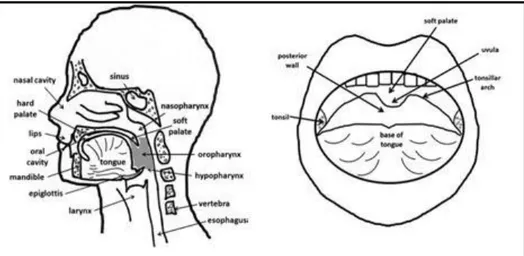
The term “head and neck cancer” (HN) includes lesions at several anatomic sites, such as the lip, oral cavity, nose and paranasal sinuses, nasopharynx, oropharynx, hypopharynx, larynx, oesophagus, salivary glands, as well the soft tissues of the neck and ear and more than 95% of them are SCC (HNSCC) (Fig. 7). It is the sixth leading cancer by incidence worldwide. It is likely that approximately 600 000 cases will arise this year worldwide, and that only 40–50% of patients with HNSCC will survive for 5 years (Peter KC Goon, 2012). On clinical
15
examination, oral SCC lesions may be preceded by mucosal alterations with histologically detectable dysplastic changes. However, a malignancy involving a complex genetic process may also occur directly “de novo” without any pre-existing clinically detectable mucosal changes. All HNSCCs tend to be diagnosed late because there is no pain until the late stages. The most important risk factor of head and neck cancer worldwide is smoking, with alcohol coming second.
In 1983, Syrjanen and colleagues provided the first evidence on the presence of HPV infection in HNSCC, by analyzing the presence of HPV antigens in 40 oral carcinomas using immunohistochemistry, but, up to date, scientists have been able to support only an association, not an aetiological role (Syrjänen et al, 1983). Although, at present, HPV infection has been established in 20-25% of all HNSCC, the role of HPV in the pathogenesis of HNSCC has been controversial mainly because the detection rates of HPV DNA have been highly variable among the studies, ranging from 0% to 100% and HPV 16 has been identified in 20–90% of the oropharyngeal carcinomas (OPSCCs). These highly variable detection rates can be partly explained by variations in the sampling techniques and different HPV detection methods.
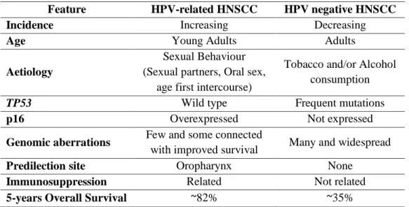
Several studies indicate that oral HPV infection is likely to be sexually acquired and it seems that HPV-positive tumours form a distinct group within HNSCCs. The aetiological factors differ, the tumours are different at the molecular level and the clinical outcome is different, in general HPV infected HNSCCs have a more favourable prognosis and response to chemo/radiotherapy (Tab. 3) (Leemans et al, 2011; Syrjanen, 2010; Worden et al, 2008). Oropharyngeal carcinomas, tonsillar cancers in particular, showed the strongest association with HPV, with some 60% being ascribed to HPV (Syrjanen, 2010).
16
HPV involvement in HNSCCs is supported by a series of observations. First, HPV is a virus with broad and essential tropism for epithelial tissues. In vitro studies clearly proved that viral oncoproteins belonging to HR-HPVs immortalize human keratinocytes, including oral keratinocytes. The HR-HPV genotypes can be detected in HNSCC with PCR techniques and fluorescence in situ hybridization. In addition, genotype-concordant viral DNA can be found in the lymph nodes of patients with metastatic oropharyngeal SCC (OPSCC) (Miller et al, 2012).
An interesting study compared gene expression profiles of HPV+ and HPV− oropharyngeal cancer and oral cavity cancer (Lohavanichbutr et al, 2009). In oral cavity tumours, no significant difference in gene expression was noted when comparing HPV+ and HPV− specimens. However, analysis of oropharyngeal tumours shows significant differences (347 differentially expressed genes) in HPV+ compared with HPV− lesions. Differences were particularly common among genes involved in DNA regulation and repair, cell cycle, and chemotherapy/radiotherapy sensitivity. These results underscore the observation that HPV+ oropharyngeal disease represents a divergent biological entity from HPV− disease.
Table 3 – Different clinical and biological characteristics of HPV-positive and HPV-negative HNSCCs (Kostareli et al, 2012; Leemans et al, 2011; Syrjanen, 2010).
Feature HPV-related HNSCC HPV negative HNSCC
Incidence Increasing Decreasing
Age Young Adults Adults
Aetiology
Sexual Behaviour (Sexual partners, Oral sex,
age first intercourse)
Tobacco and/or Alcohol consumption
TP53 Wild type Frequent mutations
p16 Overexpressed Not expressed
Genomic aberrations Few and some connected
with improved survival Many and widespread
Predilection site Oropharynx None
Immunosuppression Related Not related
5-years Overall Survival ~82% ~35%
However, according to data from different studies recently reviewed by Kostareli (Kostareli et al, 2012), also HPV-positive OPSCC are heterogeneous in both biological and clinical behaviour, possibly due to differences in viral load and/or viral oncoproteins expression. In fact, it has been recently shown that HPV DNA+/RNA- tumours have a worst prognosis compared to those HPV DNA+/RNA+ ones (Holzinger et al, 2012). These HPV-related
17
tumours have better survival maybe for the combined effect of phenomena occurring in epigenome, genome and expression pattern, which drive alterations in the intracellular signalling networks, and the components of tumour microenvironment (angiogenesis, immune system, inflammation, etc.) (Fig. 8). For example, wild type p53 enables HPV-related OPSCC to respond to DNA damage via apoptosis, making these tumours more sensitive to ionizing radiations therapy. On the other hand, infection by HPV may also render these cells more visible to the innate immune system leading to a synergistic effect with radiotherapy. But further studies are needed to confirm these hypothesis.
1.3 Control and prevention of cervical cancers: diagnosis, screening and vaccination
Cervical cancer is easily manageable through early diagnosis and treatment, which can drastically reduce incidence and mortality. More importantly, cervical cancer can be avoided to a large extent by action of both primary and secondary prevention.
1.3.1 Cervical cancer screening
Cancer screening is a public health intervention undertaken in an asymptomatic population to prevent invasive disease and resulting mortality through the early detection of precancerous lesions. It is generally accepted that organized screening is more effective and cost-effective than opportunistic screening and cervical cancer is the only gynaecologic malignancy that currently meets the criteria for screening: a) the time between the appearance of precancerous lesions and the occurrence of invasive cervical cancer is long (10-30 years), leaving time for detection and treatment, b) treatment of precancerous lesions is less expensive and more successful in avoiding death, as compared to the management of invasive cervical cancer. A good screening test should be accurate, reproducible, inexpensive, easy to perform and follow-up, acceptable and safe and several tests are available for cervical cancer screening: cytology (conventional or liquid-based), visual inspection (with acetic acid or Lugol’s iodine) and HPV testing (Cuzick et al, 2012). In 1952, Georgios Papanicolau described a cervical smear technique capable of detecting abnormal cervical cytology suggestive of cervical neoplasia, the Pap test (conventional cytology) (Fig. 9).
Guidelines for cervical cancer screening program are formulated depending on whether or not there is a program already in place, and if so, whether or not this program is successful. The most appropriate target age group for a screening program is 25-30-years, because younger women may have abnormalities, but they are likely to resolve spontaneously. An interval of 3
18
to 5 years between screening visits is considered appropriate in women with previous negative screening results. Women whose previous Pap smear was abnormal should follow the cervical diagnostic algorithm established locally.
Figure 9 - Georgios Papanicolau (left) and a scheme showing how to collect cervical specimen for the Pap test (right).
1.3.2 Techniques for HPV detection in clinical samples: HPV-DNA test
Recently established guidelines recommend HR-HPV DNA testing in order to improve the efficacy of primary cytological screening programs or as triage tests. At present the Hybrid Capture 2 (HC2) assay (QIAGEN GmbH, Hilden, Germany), the Cervista test (Hologic, Madison, WI, USA), and the Roche Cobas 4800 HPV Test (Roche Inc., Branchburg, NJ, USA) are the only tests for the detection of HR-HPV DNA approved by U.S. Food and Drug Administration for cervical cancer screening (Tab. 4) (Poljak & Kocjan, 2010).
Furthermore, specific HPV typing is important for epidemiological studies, assessment of the clinical behaviour of particular genotypes, clinical management of women, clinical follow-up studies, evaluating prevention strategies, development of new therapies and prophylactic/therapeutic vaccines.
Difference in the carcinogenic potential between the different HR-HPV types and indicate the potential value of genotyping in cervical cancer screening. HPV 16, 18, 31 and 33 infection and especially HPV 16 persistence were associated with high absolute risks for progression to high-grade cervical lesions (Kjær et al, 2010). Moreover, clinical trials conducted to test the efficacy of prophylactic vaccines that target two HR-HPV types, HPV 16 and HPV 18, as well as the low-risk (LR) HPV 6 and 11 (see next paragraph), require accurate detection of genotype-specific HPV infections associated with cancer and precancerous lesions.
Sequencing of DNA is the gold standard method for accurate viral typing. However, DNA sequencing techniques have been facing limitations in typing HPV when the specimen
19
harbours multiple genotypes resulting in non-interpretable sequence data. (Gharizadeh et al, 2005).
The most widely used PCR-based methods employ consensus primers that amplify highly conserved regions of the L1 or E1 gene or E6 gene, followed to genotype specific hybridization by reverse line blot hybridization or microchip format. All hybridization-based methods can discriminate HPV types in multiple infections, but can identify only HPV types represented by probes.
Actually, many commercially available CE-marked genotyping HPV DNA tests exist. HPV-typing assays, such as INNO-LiPA HPV (Innogenetics), Linear Array (Roche) and PapilloCheck (Greiner Bio-One GmbH), are commonly used in the follow-up of persistent infections to monitor the presence of specific HPV genotypes. Many studies comparing different methods for HPV typing noted considerable differences in the type specific sensitivities as well as the ability to detect multiple infections between the individual test systems (Qu et al, 1997; van Doorn et al, 2002). Moreover, although more sensitive than cytology, HPV testing has modest specificity and positive predictive value (PPV) for detection of pre-cancerous lesions, and cannot distinguish infections that will resolve from those that will progress. Thus, an important question is how to triage HPV-positive women and further specific and sensitive biomarkers are necessary to answer.
Table 4- Most important currently available commercial assays for the multiple detection of -HPV (Poljak & Kocjan, 2010).
HR-HPV DNA-based screening tests HPV DNA-based genotyping assays
Hybrid Capture 2 HPV DNA (HC2) test (Qiagen) INNO-LiPA HPV Genotyping (Innogenetics) Cervista HPV HR test (Hologic) Linear Array HPV Genotyping Test (Roche) Amplicor HPV test (Roche) EasyChip HPV Blot Kit
Care HPV test (Qiagen) REBA-HPV-ID
HR-HPV-DNA-based screening assays with concurrent or reflex HPV 16 and
HPV 18 genotyping
PapilloCheck HPV-Screening Test (GreinerBio) Clart HPV 2 – papillomavirus clinical arrays HPV GenoArray Test Kit
RealTime High Risk HPV test (Abbott) GeneTrack HPV DNA Chip Cobas 4800 HPV Test (Roche) GeneSQUARE HPV Microarray Cervista HPV 16/18 Test (Hologic) Infiniti HPV Assays
HR-HPV 16/18/45 Probe Set Test PANArray HPV Genotyping Chip
In situ hybridization HPV DNAChip
INFORM HPV (Ventana Medical) GG HPVCHIP GenPoint HPV Biotinylated DNA Probe (Dako)
ZytoFast HPV Probes (ZytoVision)
Multiplex HPV Genotyping Kit (xMAP, Luminex)
HPV OncoTect Test Kit (InCellDx) HR-HPV E6/E7 mRNA-based screening assays
PreTect HPV-Proofer
NucliSENS EasyQ HPV (bioMérieux) APTIMA HPV Assay (Roche)
20
1.3.3 The prophylactic vaccine
Antibody generally binds to conformational determinants on structural components of a microorganism, and assists with pathogen clearance or protects against reinfection by promoting phagocytosis and by neutralizing infectivity. Demonstration that the L1 protein of HPV was the most immunogenic viral protein and could self assemble into virus-like particles (VLPs) eliciting host protective neutralizing antibody, enabled development of prophylactic vaccines designed to prevent HPV associated disease. The commercially available vaccines are Gardasil® (Merck & Co.) and Cervarix® (GlaxoSmithKline, GSK). These vaccines are produced in yeast or insect cells and induce a polyspecific antibody response which recognizes a range of conformational determinants on the viral capsid that are generally genotype-specific. In fact, Gardasil® has been developed to protect against HPV 16, 18, 6 and 11 and Cervarix® against HPV 16 and 18. In several phase III clinical trials, vaccines have been shown effective at preventing infection with the HPV types in the vaccine and associated premalignant disease of the genital tract for periods of up to five years in previously uninfected women. Data on longer term protection, and on protection in men, are awaited from ongoing clinical trials (Frazer, 2009).
Although HPV vaccination provides an opportunity to diminish the global cervical cancer incidence and successful vaccination programs are expected to substantially reduce HPV-related diseases burden, screening programs based on cytology or HPV testing will continue as a secondary preventative measure.
1.4 HPV AND IMMUNE RESPONSE: ANTIGENS PRESENTATION,
IMMUNOPROTEASOME AND THE ROLE OF INTERFERONS (IFNs)
Both cellular and humoral immune response are essential for the clearance of HPV in the infected epithelium. The adaptive immune response to HPV appears slower compared with other pathogenic virus infections and recognizes predominantly conformational determinants displayed only when the capsidic protein L1 is correctly configured into pentamers as in the native virus. Antibodies titres against L1 following natural infection are low, and although mucosal antibodies (secretory IgA and IgG) are detected in the previous 12 months of HPV detection and may protect against HPV infection, assays are difficult to standardize, limiting the utility of serology in HPV diagnosis (Sheu et al, 2007). Moreover, for most of the duration of the HPV infectious cycle, there is little or no release into the local milieu of pro-inflammatory cytokines that are important for dendritic cells (DC) activation and migration,
21
and the essential signals to kick start the immune response in squamous epithelia are absent. Together, these facts suggest that HPV may have developed strategies to evade host immune mechanisms and there are different possibilities.
The primary mechanism of viral immune evasion for HPV infection is avoidance of antigen presentation, which takes place via the MHC Class I pathway (Fig. 10A). Briefly, the endogenous antigentic peptides (8-11 amino acid products) generated by the intracellular proteolysis machinery (i.e. proteasome) are translocated by the transporter associated with antigen processing (TAP1/2 heterodimer) into the endoplasmatic reticulum (ER), where the chaperons-dependent assembly of MHC class I molecules occurs. Functional molecules are composed by a heavy chain (H), a light chain (2-microglobulin, 2m) and the antigenic
peptide. The chaperon protein involved in this step is Tapasin. Once the trimolecular complex is formed, it is delivered to the cell surface via an exocytic pathway, where cytotoxic T cells (CD8+ CTLs) are responsible of the sensing of these presented antigens (Hwang et al, 2001). The proteasome is a cylindric-shaped protease complex arranged as four axially stacked heptameric rings. The subunits of the catalytic core are represented by two homologous gene products, (two outer rings, highly conserved) and (inner rings, divergent, with enzymatic activity). Protein degradation may be performed in ubiquitin or ATPdependent as well as -independent manner. Upon IFNs induction (especially IFN-), the subunits LMP7, LMP2 and MECL-1 displace its homologues in the “constitutive” proteasome and assemble in the so-called “immunoproteasome” (Fig. 10B). This altered catalytic specificity of the proteasome is important for generating peptides that are optimal for binding to MHC class I molecules.
IFNs-induced MHC class I pathway activation can be mediated also by proteins belonging to the NLR family (nucleotide-binding domain, leucine-rich repeat) such as the recently characterized NLRC-5, which acts as nuclear transcriptional transactivator (CITA) (Meissner et al, 2012).
Absence of cell lysis and systemic viremia during HPV infection and production of immunogenic proteins in terminally differentiated layers of the epidermis minimize antigen availability for presentation, and also ensure that few pro-inflammatory signals are given to generate adaptive immune responses. Moreover, the genomic instability which characterizes HPV persistent-infected cells and cervical cancer may alter also the complex genomic HLA region affecting genes encoding HLA class I-II molecules (Sheu et al, 2007).
22
Figure 10 – MHC Class I antigen presentation pathway (A) and presenting immunoproteasome-derived peptides (B) (Neefjes et al, 2011; Spaapen & Neefjes, 2012).
HPV evolved anti-inflammatory and immune inhibitory mechanisms to suppress key steps of the type I IFNs pathway, impairing the antigen presentation and T-cells activation (Frazer, 2009).
Human type I IFNs (i.e. IFN-α, -β, -and -) have antiviral, antiproliferative, antiangiogenic and immunostimulatory properties. Viral functional oncoproteins E6 and E7 interact directly with molecules involved in IFNs signalling (Tab. 5 and Fig. 11). For example, E7 inhibits IFN-α-mediated signal transduction by binding to IRF-9 (IFN regulatory factor-9), preventing its translocation to the nucleus, thereby inhibiting the formation of the ISGF-3 (IFN-stimulated gene factor 3) transcription complex that usually binds ISRE (IFN-specific response element) in the nucleus. Moreover, E7 physically interacts with IRF-1, inhibiting IFN- promoter (Koromilas et al, 2001). On the other hand, E6 binds to IRF-3 homodimer and inhibits its transcript activation function, preventing transcription of IFN-; binding to Tyk2, prevents binding to the cytoplasmic portion of the IFN receptor inhibiting phosphorylation of Tyk2, STAT (signal transducer and activator of transcription) 1 and STAT2, impairing JAK (Janus kinase)/STAT activation and therefore inhibiting specific IFNα-mediated signaling (Stanley et al, 2007).
23
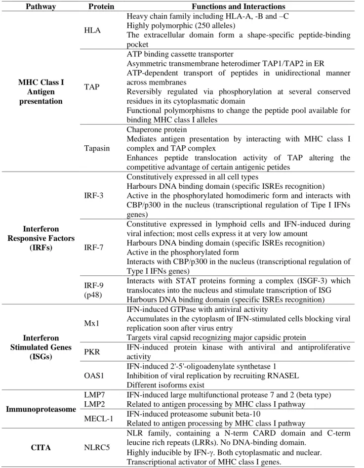
Table 5- List of the proteins involved in MHC class I antigen presentation pathway and IFNs-mediated antiviral response which are considered in this study and relative functions (Génin et al, 2009; Hwang et al, 2001; Meissner et al, 2012).
Pathway Protein Functions and Interactions
MHC Class I Antigen presentation
HLA
Heavy chain family including HLA-A, -B and –C Highly polymorphic (250 alleles)
The extracellular domain form a shape-specific peptide-binding pocket
TAP
ATP binding cassette transporter
Asymmetric transmembrane heterodimer TAP1/TAP2 in ER
ATP-dependent transport of peptides in unidirectional manner across membranes
Reversibly regulated via phosphorylation at several conserved residues in its cytoplasmatic domain
Functional polymorphisms to change the peptide pool available for binding MHC class I alleles
Tapasin
Chaperone protein
Mediates antigen presentation by interacting with MHC class I complex and TAP complex
Enhances peptide translocation activity of TAP altering the competitive advantage of certain antigenic petides
Interferon Responsive Factors
(IRFs)
IRF-3
Constitutively expressed in all cell types
Harbours DNA binding domain (specific ISREs recognition)
Active in the phosphorylated homodimeric form and interacts with CBP/p300 in the nucleus (transcriptional regulation of Tipe I IFNs genes)
IRF-7
Constitutive expressed in lymphoid cells and IFN-induced during viral infection; most cells express it at very low amount
Harbours DNA binding domain (specific ISREs recognition) Active in the phosphorylated form
Interacts with CBP/p300 in the nucleus (transcriptional regulation of Type I IFNs genes)
IRF-9 (p48)
Interacts with STAT proteins forming a complex (ISGF-3) which translocates into the nucleus and stimulate transcription of ISG Harbours DNA binding domain (specific ISREs recognition)
Interferon Stimulated Genes
(ISGs)
Mx1
IFN-induced GTPase with antiviral activity
Accumulates in the cytoplasm of IFN-stimulated cells blocking viral replication soon after virus entry
Targets viral capsid recognizing major capsidic protein
PKR IFN-induced protein kinase with antiviral and antiproliferative activity
OAS1
IFN-induced 2'-5'-oligoadenylate synthetase 1 Inhibition of viral replication by recruiting RNASEL Different isoforms exist
Immunoproteasome
LMP7 LMP2
IFN-induced large multifunctional protease 7 and 2 (beta type) Related to antigen processing by MHC class I pathway
MECL-1 IFN-induced proteasome subunit beta-10
Related to antigen processing by MHC class I pathway
CITA NLRC5
NLR family, containing a N-term CARD domain and C-term leucine rich repeats (LRRs). No DNA-binding domain.
Highly inducible by IFN-. Both cytoplasmatic and nuclear. Transcriptional activator of MHC class I genes.
24
Figure 11 – Intracellular type I IFNs signalling (Haller et al, 2007). Briefly, type I IFN binds to the receptor (IFNR1 and 2) and transducers signals through the sequential activation of receptor-associated kinases Jak and Tyk-2 leading to tyrosine phosphorylation and activation of STAT proteins. They associate to IRF-9 in the nucleus, forming the ISGF-3 complex and activating IFN-stimulated genes (ISGs) transcription.
1.4.1 IFN-
IFN- is the last discovered type I IFN which showed constitutively expression in keratinocytes and inducible in monocytes and DCs (LaFleur et al, 2001). The gene encoding the protein is located in the chromosome 9, adjacent to the type I IFNs cluster and analysis of cDNA and genomic sequences from other species failed to identify an orthologous, suggesting it may evolved later to play a specific role in humans. Most type I IFNs are expressed only upon viral infection and the constitutive expression of IFN-in resting keratinocytes is an important different characteristic which may provide a new mechanism of host defence. During viral infection, IFN- is up-regulated in keratinocytes. Another distinct feature compared to other type I IFNs is that IFN- seems to have a cell surface expression, carrying out its signalling and functions in a cell-associated manner, rather than being completely secreted (autocrine and juxtacrine, not paracrine effect) (Buontempo et al, 2006). Concerning HPV infected cells, it has been recently shown that IFN-expression is inhibited in HPV 16 or 18-positive cervical cancer cells by de novo promoter methylation and the viral oncoprotein E6, not E7, seems to be involved (Rincon-Orozco et al, 2009). Moreover, since its expression is decreased in precursor lesions and abolished in cancers, it has been concluded that IFN- repression may be an event occurring early in cervical carcinogenesis.
25
1.4.2 Type I IFNs in cervical cancer therapy
IFNs are used to treat a broad spectrum of diseases including multiple sclerosis, melanoma, some solid tumours, leukaemia and hepatitis. Unfortunately, for cervical cancer and pre-cancerous lesion treatment, controversial results have been observed in clinical trials (Beglin et al, 2009; Koromilas et al, 2001). Theoretically, IFN treatment should result in the clearance of HPV lesions and elimination of the virus, even in cases of latent infection. However, it is still far from widespread therapy. IFNs have been used successfully in treating patients with GW induced by LR-HPV types, but showed mixed results in treating low-grade lesions and cancers induced by HR-HPVs. A compilation of studies in vivo indicates that IFN- is more effective than IFN- and generally type II IFNs (i.e. IFN-) are more effective than type I. From later experiments it has been shown that treatment with IFNs can result in selecting cells with integrated copies of HPV DNA making it an ineffective methodology unless it can be combined with other therapeutic agents. Since it has been reported that the expression of viral oncoproteins, particularly E7, is significantly higher in non responsive patients, it is clear that the efficacy of IFNs is strictly dependent upon the level of viral oncogenes and the complex interactions between E6/E7 and cellular factors that affect both viral and host gene expression.
The discovering of the new IFN- with similarities, but also differences compared to other well known type I IFNs needs further investigation to better define its role in HPV infection and carcinogenesis and to assess its potential role as new treatment.
27
2. Aims of the thesis
The work presented in this thesis spaces between applied diagnostic and basic research on clinical samples and cell lines of cervical cancer and HPV-associated HNSCCs. The main aims are the following:
1) evaluate and improve diagnostic procedures for HPV-associated diseases by both assessing the performance of a new commercially available type-specific multiple-primer DNA sequencing method for HPV DNA genotyping on clinical samples from cervical lesions/cancers compared to a well known and diffuse reverse hybridization-based assay, in order to evaluate advantages and disadvantages in the clinical diagnostic practice, and determining the potential diagnostic value of virological markers, such as viral oncoprotein expression, viral load, physical state and DNA methylation, in HPV 16-positive cervical adenocarcinoma and HNSCCs in function of the available clinical and epidemiological data;
2) deep the role of HPV 16 in carcinogenesis by using data obtained from the analysis of virological markers (in particular viral DNA methylation) in clinical samples and analyzing the response to ectopic expression of IFN- in both cervical cancer and HPV-positive HNSCCs cell lines.
29
3. Materials and Methods
PART 1: COMPARISON OF HPV SIGN GENOTYPING TEST WITH INNO-LIPA HPV GENOTYPING EXTRA ASSAY ON HISTOLOGIC AND CYTOLOGIC
CERVICAL SPECIMENS (Barbieri et al, 2012) 3.1 Clinical specimens
Eighty-seven human cervical samples, previously tested by the INNO-LiPA assay, were analyzed by the new HPV sign Genotyping Test. Among these, 34 were 10 m thick paraffin-embedded biopsies belonging to patients with a histological diagnosis of cervical adenocarcinoma in situ (AIS) or invasive adenocarcinoma (invasive AdCa) and 53 cervical swabs, belonging to patients with a histological diagnosis of squamous cervical intraepithelial neoplasia of low (CIN1) or high grade (CIN2+), collected in PreservCyt® medium (Hologic, Marlborough, MA, USA). Histological diagnosis was confirmed based on pathology consensus review of tissue samples.
Clinical samples and data used in this study were obtained in the course of institutional diagnostic service, investigation described in this study could be carried out on residual specimens following diagnostic analysis, provided that all data would be kept anonymous.
3.2 DNA isolation
Total nucleic acids were extracted following different protocols. For biopsies, after heat deparaffinization at 95°C for 10 min and centrifugation at 12000 rpm for 10 min, tissue samples were digested with 20 l of proteinase K in 200 l of lyses solution at 56°C for 3 hours. DNA was extracted with QIAmp DNA Mini Kit (Qiagen, Hilden, Germany) following manufacturer’s instructions and eluted in 100 l. For cytological samples, 200 l of each specimens was processed with NucliSENSE EasyMag system (bioMérieux, Marcy l’Etoile, France), following manufacturer’s instructions, and eluted in 100 l of Elution Buffer. Extracted DNA was stored at -80°C.
3.3 INNO-LiPA HPV Genotyping Extra assay (Innogenetics, Ghent, Belgium)
This assay detects 28 different HPV genotypes, including 18 high-risk HPV (16, 18, 26, 31, 33, 35, 39, 45, 51, 52, 53, 56, 58, 59, 66, 68, 73, 82), 6 low-risk HPV (6, 11, 40, 43, 44, 54,