SAPIENZA University of Rome
Faculty of Pharmacy and Medicine
PhD in
Morphogenesis and Tissue Engineering XXX Cycle
(A.A. 2016/2017)
Unravelling the role of NAADP/TPC2 Ca
2+signalling
during angiogenesis and tumor progression
PhD student Francesca Papacci
Tutor Coordinator
INDEX
1.THE THESIS EXPLAINED pag 6
2. INTRODUCTION pag 8
2.1Calcium signalling complexity in health and diseases pag 8 2.2 Calcium signalling pathway mediated by
NAADP: the emerging role of Two-Pore Channels pag 11 2.3 Role of NAADP and TPCs in pathophysiological
processes pag 16
2.4 Biological properties of flavonoids: focus on
Naringenin pag 18
2.5 Physiological angiogenesis versus tumoral
angiogenesis: a general overview pag 22
3. AIMS pag 28
4. RESULTS AND DISCUSSION pag 30
4.1 Naringenin affects the activity of the human TPCs directly impairing hTPC2 functional activity and
decreasing hTPC1 activity pag 30
4.2 Naringenin significantly inhibits VEGF-dependent
calcium mobilization in a dose-dependent manner pag 36 4.3 Naringenin impairs VEGF-induced angiogenic process
4.4 Naringenin does not impair NAADP/TPC2-independent
angiogenic responses pag 42
4.5 Conclusions (1) pag 46
4.6 Naringenin impairs B16 melanoma cells
VEGF-dependent Ca2+ responses pag 48 4.7 Naringenin affects the proliferative/migratory behavior
of B16 melanoma cells pag 51 4.8 Naringenin interferes with B16 melanoma ability
to form vascular-like tube network pag 56
4.9 Concluding remarks pag 57
5. MATERIALS AND METHODS pag 59
5.1 Arabidopsis thaliana tpc1–2 mutants pag 59
5.2 Patch-clamp recordings pag 59
5.3 Analysis of electrophysiological data pag 60
5.4 Cell culture pag 60
5.5 Calcium imaging pag 60
5.6 Western Blot pag 61
5.7 In vitro angiogenesis assay pag 62
5.9 Scratch Assay pag 63
5.10 Cell cycle analysis pag 63
5.11 Vascular-like structure formation:
Vasculogenic Mimicry pag 63
5.12 Statistical analysis pag 64
5.13 Ethical Standards pag 64
6. APPENDIX pag 65
7. REFERENCES pag 67
1. THE THESIS EXPLAINED
Nicotinic acid adenine dinucleotide phosphate (NAADP) mediated calcium (Ca2+) signalling controls major cellular functions and involves an emerging family of intracellular channels specifically expressed on acidic compartments: Two-pore channels (TPCs). In particular, we have previously demonstrated that TPC2 isoform has a key role during angiogenesis and is likely involved in melanoma progression. Our earlier results were obtained using a selective membrane-permeant noncompetitive antagonist Ned-19 that is able to block NAADP- mediated Ca2+ signalling[1, 2].
In order to investigate a novel pharmacological tool able to interfere with the activity of TPC2 channel we have explored the potentiality of a natural flavonoid, Naringenin (Nar), in our models.
In accordance to electrophysiological evidences in a heterologous system, i.e. Arabidopsis vacuoles lacking endogenous TPCs, the present research has demonstrated that Nar dampens intracellular Ca2+ responses of human endothelial cells stimulated with VEGF, histamine or NAADP-AM, but not NAADP-independent stimuli, ATP and Angiopoietin-1. Moreover, Nar is able to impair angiogenesis in vitro and in an in vivo murine model.
On the basis of these and previous data we have also investigated Nar effects on B16 melanoma cells. We have observed that Nar impairs NAADP-controlled Ca2+ signalling (stimulated both directly and indirectly) and, interestingly, affects cells ability to proliferate/and or migrate. Furthermore, B16 melanoma cells capability to form vascular-like structures is impaired by Nar. Our present data suggest that Nar inhibition of TPC2 activity and the observed inhibition of angiogenic response to VEGF are linked by impaired intracellular Ca2+ signalling in endothelial cells. In addition, Nar effects on B16 melanoma cells strengthen the hypothesis that TPC2 represent a potential target for the control of the progression of melanoma and possibly of other tumors. The relationship we describe in endothelial cells between Nar and
TPC2 is therefore likely to have wider implications representing a novel tool for experimental, and possibly even clinical, research purposes.
2.INTRODUCTION
2.2 Calcium signalling complexity in health and diseases
Calcium (Ca2+) represents the most abundant and ubiquitous intracellular second messenger able to mediate different cell functions transducing several environmental signals into biological responses. Indeed, it’s known that Ca2+ plays an important role in some physiological processes such as fertilization, development, differentiation, proliferation, contraction, secretion, exocytosis, metabolism, gene transcription, apoptosis [3, 4] and angiogenesis [1, 5]. Spatiotemporal changes in intracellular Ca2+ concentration are coordinated by the intrinsic linking between internal compartments and plasma membrane channels. Although the maintenance of intracellular Ca2+ homeostasis represents a pivotal aspect for cell survival different mechanisms have evolved aimed to regulate the Ca2+ removal from the cytoplasm extruding it across the cell plasma membrane or sequestering it in intracellular stores. Plasma membrane Ca2+ ATPase (PMCA), Na+/ Ca2+ exchanger (NCX) and Na+/ Ca2+/ K+ (NCKX) represent the major proteins involved in withdrawing Ca2+ from the cytosol into extracellular medium. Instead, sarcoplasmic/endoplasmic reticulum Ca2+ ATPase (SERCA) pumps and ATPases closely related to SERCA contribute to sequestering Ca2+ in the sarco-endoplasmic reticulum and nuclear envelope, respectively. Furthermore, also mitochondria behave as buffers and stores Ca2+ in particular through the action of the inner mitochondrial membrane-spanning mitochondrial Ca2+ uniporter (MCU), mitochondrial Na+/ Ca2+ exchanger and the permeability transition pore (PTP) [6]. Likewise, Ca2+ influx from extracellular space is mediated by channels, pumps and exchangers which control Ca2+ entry in response to stimuli such as membrane depolarization, stretch, noxious stimuli, extracellular agonists, intracellular messengers and depletion of internal compartments [4]. For example, the voltage-operated Ca2+ channels (VOCCs) are present in excitable cells plasma membrane; consequently to membrane depolarization these channels undergo conformational
changes that cause Ca2+ influx from the extracellular environment down an electrochemical gradient. Finally when intracellular stores are empty a process known as store-operated Ca2+ entry (SOCE) induces the activation of ORAI1/STIM1 complex that allows Ca2+ entry across the plasma membrane [6].
Instead, Ca2+ release from intracellular stores is controlled by Ca2+ itself or by different Ca2+ mobilizing messengers such as inositol-1,4,5-trisphosphate (Ins(1,4,5)P3), cyclic ADP ribose (cADPR),
which mobilize Ca2+ from endoplasmic reticulum stores (ER) and nicotinic acid adenine dinucleotide phosphate (NAADP) which modulates Ca2+ release from acidic compartments such as endosomes and lysosomes [4] (Figure 1).
Parrington J. and Tunn R. Acta Physiol 2014, 211, 285-296 +2
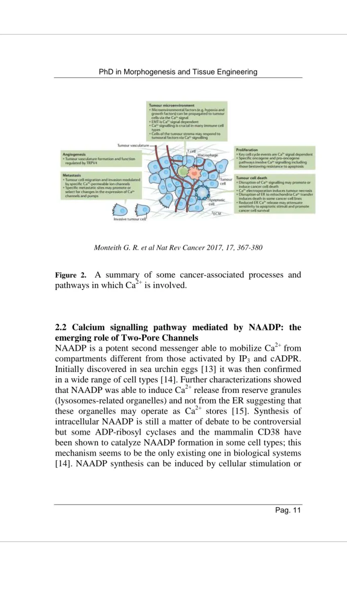
Global Ca2+ increases that may be transient or sustained, highly localized or appear as waves or oscillations represent changes in cytosolic free Ca2+ that can be transduced by the cell in a signal able to specifically regulate cell processes. Ca2+ signalling complexity due to the numerous pathways in which Ca2+ is involved implies that Ca2+ signalling deregulation can be a characteristic of different pathological states, including cancer [7]. Ca2+ signalling alterations are not necessary for cancer initiation but represent a pivotal point for tumor progression because Ca2+ is involved in relevant biological cancer hallmarks such as the ability to avoid apoptosis, self–sufficiency in growth regulation, invasion and metastasis, unlimited replication potential, resistance to anti-mitotic and anti-angiogenic stimuli, (Figure 2) [8-12]. Ca2+ toolkit used by cancer cells is not different from that of non-malignant cells but there may be some key alterations. Compared to non-malignant cells, cancer cells may express Ca2+ channels, pumps, or their specific isoforms, not generally present in normal cells of the same cell type; they may have important changes in the level of the expression, modified cellular localization, post-translational modifications, that can induce altered activity, gene mutations or changes in activity or expression associated with specific cancer-relevant processes. Furthermore, these alterations can result in changes in Ca2+ flux across intracellular organelles or across the plasma membrane [11]. Although Ca2+ signalling represent a new recent field of research in oncology, understanding how targeting Ca2+ channels pumps or exchangers and the pathways regulated by them may represent a novel area of interest to develop drugs and oncological therapy aimed to contrast cancer progression.
Monteith G. R. et al Nat Rev Cancer 2017, 17, 367-380
Figure 2. A summary of some cancer-associated processes and
pathways in which Ca2+ is involved.
2.2 Calcium signalling pathway mediated by NAADP: the emerging role of Two-Pore Channels
NAADP is a potent second messenger able to mobilize Ca2+ from compartments different from those activated by IP3 and cADPR.
Initially discovered in sea urchin eggs [13] it was then confirmed in a wide range of cell types [14]. Further characterizations showed that NAADP was able to induce Ca2+ release from reserve granules (lysosomes-related organelles) and not from the ER suggesting that these organelles may operate as Ca2+ stores [15]. Synthesis of intracellular NAADP is still a matter of debate to be controversial but some ADP-ribosyl cyclases and the mammalin CD38 have been shown to catalyze NAADP formation in some cell types; this mechanism seems to be the only existing one in biological systems
by activation of cell surface receptors to regulate a variety of processes such as fertilization, production of nitric oxide (NO) and glucose metabolism [14, 16].
The specificity of Ca2+ release from endolysosomal system induced by NAADP is still debated because it has been demonstrated that it can also mobilze Ca2+ from sarco-endoplasmic reticulum by directly activating RyRs. In particular, in cardiac and skeletal muscle, NAADP has been shown to be able to discharge Ca2+ through RyR activation [16-18]. In addition, NAADP can also elicit Ca2+ release from the same thapsigargin-sensitive pool that is sensible to IP3 and cADPR [16, 19-22]. In sea urchin egg
homogenate IP3, cADPR and NAADP seem to be involved in Ca2+
release mechanisms of functionally and pharmacologically distinct organelles [23]; in intact cells, instead, an interaction between these messengers has been usually observed. Pharmacological approaches in sea urchin eggs using IP3R and RyR antagonists
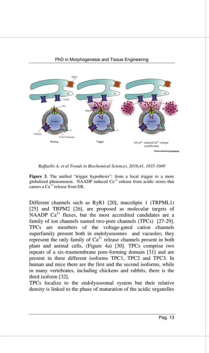
showed that Ca2+ release induced by NAADP appeared as a diffusional gradient [24]. On the basis of these evidences a unified hypothesis known as “trigger hypothesis” was proposed. Ca2+ acidic stores may trigger a further Ca2+ release from ER by a mechanism dependent on functional IP3Rs or RyRs. Local Ca2+
released from acidic compartments is amplified in a more globalized phenomenon that involves the larger and more extensive ER Ca2+ stores, event known as Ca2+-induced Ca2+ release (CICR) [14], (Figure 3).
Raffaello A. et al Trends in Biochemical Sciences, 2016,41, 1035-1049
Figure 3. The unified “trigger hypothesis”: from a local trigger to a more
globalized phenomenon. NAADP induced Ca+2 release from acidic stores that causes a Ca+2 release from ER.
Different channels such as RyR1 [20], mucolipin 1 (TRPML1) [25] and TRPM2 [26], are proposed as molecular targets of NAADP Ca2+ fluxes, but the most accredited candidates are a family of ion channels named two-pore channels (TPCs) [27-29]. TPCs are members of the voltage-gated cation channels superfamily present both in endolysosomes and vacuoles; they represent the only family of Ca2+ release channels present in both plant and animal cells, (Figure 4a) [30]. TPCs comprise two repeats of a six-trasmembrane pore-forming domain [31] and are present in three different isoforms TPC1, TPC2 and TPC3. In human and mice there are the first and the second isoforms, while in many vertebrates, including chickens and rabbits, there is the third isoform [32].
and to the different pH. Indeed, TPC1 is generally expressed in recycling endosomes, early and late endosomes and lysosomes while TPC2 is predominantly localized into late endosomes and lysosomes, (Figure 4b) [33].
a
Parrington J. et al Int. J. Dev. Biol, 2015, 59:341-355
b
Morgan A. J. et al Biochem. J., 2011, 439, 349-374 [34]
Figure 4. [a] A schematic representation of endolysosomal membrane that
shows TPCs transmembrane localization. [b] TPCs relative density linked to pH and endolysosomal system maturation.
Although both TPC1 and TPC2 activation are regulated by NAADP, TPC1 seems to be also activated by Ca2+. Moreover, also ion conduction and permeability differ considerably. TPCs are permeable to a wide range of cations although biophysical data propose that TPC2 is slightly more selective for Ca2+ over K+ than TPC1 and consequently able of discharging considerable quantities of Ca2+ from acidic compartments. It has been shown that TPC1 is also permeable to H+ and for this reason it may be involved in the regulation of lysosomal and cytosolic pH. These differences in gating and ion conducting properties suggest that TPC1 and TPC2 may accomplish complementary physiologal roles as Ca2+ release channels of endo-lysosomal compartments [35].
Several studies support the evidence that TPCs mediate Ca2+ release evoked by NAADP [28, 35, 36] from acidic stores although has been reported evidences indicating that TPCs are not activated by NAADP but rather than PI(3,5)P2 [37, 38]. A very recently
work by Ruas and co-workers shows that NAADP–induced Ca2+ release were significantly impaired in cells TPC1 and TPC2 double knock out corroborating the hypothesis that both TPC1 and TPC2 are NAADP targets [39]. Finally, the possible explication of such contrasting data is that NAADP may not directly interact with TPCs, but through an accessory binding protein [40, 41] element that might be lost in the different experimental conditions [42].
2.3 Role of NAADP and TPCs in pathophysiological processes Ca2+ signalling from TPCs evoked by NAADP is involved in different physiological processes including cellular differentiation, muscle contraction, endothelial activation, membrane trafficking, autophagy, nutrient sensing exocytosis, fertilization and embryogenesis, cytokinesis [42] and pigmentation [43-45]. Furthermore, in our laboratory we have recently demonstrated the emerging role of NAADP elicited pathway, in particular mediated
by TPC2, in angiogenesis. Indeed, in angiogenic process Vascular Endothelial Growth Factor (VEGF) signalling pathway play a pivotal role. Activation of Vascular Endothelial Growth Factor receptor 2 (VEGFR2) results in an increase in intracellular free Ca2+ mediated by NAADP that controls proliferation, migration and sprouting of endothelial cells (ECs). In vitro experiments in human umbilical vein endothelial cells (HUVECs), using a selective NAADP antagonist (Ned-19) or anti-TPC2 shRNA, showed a drastic inhibition of angiogenesis due to the significant reduction of VEGF-induced intracellular Ca2+ mobilization. Moreover, in in vivo experiments Ned-19 inhibited VEGF-induced vessels formation in matrigel plugs and accordingly in TPC2 knockout mice, but not TPC1 knockout mice, VEGF-induced vessels formation in matrigel plugs failed to occur [1].
Interestingly, recent studies reported the emerging role of TPCs in several pathologies such as myocardial ischemia [46], Parkinson’s disease [47] and Ebola virus infection [48].
Recently TPCs mediated Ca2+ signalling has been described also in cancer. Indeed, a genetic analysis aimed to indentify genetic factors implicated in development and metastasis of human oral squamous carcinoma showed an over-expression of TPC2 gene as a possible driver of the amplification of 11q13 genomic region detected in distinct tumors but the mechanism that links TPC2 overexpression to changes in growth and metastatic potential still remains unclear.
Moreover, studies about the relationship between genes involved in skin pigmentation and the tendency for malignant melanoma have shown the possible correlation between cancer and TPC2. TPC2 gene polymorphisms has been identified as potential risk factors in this pathology. Furthermore, melanosomes are modified lysosomes and changes in TPC2 expression or function induces abnormalities on melanocytes biology that could enhance tumorigenity [49]. Involvement of NAADP-mediated responses in melanoma progression have been demonstrated also in my laboratory. Indeed,
signalling is significantly reduced in B16 murine melanoma cells treated with NAADP-inhibitor Ned-19 and also in in vivo experiments in mice we have demonstrated a significant reduction of tumor growth and metastasis formation after four weeks of intraperitoneal Ned- 19 treatment [2]. In addition, data from Nguyen and co-workers indicate TPCs as possible targets of invasive cancers. In a very recent article they have shown that TPCs inhibition impairs migration and adhesion of T24 (bladder cancer), HUH7(hepatocarcinoma) and 4T1 (breast cancer) cancer cells in vitro and metastasis formation in a in vivo mouse model as result of altered integrin trafficking in the endolysosomal compartment [50].
The biological relevance of TPCs-mediated signalling in diseases, and in particular in cancer, make them possible targets for drugs development aimed to fight cancer development and progression.
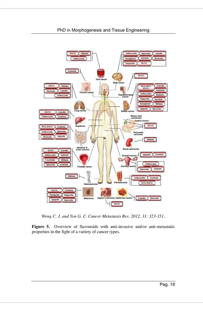
2.4 Biological properties of flavonoids: focus on Naringenin Flavonoids have aroused researchers interest for their indisputable biological properties supported by epidemiological studies that propose a direct link between the consumption of food rich in flavonoids and poor in fatty acids and a decreased risk of certain diseases in humans such as cardiovascular diseases, diabetes and cancer. Due to their antioxidants properties flavonoids seem to have also a pivotal role in the aging process, tissue damage and inflammatory processes [51]. Moreover, they exert a potential anti-invasive and/or anti-metastatic activities in a wide range of cancer types such as brain, thyroid, lung, prostate, ovarian, cervical, oral, hypopharyngeal, breast, liver, biliary tract, gastric, pancreatic, intestinal colon, renal and epidermal cancers, melanoma, fibrosarcoma, osteosarcoma and Kaposi’s sarcoma, (Figure 5) [52].
Weng C. J. and Yen G. C. Cancer Metastasis Rev, 2012, 31: 323-351.
Figure 5. Overview of flavonoids with anti-invasive and/or anti-metastatic
Naringenin (Nar) (5,7-dihydroxy-2-(4-hydroxyphenyl)chroman-4-one) is one of the main flavonoids present in human diet, (Figure 6).
Figure 6. Chemical structure of Naringenin.
Evidences from epidemiological reports showed that the consumption of vegetables and fruit with a high Nar content, such as citruses and tomatoes or their food products, correlate with a reduced incidence of metabolic and chronic-degenerative diseases [53, 54]. Several in vitro studies have shown that Nar interacts with many cellular pathways, point out its properties as an antioxidant, antinflammatory, chemopreventive an antidegenerative agent [55]. In addition, it has been shown that Nar is able to exert anti-angiogenic effects in vitro impairing endothelial cell proliferation, survival, migration and capillary-like structures formation of HUVECs and in vivo reducing neovascularization in an avian chorio-allantoid membrane model [56]. Furthermore, in rats nourished with high-cholesterol diet Nar is able to lower plasma and hepatic cholesterol concentrations by suppressing HMG-CoA reductase and ACAT [57]. The potential of Nar and its precursor naringin emerged from pre-clinical studies which described their activity in treatment of metabolic and cardiovascular disorders such as hyperlipidemia, hypertension, cardiac toxicity, hyperglycemia and diabetes, hepatic steatosis, atherosclerosis [54] and cancer [55]. Indeed, Nar chemopreventive and anticancer
activity has been described; it has been shown that Nar is able to block the progression and the formation of metastasis in different experimental models of oral [58], melanoma [59], breast [60, 61], colon[62], lung [63], and liver [64] cancers. It seems to act by upregulating many different cell survival proteins or arresting cell cycle, by inducing p53-dependent apoptosis or inhibiting inflammatory processes and, in some cases, by exploiting all of these mechanisms [52, 65, 66]. Interestingly, it is known that Nar affects also the activity of different activated or calcium-permeable ion channels. Nar 100 µM induces a threefold increase in the currents mediated by large conductance Ca2+-activated K+ (BKCa) channels, which can be closely correlated with the significant vasorelaxant effect mediated by Nar on rat endothelium-denuded vessels [67]. Moreover, the relaxant effect of Nar on rat colon smooth muscle has been attributed to the direct activation of BKCa channels, resulting in hyperpolarization of smooth muscle cells, which in turn reduces the Ca2+ influx through voltage-dependent calcium channels [68]. Furthermore, Nar is a powerful inhibitor of the melastatin-related transient receptor potential TRPM3 belonging to the TRP family, a calcium-permeable non-selective cation channel expressed in various neural and non-neural tissues, activated by neurosteroids and heat [69]. Since TRPM3-deficient mice display an impaired perception of noxious heat, the inhibition of TRPM3 may represent a novel tool for analgesic therapy [70]. Another member of the TRP family, TRPP2 or polycystin-2, a Ca2+-permeable non-selective cation channel located in the ER and in the primary cilium, is modulated by Nar concentrations ranging from 50 to 200 µM. This channel is implicated in the development of autosomal dominant polycystic kidney disease (ADPKD). It has been suggested that Nar may activate TRPP2 to cause Ca2+ influx and a decrease in cellular proliferation, thereby providing a novel therapeutic approach to ADPKD [71]. Despite the fullness of evidence on Nar efficacy, an exhaustive explanation of its mechanism of action is still lacking,
as is the experimental evidence needed to bridge the gap between multiple cellular targets and biological response.
2.5 Physiological angiogenesis versus tumoral angiogenesis: a general overview
Angiogenesis is the physiological process that foresees the formation of the new blood vessels from pre-existing ones. Although angiogenesis is a phenomenon which occurs throughout life, in the adult the vasculature is generally quiescent and physiological angiogenesis is restricted to some adult tissues that require regeneration or restructuring (including female reproductive organs or injured tissue) [72, 73].
Angiogenesis is characterized by two different mechanisms: sprouting and splitting (or intussusceptive angiogenesis).
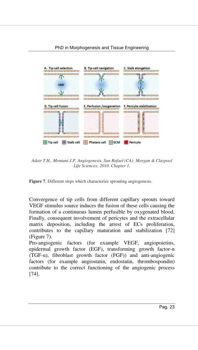
In sprouting angiogenesis enzymatic mechanisms induce degradation of capillary basement membrane, ECs proliferate and migrate, there is a tube formation, vessel fusion and pruning and the stabilization of pericytes. This process begins when tissues are poorly perfused and critical hypoxic levels require blood vessels formation aimed to meet the metabolic needs of parenchymal cells. As a response to the hypoxic conditions several parenchymal cells produce VEGF, a key factor that plays a pivotal role during early stages of angiogenesis. The sprouting process is guided by a tip cell which displays filopodia that moves toward VEGF gradient concentration through VEGF-VEGFR2 interaction. Tip cell determination is controlled by the cell-cell signalling pathway Delta-Notch that is activated by interaction of VEGF with ECs. Capillary sprout elongation is determined by contraction of filaments within the filopodia that draw the tip cell along toward the VEGF source causing consequent proliferation of endothelial stalk cells [72].
Adair T.H., Montani J.P. Angiogenesis. San Rafael (CA): Morgan & Claypool Life Sciences; 2010. Chapter 1.
Figure 7. Different steps which characterize sprouting angiogenesis.
Convergence of tip cells from different capillary sprouts toward VEGF stimulus source induces the fusion of these cells causing the formation of a continuous lumen perfusible by oxygenated blood. Finally, consequent involvement of pericytes and the extracellular matrix deposition, including the arrest of ECs proliferation, contributes to the capillary maturation and stabilization [72] (Figure 7).
Pro-angiogenic factors (for example VEGF, angiopoietins, epidermal growth factor (EGF), transforming growth factor-α (TGF-α), fibroblast growth factor (FGF)) and anti-angiogenic factors (for example angiostatin, endostatin, thrombospondin) contribute to the correct functioning of the angiogenic process [74].
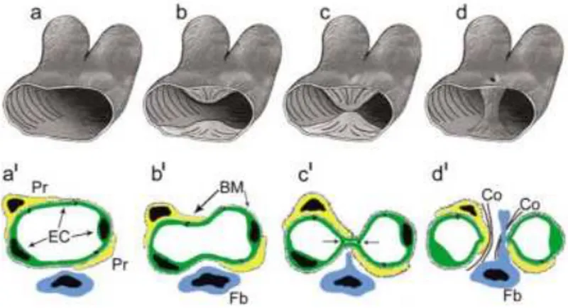
Splitting angiogenesis or intussusceptive angiogenesis, instead, is a phenomenon that plays a key role in the growth and remodelling of most vascular beds, including those of the tumor. During this process a single vessel splits in two different vessels. Invagination of the capillary walls into the vascular lumen induces the formation of intraluminal tissue pillars. The vessel wall of the opposite sides of a vessel extend toward each other forming an intraluminal pillar. In the following phases of the splitting, reorganization of the interendothelial junctions and the formation of the core of the pillar occur and afterwards pericytes and myofibroblsts invade the pillar. Finally fusion of different pillars induces the splitting of the initial capillary in two different ones, (Figure 8) [75].
Adair T.H., Montani J.P. Angiogenesis. San Rafael (CA): Morgan & Claypool Life Sciences; 2010. Chapter 1.
Figure 8. Representation in three dimensions (a-d) and two dimentions (a’-d’)
of different steps of intussusceotive angiogenesis. Endothelial cells (EC); basement membrane (BM); fibroblasts (Fb); pericytes (Pr); collagen fibrils (Co).
Moreover, intussusceptive angiogenesis has been recognized in three different forms: intussusceptive microvascular growth,
intussusceptive arborisation and intussusceptive branching remodelling [75, 76].
Intussusceptive angiogenesis seems to be a fast and efficient process that occurs in pre-existing vascular systems previously formed by vasculogenesis or sprouting angiogenesis without interfering with the local physiological conditions. Indeed, this type of angiogenesis occurs throughout life but plays a pivotal role during embryonic vascular formation in a context in which growth is fast and resources are limited [72]. Furthermore, splitting angiogenesis seems to be regulated both by hemodynamic forces and pro- angiogenic factors such as VEGF, angiopoietins, FGF and PDGFβ [75].
In general, loss of the appropriate balance between pro- and anti- angiogenic factors induces abnormalities in physiological angiogenic process toward a pathological angiogenesis, phenomenon known as “angiogenic switch” [73]. In cancer, for example, angiogenesis has a key role. Indeed tumor vessels supply tumor with oxygen, nutrients and subsequently remove waste. When tumors arise in a well vascularized region co-option of pre-existing vessels might be exploited. On the contrary, angiogenesis or the formation of new blood vessels from a pre-existing system occurs to guarantee tumor expansion. In this context the creation of a pro-angiogenic environment promotes the induction of growth vascular. Tumor secrete growth factors that produce a chemotactive gradient able to recruit ECs and perycites while matrix metalloproteinases generate remodelling of extracellular matrix aimed to facilitate migration and vascular morphogenesis [77].
Tumor blood vessels are architecturally different to the normal counterpart and show some characteristic hallmarks. There is not a specific organization in venules, arterioles and capillaries but blood vessels are usually chaotic, irregular, dilated, tortuous and can have dead end. The vascular system is leaky and haemorrhagic and in addition tumor vessels may have cancer cells integrated in the
vessel wall. Moreover, blood perfusion may be irregular and slow, (Figure 9) [73].
a b
Modified from McDonald D.M. and Choyke P.L. Nat Med 2003, 9(6):713-25
Figure 9. Representative microscopic imaging of normal [a] and tumor
vasculature [b]. [a] In normal blood vessels it is possible to observe a precise vascular network organization in arterioles, capillaries and venules. [b] Tumor blood vessels appear chaotic and not hierarchically organized [78].
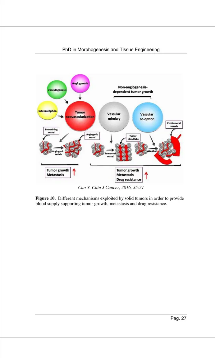
Recently, several evidences show alternative mechanisms through which vascular supply can be formed in solid tumors, as represented in Figure 10. Co-option process foresees that tumors adopt pre-existing blood vasculature of the surrounding environment. In vascular mimicry, instead, aggressive tumor cells themselves, rather than endothelial cells, form capillary-like structures perfusible by blood able to provide support to cancer growth [79]. Initially discovered in melanoma, additional studies have demonstrated that several aggressive cancers such as some sarcomas, carcinomas or gliomas can display vascular mimicry [80, 81].
Cao Y. Chin J Cancer, 2016, 35:21
Figure 10. Different mechanisms exploited by solid tumors in order to provide
3. AIMS
In our previous studies we have demonstrated the emerging role of Two-Pore Channel 2 in angiogenesis and its likely involvement in melanoma progression. Indeed, we have described that HUVECs treated with the NAADP inhibitor Ned-19 or anti-TPC2 shRNA showed a drastic inhibition of angiogenesis due to the significant reduction of VEGF-induced intracellular Ca2+ mobilization. Moreover, in in vivo experiments Ned-19 inhibits VEGF-induced vessels formation in matrigel plugs and in TPC2 knockout mice, but not TPC1 knockout mice, VEGF-induced vessels formation in matrigel plugs failed to occur [1]. Furthermore, we have also demonstrated that Ned-19 is able to block growth, migration, adhesivity and VEGFR2 expression in B16 melanoma cells in vitro and tumor growth, metastasis formation and vascularization in vivo. Collectively, these observations propose NAADP/TPC2 signalling pathway as a key point not only for neoangiogenesis but also for the direct contol of tumor cells [2].
On this basis our laboratory is presently engaged in a number of experimental approaches with the ultimate aim of assessing whether TPC2 can be a suitable target for potential melanoma therapy. My specific contribution to this aim has been characterize a novel pharmacological tool exploring the potentiality of a natural flavonoid, Naringenin, in our models.
To this purpose we have focused our attention on Nar ability to modulate TPCs activity, given that Nar interferes different Ca2+ channels [67-71], in order to investigate its effects on VEGF-dependent angiogenesis and in melanoma behavior.
In particular the aims of this work are:
- In HUVECs : to evaluate Nar effects on NAADP-controlled Ca2+ responses through calcium imaging experiments and investigate Nar effects on VEGF-dependent angiogenesis in vitro and in vivo. - In B16 melanoma cells: to evaluate Nar effects on NAADP-controlled Ca2+ responses through calcium imaging experiments and to investigate Nar effects on biological cancer processes such
as migration, proliferation and non-angiogenic vasculogenic mimicry.
4. RESULTS AND DISCUSSION
4.1 Naringenin affects the activity of the human TPCs directly impairing hTPC2 functional activity and decreasing hTPC1 activity
Mature plant cells have a particular intracellular compartment that is lacking in animal cells: the vacuole. Its large dimension and its easy isolation allow researchers to study it using patch-clamp techinique. Through this method it is possible to isolate a specific small patch of a membrane and precisely detect the current flowing through a single ion channel. Interestingly, in this way is possible to exploit A. Thaliana vacuole as a novel heterologous system for the expression and functional characterization of animal intracellular channels and transporters [82].
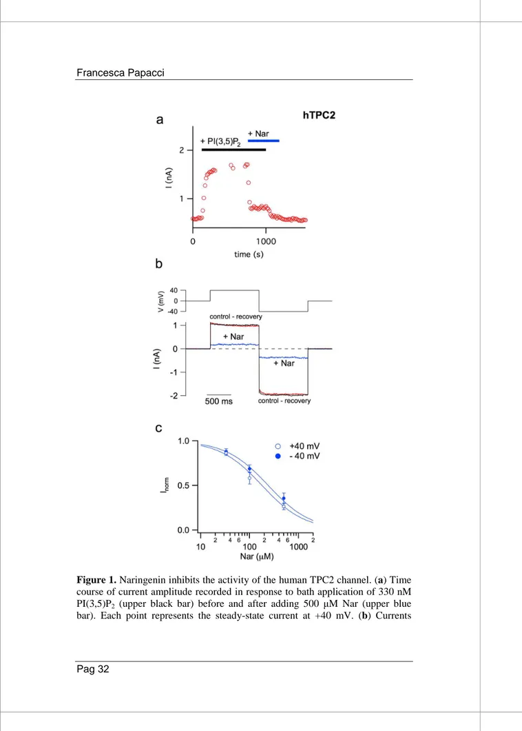
In collaboration with Armando Carpaneto group from CNR of Genoa, we have employed this heterologus system to assess the ability of Nar to bind and regulate the activity of human TPC2 (hTPC2). Isolated protoplasts of a mutant A. thaliana, lacking endogenous TPCs, were transiently transformed with hTPC2 equipped with an EGFP fused to its C-terminus [83]; we applied the patch-clamp technique to vacuoles in which GFP fluorescence was clearly detectable on the tonoplast. The addition of 330 nM PI(3,5)P2 in the external (cytosolic) solution induced robust
hTPC2-mediated currents, as shown in Figure 1a where stationary currents recorded at +40 mV were plotted versus time. When 500 µM Nar was also added in the cytosolic solution a strong decrease in the activity of hTPC2 currents was evident. Figure 1b shows control and recovery currents (∆I) recorded in the presence of PI(3,5)P2 subtracted by the correspondent background recorded
without the phosphoinositide. Upon Nar application, currents were drastically reduced at both +40 and −40 mV voltages. Even if recorded in symmetrical concentration of sodium, due to a voltage-dependent block by cytosolic magnesium [84] the currents at +40 mV were smaller than those recorded at −40 mV. Interestingly,
Nar inhibition was fully reversible (Figure 1b). The dose-response was well described by a Michaelis-Menten hyperbolic function (Figure 1c), with apparent affinity constants of 180 ± 20 µM at +40 mV and of 240 ± 40 µM at −40 mV, therefore not significantly different.
Figure 1. Naringenin inhibits the activity of the human TPC2 channel. (a) Time
course of current amplitude recorded in response to bath application of 330 nM PI(3,5)P2 (upper black bar) before and after adding 500 µM Nar (upper blue
mediated by hTPC2 and induced by the application of 330 nM PI(3,5)P2 in the
cytosolic bath solution were recorded at +40 and −40 mV (shown in the upper voltage profile) in the absence or presence of 500 µM cytosolic Nar. For the sake of clarity, background currents in the absence of the phosphoinositide were directly subtracted. (c) Dose–response analysis of Nar inhibition. Inorm, the
normalised current, was obtained by the ratio of currents recorded respectively in the presence and absence of cytosolic Nar. Both at +40 and −40 mV, normalised currents at different Nar concentrations were fitted with a Michaelis– Menten function (continuous lines). Data from 3 (Nar33 µM), 6 (Nar 100 µM) and 9 (Nar 500 µM) different vacuoles were shown as mean ± s.e.m. Values
at positive and negative voltages, at a defined Nar concentration, were not significant (P > 0.2); values at different Nar concentrations and at a defined voltage (+40 or −40 mV), were statistically significant (at least P < 0.01).
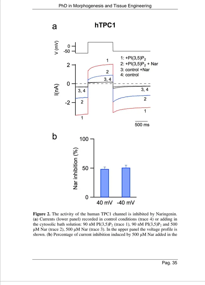
We wondered if Nar could modulate the other member of the TPC family in humans, namely hTPC1. We therefore performed a similar approach as for hTPC2 and expressed hTPC1 fused to an EGFP at its C-terminus in Arabidopsis Thaliana vacuoles lacking the endogenous TPC1. Suppl. Figure 1a, left panel, shows that the background currents recorded in the absence of PI(3,5)P2 are very
small in comparison to those elicited by the addition of the phosphoinositide (right panel). We chose a concentration of PI(3,5)P2 equal to 90 nM and therefore near to the saturation value
since the apparent binding constant in this experimental condition was about 20 nM [85]; in comparison the affinity binding constant of PI(3,5)P2 for hTPC2 is about 140 nM [83]. The current-voltage
relationship in the absence (empty symbols) and in the presence (filled symbols) of PI(3,5)P2 was plotted in Suppl. Figure 1b. In a
recent paper [85] we demonstrated that the functionality of hTPC1 was strongly affected by both cytosolic and luminal Ca2+, namely an increase in cytosolic and luminal Ca2+ induced an increase and a decrease of the activity of the channel, respectively.
Here, our standard pipette and ionic bath solutions (see Materials and Methods) allowed us to record hTPC1-mediated currents at both negative and positive potentials and therefore to evaluate the effect of Nar at different voltages. In Suppl. Figure 1b, at positive
currents due to the presence of cytosolic magnesium was also apparent [85]; a similar effect was recorded for hTPC2 in Figure 1band by other authors [84]. Figure 2ashowed that the addition to the cytosolic solution of 500 µM Nar (trace 2) induced a significant decrease of hTPC1-elicited currents (trace 1) at both negative and positive voltages and had almost no effect in control conditions (compare trace 3 and 4). The inhibition of hTPC1 by Nar was quantified in the histogram of Figure 2b; the effect was not voltage-dependent. Moreover, we verified that Nar decreased the activity of the endogenous Arabidopsis TPC1 (Suppl. Figure 2) showing that the inhibition of TPC channels by Nar was a mechanism conserved during evolution.
Figure 2. The activity of the human TPC1 channel is inhibited by Naringenin.
(a) Currents (lower panel) recorded in control conditions (trace 4) or adding in the cytosolic bath solution: 90 nM PI(3,5)P2 (trace 1), 90 nM PI(3,5)P2 and 500 µM Nar (trace 2), 500 µM Nar (trace 3). In the upper panel the voltage profile is
cytosolic solution at −40 and + 40 mV. Data from 6 different vacuoles, shown as mean ±s.e.m., were not statistically significant (P > 0.6).
4.2 Naringenin significantly inhibits VEGF-dependent calcium mobilization in a dose-dependent manner
In HUVECs, we have previously demonstrated that VEGF- dependent Ca2+ mobilization evoked by NAADP is impaired pretreating cells with Ned-19 (a noncompetitive antagonist of NAADP) [1]. In order to evaluate the possible inhibitory effect of Nar on the regulation of VEGF-dependent Ca2+ signalling in human cells, calcium imaging experiments were performed pretreating HUVECs for 30 min with different concentrations of Nar and stimulating them with 100 ng/ml VEGF. Figure 3a,b shows that Ca2+ mobilization was significantly reduced in a dose-dependent manner without affecting VEGFR2 phosphorylation at Tyr1175 demonstrating that the inhibition of the responses occurs downstream of the receptor , (Figure 4). Furthermore, opting for 1000 µM Nar, for its highest effect, we have analyzed the specificity of this response for NAADP/TPC2- dependent Ca2+ signalling using a positive and a negative control. Bar charts in Figure 3c,d show that Nar fails to block Ca2+response to ATP, known to be IP3-dependent, but blocks histamine-evoked Ca2+
release known to be NAADP/TPC2-dependent [86]. These data are a strong indication that Nar is able to reduce Ca2+ release inhibiting hTPC2. Finally, to better correlate the specific inhibitory effect of Nar treatment on TPC2, calcium imaging experiments with the direct channel activator were performed. HUVECs Ca2+ mobilization in response to 500 nM NAADP-AM was significantly impaired pretreating cells with 500 µM or 1000 µ M Nar. These set of experiments were accomplished in a Ca2+ free buffer to tightly evaluate the intracellular Ca2+ involvement alone thus limiting possible off-target responses (Figure 3e).
Taken as a whole, these data strongly suggest a direct effect of Nar on TPC2 given that it affects specifically Ca2+ released from acidic intracellular compartments.
cells. (a,b) HUVECs were pretreated for 30 min with different concentrations of Nar and then stimulated with 100 ng/ml VEGF; (a) Bar chart showing maximum Ca2+ concentrations; (b) Changes in Ca2+ levels shown as representative traces. (c,d) Cells were pretreated with 1000 µM Nar for 30 min and then stimulated with (c) 10 µM ATP (negative control) or (d) 100 µM histamine (positive control). (e) Cells were pretreated with 500 µM or 1000 µM Nar and then stimulated with 500 nM NAADP-AM. Data in bar charts were from 3 independent experiments, n = 41–180 cells. *P < 0.05; **P < 0.01; ***P < 0.001.
Figure 4. Phosphorylation state of VEGFR2 at Tyr1175, evaluated by western
blot in untreated (NT) or VEGF-treated HUVECs. (a) Cells were preincubated with Nar (200 µM, 600 µM and 1000 µM) or vehicle for 30 min, or with TSU-68 (2,1 µM), or its control (vehicle-2) for 1h. Samples were then stimulated with 100 ng/ml VEGF for 15 min. The intensity of pVEGFR2 bands was quantified and normalized to α-tubulin content. (b) Data in bar chart represent mean ± s.e.m. from three independent experiments. As apparent, VEGF-induced receptor phosphorylation is significantly inhibited by TSU-68 but not by Nar.
4.3 Naringenin impairs VEGF-induced angiogenic process in
vitro and in vivo
Li et al recently have demonstrated the antiangiogenic effects of Nar on HUVECs in vitro and in the avian chorio-allantoid membrane model in vivo. Although its antiangiogenic effects have
been reported to involve impairment of estrogen-related receptor α (ERR α) and VEGF secretion, the specific pathway involved has yet to be understood [56]. Previously, we have demonstrated that NAADP-mediated Ca2+ signalling has a key role in the regulation of in vitro and in vivo angiogenesis. VEGF-induced angiogenesis was inhibited by Ned-19 in vitro and in an established in vivo murine model [1]. In order to explore the anti-angiogenic effects of Nar we have evaluated VEGF-dependent angiogenesis in vitro and in vivo.
HUVECs have the ability to form capillary-like tubes in vitro, characteristic that is commonly considered as representative of later, differentiative, steps of angiogenesis. In this way exploiting this assay it is possible to analyze the pro or anti-angiogenic effects of a compound. ECs plated onto matrigel matrix adhere, migrate and, within a few hours, differentiate into capillary- like structures. Figure 5 shows that both 1000 µM or 500 µM Nar significantly impaired the number of closed polygons formed by cells stimulated with VEGF compared with samples stimulated with VEGF alone. This evidence points out that Nar regulates VEGF-induced capillary-like formation in vitro specifically inhibiting the NAADP/TPC2- mediated Ca2+ signalling.
Figure 5. HUVECs ability to form capillary-like structures induced by VEGF
in vitro were impaired by Naringenin. (a,c) Representative images of one of
three independent experiments. HUVECs were plated in Matrigel-coated dishes and incubated for 3–4 h in EBM-2 + 2% FBS supplemented or not with VEGF or Nar 1000 µM (a) and 500 µM (c) or in medium containing both VEGF and Nar. (b,d) Quantitative evaluation of tube formation as the number of closed polygons formed in 6 fields for each experimental condition. Data in bar charts represent mean ± s.e.m. of three independent experiments. *P < 0.05
In our in vivo murine model we have previously demonstrated that VEGF-treated plugs implanted in the flank of wild type mice not only failed to be vascularized if treated with Ned-19, but more strikingly remained avascular in TPC2 - /- mice. In addition, the
specific role of TPC2 was supported by the evidence that vascularization was not inhibited in plugs from TPC1 -/- mice [1]. To verify the inhibitory effect of Nar on in vivo angiogenesis C57BL/6 were injected with matrigel plugs containing either Nar or vehicle, or Nar plus VEGF; five day later the extent of plug vascularization under the different experimental conditions was evaluated by measuring the hemoglobin content. Figure 6 shows that while VEGF alone induces significant vascularization, plugs treated with VEGF plus Nar remain virtually avascular. Interestingly, our observations in vivo open a new perspective toward a potential therapeutic approach although we cannot exclude the possibility that targets other than TPC2 may contribute to this antiangiogenic effect.
Figure 6. Naringenin affects vascularization induced by VEGF in a in vivo
murine model. Male/female C57BL/6 mice 5 weeks old were injected subcutaneously with matrigel plugs containing either vehicle or VEGF or VEGF
vascularization was evaluated both macroscopically, as shown in two representative images (a) and as hemoglobin content expressed as absorbance (OD)/1 g matrigel plug (b); values from three independent experiments are expressed as mean ± s.e.m. *P < 0.05. n = 14–15 plugs for each experimental condition.
4.4 Naringenin does not impair NAADP/TPC2-independent angiogenic responses
We have previously shown and analyzed that intracellular Ca2+signalling is involved in HUVECs angiogenic responses to Angiopoietins. Evidence from pharmacological inhibition of different Ca2+ release channels showed that neither the increase of intracellular Ca2+ nor the angiogenic responses elicited by Angiopoietin-1 (Ang-1) are dependent on NAADP-TPC2 pathway. In fact, both response were significantly impaired by thapsigargin, which inhibits SERCA pumps, but not by bafilomycin A1, which inhibits pH-dependent Ca2+ uptake into acidic stores [5].
To further validate the hypothesis that Nar specifically impairs angiogenic responses inhibiting NAADP/TPC2 signalling pathway we tested Nar effect on HUVECs stimulated with Ang-1, as an angiogenic stimulus that does not couple with TPC2, using Ned-19 as positive control. To this aim calcium imaging experiments were performed. Cells were pretreated for 30 min with Nar (Figure 7a) or Ned-19 (Figure 7b) and stimulated with 100 ng/ml Ang-1. Both Nar and Ned-19 failed to impairs Ang-1-dependent Ca2+ mobilization corroborating our aforementioned hypothesis.
Figure 7. Neither Naringenin nor Ned-19 impairs Ang1-dependent Ca2+ release. Live imaging in single FURA2-AM loaded cells. (a) HUVECs were pretreated for 30 min with 500 µM or 1000 µM Nar and then stimulated with 100 ng/ml Ang-1. Both Nar concentrations failed significantly inhibit Ang-1 mediated Ca2+ release (P >0,2). (b) Calcium imaging experiments in HUVECs pretreated for 30
shows that Ang-1induced-increase in [Ca2+]i is not impaired by Ned-19. Data
were from 3 independent experiments ***P<0,001.
Likewise we have analyzed whether Nar was able to impair NAADP/TPC2-independent angiogenic responses in vitro and in vivo. As expected, Ang-1 stimulated HUVECs capability to form capillary-like structures on matrigel, but both Nar (Figure 8a,b) and Ned-19 (Figure 8c) failed to interfere with this phenomenon in vitro.
Figure 8. Naringenin does not affect angiogenic responses to Ang-1 in vitro. (a)
Representative images of one of three independent experiments. HUVECs were plated in Matrigel-coated dishes and incubated for 2–3 h in EGM-2 supplemented or not with Ang-1 or Nar, or in medium containing both Ang-1 and Nar. (b) Quantitative evaluation of tube formation as the number of closed polygons formed in 6 fields for each experimental condition. Data in bar charts represent mean ± s.e.m. of three independent experiments. *P < 0.05. (c) Cells were plated in Matrigel-coated dishes and incubated for 2-3 h in EGM-2 supplemented with Ang-1 or Ang-1 + Ned-19. Quantitative evaluation of tube formation as the number of closed polygons formed in 9 fields for each experimental condition. Data in bar charts represent percentage of the control (mean ± s.e.m. of three independent experiments).
In addition in vivo matrigel assay were performed. Matrigel plugs containing either Nar or vehicle or Nar plus Ang-1from injected mice were evaluated in hemoglobin content 5 days after inocolum.
Bar charts in Figure 9 show that the vascularization of Ang-1 plugs did not significantly differ from that in Ang-1 plus Nar plugs. Taken together these data indicate that IP3-dependent Ca2+
responses and angiogenic process (in vitro and in vivo) stimulated by Ang-1 are not affected by Nar strongly indicating that Nar specifically impairs NAADP/TPC2- dependent signalling pathway.
Figure 9. Naringenin does not affect angiogenic responses to Ang-1 in vivo. In
vivo vessel formation was assessed after subcutaneous injection of 5 weeks old
male/female C57BL/6 mice with Matrigel plugs containing either vehicle or Ang-1 (150 ng/ml) or Ang-1 plus 1000 µM Nar. Five days after injection the mice were sacrificed and plug vascularization was evaluated as hemoglobin content expressed as absorbance (OD)/1 g matrigel plug. Hemoglobin content in Ang-1 plugs (n=8) did not significantly differ from that in Ang-1 + Nar plugs (n=8) (P>0.2).
4.5 Conclusions (1)
In the present study we demonstrated that Nar can specifically inhibit the ability of TPC2 to release Ca2+ from acidic stores in
NAADP-mediated responses. Electrophysiological analysis shows that hTPC2-current is specifically reduced by Nar. Furthermore, we assess the possible effects of Nar on TPC2 in human endothelial cells analyzing VEGF-dependent Ca2+ release and VEGF-dependent angiogenesis in vitro and in vivo. Both Ca2+ -activated responses of HUVECs to VEGF and in vitro and in vivo angiogenesis are inhibited by Nar. In addition, Ang-1-dependent angiogenic responses, known to be TPC2-independent, are not affected by Nar. Figure 10 describes schematically the specific role of Nar in the control of VEGF-induced angiogenesis, based on present and previous data.
Figure 10. Schematic representation of specific Ca+2 signalling pathway involved in the control of angiogenesis by VEGF and Ang-1. Nar inhibits Ca+2 mobilization when mediated by TPC2, but not by RyR/InsP3R. AC: acidic
compartments, ER: endoplasmic reticulum.
VEGF-light on the mechanisms underlying this process by revealing the pivotal role played by TPC2 and provide the first evidence of the antiangiogenic properties of Nar in an in vivo murine model.
Nar is known to exert a wide range of effects induced by a number of mechanisms that have yet to be discovered.
By contrast, a limited number of factors are known to modulate TPC2 activity, and our data shed light on a novel, naturally-occurring molecule that targets this channel which is attracting a considerable amount of interest owing to its relevance to important pathophysiological processes. The relationship we describe here between naringenin and TPC2 is therefore likely to have wider implications in systems other than the vascular system, thus representing a novel tool for experimental, and possibly even clinical, research purposes.
4.6 Naringenin impairs B16 melanoma cells VEGF-dependent Ca2+ responses
In tumor biology Ca2+ signaling pathway represents an important aspect. Changes in Ca2+ toolkit lead cancer cells to acquire some distinctive hallmarks such as alteration in the expression of proteins involved in the mobilization of Ca2+ across the plasma membrane and sub-cellular organelles. Many studies have shown that Ca2+ is involved in basic processes such as proliferation, migration, survival of tumors cells, invasiveness and metastasis formation [9-12]. On the basis of this evidence we have recently focused our attention on the study of VEGF-dependent Ca2+ signaling in cancer cells, starting with murine melanoma. We have demonstrated a role for NAADP-evoked Ca2+ signalling in a number of melanoma features in vitro and in a murine melanoma in vivo model. Using the NAADP inhibitor Ned-19 in B16 melanoma cell line cultured in vitro or inoculated in C57BL/6 mice in vivo we have observed that NAADP controls cellular migratory ability, cellular proliferation, tumor vascularization, growth and
metastatic dissemination. These data strongly suggest that NAADP/TPC2 Ca2+ pathway can be relevant not only for neoangiogenesis, but also for a direct control of tumor cells [2]. Exploiting Nar ability to block TPC2-mediated responses in HUVECs we have investigated whether Nar could interfere with the response of B16 melanoma cells to VEGF. Ca2+ imaging experiments were performed in B16 FURA2-AM loaded single cells pretreated for 30 min with different concentrations of Nar and stimulated with 100 ng/ml VEGF. Histograms in Figure 11a, shows that Nar is able to inhibit VEGF-related Ca2+ signalling in a dose-dependent manner. Furthermore, to directly assess the specific inhibitory effect of Nar treatment on TPC2 in this experimental system we have performed Ca2+ imaging experiments pretreating cells with Nar and stimulating them with 100 nM NAADP-AM. Data in bar charts show that NAADP-mediated Ca2+ mobilization is inhibited by Nar, (Figure 11b). In addition, to further support our observation we have also performed Ca2+ imaging experiments pretreating cells with different concentration of Ned-19 (previously demonstrated to inhibit NAADP-dependent responses in B16 melanoma cells [2]) and stimulating them with 100 nM NAADP-AM. As expected, Figure 11c shows that also Ned-19 was able to significantly inhibit Ca2+ responses to NAADP-AM. Taken as a whole these observations strongly suggest a specific inhibitory effect of Nar on TPC2-mediated Ca2+ release also in B16 melanoma cells.
Figure 11. Naringenin inhibits NAADP-controlled Ca2+ mobilization in B16 melanoma cells. Ca2+ imaging experiments. Cells pretreated for 30 min with different concentration of Nar were stimulated with 100 ng/ml VEGF (a) or 100 nM NAADP-AM (b). (c) B16 cells pretreated with Ned-19 and stimulated with 100 nM NAADP-AM.
4.7 Naringenin affects the proliferative/migratory behavior of B16 melanoma cells
Migration and proliferation are characteristic hallmarks of cancer; we have previously demonstrated that NAADP- mediated Ca2+ release is required for the migratory ability of B16 cells in their own conditioned medium. Indeed, we have shown that Ned-19 significantly reduces cells migration and focal adhesion kinase (FAK) activation, protein known to have an important role in the regulation of dynamic changes in actin cytoskeleteon reorganization [2]. In order to investigate the possible inhibitory effect of Nar on B16 melanoma cells migratory ability, scratch assay was performed on confluent monolayers treated or not with the inhibitor and cultured for 18 hours in the absence of serum in order to minimize the contribution of growth versus migration. Figure 12a,b shows that B16 melanoma cells capability to move on the wound is inhibited by Nar. In addition western blot analysis of P-FAK and P-AKT levels at the same time and conditions shows that Nar significantly impairs FAK and AKT activation allowing to attribute, at least in part, the observed behavior to changes in cell motility (Figure 12c,d). This point, of potential relevance for the control of invasive and metastatic behavior, opens the way to further more direct and elaborate approaches, presently ongoing in our laboratory.
We also asked whether in this experiment Nar had interfered with the proliferation. To this purpose cell cycle analysis were performed in the same experimental conditions (18h, no serum) and % G1 and S phase was evaluated in a cytometer. Data in figure 13 a, b show that Nar treatment significantly abates the percentage of cells undergoing S phase and increases the percentage of cells undergoing G1 phase. Analyzing these results we cannot exclude the contribute of proliferation inhibition on the migration process, but this indications suggest that both of them may be affected by Nar. We previously demonstrated in B16 melanoma cells the
of migration and proliferation [2]. Taken together these data suggests that Nar can interfere with this control most likely through TPC2, a hypothesis which we are presently investigating by means of genetic approaches.
Figure 12. Naringenin impairs melanoma migration. (a) Scratch assay to
investigate B16 melanoma cells ability to move on wound. Wounded monolayer were maintained in serum free medium in presence or absence of Nar for 18h. Representative images of three independent experiments at the time of manual damage t=0 and after 18h. (b) Quantitative evaluation of the percentage of free area versus t=0 after 18h. (c,d) Western blot assessment of phosphorylation levels of FAK (c) and AKT (d). Cells, treated or not with Nar in a serum free medium for 18h, were lysed and western blot analysis was performed.
Figure 13. Naringenin interferes with B16 melanoma cell cycle increasing
G1phase and decreasing S phase. (a,b) Data are representative of three independent experiments. Evaluation of (a) % G1 phase (b) % S phase 18h after Nar treatment in a serum free medium.
4.8 Naringenin interferes with B16 melanoma ability to form vascular-like tube network
The ability of aggressive tumor cells to form vascular channels, a phenomenon known as vasculogenic mimicry (VM) has been described. Through this process tumor cells, without implication of ECs, form tubes de novo independently of angiogenesis. Interestingly, channels are not true blood vessels formed by preexisting ones, but mimic capillary function. In addition, a link has been highlighted between VM and poor clinical outcomes and suggesting that this could explain why some tumors poorly respond to angiogenic inhibitors [81, 87].
The initial characterization of this tumor plasticity was made in melanoma in which cells coexpression of endothelial, tumor markers and perfusible vascular-like structures rich in laminin, collagens IV and VI and heparin sulfate proteoglycans were described [80]. Furthermore, subsequent comparative studies between melanoma VM networks versus endothelial-dependent angiogenic vasculature reported upregulated expression of key angiogenesis-related and stem-cell–associated genes by the melanoma cells [81].
Interestingly, we have observed that Nar interferes with melanoma cells VM. We have seeded B16 melanoma cells on Matrigel and culturing them for 18h to allow for the development of capillary-like tubes. The cells treated in a serum free medium with the vehicle alone underwent a rearrangement and formed a vasculogenic network within 18h. Otherwise, cells treated with 200
µM Nar showed a significant inhibition of these structures
suggesting a possible role for TPC2 in VM, (Figure 14).
In this study we have introduced the natural flavonoid Nar as a tool to directly block TPC2-mediated angiogenic response in HUVECs. In addition, VM of B16 melanoma cells is also impaired by Nar; although we cannot exclude the possibility that in B16 melanoma cells Nar targets pathways different from TPC2, these data suggest that this ion channel may have a possible role in endothelial and melanoma pathways in the formation of angiogenic and
non-angiogenic vascular system respectively: a novel promising field worth of further insight.
Figure 14. Naringenin inhibits B16 melanoma cells ability to form vascular-like
structures. Cells were seeded on Matrigel in presence or absence of Nar in a serum free medium for 18h. (a) Representative images of one of five independent experiments. (b) Quantitative evaluation of tube formation as the number of closed polygons formed in 9 fields for each experimental condition. Data in bar charts represent fold of treated samples compared with control set as 1 (mean ± s.e.m. of five independent experiments).
4.9 Concluding remarks
Investigating cancer behavior in the context of its microenvironment represents an important aspect in the fight against cancer. In particular, angiogenesis has a pivotal role in tumor progression and the communication with the surrounding environment.
In literature several studies point out the emerging role of TPCs in patophysiological processes [1, 42-48] including cancer [2, 49, 50].
Indeed, we have previously demonstrated TPC2 involvement in angiogenesis and NAADP signalling [1, 2].
In this study we introduce Nar as a novel natural pharmacological tool able to interfere with TPC2 activity. In particular, we have demonstrated that in endothelial cells Nar specifically inhibits both
VEGF/NAADP/TPC2-dependent Ca2+ mobilization and
angiogenesis in vitro and in vivo. On the other side, also data from melanoma, although lacking a direct correlation, strongly suggest that Nar effects on B16 biological processes may be dependent upon TPC2 inhibition.
Finally, on the basis of this evidence, we can conclude that Nar may represent an important natural tool that potentially capable to impair NAADP/TPC2 Ca2+ signalling not only in angiogenesis but also in melanoma progression pointing out the relevance, for clinical application, to develop drugs able to target at the same time both processes.
5. MATERIALS AND METHODS
5.1 Arabidopsis thaliana tpc1–2 mutants [88]were grown in soil in a growth chamber at 22 °C and 8 h light/16 h dark regime. Well-expanded leaves from 4 weeks old plants were used for mesophyll protoplast preparation as described in refs [89] and [90]. The transient transformation was performed as described previously [83, 85] using the pSAT-hTPC2-EGFP or pSAT-hTPC1-EGFP plasmid as reported therein.
The transformed cells were maintained in the dark at 23 °C in W5 ionic solution (in mM: 154 NaCl, 125 CaCl2,KCl 5, MES 2, pH 5.6
with KOH) plus 50 µg/ml Ampicillin as described in ref. [89]. Vacuoles were efficiently released from the protoplasts using the following vacuole release solution (in mM): 100 malic acid, 5
EGTA, 3MgCl2, pH 7.5 using 160 1,3-bis
(tris(hydroxymethyl)methylamino) propane (BTP), 450 mOsm with D-sorbitol.
5.2 Patch-clamp recordings. Patch-clamp experiments on vacuoles from fluorescent protoplasts expressing the hTPC2-EGFP fusion on the tonoplast were performed in whole-vacuole configuration ≥40 h after the transformation.
The bath solution (cytoplasmic side) contained (in mM): 100 NaCl, 2 MgCl2, 10 Hepes-Tris, pH 7.5. The pipette solution (vacuolar
side) was (in mM): 100 NaCl, 2 MgCl2, 10 MES-Tris, pH 5.5. The
bath solution and the pipette solution were adjusted to 580 mOsm and 618 mOsm by the addition of D-sorbitol. Dithiothreitol (DTT; 2 mM) was added to the vacuole release solution before the patch-clamp experiments. DTT was prepared as 1 M stock solution the day of the experiment and kept on ice until use. Nar was also prepared fresh as a stock solution in DMSO. Since the usual maximum concentration of Nar used in electrophysiological studies (100 µM) induced about 40% inhibition of hTPC2 and even less of AtTPC1, we decided to increase [Nar] up to 1000 µM. However, some precipitate was present in these conditions possibly
that the maximum DMSO concentration used in this study (up to 0.1%) were not effective on channel activity. PI (3,5)P2, purchased
as dioctanyl ester (diC8) from AG Scientific or Echelon Biosciences Inc, was prepared as 1 mM stock solution and stored at −20 °C. The other chemicals were purchased from Sigma-Aldrich and Carl Roth.
5.3Analysis of electrophysiological data. Positive currents correspond to cations flowing from the cytoplasmic side to the lumen of the vacuole. Data analysis and figure preparation were done with Igor Pro software (Wavemetrics) or Photoshop (Adobe Inc.).
5.4 Cell culture. HUVECs were obtained from Lonza Sales Ldt, cultured in EGM-2 Endothelial Cell Growth Medium-2 (Endothelial Basal Medium EBM-2 + EGM-2 Bullet Kit, Lonza), + 100 mM Penicillin/Streptomycin (P/S) (Sigma); Cells were used at passage 1 to 6.
B16 F0 melanoma cells ATCC CRL-6322 were cultured in Dulbecco’s Modified Eagle Medium (DMEM) supplemented by 10% FBS, 2% P/S and 2% L-glutamine (Sigma).
Both cell lines were maintained at 37 °C in a humidified 5% CO2
incubator.
Reagents used are: VEGF-A165 (Peprotech), Ang-1 (Peprotech), Histamine (Sigma), ATP (Sigma), Ned-19 (Tocris Bioscience) Naringenin (Sigma); NAADP-AM was a generous gift of G. C. Churchill (Oxford University).
5.5 Calcium imaging. Cells were incubated in EGM-2 or DMEM containing 3.5 µM Fura-2-AM (Invitrogen) for 1 h at 37 °C and then rinsed with either Hanks’ Balanced Salt Solution (HBSS, Sigma) or Ca2+ free Krebs Henseleit Hepes buffer (KHH). Dishes were placed into a culture chamber kept at 37 °C controlled temperature on the stage of an inverted microfluorimeter (Nikon
![Figure 4. [a] A schematic representation of endolysosomal membrane that](https://thumb-eu.123doks.com/thumbv2/123dokorg/2892914.11362/15.892.110.795.3.1221/figure-schematic-representation-endolysosomal-membrane.webp)
![Figure 9. Representative microscopic imaging of normal [a] and tumor](https://thumb-eu.123doks.com/thumbv2/123dokorg/2892914.11362/26.892.194.680.322.559/figure-representative-microscopic-imaging-of-normal-and-tumor.webp)