SAPIENZA UNIVERSITY OF ROME
SCHOOL OF BIOLOGY AND MOLECULAR MEDICINE
PhD THESIS
Targeted Resequencing as a diagnostic tool
in patients with epilepsy
Human Biology and Medical Genetics PhD course
Medical Genetics curriculum
XXXII Cycle
Coordinator: Prof. Antonio Pizzuti
Tutor: Dr. Massimo Carella
Candidate: Dr.ssa Ester Di Muro
3
Index
Abstract 5
1. INTRODUCTION 6
1.1
Epilepsy: definitions and epidemiology 6
1.2
Classification of seizure and epilepsy 8
1.3
Etiology of epilepsy 10
1.4
Genetic basis of epilepsy 11
1.4.1 Genetics of Epilepsy Syndromes 13
2. NEXT GENERATION SEQUENCING TECHNOLOGY 19
2.1
Workflow of next generation sequencing analysis 21
2.2
Next generation sequencing and epilepsy 25
3. AIMS OF THE STUDY 27
4. MATERIALS AND METHODS 28
4.1 Cohort of the study 28
4.2
Targeted Resequencing (TRS) 30
4.2.1 Gene panel design 30
4.2.2 Library preparation 33
4.2.3 Sequencing 35
4
5. RESULTS 39
6. DISCUSSION 70
7. CONCLUSIONS 76
Reference 77
Publications 96
5
Abstract
Epilepsy is one of the most common neurological disorder, affecting 5–8/1.000 individuals worldwide. Approximately 20–30 % of epilepsy cases are caused by acquired conditions such as stroke, tumor or head injury, but the remaining 70–80 % of cases are believed to be due to one or more genetic factors. In the last decade, advances in genomic technologies have led to a rapid increase in understanding of epilepsy genetics and to date, to the best of our knowledge, about 1000 genes have been associated with epilepsy. The aim of this study is to determine the contribution of some currently known disease-causing genes in a cohort of Italian patients affected by syndromic or non-syndromic forms of epilepsy. We designed a genes panel for Targeted Resequencing (TRS) containing 85 relevant epilepsy genes responsible for the most common epilepsy phenotypes known so far. A cohort of 49 patients (23 male and 26 female) with a clinical diagnosis of epilepsy, including both sporadic and familial cases, has been enrolled for the study and analyzed by TRS. This approach allowed us to identify variants in 25/49 (51%) patients analyzed. In detail, disease-causing mutations (classified as pathogenic or likely pathogenic following the American College of Medical genetics guidelines), has been identified in 10/25 (40%) affecting the genes ARX, GAMT, KCNQ2, MECP2, SCN1A, POLG, SPTAN1, STXBP1 and TCF4, while variants of uncertain clinical significance (VUS) has been identified in the remaining 15/25 patients (60%) affecting the genes ATP1A2, CACNB4, CLN3, CLN6, CNTN4, CACNA1H, CNTNAP2,
GRIN2A, GRIN2B, KCNMA1, LIAS, POLG, PNKP, PRICKLE2, SCN1A, SCN2A, SPTAN1, SCN9A, TSC1. Next Generation Sequencing technologies have revolutionized our approach to genetic
epilepsies both from research than clinical perspective. The identification of novel mutations in known epilepsy associated genes is useful to increase our knowledge about the molecular mechanisms of the disease. More importantly, our study highlight once again the utility of next generation sequencing in establishing an etiological basis in clinically and genetically heterogeneous conditions such as epilepsy. Knowing the genetic basis of the disease can be valuable not only for diagnosis but also for guiding treatment and, above all, estimating recurrence risk.
6
1. INTRODUCTION
1.1 Epilepsy: definitions and epidemiology
Epilepsy is one of the most common neurological disorders characterized by recurrent unprovoked seizures due to neuronal hyperexcitability and abnormal synchronization.
The term ‚seizure‛ describes a paroxysmal alteration of neurologic function caused by the excessive, hypersynchronous discharge of neurons in the brain, while the term ‚epileptic seizure‛ is used to distinguish a seizure caused by abnormal neuronal firing from a nonepileptic event, such as a psychogenic seizure. ‚Epilepsy syndrome‛ refers to a group of clinical characteristics that consistently occur together, with similar seizure type(s), age of onset, EEG findings, triggering factors, genetics, natural history, prognosis, and response to antiepileptic drugs (AEDs) (Stafstrom and Carmant, 2015). Approximately 3% of the general population is affected by epilepsy (Hauser et al., 1993).
The WHO's 2010 Global Burden of Disease study classify epilepsy as the second most heavy neurologic disorder worldwide in terms of disability (Cross, 2011).
Incidence and prevalence studies are critical to provide measures of frequency and therefore the burden of disease, and allow for proper planning of services. Prevalence is an estimate of the number of people with epilepsy in a given population at a specified time (point prevalence), or during a defined time interval (period prevalence). In most countries worldwide, the prevalence of epilepsy ranges from 4 to 10/1000. The incidence, instead, is the number of new cases per year. The incidence of epilepsy ranges from 40 to 70/100.000 in most developed countries and is nearly double in developing countries (Fiest et al., 2017).
In particular 2–5% of the general population will suffer an epileptic seizure, while a third of those patients will eventually develop epilepsy according to its historical definition (at least two unprovoked seizures 24 hours apart). It was recently proposed that a diagnosis of epilepsy could be performed after the first seizure, when the risk of seizure recurrence is at least 60%. This complex relationship between seizure and epilepsy explains some of the challenges encountered in their epidemiology (Behr et al., 2016).
7
8
1.2 Classification of seizure and epilepsy
The classification of seizures, epilepsies, and epilepsy syndromes creates a framework for clinicians, researchers, and patients and their families. This classification has evolved over the years, and in 2017 the International League Against Epilepsy (ILAE) published a classification of seizures, epilepsies and epilepsy syndromes. Understanding this classification is important in the diagnosis, prognosis and treatment (Pack, 2019).
Seizure classification is based on the propagation and localization of the neurological event and, taking into account this, seizures can be divided in focal and generalized. Focal seizures originate within a neuronal network limited to one hemisphere, whereas generalized seizures originate at some point within the brain and rapidly engage neuronal networks located on both the hemispheres. If the onset of the seizure is missed or is unclear, the seizure is classified as unknown etiology (Pack, 2019).
The second level of classification is based on the epilepsy type which is clinically determined (characteristic EEG findings provide supportive evidence). Also in this case, as for the seizure classification, the epilepsies are distinguished in generalized and focal. The new classification system additionally recognizes two new categories: combined generalized and focal epilepsy (examples of combined generalized and focal epilepsy include Dravet syndrome and Lennox-Gastaut syndrome), and unknown epilepsy.
Finally, the group ‘epilepsy syndromes’ has been recently added to the current classification system and is defined as ‚a cluster of features incorporating seizure types, EEG, and imaging features that tend to occur together.‛ Factors that contribute to epilepsy syndrome include age of onset, remission, triggers, diurnal variation, intellectual and psychiatric dysfunction, EEG findings, imaging studies, family history, and genetics (Pack, 2019).
In 2017, the ILAE released a new classification of seizure types, largely based upon the existing classification formulated in 1981. The differences include new focal seizure types, classifying focal seizures by the first clinical manifestation, a few new generalized seizure types, ability to classify some seizures when onset is unknown, and renaming of certain terms to improve clarity of meaning.
At the level of epilepsy syndrome, the 2017 ILAE document introduced the concept of ‚development and epileptic encephalopathies‛, an important distinction for geneticists.
9
10
1.3 Etiology of epilepsy
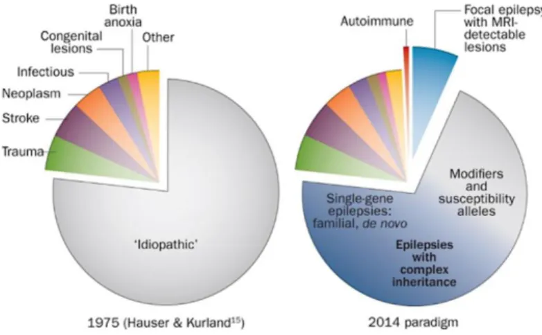
The etiology of epilepsy is emphasized in the new classification system; in particular, six etiological categories (structural, genetic, infectious, metabolic, immune, unknown) have been defined, each reflecting underlying brain dysfunction (Shorvon et al., 2011).
About 1% of all people develop recurrent unprovoked seizures for no obvious reason and without any other neurological abnormalities. These are named ‘idiopathic epilepsies’, and they are assumed to be mainly genetic in origin (Steinlein, 2004).
Fig. 3 Advances in understanding the causes of epilepsy.
In particular, about 20–30% of epilepsy cases are due to acquired conditions such as stroke, tumor or head injury, but the remaining 70–80% of cases are believed to be due to one or more genetic factors (Myers and Mefford, 2015).
Genetic etiologies are determined if there is a genetic mutation in one or more genes known to be associated or responsible for a clinical condition in which the epilepsy is the main symptom of the disorder. Although some epilepsies are inherited, many occur secondary to a de novo mutation in the affected individual. In some cases, the genetic mutation is not identified, but the clinical presentation, EEG findings, and family history suggest a genetic etiology (Pack, 2019).
11
1.4 Genetic basis of epilepsy
A genetic basis for epilepsy has been hypothesized for decades, but the first evidence of a genetic component emerged from epidemiological studies that reported an increased risk of epilepsy in relatives of affected individuals. Studies of twins showed that monozygotic twins have a higher concordance rate for both genetic generalized epilepsy (GGE) and focal epilepsy than dizygotic twins, supporting the hypothesis that epilepsy has a genetic basis (Myers and Mefford, 2015). In particular, twin studies suggested that the heritability of epilepsy is ~25%-70%. While genetic linkage analyses have identified several susceptibility loci for epilepsy, recent advances in genomic technology have made it feasible to identify single nucleotide variants, and copy number variants (CNVs) associated with epilepsies (Chen et al., 2017).
To date, extensive research has identified different genetic component of epilepsy including genetic aberrations now known to cause or contribute to the condition. In particular, ring chromosome 20 syndrome is a rare but well-known cause of epilepsy. Atkins et al. (1972) first described this condition in a 7 year old boy with behavioral issues, mental retardation and grand mal seizures. Other examples of chromosomal aberrations associated with epilepsy include Klinefelter Syndrome (47,XXY) (Elia et al., 1995; Tatum et al., 1998) and Pallister-Killian Syndrome (OMIM #601803) (12p tetrasomy) (Pallister et al., 1977; Peltomaki et al., 1987).
Copy number variations (CNVs) have also been associated with epilepsy and other neurological disorders (Mullen et al., 2013; Mefford, 2015; Borlot et al., 2017). Briefly, CNVs are classified as deletions or duplications of DNA, larger than 1 kb in size, which can be recognized as either a normal variation of the genome or to be pathogenic based on the location and number of genes encompassed by the rearrangement (Mefford, 2014). Examples of CNVs associated with epilepsy include deletions at Xp22.31, 1q21.1, 15q11.2, 15q13.3, and 16p13.11 chromosome regions respectively as well as duplications involving the 1p36.33 and 22q11.2 chromosome regions which have been all previously identified as risk factors for genetic generalized epilepsy (Mefford, 2014; Addis et al., 2016).
Other factors, such as uniparental disomy (UPD) or genetic imprinting, have also been associated with epilepsy as reported, for example, for the Angelman syndrome (OMIM #105830) involving the chromosome region 15q11q13 (Lalande et al., 1999; Valente et al., 2005).
In addition, epigenetic factors, including DNA methylation (Kobow and Blumcke, 2012; Wang et al., 2016), histone modification, transcriptional regulation (Hwang et al., 2013; Jagirdar et al., 2015),
12 and microRNAs involvement (Henshall, 2014; Raoof et al., 2017), have been implicated in epilepsy. They are able to regulate the neuro-inflammatory responses, neuronal cell growth and other relevant cellular process (Roopra et al., 2012; Boison, 2016; Kobow and Blumcke, 2017).
Mutations of mitochondrial DNA (mtDNA) have also been identified in patients affected by epilepsy and/or other neurological diseases (Wallace et al., 1994). The first example was described by Shoffner et al., in 1990, when they reported a mutation in tRNALys as cause of myoclonic epilepsy with ragged red fibers (MERFF; OMIM #545000). Subsequently, Tatuch et al. (1992) reported a mutation in ATPase6 that cause Leigh syndrome (OMIM #256000) if present in a high percentage of cells. Mosaic mutations in well-known epilepsy genes, such as SCN1A and SLC6A1, have also been identified to cause the epilepsy phenotype (Shi et al., 2012; Halvorsen et al., 2016). In a recent study by Stosser and collaborators (2017), they found a 3.5% overall frequency of mosaicism in 893 affected patients across 9 different nuclear genes (CDKL5, GABRA1, GABRG2,
GRIN2B, KCNQ2, MECP2, PCDH19, SCN1A, and SCN2A). Mosaicism is thought to be an
underreported cause of genetic disorders, due to detection challenges, although there are numerous studies aimed at improving this using Next Generation Sequencing (NGS) technology (Stosser et al., 2017). Furthermore, mosaicism is not limited to point mutations regarding mtDNA or nuclear DNA, but it can be also observed at chromosomal level (i.e. aneuploidies and/or CNVs) (Gajecka, 2016).
To date, approximately 977 genes have been associated with epilepsy grouped into the following categories: i) 84 genes causing epilepsy as a core symptom; ii) 73 neurological genes associated with brain gross development and epilepsy; iii) 536 epilepsy-associated genes where epilepsy is a symptom of another neurological disorder; iv) 284 potential-epilepsy genes (Wang et al., 2017). Some of these genes are associated, not only with epilepsy as the only symptom, but also with different phenotypes including well-characterized syndromes with variable clinical manifestations.
13
1.4.1 Genetics of Epilepsy Syndromes
Although the definition of epilepsy suggests that it is a single disorder, it is more accurate to describe epilepsy as a group of disorders clinically and etiologically heterogeneous.
A crucial issue underpinning gene discovery in epilepsy is that each gene shows phenotypic pleiotropy, and that each epilepsy syndrome shows genetic heterogeneity. Phenotypic heterogeneity or pleiotropy, in which mutations in a single gene cause different phenotypes, is increasingly recognised in epilepsy and across many neurological disorders. Many factors contribute to phenotypic heterogeneity, including the following: type of mutations during development, gene expression, epigenetic factors and modifier genes (McTague et al., 2016). Channelopaties are such an example. SCN1A mutations are associated with Dravet syndrome (OMIM #607208) but also with milder phenotypes such as genetic epilepsy with febrile seizures plus (GEFS+; OMIM 604233, 604403, 609800, 611277, 612279, 613060, 613828 613863, 616172, 618482). KCNQ3 mutations are associated with benign neonatal seizure while KCNQ2 can be associated with early-onset epileptic encephalopathy or benign neonatal seizure, highlighting phenotypic variations resulting from mutations in the same gene (El Achkar et al., 2015).
Fig.4 Clinical heterogeneity of epilepsy genes.
According to the ILAE, epilepsy syndromes are classified based on the electroclinical features and age of epilepsy onset. It is therefore clinically relevant to understand the diverse genetic
14 mechanisms of each of these syndromes as they might shed a light on diagnosis, treatment and prognosis.
I) Early-onset epileptic encephalopathies (EOEEs)
Epileptic encephalopathies are disorders caused by recurrent clinical seizures or prominent interictal epileptiform discharges and usually seen during the early infantile period. They are associated with impaired cognitive, sensory, and motor development. The most common epileptic encephalopathies are Ohtahara syndrome (OS), early myoclonic encephalopathy, epilepsy of infancy with migrating focal seizures (EIMFS), West syndrome, and Dravet syndrome (Mastrangelo et al., 2012). Based on current literature, single gene variants explain at least 20-30% of epileptic encephalopathies (Olson et al., 2017).
Ohtahara syndrome is a rare form of epilepsy, characterized by intractable seizures within the first few weeks to months of neonatal period. It is usually associated with poor developmental outcome. Infants may develop acute generalized or lateralized tonic spasms that can occur either singly or in clusters and are independent of the sleep cycle. The seizure frequency may be very high, ranging from 10 to 300 spasms in 10 to 20 clusters per day. To date, various genes, which have essential roles in lower brain’s neuronal and interneuronal functions, have been reported to be associated with Ohtahara syndrome. For instance, syntaxin binding protein 1 (STXBP1) regulates synaptic vesicle release; aristaless-related homeobox (ARX) acts as a regulator of proliferation and differentiation of neuronal progenitors; solute carrier family 25 member 22 (SLC25A22) encodes a mitochondrial glutamate transporter; and potassium voltage-gated channel, KQT-like subfamily, member 2 (KCNQ2) plays a key role in a cell’s ability to generate and transmit electrical signals. (Beal et al., 2012).
Dravet syndrome is another genetic form of early-onset epileptic encephalopathies. It affects between 1 of 20 000 and 1 of 40000 live births. A positive family history is usually present in 25% to 71% of patients, while mutations in SCN1A are identified in 70-80% of cases. To date, more than 500 mutations of SCN1A have been associated with Dravet syndrome. However, other genes have been found as cause Dravet or Dravet-like clinical syndrome, including PCDH19, SCN1B, STXBP1,
GABRA1, CHD2, HCN1 and GABRG2 (Chopra and Isom, 2014).
EIMFS is an even more heterogeneous syndrome. While KCNT1 mutations account for about one third of reported cases of EIMFS, the other cases are caused by mutations in a number of other genes (SCN1A, SCN8A, SLC25A22, SCN2A) (Barcia et al. 2012; Ohba et al., 2014).
15 Infantile spasms are another form of epileptic disorder that occur during the first year of life. Genetic analysis of children with unexplained infantile spasms have demonstrated mutations in diverse genes including ARX, CDKL5, ALG13 as well as de novo mutations in autosomal genes, including PDZ, MAGI2, STXB1, SCN1A, SCN2A, GABRB3 and DMN1 (Mastrangelo et al., 2012).
16 II) Benign familial neonatal/infantile seizures
Benign familial neonatal (BFNC), neonatal infantile, and infantile seizures (BFIS) are genetically distinct syndromes, although it has been proposed patients to be grouped into a single group broad referred to as benign familial infantile epilepsy (BFIE). Benign familial neonatal seizures are characterized by convulsions occurring shortly after birth and continuing only in the first month of life (Pandolfo, 2011). Genetic causes include channelopathy causing mutations (e.g., KCNQ2, KCNQ3, and SCN2A), as well as PRRT2 present in the 16p11.2 chromosome region which is also associated with choreoatheosis syndrome (M neret et al., 2013).
III) Progressive myoclonus epilepsies (PMEs)
The progressive myoclonus epilepsies (PMEs) comprise a group of rare and heterogeneous disorders defined by the combination of myoclonus, epileptic seizures, and progressive neurologic deterioration. The gene defects for the most common forms of PME (Unverricht– Lundborg disease, Lafora disease, several forms of neuronal ceroid lipofuscinoses, myoclonus epilepsy with ragged-red fibers (MERRF), and type 1 and 2 sialidoses) have been identified (Kälviäinen, 2015). The most common subtype, Unverricht-Lundborg disease (ULD), can be caused by mutations in CSTB, SCARB2, PRICKLE1, and GOSR2 (Lalioti et al., 1997; Corbett et al., 2011). While, Lafora disease can be caused by EPM2A or EPM2B (Ferlazzo et al., 2014). Neuronal ceroid lipofuscinosis (NCL) can be caused by numerous mutations: CLN1 in classic infantile-onset form, CLN2, CLN5, CLN6, CLN7, and CLN8 in late infantile-onset forms, CLN3 in classic juvenile-onset form, CLN4 and CLN6 in adult-onset forms, and CLN10 in congenital NCL. A recently identified PME mutation in KCNC1, encoding a subunit of a voltage gated potassium channel, was found in 11 individuals with phenotype resembling classic ULD (Muona et al., 2014).
IV) Genetic generalized epilepsies (GGE)
The genetic generalized epilepsies (GGE), characterized by generalized seizures that involve both sides of the brain, include juvenile myoclonic epilepsy (JME), childhood absence epilepsy (CAE), juvenile absent epilepsy (JAE) and epilepsy with generalized tonic-clonic seizure alone
17 (IGE-TCS) (Guerrini et al., 2019). The GGEs tend to start in childhood or adolescence and are usually associated with normal development and intellect.
Fig.6 List of genes associated to GGE
An important clinical example is mutations in SLC2A1 gene that cause glucose transporter 1 deficiency, and can present early-onset absence epilepsy or other generalized epilepsies such as typical childhood absence or juvenile myoclonic epilepsy. The treatment of choice is ketogenic diet. Copy number variations (CNV) have been frequently found in this type of epilepsies. These include 1q21.1, 15q11.2, 15q13.3, 15q11-q13, 16p11.2, 16p13.11, and 1q21.1 (Mefford et al., 2010; Helbig I et al. 2009). Other identified genes are likely susceptibility genes for generalized epilepsies and include CACNA1H, CACNB4, and CLCN2, in addition to CACNA1A, which is associated with both focal and generalized seizures (Lu JJ et al., 2005; Chioza et al., 2001).
V) Genetic epilepsy with febrile seizures plus (GEFS+)
Genetic epilepsy with febrile seizures plus (GEFS+) is a familial epilepsy syndrome in which affected individuals within a family show a variety of epilepsy phenotypes, including varying from simple febrile seizures and febrile seizures plus to severe epileptic encephalopathy. The GEFS+ is associated with mutations mainly in SCN1A, SCN1B, GABRG2 and GABA. Other genes have been implicated in GEFS+, including STX1B, SCN9A, GABRD and FGF13 (Bonanni et al., 2004).
18 VI) Focal epilepsy
Fig.7 List of Familial Focal Epilespy Syndrome.
The first epilepsy gene, with autosomal dominant inheritance, identified in a SHE family has been CHRNA4. Subsequently, two other genes were implicated in SHE, CHRNB2 and
CHRNA2. In addition, mutations in CRH, which encodes corticotropin-releasing hormone, have
been implicated in families with autosomal dominant SHE. The underlying molecular mechanism in these cases is unclear, although corticotropin-releasing hormone is known to have a potentially pro-convulsive properties. The sodium-gated potassium channel, KCNT1, has also been associated with severe familial autosomal dominant SHE, as well as sporadic cases with de novo mutations. (McTague et al., 2018).
To date, the use of next generation sequencing technologies in research and diagnostic laboratories has given rise to the rapid identification of genes associated with epilepsy syndromes.
Focal seizures originate in one hemisphere of the brain. Examples of focal epilepsy syndromes are autosomal dominant sleep-related hypermotor Epilepsy (ADSHE), temporal lobe epilepsy (TLE), and autosomal dominant epilepsy with auditory features. (Myers and Mefford, 2015). ADSHE (previously known as ‚autosomal dominant nocturnal frontal lobe epilepsy‛) is characterized by seizures beginning in the first 2 decades of life. A severe form of ADSHE includes drug-resistant, and intellectual disability and displays autosomal dominant inheritance, with a penetrance of ∼70% (Perucca, 2018).
19
2. NEXT GENERATION SEQUENCING TECHNOLOGY
Next generation sequencing (NGS), massively parallel or deep sequencing are related terms that describe a DNA sequencing technology which revolutionised genomic research. NGS has the potential to find causal mutations, including de novo, new and familial mutations associated with both epilepsy and epilepsy-related phenotypes, providing high performance in very few times (Dunn et al., 2018).
Conventional DNA sequencing developed by Sanger in 1977 (Sanger et al., 1977), led to many genetic discoveries and has been widely used for over 30 years in research and diagnostic laboratories. Although considered a major technological breakthrough, and still finding utility today for variant verification, the technique has limitations, in particular when examining large regions of the genome. More recently, NGS technology has begun to replace Sanger sequencing due its ability to quickly sequence large numbers of genes, the whole exome (protein-coding regions) or entire genome at once.
The applications of NGS include targeted gene panels, whole exome sequencing (WES) and whole genome sequencing (WGS). Custom gene panel testing allows to screen multiple clinically relevant genes and for more flexibility in phenotype–genotype correlations than required when testing individual genes (Poduri et al., 2014). WES focuses on the protein coding regions in the genome, comprising approximately 1–2% of the genome, attributable to ~85% of disease related mutations. In contrast, WGS provides information on the entire genome (both coding and non-coding regions), providing additional information on mutations in regulatory regions. The ~99% of the genome contains untranslated regions which may have a regulatory role (e.g., non-coding RNAs or transcription binding sites) along with potential protein coding sites yet to be annotated as genes. The impact of variants found in non-coding regions are not currently well understood, however it is feasible that a single or a combination of variants could have a significant impact on the pathology conditions such as epilepsy. This is most evident for non-coding variants that may influence expression levels or mRNA splicing, affecting protein abundance or isoforms (Dunn et al., 2018).
20 i. Targeted resequencing: a set number of genes at a higher sequencing depth and lower cost when compared to whole exome and whole genome sequencing, however the number and specificity of genes included in the panel may influence the success of diagnosis.
ii. Whole exome sequencing is associated with a high sequencing depth of the protein coding regions at a lower cost compared to whole genome sequencing.
iii. Whole genome sequencing can theoretically provide the coverage of the full genome, compared to whole exome sequencing and gene panels, but it shows a lower sequencing depth and a higher cost per sample.
One key analytical difference between the three NGS methods is the number of variants identified. Approximately 3–4 million variants per individual are commonly identified through WGS and approximately 30,000–40,000 variants that differ to the reference genome per person are obtained by WES. Although the increased content generated from WGS allows for a better chance at finding pathogenic variants, it also increases the number of incidental findings (Dunn et al., 2018).
21
2.1 Workflow of next generation sequencing
The NGS workflow consists of multiple steps including: library preparation, sequencing and bioinformatics analysis.
Library preparation
In all NGS approaches, DNA is fragmented prior to sequencing. This is performed in several ways and is dependent upon the specific kit used for library preparation and sequencing platform. DNA can be sheared using high frequency soundwaves (sonication), via enzymatic digestion or transposase. The key differences between the different assays is summarized in figure 8 which includes the current commonly utilized fragmentation and hybridisation techniques.
Fig.8 Common assays for library preparation.
An important step of the library preparation is amplification. Amplification is needed so that the ensuing sequencing reactions produce sufficient signal for detection by the instruments optical system.
22
Sequencing
NGS technologies can perform massively parallel sequencing of millions of DNA strands. This phenomenal sequencing capability of NGS, have made this method important both for research and clinical diagnostic applications (Ballester et al., 2016). The characteristics of the DNA sequencing reaction are different for each platform, emphasizing the range of innovation in chemistry, molecular biology and engineering required to produce sequence (Mardis, 2014). In particular, the NGS sequencing technologies are based on two different sequencing systems: optical imaging and non-optical imaging. The optical imaging-based NGS technologies use sensitive optical imaging to detect and identify the nucleotides being incorporated during the sequencing. This is the most common NGS technology used by commercially available platforms. In figure 9 are summarized the principal sequencing platforms.
Fig.9 Overview of major next-generation sequencing platforms.
Bioinformatics analysis
The detection of Single Nucleotide Variants (SNVs) and small insertions and deletions (indels) from raw NGS data consists of the following major steps:
- Sequence generation: Sequence generation (signal processing and base calling) is the process that converts sensor (optical and non-optical) data from the sequencing platform and identifies the sequence of nucleotides for each of the short fragments of DNA in the sample prepared for analysis. For each nucleotide sequenced in these short fragments (ie, raw reads), a corresponding Phred-like quality score is assigned, which is sequencing
23 platform specific. The read sequences along with the Phred-like quality scores are stored in a FASTQ file, which is a de facto standard for representing biological sequence information. After read quality control, alignment of the sequence reads to the human reference genome is performed.
- Sequence alignment is the process of alignment of each short DNA sequence (each typically <250 bp) with a reference genome (eg, the human reference genome used in clinical laboratories). This computationally process assigns a Phred-scale mapping quality score to each of the short sequence reads, determining the quality of the alignment process. This step also can be used to calculate the proportion of mapped reads and depth (coverage) of sequencing for one or more regions of interest. The sequence alignment data are usually stored in a de facto standard binary alignment map (BAM, binary alignment map) file format, which is a binary version of the sequence alignment/map format. Sequence alignments (BAM file formats) usually undergo quality control and alignment recalibration steps.
- Variant calling: is the process of accurately identifying the differences or variations between the sample and the reference genome sequence. Variant calling is based on numerous algorithmic strategies in order to create a list of sequence variants such as single nucleotide variants (SNVs), small insertions and deletions (indels), copy number alterations, and large structural alterations (insertions, inversions, and translocations). Sequence variants identified are written onto standard format files called VCF (Variant Call Format). The accuracy of variant calling is highly dependent on the quality of called bases and aligned reads. Therefore, variant calling processing, such as local realignment around expected indels and base quality score recalibration, is routinely used to ensure accurate and efficient variant calling. Typically 15,000 to 20,000 variants are discovered per exome, by contrast, about 3 million human SNV per genome are discovered using whole-genome sequencing (Ng et al., 2009; Koboldt et al., 2010).
- Variant Filtering: variant filtering is the process by which variants representing false-positive artifacts of the NGS method are flagged or filtered from the original VCF file on the basis of several sequence alignment and variant calling associated metadata (eg,
24 mapping quality, base-calling quality, strand bias, and others). This is usually a post-variant calling step, although some post-variant callers incorporate this step as part of the variant calling process. This automated process may be used as a hard filter to allow annotation and review of only the assumed true variants.
- Variant Annotation: variant annotation performs queries against multiple sequence and variant databases in order to characterize each called variant such as variant location, predicted cDNA and amino acid sequence change (HGVS nomenclature), minor allele frequencies in human populations, and prevalence in different variant databases (eg, Catalogue of Somatic Mutations in Cancer, The Cancer Genome Atlas, Single-Nucleotide Polymorphism (SNP) Database, and ClinVar). This information is used to further prioritize or filter variants for classification and interpretation.
- Variant Prioritization: variant prioritization is central to every Mendelian disease discovery and diagnosis effort. It is the process of determining which variants identified in the course of genetic testing are the most likely to damage gene function and underlie the disease phenotype (Karen Eilbeck et al., 2017). To perform an accurate and detailed clinical classification of each variants, the American College of Medical Geneticists guidelines (Richards et al., 2014) are follow. These recommendations suggest to assign one of the following six categories: 1) pathogenic: i.e. disease causing, sequence variation has previously been reported and is a recognized cause of the disorder; 2) Likely pathogenic: i.e. probably disease causing, sequence variation has not previously been reported and it is of a type expected to cause the disorder, usually in a known disease gene (for example, a nonsense mutation in a gene for which other mutations of this type, but at a different residue, have been reported); 3) Benign: sequence variation has previously been reported and is a recognized neutral variant; 4) Likely benign: sequence variation has not previously been reported and is probably not causative of disease; 5) Variant of unknown clinical significance (VUS): the variant is not described as pathogenetic in the literature or public database (such as ClinVar, dbSNP etc.) and/or involves a gene that could be functionally-related with the patient's clinical phenotype but animal models and/or functional validation studies have not yet been performed.
25
2.2 Next generation sequencing and epilepsy
The advent of next-generation (massively parallel) sequencing technology has revolutionized gene discovery in many disorders, including epilepsy. The most significant recent advance in understanding the genetics of epilepsy has come from exome sequencing in the EEs. The discovery in 2001 that de novo mutations in SCN1A gene cause Dravet syndrome set the stage for a better comprehension for this class of disorders (Claes et al., 2001). Targeted resequencing and the exome sequencing has proven to be an essential tool to confirm the importance of de novo mutations, facilitate rapid gene discovery and highlight the genetic heterogeneity of epilepsy.
In 2012, whole-genome sequencing in a family with a severely affected child revealed a de novo
SCN8A mutation in the proband (Veeramah et al., 2012). Subsequent studies have confirmed the
importance of this gene in the etiology of EE, with more than 25 cases reported. In a slightly larger study, using exome sequencing in 39 patients with fever-associated epilepsies similar to Dravet syndrome, Nava and colleagues identified two de novo mutations in HCN1. Subsequent sequencing of HCN1 in 157 affected individuals has identified causative mutations in this gene (Nava et al., 2014). In particular, HCN1 belongs to a family of hyperpolarization-activated, cyclic-nucleotide-gated channels that regulate neuronal excitability. Previous studies suggested that rare variants in
HCN1 play a significant role in the onset of GGE, while HCN2 and HCN4 variations can predispose
to the disease (Tang et al., 2008). Another study of 13 patients with Dravet syndrome revealed the importance of mutations in GABRA1 and STXBP1 (Carvill et al., 2014), two genes previously implicated in other EEs, in addition to the Dravet-related gene SCN1A. GABRA1 encodes the α1 subunit of the GABAA receptor, a multi subunit chloride channel that serves as the receptor for the GABA inhibitory neurotransmitter. STXBP1 encodes a syntaxin-binding protein that is critical for presynaptic vesicle docking and fusion. Another gene identified by exome sequencing as causative for epilepsy is KCNB1, a voltage-gated potassium channel, in which new pathogenic variants were detected in three affected families (Torkamani et al., 2014).
A growing class of genes implicated in epilepsy and related disorders are those that encode for proteins involved in chromatin remodeling and transcriptional regulation.
Through targeted sequencing of candidate genes, have been identified 5 of 500 patients with EE who had a de novo mutation in CHD2 (Carvill et al., 2013). To date, more than 20 patients with mutations in CHD2 have been identified, with the majority of mutations arising as de novo events (Thomas et al., 2015; Suls et al., 2013). In addition, a massive study on 580 patients with GGE
26 (photosensitive epilepsy or photoparoxysmal response as determined by EEG ) sequenced by TRS and WES showed that variants in CHD2 are a risk factor for photosensitivity in the GGEs, in particular disruptive variants (Galizia et al., 2015).
Another gene that causes a specific neural phenotype is the transcription factor myocyte enhancer factor 2C (MEF2C). Although MEF2C has important regulatory roles in other tissue types such as cardiac and skeletal muscle, it is a critical gene in neural progenitor cell differentiation and maturation, and the causative gene in 5q14 deletion syndrome. In fact, its haploinsufficiency can cause a range of features, including hyperkinesis, variable epilepsy, ID and autism, as well as atypical Rett syndrome (Zweier et al., 2012; Lambert et al., 2012).
The figure 10 show the timeline of gene discovery.
27
3. AIMS OF THE STUDY
Epilepsies have a highly heterogeneous background with a strong genetic contribution. The variety of unspecific and overlapping syndromic and non syndromic phenotypes often hampers a clear clinical diagnosis and prevents straight forward genetic testing. Knowing the genetic basis of a patient’s epilepsy can be valuable not only for diagnosis but also for guiding treatment and estimating recurrence risk.
The aim of my project is to use TRS on a clinically well-selected cohort of patients affected by epilepsy and neurodevelopmental disorders in order to:
i) identify novel variants in candidate genes and some currently known disease-causing genes useful to expand our knowledge about the molecular mechanisms of the disease; ii) characterize, both from molecular than from clinical point of view, novel syndromic and
non-syndromic forms of the disease useful for diagnosis purpose, counseling and
28
4. MATERIALS AND METHODS
4.1 Cohort of the study
A total of 49 (23 male and 26 female) Caucasian patients were recruited at the Medical Genetic Unit of IRCCS Casa Sollievo della Sofferenza (San Giovanni Rotondo, Foggia, Italy) for genetic study by targeted resequencing.
All participants had a clinical diagnosis of concise epilepsy phenotype, often with additional symptoms such as myoclonus or intellectual disability (with or without another medical condition).
The patients were selected according to the following criteria:
• Cases with similar clinical characteristics in at least one aspect between clinical, cognitive phenotype, behavioral, pregnancy type, major abnormalities.
• Negatives to genetic tests including karyotype and high resolution SNP-array (CytoScan HD and CytoSca XON arrays).
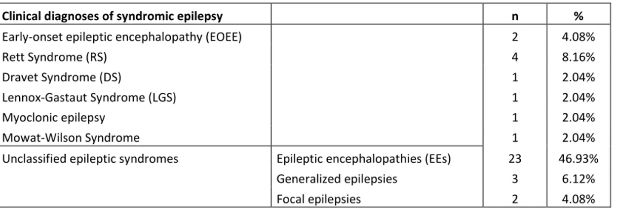
From a clinical point of view, the patients enrolled for the study were subdivided into two groups: - Patients affected by a syndromic form of epilepsy (table 1);
- Patients affected by a non-syndromic form of epilepsy (table 2).
Clinical diagnoses of syndromic epilepsy n %
Early-onset epileptic encephalopathy (EOEE) 2 4.08%
Rett Syndrome (RS) 4 8.16%
Dravet Syndrome (DS) 1 2.04%
Lennox-Gastaut Syndrome (LGS) 1 2.04%
Myoclonic epilepsy 1 2.04%
Mowat-Wilson Syndrome 1 2.04%
Unclassified epileptic syndromes Epileptic encephalopathies (EEs) 23 46.93%
Generalized epilepsies 3 6.12%
Focal epilepsies 2 4.08%
Tab. 1. n: number of patients; %: percentage of patients
Clinical diagnoses of non syndromic epilepsy n %
Epilepsy 11 22.44%
29 Clinical data was provided by the referring physician and included medical history (i.e. anamnesis, personal and family histories, physical and dysmorphological examination) and basic complementary tests, including electroencephalogram test, brain magnetic resonance imaging (MRI).
Written informed consent for genetic testing was obtained from all individuals enrolled for the study.
30
4.2 Targeted Resequencing (TRS)
4.2.1 Gene selection and panel design
A list of known and candidate genes associated with epilepsy was compiled on the basis of current literature (PubMed), clinical procedures suggested and adopted by the LICE (Lega Italiana Contro l’Epilessia) and in-house data. We selected 85 genes reported to cause different genetic forms of epilepsy and NDD. In particular we subdivided the genes into six categories: epilepsy genes (71 genes), neurodevelopment-associated epilepsy genes (13 genes), epileptic encephalopathy genes (16 genes) and (table 3).
Tab 3. Epilepsy genes: mutations in these genes cause pure or relatively pure epilepsies, or syndromes with epilepsy as
the core symptom; Neurodevelopment-associated epilepsy genes: mutations in these genes produce gross neurodevelopmental malformations and epilepsy, which may vary in severity; Epileptic encephalopathy: are severe brain disorders in which the epileptic electrical discharges may contribute to progressive psychomotor dysfunction.
From a functional point of view, the majority of selected genes encodes for proteins involved in membrane protein (i.e ion channel), enzyme, synaptic formation/remodeling/maintenance, neurotransmission (or DNA methylation/chromatin remodeling). Table 4 summarized the selected genes to be included in the gene panel as well as their function, associated disorders, chromosomal position and sequencing details.
Phenotype Genes
Epilepsy genes (71 genes)
ADSL ALDH7A1 ARHGEF9 ATP1A2 ATP6AP2 CACNA1H CACNB4 CDKL5 CHD2 CHRNA2 CHRNA4 CHRNA7 CHRNB2 CLN3 CLN5 CLN6 CLN8 CPA6 CSTB DNAJC5 EFHC1 EPM2A FOLR1 FOXG1 GAMT GATM GABRA1 GABRD GABRG2 GOSR2 GRIN2A GRIN2B KCNJ10 KCNMA1 KCNQ2 KCNQ3 KCTD7 LGI1 LIAS MAGI2 MBD5 MECP2 MEF2C MFSD8 NRXN1 NHLRC1 PCDH19 PLCB1 PNPO PRICKLE1 PRRT2 PRICKLE2 POLG PPT1 SCARB2 SCN1A SCN1B SCN2A SCN8A SCN9A SLC25A22 SLC2A1 SPTAN1 ST3GAL3 STXBP1 SLC9A6 TCF4 TPP1 TBC1D24 UBE3A ZEB2
Neurodevelopment-associated epilepsy genes (13 genes)
ARX CNTN4 CNTNAP2 CRBN IER3IP1 KANSL1 PNKP SRPX2 SYN1 TMLHE TSC1 TSC2 TSEN2
Epileptic encephalopathy genes (16 genes) ARX CDKL5 CHD2 GABRA1 GRIN2B KCNQ2 PCDH19 PLCB1 SCN1B SCN2A
31
Gene Inheritance Description Cellular Function Phenotype Chr. Location Coverage (%) Regions
ADSL AR adenylosuccinate lyase Enzyme adenylosuccinase lyase deficiency (ADSLD) 22q13.1 100.00% 15
ALDH7A1 AR aldehyde dehydrogenase 7 family, member A1 Enzyme Pyridoxine-dependent epilepsy (EPD) 5q23.2 100.00% 19
ARHGEF9 XLR RHO guanine nucleotide exchange factor 9 Adaptor protein Early infantile epileptic encephalopathy (EIEE) Xq11.1 100.00% 12
ARX XLR aristaless related homeobox Nucleic acid binding Early infantile epileptic encephalopathy (EIEE) Xq21.3 100.00% 5
ATP1A2 AD ATPase Na+/K+ transporting subunit alpha 2 Membrane protein Enzymatic deficiency 1q23.2 98.81% 24
ATP6AP2 XLR ATPase H+ transporting accessory protein 2 Membrane protein Seizures, generalized tonic-clonic Xp11.4 99.19% 9
CACNA1H UN calcium channel, voltage-dependent, T type, alpha-1H subunit Membrane protein Childhood absence epilepsy (CAE) 16p13.3 99.91% 34
CACNB4 AD calcium channel, voltage-dependent, beta-4 subunit Membrane protein Juvenile myoclonic epilepsy (JME)/Epilepsy, idiopathic generalized 2q23.3 99.62% 18
CDKL5 XLD cyclin-dependent kinase-like 5 Enzyme Early infantile epileptic encephalopathy (EIEE) Xp22.13 100.00% 20
CHD2 AD chromodomain helicase DNA-binding protein 2 Nucleic acid binding Childhood-onset epileptic encephalopathy (COEE) 15q26.1 99.83% 39
CHRNA2 AD cholinergic receptor,neuronal nicotinic, alpha polypeptide 2 Neuronal receptors Nocturnal frontal lobe epilepsy (NFLE) 8p21.2 100.00% 6
CHRNA4 AD cholinergic receptor, neuronal nicotinic, alpha polypeptide 4 Acetylcholine receptor Nocturnal frontal lobe epilepsy (NFLE) 20q13.33 99.32% 8
CHRNA7 AR cholinergic receptor nicotinic alpha 7 subunit Acetylcholine receptor Epilepsy, juvenile myoclonic/Epilepsy, idiopathic generalized 15q13.3 89.53% 15
CHRNB2 UN cholinergic receptor, neuronal nicotinic, beta polypeptide 2 cholinergic receptor, neuronal nicotinic, beta polypeptide 2Nocturnal frontal lobe epilepsy (NFLE) 1q21.3 100.00% 6
CLN3 AR CLN3 lysosomal/endosomal transmembrane protein lysosomal function Ceroid lipofuscinosis, neuronal 16p12.1 100.00% 16
CLN5 AR CLN5 intracellular trafficking protein lysosomal function Ceroid lipofuscinosis, neuronal 13q22.3 100.00% 4
CLN6 AR CLN6 transmembrane ER protein lysosomal function Ceroid lipofuscinosis, neuronal 15q23 100.00% 8
CLN8 AR CLN8 transmembrane ER and ERGIC protein recycle between the ER and ER-Golgi Ceroid lipofuscinosis, neuronal 8p23.3 100.00% 2
CNTN4 AD contactin 4 cell adhesion molecule 3p- syndrome/Seizures (rare) 3p26.3-p26.2 100.00% 24
CNTNAP2 AR contactin associated protein like 2 Cell adhesion molecule Cortical dysplasia-focal epilepsy syndrome 7q35-q36 99.83% 25
CPA6 AR, AD carboxypeptidase A6 Enzyme Familial febrile seizures (FFS)/Familial temporal lobe epilepsy (FTLE) 8q13.2 100.00% 11
CRBN AR cereblon Cytoplasm localized with a calcium channel membrane proteinNonsyndromic cognitive disability 3p26.2 100.00% 11
CSTB AR cystatin B Enzyme modulator Progressive myoclonic epilepsy (PME) 21q22.3 100.00% 3
CTSD AR cathepsin D Enzyme Ceroid lipofuscinosis, neuronal 11p15.5 100.00% 9
DNAJC5 AD DnaJ heat shock protein family (Hsp40) member C5 membrane trafficking and protein folding Ceroid lipofuscinosis, neuronal 20q13.33 99.88% 5
EFHC1 AD EF-hand domain (C-terminal)-containing protein 1 Signal transduction/molecule Juvenile absence epilepsy (JAE) 6p12.2 99.40% 13
EPM2A AR EPM2A gene, encodes laforin Enzyme Progressive myoclonic epilepsy (PME) 6q24.3 100.00% 10
FOLR1 AR folate receptor alpha Receptor protein Neurodegeneration due to cerebral folate transport deficiency 11q13.4 100.00% 4
FOXG1 AD forkhead box G1 Repressor protein Rett syndrome 14q12 99.80% 1
GABRA1 AD gamma-aminobutyric acid receptor, alpha-1 GABA-A receptor Early infantile epileptic encephalopathy (EIEE)/Childhood absence epilepsy (CAE)/Juvenile myoclonic epilepsy (JME) 5q34 100.00% 10
GABRD AD gamma-aminobutyric acid receptor, delta GABA-A receptor Generalized epilepsy with febrile seizures plus (GEFS + ) 1p36.33 100.00% 9
GABRG2 AD gamma-aminobutyric acid receptor, gamma-2 GABA-A receptor Familial febrile seizures (FFS)/Generalized epilepsy with febrile seizures plus (GEFS + )/Childhood absence epilepsy (CAE) 5q34 98.09% 12
GAMT AR guanidinoacetate N-methyltransferase Enzyme Cerebral creatine deficiency syndrome 19p13.3 100.00% 6
GATM AR glycine amidinotransferase Enzyme Cerebral creatine deficiency syndrome 15q21.1 100.00% 9
GOSR2 AR golgi snap receptor complex member 2 Membrane trafficking Progressive myoclonic epilepsy (PME) 17q21.32 100.00% 9
GRIN2A AD glutamate receptor, ionotropic, N-methyl-D-aspartate, subunit 2A NMDA receptor Focal epilepsy, with speech disorder and with or without mental retardation 16p13.2 99.04% 12
GRIN2B AD glutamate receptor, ionotropic, N-methyl-D-aspartate, subunit 2B NMDA receptor Early infantile epileptic encephalopathy (EIEE) 15p13.1 99.86% 12
IER3IP1 AR immediate early response 3 interacting protein 1 Cell differentiation and apoptosis Microcephaly, epilepsy, and diabetes syndrome 18q21.1 100.00% 3
KANSL1 AD KAT8 regulatory NSL complex subunit 1 Nuclear protein Koolen-De Vries syndrome 17q21.31 100.00% 14
KCNJ10 AR potassium inwardly rectifying channel subfamily J member 10 Potassium channel SESAME syndrome 1q23.2 100.00% 1
KCNMA1 AR, AD potassium channel, calcium-activated, large conductance, subfamily M, alpha member 1Potassium channel Generalized epilepsy and paroxysmal dyskinesia (GEPD) 10q22.3 100.00% 41
KCNQ2 AD potassium channel, voltage-gated, KQT-like subfamily, member 2 Potassium channel Benign familial neonatal seizures (BFNS)/Early infantile epileptic encephalopathy (EIEE) 2oq13.33 100.00% 19
KCNQ3 AD potassium channel, voltage-gated, KQT-like subfamily, member 3 Potassium channel Benign familial neonatal seizures (BFNS) 8q24.22 99.78% 18
KCTD7 AR potassium channel tetramerization domain-containing protein 7 Potassium channel Epilepsy, progressive myoclonic, with or without intracellular inclusions 7q11.21 98.91% 16
LGI1 AD leucine-rich gene, glioma-inactivated,1 Neuronal growth regulation and cell survival Familial temporal lobe epilepsy (FTLE) 10q23.33 100.00% 8
LIAS AR lipoic acid synthetase Enzyme Hyperglycinemia, lactic acidosis, and seizures 4p14 99.41% 11
MAGI2 AR membrane associated guanylate kinase, WW and PDZ domain containing 2 Enzyme Nephrotic syndrome 7q21.11 98.57% 24
MBD5 AD methyl-CpG binding domain protein 5 Enzyme Kleefstra syndrome 2q23.1 99.60% 10
MECP2 XLR methyl-CpG binding protein 2 Enzyme Encephalopathy, neonatal severe Xq28 95.76% 5
MEF2C AD myocyte enhancer factor 2C Transcription factors Mental retardation, stereotypic movements, epilepsy, and/or cerebral malformations 5q14.3 100.00% 11
MFSD8 AR major facilitator superfamily domain containing 8 Membrane protein Neuronal ceroid lipofuscinosis 4q28.2 100.00% 12
NHLRC1 AR NHL repeat-containing 1 gene Enzyme Progressive myoclonic epilepsy (PME) 6p22.3 100.00% 1
NRXN1 AR neurexin 1 Membrane protein Pitt-Hopkins-like syndrome 2p16.3 97.44% 38
PCDH19 XL protocadherin 19 Cell adhesion molecule Early infantile epileptic encephalopathy (EIEE) Xq22.1 100.00% 6
PLCB1 AR phospholipase C, beta-1 Enzyme Early infantile epileptic encephalopathy (EIEE) 20p12.3 99.90% 36
PNKP AR polynucleotide kinase 3'-phosphatase Enzyme Microcephaly, seizures, and developmental delay 19q13.33 100.00% 16
PNPO AR pyridoxamine 5-prime-phosphate oxidase Enzyme Pyridoxamine 5'-phosphate oxidase deficiency 17q21.32 100.00% 7
POLG AR DNA polymerase gamma, catalytic subunit Enzyme Mitochondrial disease 15q26.1 99.67% 22
32
Tab.4 Genes and characteristic included in the 85 NGS panel.
Gene Inheritance Description Cellular Function Phenotype Chr. Location Coverage (%) Regions
PRICKLE1 AR prickle planar cell polarity protein 1 nuclear receptor Progressive myoclonic epilepsy (PME) 12q12 100.00% 7 PRICKLE2 AR prickle planar cell polarity protein 2 Unclassified Progressive myoclonic epilepsy (PME) 3p14.1 100.00% 7 PRRT2 AD proline-rich transmembrane protein 2 transmembrane protein Benign familial infantile seizures (BFIS) 16p11.2 100.00% 2 SCARB2 AR scavenger receptor class B, member 2 Receptor Progressive myoclonic epilepsy (PME) 4q21.1 100.00% 13 SCN1A AD sodium channel, neuronal type I, alpha subunit Sodium channel Dravet syndrome (DS) 2q24.3 99.10% 26 SCN1B AR sodium channel, voltage-gated, type I, beta subunit Sodium channel Generalized epilepsy with febrile seizures plus (GEFS + ) 19q13.11 99.68% 5 SCN2A AD sodium channel, voltage-gated, type II, alpha subunit Sodium channel Benign familial infantile seizures (BFIS)/Early infantile epileptic encephalopathy (EIEE) 2q24.3 99.48% 27 SCN8A AD sodium channel, voltage-gated, type VIII, alpha subunit Sodium channel Benign familial infantile seizures (BFIS)/Early infantile epileptic encephalopathy (EIEE) 12q13.3 100.00% 28 SCN9A AD sodium channel, voltage-gated, type IX, alpha subunit Sodium channel Familial febrile seizures (FFS) 2q24.3 98.87% 27 SLC25A22 AR solute carrier family 25 (mitochondrial carrier, glutamate), member 22 Transporter Early infantile epileptic encephalopathy (EIEE) 11p15.5 100.00% 9 SLC2A1 AD solute carrier family 2 (facilitated glucose transporter), member 1 Transporter Idiopathic generalized epilepsy (IGE) 1p34.2 100.00% 11 SLC9A6 XLD solute carrier family 9 member A6 Membrane transport proteins Mental retardation Xq26.3 100.00% 16 SPTAN1 AD spectrin, alpha nonerythrocytic 1 Cytoskeletal protein Early infantile epileptic encephalopathy (EIEE) 9q34.11 99.71% 57 SRPX2 UN sushi repeat containing protein X-linked 2 Enzyme Rolandic epilepsy, speech dyspraxia, and mental retardation Xq22.1 100.00% 10 ST3GAL3 AR ST3 beta-galactoside alpha-2,3-sialyltransferase 3 Enzyme Early infantile epileptic encephalopathy (EIEE) 1q34.1 100.00% 13 STXBP1 AD syntaxin-binding protein 1 Membrane trafficking Early infantile epileptic encephalopathy (EIEE) 9q34.11 99.52% 20 SYN1 XLR, XLD synapsin I Membrane trafficking X-linked epilepsy with variable learning disabilities and behavior disorders Xp11.3-p11.2 99.67% 13 TBC1D24 AR Tre2-Bub2-Cdc16/TBC1 domain family, member 24 Enzyme modulator Familial infantile myoclonic epilepsy (FIME) 16p13.3 100.00% 7
TCF4 AD transcription factor 4 Transcription factor Pitt-Hopkins syndrome 18q21.2 99.80% 25
TMLHE XLR trimethyllysine hydroxylase, epsilon Enzyme Autism Xq28 99.50% 8
TPP1 AR tripeptidyl peptidase 1 Enzyme Neuronal ceroid lipofuscinosis 11p15.4 100.00% 13
TSC1 AD TSC complex subunit 1 Enzyme Tuberous sclerosis 9q34.13 100.00% 21
TSC2 AD TSC complex subunit 2 Enzyme Tuberous sclerosis 16p13.3 97.92% 42
TSEN2 AR tRNA splicing endonuclease subunit 2 Enzyme Pontocerebellar hypoplasia 3p25.2 99.56% 12
UBE3A AD ubiquitin protein ligase E3A Enzyme Angelman syndrome 15q11.2 99.88% 13
33 The specific targeted gene panel has been defined by using Agilent Sure Design tool (Agilent Technologies, CA, USA), the total amplicons obtained were 14.971, with a targeted region of 268,42 Kbp corresponding to the 85 selected genes protein-coding exons (CCDS) plus 25 bp of exons/introns boundaries. This design ensures an overall coverage greater than 98.5% of the targeted region.
4.2.2 Library preparation
Genomic DNA of each patients enrolled for the study was extracted from the peripheral blood leukocytes by using BioRobot EZ1 (Qiagen, Solna, Sweden).
The quality of DNA was tested on 1% electrophorese agarose gel, and the DNA concentration was measured using a Qubit DNA BR Assay Kit in a Qubit 2.0 Fluorometer (Life Technologies, San Diego, CA, USA).
A library of all coding exons and intron-exon boundaries was prepared using a HaloPlex target enrichment kit following the manufacturer's instructions (Agilent, Santa Clara, USA). The experimental workflow is represented in Figure 11.
34 Briefly, we fragmented the human genome (the samples were digested by 16 different restriction enzymes to create a library of gDNA restriction fragments) and enriched for the coding regions of genes by using complementary highly specific biotinylated probes. HaloPlex probes are designed to hybridize selectively to fragments originating from target regions of the genome and to direct circularization of the targeted DNA fragments.
Hybridized probes were captured with magnetic beads and target fragments were ligated to create circular DNA molecules. Subsequently, libraries were amplified by PCR, introducing unique index sequences that allow all pools to be sequenced together. Sequencing was performed using the NGS MiSeq Illumina sequencer (Illumina, Inc.). We selected a Q-score of 30, corresponding to a 1:1000 error rate, as an acceptance threshold value.
35
4.2.3 Sequencing
The libraries were pooled, and sequencing was performed on a Miseq sequencer (Illumina, San Diego, CA, USA) using Miseq Reagent Kit V3 300 cycles flow cell. For the Illumina platform, adapter modified library fragments are automatically dispensed onto a glass slide flow cell that displays oligonucleotides complementary to Illumina adapter sequences. Subsequently, a process called bridge amplification is used in order to generate clonal ‚clusters‛ of approximately 1000 identical molecules per cluster. Single-stranded, adapter-ligated fragments are bound to the surface of the flow cell exposed to reagents for polymerase-based extension. Priming occurs as the free/distal end of a ligated fragment ‚bridges‛ to a complementary oligo on the surface. Repeated denaturation and extension result in localized amplification of single molecules in ‚clusters‛. The Illumina sequencing platform is based on a sequencing-by-synthesis approach, in which all four nucleotides are added simultaneously to the flow cell channels, along with DNA polymerase, for incorporation into the oligo-primed cluster fragments. The nucleotides carry a base unique fluorescent label and the 3’-OH group is chemically blocked (Figure 12), so that each incorporation is a unique event.
Fig.12 3’-blocked reversible terminator.
Once the base is incorporated, a fluorescence signal is emitted and this event is captured by a fluorescence detector. Subsequently, the 3’ blocking group is chemically removed to prepare each strand for the next incorporation. The cycle is repeated, one base at a time, generating a series of images each representing a single base extension at a specific cluster.
36
37
4.2.4 Bioinformatic analysis and variants prioritization
The analysis of NGS sequencing data was performed according standard bioinformatics procedures. Raw individual sequence files (*.fastq) were checked for their quality through the FastQC tool (Andrews, 2010), in order to detect trends in base call quality scores according to read position, adapter content, base compositional bias, etc. Quality-checked reads were mapped against the hg19 version of the human reference genomic sequence through the BWA align (Li et al., 2009); the produced alignments (*.bam) were also processed through the GATK suite (McKenna et al., 2010). Refined *.bam alignments were used to calculate on-target and off-target sequencing coverage statistics (in particular, average target depth of coverage and proportion of target sites at 20X minimal coverage) by using the TEQC R Package (Hummel et al., 2011). Finally, variants were detected by means of the GATK v3.7 Haplotype Caller, and annotated by using ANNOVAR package respect to the hg19 RefSeq gene and transcript annotation (Wang et al., 2010). Functionally annotated variants were also checked for their presence in public variant collections, like dbSNP v151 and its ClinVar specialized database (Sherry et al., 2001), ExAC v0.3 (Exome Aggregation Consortium 2016) and gnomAD (Konrad et al., 2019). Furthermore, variants with a missense functional impact were also annotated with pathogenicity predictors, by querying the dbNSFP resource (Liu et al., 2011).
In order to prioritization variants the following pipeline was applied:
- Allele frequency. The use of a MAF threshold >1% allowed a removal of a large number of variants not connected with the phenotype.
- Predicted consequence. Predicted effect on protein function: this step is particularly critical for non-synonymous or missense variants in which the amino acid sequence is determined the functional consequences and of the biochemical conservation respect to the replaced amino acid. During this step we used SIFT (Kumar et al., 2009), PolyPhen2 (Adshubei et al., 2010), Mutation Taster (Schwarz JM et al., 2014) FATHMM (Rogers FM et al., 2018) and CADD (Rentzsch P et al., 2019). Splice-site variants were predicted by Human Splicing Finder (Desmet FO et al., 2009). - Inheritance. Variants were evaluated based on the family history of the disease and the inheritance of the gene (dominant/recessive/X-linked), including de novo mutations in dominant and X-linked genes and variants in recessive genes (homozygous and compound heterozygous). Inherited missense variants predicted to be benign were excluded.
38 In order to exclude the presence of mosaic variants, we focused our attention on the following values: Total Depht (filtered), Genotype Depht (filtered) and Ref/Alt (filtered reads). We used these parameters to evaluate the heterozygous or homozygous state for each patient.
For patients in which no variants of interest were found, the filtering criteria were relaxed in order to include variants with an MAF up to 0.05, distant from the canonical splicing site for around 8 bp, and potentially pathogenic synonymous variants based on the annotations predicted by the TraP (Gelfman S. et al., 2017) and absent in dbDSM database. (Database of Deleterious synonymous mutation).
All variants were visualized through tool the Integrated Genomics Viewer (Thorvaldsdottir et al., 2013) and confirmed through Sanger sequencing in order to detecting false positives.
39
5. RESULTS
In total, we recruited 49 patients with a phenotype of epilepsy and NDD. All samples have been analyzed through targeted resequencing of a gene panel containing 85 genes (10 X-linked and 75 autosomal genes, respectively) related to various form of epilepsy and NDD, and selected on the most updated literature survey.
All the sequencing run obtained more than 90% of targeted regions covered at least 100X and a minimum average coverage of 250x. For the first variants prioritization, we excluded intronic variants outside +/- 8 bp from the splice sites , as well as synonymous exonic variations outside the highly conserved acceptor and donor splice site. The candidates were filtered by frequency and submitted to an in silico prediction analysis of their functional effect at the amino acid level (i.e. protein folding, evolutionary conservation, loss of function or gain of function, etc.) and confirmed by Sanger sequencing.
This approach allowed us to identify variants in 25 of 49 patients (51%), including sequence alterations in frequently as well as in less commonly affected genes. In the remaining 24 affected individuals, the analysis revealed no significant variant at all.
In particular, we obtained a molecular diagnosis (pathogenic or likely pathogenic variants) in 10/49 probands (diagnostic yield 20.4%) while we identified variants of uncertain significance in 15/49 patients (30.6%). In details, causative variants (pathogenic or likely pathogenic) were related to 9 genes (ARX, GAMT, KCNQ2, MECP2, POLG, SCN1A, SPTAN1, STXBP1, TCF4) while the variants of uncertain significance were related to 19 genes (SCN9A, TSC1, SCN2A, SCN1A, GRIN2B,
GRIN2A, CLN3, CLN6, CACNA1H, CNTN4, LIAS, PRICKLE2, PNKP, SPTAN1, POLG, CACNB4, ATP1A2, CNTNAP2, KCNMA1).
In total, we found 31 rare exonic variants, 7 were de novo of which 6 mutations in autosomal genes (GAMT, KCNQ2, SCN1A, STXBP1, TCF4) and 1 mutation in X-linked gene (MECP2), 7 inherited variants and 17 with unknown inheritance since parents DNA was not available. Furthermore, of these 31 variants, 10 were new, i.e. not reported in literature or public database, and 21 were previously reported.
X-linked disorders were diagnosed in 2 patients (1 male and 1 female), including ARX, epileptic encephalopathy, early infantile (#308350) and MECP2, encephalopathy, neonatal severe (#300673), respectively.
40 With regard to the types of the variants, we identified: 26 missense variant, 4 frameshif, 1 nonsense.
Frameshift variants were more common in patients with mutations in autosomal dominant genes (such as TCF4, SPTAN1, KCNMA1).
Missense variants were found to be disease causing in both dominant and recessive disorders.
The 31 variants were related to 25 genes. The genes included ARX (n = 1), ATP1A2 (n = 1),
CACNA1H (n = 2), CACNB4 (n = 2), CLN3 (n = 1), CLN6 (n = 1), CNTN4 (n = 1), CNTNAP2 (n = 1), GAMT (n = 1), GRIN2A (n = 1), GRIN2B (n = 1), KCNMA1 (n = 1), KCNQ2 (n = 2), LIAS (n = 1), MECP2 (n = 1), POLG (n = 2), TSC1 (n = 1), PRICKLE2 (n = 1), PNKP (n = 1), STXBP1 (n = 2), SCN1A
(n = 2), SCN2A (n = 1), SCN9A (n = 1), SPTAN1 (n = 1), TCF4 (n = 1). These genes identified in our cohort were classified into the following ten categories related to the epilepsy-causing mechanism: ion channels (ATP1A2, CACNB4, CACNA1H, GRIN2A, GRIN2B, KCNQ2, KCNMA1, SCN1A,
SCN2A, SCN9A), enzyme (GAMT, MECP2, LIAS, PNKP, STXBP1, TSC1) , nucleic acid binding
(ARX), cell adhesion molecules (CNTNAP2, CNTN4),), transcription factor (TCF4), cytoskeletal protein (SPTAN1), membrane trafficking (STXBP1), lysosomal function (CLN3, CLN6), nuclear receptor (PRICKLE2) (Figure 14).
From a clinical point of view, we identified variants in 10/25 (40%) patients with a diagnosis of epileptic encephalopathy (4 pathogenic/likely pathogenic and 8 VUS), 7/25 (28%) patients with a diagnosis of syndromic epilepsy (epilepsy with other symptoms such as mental retardation) (2 pathogenic/likely pathogenic and 6 VUS), 1/25 (4%) with a diagnosis of Lennox-Gastaut syndrome (1 likely pathogenic), 1/25 (4%) with a diagnosis of GEFS/SMEI (1 pathogenic), 1/25 (4%) with a diagnosis of generalized epilepsy (4 VUS) and 5/25 (20%) with a diagnosis of epilepsy (2 pathogenic/likely pathogenic and 3 VUS).