BIOS – Research Doctorate School in BIOmolecular Sciences
Course in Biomaterial, XXIV cycle
Structure and Enzyme Kinetics Studies on Different
Forms of Cathepsin L: Implication on
Development of Anticancer Agents
CHIM/08
Candidate:
Claudio Ricci
Supervisor: Tutors:
Dr. Anna Maria Bianucci Dr. Ilaria Massarelli
Contents
The Idea Behind………. Pag. 1
INTRODUCTION
Chapter 1. Enzyme and Proteolysis………. Pag. 2 Chapter 2. Cysteine Cathepsin: Cathepsin L………..Pag. 19 Chapter 3. The Plant Chelidonius Majus……….Pag. 38
Aim of the Study………Pag 48
Chapter 4. Materials and Methods………Pag 49
RESULTS
Chapter 5. Computational Results………..Pag 68 Chapter 6 Enzyme Kinetics Studies on 29 kDa Human Liver Cathepsin L……….Pag 100
Conclusions……….Pag 113
Curriculum Vitae………..Pag. 115
Acknowledgements……….Pag. 116
Page 1
The Idea behind the Study
The idea behind the study described in this thesis arose from a bibliographic research concerning the biological behavior of several compounds present in different extracts of Chelidonium majus (C. majus), a herb widely diffused in several areas around the world. This herb is used, according to an unwritten knowledge, in the folk medicine of Northern Sardinia (Italy) both for healing severe burns and wounds, and for reducing scars.
The interest toward this herb was initially motivated by the observation that different properties of some of its extracts are apparently in contrast with each other. Indeed the ability of stimulating proliferation and migration of different cell types have been found as well as inhibitory properties. These latter properties are mostly evident in different cancer cell lines.
The bibliographic research afforded a great variety of data concerning a very wide spectrum of activities possessed by the extacts of C. majus and many of their single components. Such biological properties range from antimicrobial to immunostimulating as well as keratolytic and anticancer activity, other than stimulating activity of proliferation and/or migration of several cell types.
In particular our interest was kept by the description of a semisynthetic derivative (Ukrain) never approved by the FDA for which, nevertheless, anticancer and antimetastatic properties are reported by a quite high number of scientific publications. Ukrain is based on a particular C. maius extract added with a chemical reactant. The study described here started with the identification of one among the many biological targets potentially involved in the anticancer-antimetastatic activity of Ukrain.
Among the compounds contained in C. majus, a single-chain protein (chelidocistatin) is described to inhibit Catehpsin L, which has been recognized to be an interesting target for anticancer-antimetastatic agents. Moreover several alkaloids present in C. majus have been recognized to possess anticancer activity: among them Chelidonine is reported to interfere with the dynamic tubulin polymerization-depolymerization equilibrium and to produce cell cycle arrest in different cell lines.
After a first screening of the literature data, Cathespin L and tubulin appeared to be optimal targets for the design of new effective anticancer drugs and Cathepsin L was selected as the first target to be considered. It became the primary target to be addressed in the search of new anticancer drugs described in in this thesis.
Page 2
CHAPTER 1
ENZYME AND PROTEOLYSIS
This chapter briefly describes the proteolysis, the relevant types of protease and their regulation. The maintenance of a healthy organism largely relies upon controlled biosynthesis, maturation, function and terminal breakdown of proteins. Proteolytic enzymes contribute to these process by irreversibly cleaving peptide bonds. In particular the lysosomal proteins are important for these processes because of their high proteolytic activity.
Page 3
1.1 Basics on Enzyme Kinetics
Life depends on the existence of powerful and specific catalysts: the enzymes. Almost every biochemical reaction is catalyzed by an enzyme. Enzymes, like other proteins, have molecular weights ranging from about 12,000 to more than 1 million Da. Some enzymes do not require, other than the chemical functionalities of their amino acid residues, any additional chemical species for exerting their activity on the molecules to be acted upon. Others require an additional chemical component called cofactor — either one or more inorganic ions, such as Fe2+, Mg2+, Mn2+, or Zn2+, or a complex organic or metal-organic molecule called a coenzyme. Some enzymes require both a coenzyme and one or more metal ions for their activity. A coenzyme or metal ion that is very tightly or even covalently bound to the enzyme protein is called a prosthetic group. A complete, catalytically active enzyme together with its bound coenzyme and/or metal ions is called a holoenzyme. The protein moiety of such an enzymatic complex is called the apoenzyme or apoprotein. Many enzymes have been named by adding the suffix “-ase” to the name of their substrate or to a word or phrase describing their activity. Thus urease catalyzes hydrolysis of urea, and DNA polymerase catalyzes the polymerization of nucleotides to form DNA. Other enzymes were named based on the first discover of a broad function, before the specific reaction catalyzed was known.The enzymatic catalysis of reactions is essential to living systems. Under biologically relevant conditions, uncatalyzed reactions tend to be slow—most biological molecules are quite stable at the neutral-pH, mild-temperature, aqueous environment inside cells. Furthermore, many common reactions in biochemistry entail chemical events that are unfavorable or unlikely in the cellular environment, such as the transient formation of unstable charged intermediates or the collision of two or more molecules in the precise orientation required for reaction. Reactions required to digest food, send nerve signals, or contract a muscle simply do not occur at a useful rate without catalysis. An enzyme circumvents these problems by providing a specific environment within which a given reaction can occur more rapidly. The distinguishing feature of an enzyme-catalyzed reaction is that it takes place within the confines of a pocket on the enzyme called the active site (Figure 1). The molecule that is bound in the active site and is acted upon by the enzyme is called the substrate.
Page 4
A simple enzymatic reaction might be modeled by the following scheme:
E +S ES EP E+P
where E, S, and P represent the enzyme, the substrate, and the product respectively. ES and EP are transient complexes of the enzyme with the substrate and with the product. To understand catalysis, we must first recall the important distinction between reaction equilibria and reaction rates. The function of a catalyst is to increase the rate of a reaction. Catalysts do not affect reaction equilibria. Any reaction, such as “S to P”, can be described by a reaction coordinate diagram (Figure 2), a picture of the energy changes during the reaction. The modern notion of enzymatic catalysis, first proposed by Michael Polanyi (1921) and Haldane (1930), was subsequently enriched by Linus Pauling in 1946: in order to catalyze reactions, an enzyme must be complementary to the reactants transition state. This means that optimal interactions between substrate and enzyme occur only in the transition state. Figure 6–5cdemonstrates how such an enzyme can work. Many of these interactions involve parts of the stick that are distant from the point of breakage; thus interactions between the stickase and nonreacting parts of the stick provide some of the energy needed to catalyze stick breakage. This “energy payment” translates into a lower net activation energy and a faster reaction rate. The reaction catalysed by an enzyme uses exactly the same reactants and produces exactly the same products as the uncatalysed reaction. Like other catalysts, enzymes do not alter the position of equilibrium between substrates and products.[1] However, unlike uncatalysed chemical reactions, enzyme-catalysed reactions display saturation kinetics. For a given enzyme concentration and for relatively low substrate concentrations, the reaction rate increases linearly with substrate concentration; the enzyme molecules are largely free to catalyse the reaction, and increasing substrate concentration means an increasing rate at which the enzyme and substrate molecules encounter one another. However, at relatively high substrate concentrations, the reaction rate asymptotically approaches the theoretical maximum; the enzyme active sites are almost all occupied and the reaction rate is determined by the intrinsic turnover rate of the enzyme. The substrate concentration midway between these two limiting cases is denoted by KM. The two most important kinetic properties of an enzyme are how quickly the enzyme becomes saturated with a particular substrate, and the maximum rate it can achieve.
Page 5
Figure 2. An imaginary enzyme (stickase) designed to catalyze breakage of a metal stick. (a) Before the stick is broken, it must first be bent (the transition state). In both stickase examples, magnetic interactions take the place of weak bonding interactions between enzyme and substrate. (b) A stickase with a magnet-lined pocket complementary in structure to the stick (the substrate) stabilizes the substrate. Bending is impeded by the magnetic attraction between stick and stickase. (c) An enzyme with a pocket complementary to the reaction transition state helps to destabilize the stick, contributing to catalysis of the reaction. The binding energy of the magnetic interactions compensates for the increase in free energy required to bend the stick. Reaction coordinate diagrams (right) show the energy consequences of complementarity to substrate versus complementarity to transition state (EP complexes are omitted). _GM, the difference between the transition-state energies of the uncatalyzed and catalyzed reactions, is contributed by the magnetic interactions between the stick and stickase. When the enzyme is complementary to the substrate (b), the ES complex is more stable and has less free energy in the ground state than substrate alone. The result is an increase in the activation energy.
The knowledge of these properties suggests what an enzyme might do in the cell and enable the researchers to predict how the enzyme will respond to changes in these conditions. Enzyme assays are laboratory procedures that measure the rate of enzyme reactions. Because enzymes are not consumed by the reactions they catalyze, enzyme assays usually follow changes in the concentration of either substrates or products to measure the rate of reaction. There are many methods of measurement. Spectrophotometric assays observe change in the absorbance of light between products and reactants; radiometric assays involve the incorporation or release of radioactivity to measure the amount of product made over time. Spectrophotometric assays are most convenient since they allow the rate of the reaction to be measured continuously. Although radiometric assays require the removal and counting of samples (i.e., they are discontinuous assays) they are usually extremely sensitive and can measure very low levels of enzyme activity.[2] An analogous approach is to use mass spectrometry to monitor the incorporation or release of stable isotopes as substrate is converted into product. The most sensitive enzyme assays use lasers focused through a microscope to observe changes in single enzyme molecules as they catalyse their reactions. These measurements either use changes in the
Page 6
fluorescence of cofactors during an enzyme's reaction mechanism, or of fluorescent dyes added onto specific sites of the protein to report movements that occur during catalysis.[3] These studies are providing a new view of the kinetics and dynamics of single enzymes, as opposed to traditional enzyme kinetics, which observes the average behaviour of populations of millions of enzyme molecules.[4][5]
An example of progress curve for an enzyme assay is shown in Figure 3. The enzyme produces product at an initial rate that is approximately linear for a short period after the start of the reaction. As the reaction proceeds and substrate is consumed, the rate continuously slows (so long as substrate is not still at saturating levels). To measure the initial (and maximal) rate, enzyme assays are typically carried out while the reaction has progressed only a few percent towards total completion. The length of the initial rate period depends on the assay conditions and can range from milliseconds to hours. However, equipment for rapidly mixing liquids allows fast kinetic measurements on initial rates of less than one second.[6] These very rapid assays are essential for measuring pre-steady-state kinetics. Most enzyme kinetics studies concentrate on this initial, approximately linear part of enzyme reactions. However, it is also possible to measure the complete reaction curve and fit this data to a non-linear rate equation. This way of measuring enzyme reactions is called progress-curve analysis.[7] This approach is useful as an alternative to rapid kinetics when the initial rate is too fast to be accurately measured.
Figure 3.Progress curve for an enzyme reaction. The slope in the initial rate period is the initial rate of reaction v. The Michaelis–Menten equation describes how this slope varies with the concentration of substrate.
Page 7
1.1 General characteristics of the proteases
Proteases are enzymes present in all tissues that hydrolyze peptide bonds. Other than determining the ultimate fate of the proteins, in many cases, their activity is involved in the regulation of cellular functions, achieved through the activation or inactivation of enzymatic proteins. Although the proteolysis is required for the life of cells, for the maintenance of an optimal balance between synthesis and degradation of proteins, it is undeniable that an excessive activity of proteases is at the basis of many pathologies and cell death, depending upon the response to physiological or pathological stimuli.
The complete degradation of the proteins in the constituent amino acids takes place for a large part inside the cells and, in physiological conditions, it is in perfect balance with protein synthesis. In addition to this form of “full” proteolysis, different cases of “limited” proteolysis take place both intracellularly and extracellularly. In these latter cases the protease, acting on specific sites of hydrolysis, cause the cleavage of the target protein into one or more peptide fragments This type of proteolysis can be classified as a post-translational modification. However, unlike other covalent modifications of proteins (eg phosphorylation), proteolysis is irreversible. The proteolysis is therefore at the basis of irreversible processes such as formation of cytokines, hormones and neuro-peptides, as well as coagulation, and apoptosis.[8]
.
1.2 Classification and Naming of Protease
The hydrolysis of the peptide bond is an acid-base reaction which starts with the attack of a nucleophilic group of the enzyme active site on the carbon atom of the peptide bond carbonyl.
Proteases are classified according to two criteria [9]: a) The type of catalyzed reaction.
b) The chemical nature of the catalysis site.
The classification criterion base on the type of catalyzed reaction is the one generally used to classify the enzymes. The proteases, in their entirety, constitute a sub-class of hydrolases named as sub-class 4 which in turn belongs to the so-called class 3 of hydrolases (3.4 sub-class). The further splitting of the 3.4 sub-class in two sub-sub-classes, exopeptidases (CEs 3.4.1n) and endopeptidases or proteinases (CEs 3.4.2n), is made in relation to the site of hydrolysis (Figure 1). The suffix endo-is used for those enzymes that act of preference on
amino acid residues distant from the ends: in special conditions the endopeptidase can behave as exopeptidases. A particular subgroup of endopeptidase (oligopeptidase) is represented by shorter polypeptide chain fragments (<50 residues). The exopeptidases act near the ends of the polypeptide chain. If it is seconded to the N-terminal residue the name is “amino peptidase” (EC 3.4.11), while for the detachment of the last two or three residues at this end we use respectively the terms of dipeptidyl-and tripeptidil-peptidase (EC
Page 8
3.4.14 ). If the last two residues are located on the side of the carboxy terminus the name is peptidyl-dipeptidase (EC 4.3.15). The amino acid names are recalled in the ending-part of the amino-peptidase protein names so that they are recognized on the basis of the specific peptide bond which they are able to cleave.
Endopeptidase Exopeptidase Exopeptidase
(aminopeptidase) (carboxypeptidase)
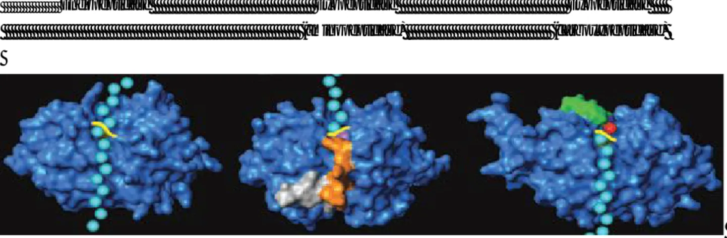
Figure 4. Modes of substrate cleavage caused by peptidases (cysteine cathepsins were used as examples): endopeptidase (cathepsin L) and
exopeptidases (left, cathepsin H, an aminopeptidase; and right, cathepsin X, a carboxypeptidase). Peptide substrate (schematically represented by cyan balls) runs through the entire length of the active site of an endopeptidase framework (blue) and is cleaved in the middle of the molecule (scissile bond marked yellow). In exopeptidases, substrate binding is structurally constrained (mini-chain in cathepsin H, orange; mini-loop in cathepsin X, green). In cathepsin exopeptidases these additional structural elements also provide negative charge (cathepsin H) to bind the positively charged amino terminus (blue) of the substrate, or positive charge (cathepsin X) to bind the negatively charged carboxyl terminus (red) of the substrat [9] .
The classification criterion base on the different types of nucleophilic attack implies that the proteases are grouped into four main classes: Serine, Cysteine, Aspartic and Metal (in most cases Zn2+) protease: (Figure 5). In the last two classes, the aspartic acid moiety or the metal ion polarize a water molecule, so that the oxygen atom can act as nucleophile species. In the other classes instead the nucleophile is represented by the serine oxydril in the form of oxyanion, or from the of cysteine sulfhydryl in the form of thiolate (Figure 6).
Page 9
Figure5.Classification of proteolytic enzyme[8].
Due to their relevance both as therapeutic targets and probe molecules for basic research, the discovery of new protease inhibitors is briskly pursued at present. Inhibitors may help in the isolation and characterization of the different types of proteases.
Figura 6.Catalytic mechanisms of mammalian proteases. The five major catalytic classes of proteases use two fundamentally different catalytic
mechanisms to stabilize the tetrahedral intermediate. In the serine, cysteine and threonine proteases the nucleophile of the catalytic site is part of an amino acid (covalent catalysis), whereas in the metalloproteinases and aspartic proteases the nucleophile is an activated water molecule (non-covalent catalysis). In (non-covalent catalysis, histidines normally function as a base, whereas in non-(non-covalent catalysis Asp or Glu residues and zinc (metalloproteinases) serve as acids and bases. A further difference between the two groups is apparent in the formation of the reaction products from the tetrahedral intermediate, which for cysteine and serine proteases requires an additional intermediate step (acyl-enzyme intermedia)[9]
Page 10
1.3 Characteristics and Regulation of Intracellular Proteolysis.
In the cell the complete degradation of proteins is carried out by lysosomal enzymes and proteasome. The first ones serve primarily to remove proteins from the extracellular and intracellular vesicles, while some of the intracellular proteins are degraded by the proteasome. The two intracellular processes require, unlike the extracellular system, the availability of ATP. The lysosomal degradation uses ATP to acidify the vesicles through a proton pump, while the degradation by proteasome requires ATP at the level of poly-ubiquitination of the substrate and the subsequent demolition. The proteolysis in general, and in particular the intracellular one, must be subject to a tight spatial and temporal control to avoid the indiscriminate protein degradation which would lead in a short time to the destruction of the tissues.
Several factors are involved in the regulation of the intracellular proteolysis [10]:
Compartmentalisation. Is the main response of the cell to the need to safeguard their structure. In addition, the compartmentalization of more enzymes participating in the same metabolic process facilitates the exchange of substrates and considerably increases the overall speed of the process. In this regard those processes of post-translational modification of the proteins that determine the addressing and activation become particularly important.
Conversion of zymogens. The proteases are generally synthesized in the form of inactive precursors (zymogens) that are activated through appropriate proteolysis. Proteolysis must be so strictly controlled, being the limiting step of the related proteolytic processes. Indeed, the zymogen-enzyme conversion usually takes place in specific conditions (e.g. at acidic pH), or in response to specific signals that may originate inside or outside the cell.
pH. Several proteases are poorly active at neutral pH, being optimized to operate in the acidic compartments (lysosomes and endosomes). In such sites the partial denaturation of the substrates, determined by the low pH, favors the proteolytic activity.
Redox State. In cysteine proteases the thiol group is readily oxidizable: because of that, this kind of enzymes require a reducing environment which is ensured in the endosomes by the accumulation of cysteine.
Page 11
Endogenous inhibitors. The activities of several intracellular proteases, the cytoplasmic protease in particular, are controlled by their equilibrium with endogenous inhibitors which are mostly of peptidic nature. They are generally present in large excess with respect to the enzyme. This is without doubt the more selective regulatory mechanism in the cell. There are several known serine proteases inhibitors of proteic type, comprised in the serpin family, located mostly in the extracellular environment. The most common protein inhibitors of cysteine proteases within the group are splitted into three families of cystatins. The oligomeric proteins comprised in family 1 are localized in the cytoplasm, while the members of other families are present in the extracellular compartment. The reversible, competitive inhibition is directed mainly to the papain-like proteases. The activity of Calpain is specifically inhibited by Calpastatin, whereas Caspases are inhibited by both endogenous proteins, and exogenous proteins introduced by infectious agents. Up to now no endogenous inhibitors of aspartic and metal proteases are known. This may explain, at least in part, the high pathogenicity of some infectious agents[11].
In addition to protein catabolism, the degradation of intracellular proteins plays a significant biochemical and physiological role completely different in the case of limited proteolysis, that is realized not only in the conversion of zymogens to active forms, but also in the formation of new enzymes or peptides with different and specialized functions compared to those of the native protein. In general the proteases may participate in the processes of transduction of the signal at three levels [12]:
a) Activation of a specific way of signal transduction. Following the appearance of a second messenger (Ca2+ for example), or the interaction with a specific effector protein, the intracellular proteolysis is activated to trigger in turn a cascade of other enzymatic activities determinants for the cellular response.
b) Modulation of the activity of the receptors. The proteolysis of the intracellular domain of these trans-membrane proteins can activate or repress specific receptors.
c) Regulation of the number of receptors. A membrane receptor, internalized by endocytosis, can be recycled to the membrane or degraded in lysosomes. The number of receptors is given by this balance.
Page 12
1.4 The Lysosome
Lysosomes are membrane-delimited organelles which occur in all mammalian cells except red blood cells. Lysosomes are defined by functional rather than structural properties. They contain a high proton concentration (pH ≤ 5) and more than 40 hydrolases with an optimal H below 6.Their limiting membrane is endowed with specific integral proteins including a vacuolar-type H+-ATPase, several highly glycosylated proteins and various types of transporters. Lysosomes are engaged in the degradation of macromolecules delivered from the cell‘s own cytoplasm as well as materials taken up from the extracellular space (by
endocytosis). Depending on the cell type and the functional state, lysosomes can considerably vary in structure. Therefore it is not surprising that the existence of lysosomes was first realized solely on the basis of biochemical results. The term lysosome was coined by DeDuve five decades ago[13]. Only thereafter, ultrastructural and enzyme-cytochemical studies of Novikoff and coworkers[14] uncovered the morphologic identity of lysosomes. Expanding research in this field gradually revealed the lysosomes as being part of the highly dynamic endosome/lysosome system (Figure 7), a collection of several categories of vesicular organelles which, to a certain extent, exchange membrane constituents and contents thus having overlapping properties. An important feature of lysosomes which serves to discriminate them from endosomes and other related vesicles is the absence of mannose-6-phosphate receptors.
1.5 Biogenesis of Lysosomes
On the basis of the subcellular distribution of acid phosphatase, as demonstrated by enzyme cytochemistry, Novikoff and coworkers[15] proposed the concept of GERL (Golgi apparatus - endoplasmic reticulum - lysosome). It implied (a) that lysosomal enzymes, after biosynthesis in the rough endoplasmic reticulum (ER), are packaged into vesicles (“primary lysosomes”) budding off from tubules which are continuous with the ER and intimately related to the Golgi apparatus, (b) that the vesicles are conveyed to preexisting lysosomes which have already been engaged in a digestive process (“secondary lysosomes”). This concept was later replaced by the concept of the trans Golgi network (TGN) as the common exit site for all products including lysosomal enzymes, secretory proteins and membrane proteins[16]. The most important modifications of the previous GERL concept were induced by data indicating that the intracellular transport of lysosomal enzymes is receptor-mediated and has to pass through a prelysosomal compartment (Figure 7).
Page 13
Figure 7. Diagrammatic summary of the endosome/lysosome system, with special reference to the biogenesis of lysosomes. After synthesis in
the rER and modification in the Golgi apparatus (not shown) precursors of soluble lysosomal enzymes decorated with mannose-6-phosphate residues meet the mannose-6-phosphate receptor (MPR) in the trans Golgi network (TGN), are packaged (1) into clathrin-coated vesicles (ccv), and are transported (2) to late endosomes (LE) either directly or indirectly via early endosomes (EE) (3). The process of enzyme transfer (4) from the LE to the lysosome (Lys) is not fully elucidated yet; possibly LE matures to become Lys, or LE and Lys fuse to form a transient hybrid organelle (for further possibilities see text). The MPR is recycled (5) from the LE to the TGN, the lysosome is devoid of MPRs. A minor portion of the enzyme precursors gets into the secretory pathway (6) and is recaptured into clathrin-coated pits (ccp) by MPRs, which may be transferred (7) from the EE to the plasma membrane. Thus the enzyme precursors can reach the lysosome via the endocytic pathway (8) as do endocytic tracer molecules (9), whose receptors are recycled from the tubular extensions of the EE (10). Autophagic vacuoles (AV) and phagocytic vacuoles (PV) acquire lysosomal enzymes by fusion with lysosomes and/or LE (11) to become autolysosomes and phagolysosomes,85 respectively. LE often resemble multivesicular bodies, i.e., they display invaginations of their membrane and internal vesicles budded off the invaginations (or representing cross sections of the invaginations). lgp/lamp, lysosomal membrane glycoproteins/lysosome-associated membrane proteins[9].
1.5 Transport of Lysosomal Enzymes
More than 50 acid hydrolases involved in the ordered lysosomal degradation of a variety of proteins, lipids, carbohydrates, and nucleic acids have been identified. The hydrolases are enclosed by a membrane containing a set of highly glycosylated lysosomal membrane proteins. Lysosomal enzymes are also components of cell type-specific compartments referred to as lysosome-related organelles which include melanosomes, lytic granules, MHC class II compartments, platelet-dense granules, and synaptic-like microvesicles.1 The biogenesis of new lysosomes or lysosome-related organelles requires a continuous substitution with newly synthesized components. The targeting of acid hydrolases depends on the presence of mannose 6-phosphate (M6P) residues that are recognized by specific receptors mediating the intracellular
Page 14
transport to an endosomal/prelysosomal compartment. The acidification of endosomes, lysosomes, and lysosome-related organelles facilitates not only the dissociation of the receptor-ligand complexes, but also the proteolytic processing required for the enzymatic activation of several hydrolases as well as the denaturation of target proteins as prerequisite for lysosomal proteolysis[17] .
1.5.1 Synthesis and Modifications of Soluble Lysosomal Proteins
Lysosomal hydrolases are synthesized with an N-terminal sequence of 20-25 amino acids recognized by the signal recognition particle which enables the nascent polypeptides to be translocated across the membrane of the endoplasmic reticulum (ER, Figure 8, step 1). After removal of the signal peptide by a signal peptidase, preformed oligosaccharides are transferred to certain asparagine residues which are part of an N-glycosylation consensus motif Asn-X-Thr/Ser[18]. Typically, the oligosaccharides are composed of 3 glucose, 9 mannose and 2 N-acetylglucosamine residues (Glc3Man9GlcNac2). The oligosaccharides undergo extensive processing before completion of translation by removal of the outmost glucose residues catalyzed by glucosidase I and II. Monoglycosylated core glycans of the newly synthesized polypeptide then bind to the molecular chaperone calnexin until the protein is properly folded[19]. Furthermore, all members of the sulfatase family responsible for the hydrolysis of sulfate esters from sulfated mono- and polysaccharides, glycolipids and hydroxysteroids, are modified in the ER generating a Cα-formylglycine (FGly) residue. The oxidation of a highly conserved cysteine occurs when the protein has not been folded yet and is catalyzed by the FGly-generating enzyme (FGE). Mutations in the gene encoding the human FGE result in the appearance of multiple lysosomal sulfatases lacking enzymatic activity[20]. Moreover, aspartylglucosaminidase, a lysosomal enzyme that hydrolyzes the amide bond between asparagines and N-acetylglucosamine, has been reported to be rapidly activated in the ER by a proteolytic cleavage of the inactive precursor into α- and β-subunits which is triggered by the dimerization of the two precursor molecules[21].
1.5.2 Formation of Mannose 6-Phosphate Recognition Marker
Upon arrival in the Golgi, the oligosaccharide chains of lysosomal enzymes are further trimmed and modified by the addition of complex sugar residues (galactose, fucose, and sialic acid), sulphate groups, and by the formation of the M6P recognition marker. This marker is generated by the sequential action of two enzymes [22]. In the first step, GlcNac-1-phosphate is added to the C6-hydroxyl group of selected mannoses on high mannose-type oligosaccharides by the enzyme UDP-acetylglucosamine: lysosomal enzyme N-acetylglucosamine-1-phosphotransferase (phosphotransferase) (Figure 8, step 2). The purified bovine phosphotransferase is a 540 kDa heterohexameric complex composed of two disulfide-linked homodimer of 166 and 51 kDa subunits and two noncovalently associated 56 kDa subunits (α2β2γ2)[23,24].
Page 15
Figure 5. Model for the intracellular transport of MPR and proteins to lysosomes. Soluble lysosomal enzymes are synthesized and translocated into the lumen of the ER (step 1). In the Golgi (step 2) the enzymes are equipped with the M6P recognition marker (step 3) followed by binding to MPRs. The receptor-ligand complexes are transported to the early endosomal compartment (EE; step 4). Due to the low pH the receptor-ligand complexes dissociate and the lysosomal enzymes are delivered to the lysosome (Lys, steps 5 and 6). The MPRs recycle either back to the TGN (steps 7 and 8) or to the plasma membrane (step 9). Exogenous M6P-containing proteins can be internalized by CI-MPR (step 10) and are transported to lysosomes along the endocytic pathway (step 11). Lysosomal enzymes escaping binding to MPR in the TGN are secreted (step 12)[12].
The phosphotransferase γ-subunit has been shown to form cysteine-linked dimers and contains two N-linked oligosaccharide. It has been proposed that the α and β-subunits harbour phosphotransferase activity and the γ-subunit functions in recognition of lysosomal enzymes. Studies with chimeric proteins between cathepsin D and pepsinogen, mutant cathepsin L and aspartylglucosaminidase, and blocking antibodies against various epitopes on arylsulfatase A suggest that 2-3 lysine residues, separated by a distance of ~ 34 Å residing in distinct regions of the enzymes, are critical phosphorylation signals[25,26]. In the second step, N-acetylglucosamine residues are removed by an N-acetylglucosamine-1-phosphodieste α-N-acetylglucosaminidase (UCE; uncovering enzyme) exposing the M6P recognition marker (Figure 4, step 3). The human UCE is a type I membrane spanning glycoprotein of 515 amino acids with a transmembrane domain and a cytoplasmic tail of 41 amino acids. UCE is mainly localized in the TGN and constitutively cycles via the plasma membrane[27] UCE is synthesized as an inactive proenzyme which is activated in the TGN by
Page 16
the cleavage of an N-terminal pro-peptide catalyzed by the endoprotease furin The bovine UCE exists as a tetramer composed of two disulfide-linked homodimers with molecular masses of 68 kDa for each monomer[28]. Following the uncovering of the M6P marker, lysosomal enzymes can be recognized by mannose 6-phosphate receptors (MPRs).
1.5.3 Mannose 6-Phosphate Receptors
In mammalian cells two MPR exist, one is a 46 kDa cation-dependent (CD-MPR, MPR46) and the other an 300 kDa cation-independent (CI-MPR, MPR300) protein[29]. Both MPRs are type I membrane glycoproteins that differ in their developmental expression pattern, subcellular localization, quarternary structure, half life of receptor protein, ligand binding properties and functions[30]. The MPRs bind mono- or diphosphorylated oligosaccharides with a Kd of 2 x 10⁻⁷ -2 x 10⁻⁹ M per mole of receptor. Subsequently the receptor-ligand complexes exit from the TGN in clathrin-coated vesicles and fuse with membranes of the early or late endosomal compartment (Figure 8, steps 4, 5). Due to the low pH, the lysosomal enzymes dissociate and are delivered to lysosomes (Figure 8, step 6); the MPRs recycle back to the TGN to mediate further rounds of transport (Figure 8, steps 7, 8)[29]. Small amounts of both MPRs are localized at the plasma membrane (Figure 4, step 10) but only the CI-MPR is capable of binding and internalizing M6P-containing lysosomal enzymes (Figure 8, step 11).
1.6 The Lysosomal Proteases
Lysosomal proteolytic enzymes catalyze the hydrolysis of proteins. Only few of the proteinases work as amino- or carboxypeptidases, while most are endopeptidases preferably cleaving peptide bonds within a polypeptide chain rather than at its ends. Accordingly, the name cathepsin, derived from the Ancient Greek word καθέψω (kathépsō,“boil down”), was originally meant to be reserved for endopeptidases. However, it turned out that cathepsin Bl which, to follow this convention, was explicitly named as such, is both, an endo- and an exopeptidase. In the late 1990s, a more recent nomenclature discussion was started by members of the International Proteolysis Society (www.protease.org). Due to the progress in genome sequencing, the scientific community was afraid of shortly running out of letters of the alphabet to be added to the term cathepsin in order to name newly identified enzymes. However, no better nomenclature system was found, and, hence, we still stick to the term cathepsin, but now, when a new member of the family is discovered, we add numbers instead of letters[31]. An excellent reference and a classification of lysosomal proteases can be found in the MEROPS database (merops.sanger.ac.uk).
Metalloproteinases, with the exception of lysosomal dipeptidase I, proteases with unknown mechanism of proteolytic cleavage, and the most recendy discovered threonine proteases are rarely found within lysosomes.
Page 17
Mainly, lysosomal proteases are members of the aspartic, cysteine (Table 1, Chapter 2) , or serine proteinase families of proteolytic enzymes.
1.7 References
[1] M. M. Cox, D.L. Nelson, Lehninger Principles of Biochemistry, Fourth Edition, W. H. Freeman 2004 [2] R. A. Harvey, D. R. Ferrier, Biochemistry, Lippincott Williams & Wilkins 2010
[3] M. Danson, R. Eisenthal, Enzyme assays: a practical approach. Oxford [Oxfordshire]: (2002) Oxford University Press. [4] X.S. Xie, H.P. Lu, “Single-molecule enzymology "J. Biol. Chem. 274 (23) (1999): 15967–70.
[5] H. Lu, Single-molecule spectroscopy studies of conformational change dynamics in enzymatic reactions, Current pharmaceutical
biotechnology 5 (3) (2004) : 261–9.
[6] J. Schnell, H.Dyson, P. Wright, "Structure, dynamics, and catalytic function of dihydrofolate reductase". Annual review of biophysics and
biomolecular structure 33 (2004): 119–40.
[7] Q.H. Gibson "Rapid mixing: Stopped flow". Methods Enzymol. Methods in Enzymology 16 (1969): 187–228. [8] C.E. Chwieralski, T. Welte, F. Buhling, Cathepsin-regulated apoptosis Apoptosis 11 (2006) 143-149
[9] B. Turk, Targeting proteases: successes, failures and future prospects, Nat. Rev. Drug Discov. 5 (2006) 785–799.
[10] D. Turk, B. Turk, V. Turk, Papain-like lysosomal cysteine proteases and their inhibitors: drug discovery targets? Biochem. Soc. Symp. 70 (2003) 15–30.
[11] J. M. Lankelma ,M. Voorend, T. Barwari, J. Koetsveld, A. H. Van der Spek, P.N.A. De Porto, G. Van Rooijen, J.F. Van Noorden, Cathepsin L, target in cancer treatment? Life Sciences 86 (2010) 225–233
[12] P. Safting Lysosomes, cap. 2 Springer Science 2005
[13] C. DeDuve, B.C. Pressman, R. Gianetto, Tissue fraction studies.Intracellular distribution patterns of enzymes in rat liver tissue. Biochem
J 60 (1955): 604-617.
[14] A.B. Novikoff, H. Beaufay, C. DeDuve, Electron microscopy of lysosome-rich fractions from rat liver. J Biophys Biochem Cytol 2(Suppl) (1956):179-184.
[15] P. Safting Lysosomes, cap. 1 Springer Science 2005
[16] P.M. Novikoff, A.B. Novikoff, N. Quintana, Golgi apparatus, GERL, and lysosomes of neurons in rat dorsal root ganglia studied in thick section and thin section cytochemistry. J Cell Biol 50 (1971); 859-886.
[17] G. Griffiths, K. Simons, The trans Golgi network: Sorting at the exit site of the Golgi complex. Science 234 (1986):438-443. [18] Lysososmes capitolo 2
[19] R. Kornfeld, S. Kornfeld, Assembly of asparagine-linked oligosaccharides. Annu Rev Biochem (1985) ; 54:631-664 [20] A. Helenius, M. Aebi, Intracellular functions of N-linked glycans. Science (2001); 291(5512):2364-2369.
[21] T. Dierks, B. Schmidt, L.V. Borissenko, Multiple sulfatase deficiency is caused by mutations in the gene encoding the human C(alpha)-formylglycine generating enzyme. Cell (2003); 113(4):435-444.
[22] A. Riikonen, J. Rouvinen, R. Tikkanen, Primary folding of aspartylglucosaminidase. Significance of disulfide bridges and evidence of early multimerization. J Biol Chem (1996); 271(35):21340-21344.
[23] M.Bao, Booth JL, Elmendorf BJ et al. Bovine UDP-N-acetylglucosamine: Lysosomal-enzyme N-acetylglucosamine-1 phosphotransferase. I. Purification and subunit structure. J Biol Chem (1996); 271(49):31437-31445.
[24] Bao M, B.J Elmendorf, J.L. Booth, Bovine UDP-N-acetylglucosamine: Lysosomal-enzyme N-acetylglucosamine-1 phosphotransferase. II. Enzymatic characterization and identification of the catalytic subunit. J Biol Chem (1996); 271(49):31446-31451.
[25] T.J. Baranski, A.B. Cantor, S. Kornfeld, Lysosomal enzyme phosphorylation. I. Protein recognition determinants in both lobes of procathepsin D mediate its interaction with UDP-GlcNAc:lysosomal enzyme N-acetylglucosamine-1-phosphotransferase. J Biol Chem (1992); 267(32):23342-23348.
[26] J.B. Warner, C. Thalhauser, K. Tao, Role of N-linked oligosaccharide flexibility in mannose phosphorylation of lysosomal enzyme cathepsin L. J Biol Chem (2002); 277(44):41897-41905.
Page 18
[27] J. Rohrer, R. Kornfeld, Lysosomal hydrolase mannose 6-phosphate uncovering enzyme resides in the trans-Golgi network. Mol Biol Cell (2001); 12(6):1623-1631.
[28] R. Kornfeld, M. Bao, K. Brewer, Purification and multimeric structure of bovine N-acetylglucosamine-1-phosphodiester alpha-N-acetylglucosaminidase. J Biol Chem (1998); 273(36):23203-23210.
[29] P. Ghosh, N.M. Dahms, S. Kornfeld, Mannose 6-phosphate receptors: New twists in the tale. Nat Rev Mol Cell Biol (2003); 4(3):202-212.
[30] T. Braulke, Origin of lysosomal proteins. In: Lloyd JB, Mason RW, eds. Subcellular Biochemistry Biology of the Lysosome. New York:
Plenum Press, (1996):27:15–49.
Page 19
CHAPTER 2
CYSTEINE CATHEPSINS: CATHEPSIN L
There are 11 human cathepsins, which were originally identified as proteases that act in the lysosome. Recent work has uncovered non traditional roles for cathepsins in the extracellular space as well as in the cytosol and nucleus. There is strong evidence that subspecialized and compartmentalized cathepsins participate in many physiologic and pathophysiologic cellular processes, in particular multiple mechanisms increase cysteine cathepsin expression in tumors.
Page 20
2.1 A brief description of Cathepsins
The name cathepsin was initially proposed for those proteases which act in a slightly acidic environment[1,2].The cysteine cathepsins are predominantly endopeptidases, which are located intracellularly in endolysosomal vesicles. Later, the name cathepsin was introduced for the serine proteases cathepsins A and G, the aspartic proteases cathepsins D and E, and the lysosomal cysteine cathepsins. There are 11 human cysteine cathepsins, i.e., the cathepsins B, C, F, H, K, L, O, S, V, X and W, the sequences of which are known. The sequences were confirmed by bioinformatic analysis of the draft sequence of the human genome. The 11 human cysteine cathepsins (Table 1,2) belong to the papain subfamily of cysteine proteases[3].
Table 1.Human Cysteine Cathepsins[3]
2.2 Localization and main properties
The majority of cathepsins are ubiquitously expressed in human tissues: their expression profile indicates that these enzymes are involved in a normal cellular protein degradation and turnover. In contrast, cathepsins K, W and S show a restricted cell or tissue-specific distribution, indicating their more specific roles. For example, cathepsin K is highly expressed in osteoclasts, in most epithelial cells and in the synovial fibroblasts in rheumatoid arthritis joints[4] (Figure 1). Among the matrix-degrading enzymes, cathepsin K is the only enzyme for which an essential role in bone resorption has been unambiguously documented in mice and humans[5]. These two enzymes might be expected to have discrete functions and studies in mice deficient in cathepsin K
Page 21
or cathepsin S have confirmed a role for cathepsin K in bone remodeling and Cathepsin S in MHC (major histocompatibility complex) class II antigen presentation.
Cathepsin W (also lymphopain) is predominantly expressed in CD8+ lymphocytes and natural killer (NK) cells[6,7]. However, when cathepsin W expression has been downregulated using the shRNA approach, its essential role in the process of cytotoxity has been excluded[8].
Table 2. General characteristics
Cathepsin S is predominantly expressed in the professional antigen-presenting cells (APCs), such as the dendritic cells (DCs) and B-cells[9]. In addition, cathepsin V (also named L2) is highly homologous to cathepsin L, but in contrast to the ubiquitously expressed cathepsin L, its expression is restricted to thymus and testis[10,11]. Moreover, there is sufficient evidence that the specific physiological functions of the cathepsins might be at least partially attributed to their differences in localization inside and outside the cells[12,13,14]. This is consistent with the recent advances in the field of proteases that led to a new concept, based on proteases as important signaling molecules involved in the controlling mechanisms which play roles in many normal and pathological processes[15]. Cysteine cathepsins are commonly considered to be optimally active in a slightly
Page 22
acidic pH and are mostlyunstable at neutral pH When cathepsins are outside the lysosomes or extracellularly they can be inactivated irreversibly in a relatively rapid way at neutral pH[16].
Figure 1. For example the cysteine cathepsins that are known to be expressed in tumour cells and tumour-associated cells, and have been identified as contributing to neoplastic progression.
The extracellular localization of cysteine cathepsins often coincides with their increased expression and/or activity, indicating that pH is not the sole factor responsible for their activity. Twenty years ago, it was demonstrated that cathepsin B, from normal or tumor tissues, degraded purified extracellular-matrix components, type IV collagen, laminin and fibronectin, both under acid and neutral pH[17]. Cathepsins with strong elastolytic and collagenolytic activities are now known to be chiefly responsible for the remodeling of the extracellular matrix (ECM), thus contributing to various pathologies. The ECM consists of elastins, collagens and proteoglycans, which have all been identified as cathepsin's substrates[18]. Elastin was found to be cleaved by the cathepsins K, L, S, F, V and B, although they differ in their elastolytic activitiy. Cathepsin V was thus found to be the most potent enzyme, followed by the cathepsins K, S, F, L and B[19]. It was further demonstrated that the elastolytic activities of the cathepsins V and K could be inhibited by ubiquitously expressed glycosaminoglycans (GAGs), such as chondroitin sulfate, through the formation of cathepsin-GAG complexes. In contrast, cathepsin S, which does not form complexes with chondroitin sulfate, is not inhibited,
Page 23
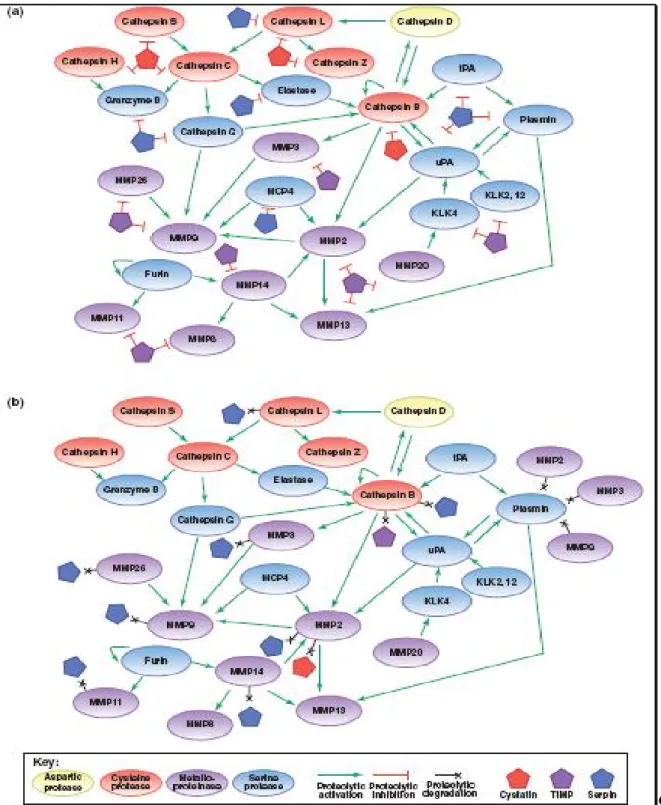
thus suggesting that there might exist some specific regulation of the elastolytic activities of cathepsins by GAGs[20]. Cysteine cathepsins such as cathepsin B and cathepsin L are expressed constitutively and are thought to participate in protein turnover. The expression of some cysteine cathepsins is regulated and is high in specific cell types. More surprisingly, studies in mice deficient in the constitutively expressed cathepsins L and B have established that these enzymes have specific functions in certain tissues. For example, cathepsin L-deficient mice exhibit phenotypes in skin and cardiac muscle[21,22] and cathepsin B-deficient mice exhibit reduced TNF (tumour necrosis factor)-induced apoptosis of hepatocytes[23]. Also unexpected is that there is redundancy of both constitutive and regulated cysteine cathepsins. In mice that are deficient in both cathepsins S and B[24], degradation of immune complexes is reduced by >80%, whereas in mice that are deficient in both cathepsins L and B[25], brain atrophy, which is induced by massive apoptosis of cerebral and cerebellar neurons, results in their death at 2–4 weeks. Cathepsins S and L are endopeptidases, but cathepsin B is both an exopeptidase and an endopeptidase, suggesting that, at least in immune-complex degradation and neuronal apoptosis, cathepsin B acts as an endopeptidase. Loss-of-function mutations of human cathepsins C and K are associated with Papillon–Lefevre syndrome and pycnodysostosis, respectively. Patients with Papillon–Lefevre syndrome have reduced activity and stability of neutrophil-derived serine proteases, but not of cytotoxic lymphocyte-derived serine proteases (granzymes)[26]. In cathepsin C-deficient mice, however, neither cytotoxic lymphocyte-derived serine proteases nor mast cell serine proteases (chymases) are activated[27]. This cysteine cathepsin therefore regulates a proteolytic pathway, in which there is the activation of other prostheses (figure 2). Cysteine cathepsins were long thought to function primarily intracellularly within endolysosomal compartments. However, there are exceptions; for example, cathepsin K, which is secreted into the resorptive pit between osteoclasts and bone, often termed an ‘extracellular’ lysosome. Studies on mice that are deficient in the cysteine cathepsins B, K and L have shown that all three of these cysteine cathepsins function both extracellularly and intracellularly to liberate thyroglobulin[28]. In cancers, as discussed in the next paragraph, there is evidence for the cysteine cathepsins functioning extracellularly as well as intracellularly. We emphasize studies on cathepsins B and L as these enzymes have been studied most thoroughly, however, even the functions of these two cysteine cathepsins in cancer are not well defined yet nor are their roles in tumor cells and the tumor-associated cells that contribute to neoplastic progression[3] (Figure 1).
Page 24
Figure 2. Mechanisms of proteolytic regulation within the network. (a) Endogenous protein inhibitors can regulate proteolytic activity at many points in the network, allowing for tight regulation (or deregulation, if inhibitor expression is downregulated) of proteolytic activity. (b) Several proteases have been shown to degrade endogenous inhibitors, giving them the ability to increase the activity of other proteases indirectly. These interactions add further complexity to the network, and demonstrate how proteolytic signals can flow in multiple directions through different mechanisms. References for proteolytic interactions not described in the main text: cathepsin B can inactivate some serpins and TIMPs[29],
serpinB13/hurpin and serpinB3/SCCA1 inhibit cathepsin L , cathepsin L inactivates serpinA1[30], and several MMPs can inactivate a variety of
Page 25
2.3 Chatepsin L
Cathepsin L, a lysosomal cysteine protease, plays an important role in the catabolism of proteins and consequently in the behavior of human cells[31]. Is produced as preprocathepsin L, transported via the Golgi apparatus as procathepsin L in secretory vesicles and then stored as mature cathepsin L in lysosomes[32].
2.3.1 Transcription and translation Genetic information[33,34]:
Enzyme Nomenclature: Cathepsin L ECnumber: 3.4.22.15
Other Aliases: RP11-65B23.1, CATL, CTSL, FLJ31037, MEP Gene Name: CTSL1
Chromosome: 9; Location: 9q21.33
Annotation: Chromosome 9, NC_000009.11 (90340974..90346384)
Troen et al. first reporting[35] on the complex manner by which mouse cathepsin L transcription is regulated. Later on, regulation of cathepsin L gene transcription has been studied mainly in reproductive tissue. The cathepsin L and ADAMTS-1 genes are targets of the progesteron receptor signaling pathway during ovulation[36]. Cathepsin L mRNA is also upregulated during the implantation of the early trophoblast[37]. In male rats, FSH induced an increase in cathepsin L mRNA in late stages of the seminiferous tubular epithelium cycle in a cAMP-dependent manner[38]. Substantial work has been done to analyze the promoter regions of the human cathepsin L gene (CTSL) as well as to understand the regulation of different splice variants within the 5′ untranslated region of the transcript. Indeedcathepsin L shows an ubiquitous organ distribution; more recent work has focused on the regulation of cathepsin L alternative translation. According to the presence of different forms of cathepsin L in distinct subcellular and extracellular compartments, cathepsin L proteins can be initiated from downstream AUG sites[39], omitting the signal peptide that is normally present at the N terminus of lysosomal cathepsin L that routes the protein to the ER during its synthesis. The enormous increase of cathepsin L mRNA after Kirsten virus transformation of NIH-3T3 fibroblasts is a long-known phenomenon. Four splice variants of human cathepsin L mRNA have been reported, hCATLA, Al, A2 and -B, all differing in the 5’-UTR and thus coding for identical proteins [40,41,42]. Later on three more isoforms were identified, all showing differences in amino acid sequences in the exon 1 moiety[43],. In any case they have all the encoding region between exon 2 and 7 (figure 3)
Page 26
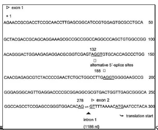
Figure 3.Structure of the various mRNAs at the 5’-UTR amplified by 5’-RACE PCR and confirmed by sequencing. The numbering for hCATLA
mRNA (Gal et al., 1988) begins at the transcription site (+) of the hCATL.
Figure 4 . Partial cDNA Sequence of hCATL: Exon 1 and Transition to Exon 2. Numbering starts from the transcriptional start site (+1) of the
hCAT-L gene. The exon/intron boundary (_), the translational start for hCAT-L (underlined) as well as the alternative splice sites (_) for both the hCATL-A2 and -A3 variants are indicated.
For these isoforms the gene sequences appear to be quite different from each other only in the exon 1 moieties where the sites in which the different alternative splicing starts are present (figure 4).Summarizing, we can say that a dominating isoform is evident, named hCATL-A3 in which the exon1 is 146 nucleotides
Page 27
shorter than the one of hCATL-A. It seems to be the dominant form of cathepsin L transcripts in various cell lines, tissues and tumor samples examined in the relevant study. Subsequent DSD-Page experiments showed that the weight of the pre-propenzyme is 42 kDa.
2.3.2 Biosynthesis, Processing and Trafficking
Under normal conditions in healthy organisms, cells use various mechanisms to prevent potentially harmful and uncontrolled proteolytic activity. They include the compartmentalization of cathepsins within the lysosome or other organelles, zymogen activation, pH effects, regulation of proteolytic activities by small-molecule inhibitors and various endogenous protein inhibitors, or a combination of all these factors for an optimal enzyme function[44]. The organization of activation of protease from proenzyme and the role of mannose-6-phosphate receptor is described in some detail in chapter 1. For cathepsins and, especially for cathepsin L, a similar activation occurs (Figure 5). As secreted proteins, cathepsins are synthesized with an N-terminal signal peptide that targets the protein to the lumen of the ER. The signal peptide is co-translationally cleaved and N-linked glycosylation occurs within the ER. Similar to other proteases, cysteine cathepsins are synthesized as inactive proenzymes and require proteolytic processing for activity. The immature protein possesses an N-terminal pro-region, which is removed to activate the enzyme, suggesting that the pro-region acts as an auto-inhibitor. Indeed, synthetic peptides corresponding to pro-regions do function as specific inhibitors of the parent cathepsin in the case of cathepsin L and cathepsin-like cysteine proteases[45]. Further progress in understanding the nature of zymogen activation was obtained from the pro-cathepsin structures. The crystal structures of human pro-cathepsin L[46] showed that the pro-peptide chain folds (arranges itself) on the surface of the enzyme in an extended conformation and runs through the active-site cleft, in the opposite direction to the substrate, thereby blocking the access of the latter to the active site, which is already formed in the zymogen. In the structure of most pro-enzymes, hydrophobic interactions, salt bridges and hydrogen-bonding interaction within the pro peptide and between the pro-peptide and the mature enzyme do exist. Some early studies suggested that autocatalytic processing is a mono-molecular process, while others studiesproposed inter- and intra-molecular mechanisms. This enigma has been recently clarified. It is now clear that the auto-activation of cathepsins is a combination of a mono-molecular and a bi-molecular process[47]. Pro-cathepsin B possesses a low catalytic activity that is, nevertheless, sufficient to trigger the autocatalytic activation of the zymogen. This activity is the result of the pro-peptide dissociation from the active-site cleft as the first step in this process, which is in fact the only mono-molecular step in the process[48]. In the next step, which is already bi-molecular, this catalytically active zymogen molecule processes and activates another pro-cathepsin molecule in one or more steps. The mature cathepsin molecules generated in this way then initiate a chain reaction leading to a rapid activation of the remaining pro-cathepsin molecules. In addition to its role in
auto-Page 28
inhibition, the N-terminal pro-region of cathepsin L appears to be necessary for the correct folding of the protein[49], a finding that is also true for papain.
Figure 5. Cathepsin structures and traditional and nontraditional protease trafficking. Cathepsins contain a signal peptide (blue) that directs insertion of the nascent polypeptide chain into the ER. Within the ER, the signal peptide is cleaved and the protein folds with the assistance of the proregion (red). Disulfide bond formation (indicated by S-S) and N-linked glycosylation with high-mannose glycans subsequently occurs in the ER. Within the Golgi, mannose residues are phosphorylated to form m6p, which is used to route the protein into the endosomal/lysosomal compartment via the m6p receptor. Upon initial acidification of the endosome, cathepsins are activated, which leads to cleavage of the proregion and further activation of the cathepsin, resulting in further proteolytic processing in the lysosome into heavy and light chains (yellow). A portion of the cathepsins is not converted to the m6p form and as a result is shunted into the exocytosis pathway. Conversion to m6p appears to be rate limiting, as overexpression of a given cathepsin greatly increases the proportion of the enzyme in this pathway. Ribbon diagrams depict the structure of mature cathepsin L in the extracellular matrix and in the lysosome. The ribbon colors correspond to the colors used in the diagram on the left.
Page 29
Mutations destabilizing the interface between the helices prevent correct folding of the protein[50]. Folding of the pro-region may precede folding of the full protein, thereby providing a scaffold which then directs the folding of the remaining domains. The full structure would be stabilized by the formation of disulfide bonds that prevent unfolding once the pro-region has been removed (Figure 4). Initial glycosylation generates high mannose glycans within the ER. Cathepsins destined for the lysosome are further processed in the Golgi apparatus by modification of mannose residues to mannose-6-phosphate (m6p). Two to three lysine residues separated by 34 Å appear to be critical in the recognition motif[51]. Upon acidification in endosomes, cathepsins become active and begin proteolytic processing, with cleavage within the pro-region, allowing pro-region dissociation from the enzyme as mentioned above. This results in an active, single chain form of the protein. Upon arrival at the lysosome, further processing cleaves the protein into two chains. Active cathepsins may also be recruited from late endosomes or lysosomes for secretion into the extracellular space via Ca2+ -mediated fusion of these organelles with the plasma membrane. In addition, a minor population of cathepsins (approximately 5%) does not travel to the lysosome but is instead secreted as a pro-enzyme. Furthermore, alternative splicing and exon skipping can lead to cathepsin forms that lack the signal peptide, and these can subsequently localize to the nucleus and mitochondrial matrix[52]. Recent data suggest that truncated forms of cathepsin L are important in regulating the cytoskeleton of kidney podocytes[53], whereas others data suggest that mature cathepsin L is present outside lysosomes, e.g., during histone processing in embryonic stem cells[54]. These cathepsin L variants have been previously shown to arise by translation from an alternate downstream AUG site[39] and to be located in the nucleus of fibroblasts, in which they can cleave the transcription factor cut-like home[39]. Cathepsin L also processes histone H3 during mouse embryonic stem cell differentiation (25). Although conventional cathepsin L cleaves various proteins very efficiently, due to the denaturing conditions and low pH of the lysosome, cytosolic and nuclear cathepsin L exhibit remarkable substrate specificity that allows a very specific enzymatic activity at cytosolic or nuclear pH[55] .
2.3.3 Structure and Specificity
Understanding the interactions between cysteine cathepsins and their substrates has been and remains the main challenge in the research on cysteine cathepsins. Papain served as the model in the pioneering work of Schechter and Berger[56], in which they proposed the nomenclature for the positions of the substrate residues (P) and the sub-sites (S) where they bind to the surface of a protease. The positions and sub-sites were numbered in both directions from the scissile bond between the residues P1 and P1′ onwards, where the non-primed side refers to the N-terminal part and the non-primed side to the C-terminal part of the substrate. In their pioneering work, they found that adding residues longer than 7 alanins to peptides does not affect the kinetics
Page 30
of their degradation and concluded that there are seven substrate residues binding into seven sub-sites from S4 to S3′. Three decades later, the definition of the substrate binding sites was revised based on an insight provided by the crystal structures of the complexes with small-molecule, substrate-mimicking inhibitors[57]. These structures show that a substrate binds along the active-site cleft in an extended conformation Figure 6.
Figure 6.3D schematic representation of the substrate-binding sites of papain-like proteases along the active-site cleft. The representation is based on the proposed revised definition of substrate-binding sites based on the crystal structures of substratemimicking inhibitors bound to the protease's active sites [57]. The substrate-binding sites of the papain-like proteases are located on the left (S1, S3, S2′) and right (S2, S1′) side of the active-site cleft in accordance with the standard view orientation. According to papain numbering, L-domain loops include residues Gln19-Cys25 and Arg59-Tyr67 whereas R-domain loops contain residues Leu134-His159 and Asn175- Ser205, respectively. The active-site residues Cys25 and His159 and the disulphide Cys22-Cys63 are indicated.
A 3D-based sequence alignment of the mature form of the nine cysteine cathepsins with a known 3D structure exhibits conservation of the active-site residues (Cys25 and His163, for cathepsin L); these residues interact with the main chain of the bound substrate, (Gln19, Gly68, Trp183), the N-terminus Pro2 and certain Cys residues do interact as well. Figure 7 shows the papain-like fold of cathepsin L. It is composed of two domains, referred to as the left (L-) and right (R-) ones, in accordance with the standard view. The L-domain contains three α-helices. The longest, i.e., the vertical helix, known also as the central helix, is over 30 residues long. The R-domain is a kind of β-barrel with the front strand(s) forming a coiled structure. At the bottom, the barrel is enclosed by an α-helix. The reactive site histidine is located at the top of the sheet forming the barrel. The two-domain interface opens at the top, forming the active-site cleft. In its center are the reactive site residues, Cys25 and His163, each coming from a different domain. The reactive site cysteine is located at the N-terminus of the central helix of the L-domain, whereas the histidine is located within the β-barrel residues of
Page 31
the R-domain. These two catalytic residues form the thiolate-imidazolium ion pair, which is essential for the proteolytic activity of the enzymes[20].
Figure 7.Fold of the mature form of cysteine cathepsins–endopeptidases. The fold of the two-chain form of native cathepsin L (1icf) is shown in
the green ribbon representation. The parts corresponding to the secondary structure elements,α-helices and β-sheets, are shown in blue and red color, respectively. The side chains of the reactive-site cysteine (Cys25) and histidine (His163) are indicated in a ball-and-stick representation.
The active-site surface is composed of the residues coming from four loops located on both domains (Figure 6). The two L-domain loops are short and cross-connected with a disulfide bond. The two R-domain loops are much larger and form the lid of the β-barrel hydrophobic core. The substrate binds along the active-site cleft in an extended conformation[57]. Its side chains make alternating contacts with the L- and R-domains. The two loops on the L-domain and their bridged interface provide the surface for the side chains of the P3, P1 and P2′ residues, whereas the loops and residues of the R-domain provide the binding surface for the P2 and P1′ residues. An exception to this is the P2 residue. According to the proposed model[58], the P2 residue interacts with both domains.
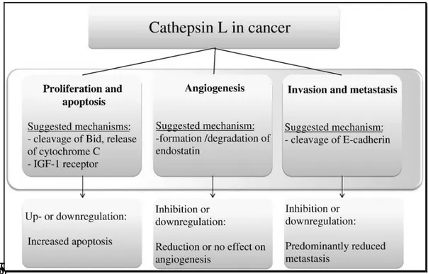
2.3.4 Cathepsin L and Cancer
Several members of the cysteine cathepsin family have been implicated in cancer progression on the basis of their increased expression, activity and mislocalization in various human and mouse tumors (Figure 8)[59,60]. Furthermore, in some of these cancers, the changes in cysteine cathepsin expression or activity have diagnostic or prognostic value[61]. In terms of which cysteine cathepsins are specifically involved in cancer, cysteine cathepsins B and L have been investigated most intensively, and invariably their increased expression and/or activity correlates with malignant progression. It is now clear that the cathepsins have an important role in both tumor progression and invasion, which is supported by the numerous clinical reports and


![Table 1. Human Cysteine Cathepsins [3]](https://thumb-eu.123doks.com/thumbv2/123dokorg/7572675.111742/22.892.86.806.404.764/table-human-cysteine-cathepsins.webp)