DOTTORATO DI RICERCA IN
TERAPIE AVANZATE BIOMEDICHE E CHIRURGICHE XXXI CICLO
Coordinatore Prof. Giovanni Di Minno
TESI DI DOTTORATO
“TYPE 2 DEIODINASE POLYMORPHISM THR92ALA:
CHARACTERIZATION ON A BIOLOGICAL MODEL AND CLINICAL IMPLICATIONS”
Relatore Candidato
Index
1 Summary 2
2 Background 4
2.1 Intracellular metabolism of thyroid hormones 4 2.2 Role of deiodinases in muscle stem cells proliferation and development 5 2.3 Clinical implications of Thr92Ala polymorphism of type 2 deiodinase 7
3 Aim of the study 10
4 Materials and methods 12
5 Results 16
5.1 D2 expression in proliferating myoblasts is critical for TH-induced
proapoptotic effect 16
5.2 The TH-induced apoptosis is dependent on TH receptor action 18 5.3 The T4 inflow in proliferating myoblasts is mediated by transporters
OATP 1C1 and 3A1 20
5.4 Endogenous D2 protein of wild-type and mutated isoforms are
dynamically located in the endoplasmic reticulum 23 5.5 D2 Ala-mutant protein has a longer stability than wild-type 28 5.6 D2-Ala mutant is less efficient than D2-WT in converting T4 into T3
in muscle stem cells and pituitary thyrotrophs 31
5.6.1 Muscle stem cells model 31
5.6.2 Thyrotrophic pituitary cells model 31
5.7 Comparison of presurgical and postsurgical thyroid hormone levels in
patients submitted to total thyroidectomy 35 5.8 Correlation between DIO2 Thr92Ala polymorphism and serum T3
levels in athyreotic patients 39
6 Discussion 42
1 Summary
The production rate of thyroid hormones (TH) by thyroid gland is regulated by the hypothalamus-hypophysis-thyroid axis in order to maintain a highly stable plasmatic hormone concentration, needed for the basal metabolic activity of target tissues (1). On the contrary, the intracellular concentration of TH, namely triiodothyronine (T3) and thyroxine (T4), is subjected to dramatic changes in response to tissue-specific metabolic requests (2). The peripheral metabolism of TH is finely regulated by the deiodinase family of selenoproteins. Type 1 and type 2 deiodinases (D1 and D2) mediate the activation of the pro-hormone T4 into the active form T3 (3-5), while the type 3 deiodinase (D3) inactivates both T4 and T3 (6, 7). The 80% of T3 production derives from peripheral T4 to T3 deiodination (8). D2 represents the major source of T3 in humans, with a 700-fold greater catalytic efficiency compared to D1 (3). No mutations in D2 have been reported yet, but over the last few years several single nucleotide polymorphisms (SNP) have been identified (9). Among these, the SNP Thr92Ala (rs225014) of DIO2 gene is founded to be present in the general population with a prevalence that ranges between 12.9 and 14.9% (10-12). Since its identification in 2002, the clinical role of the D2-Ala isoform has been a matter of debate. Particularly, it has been speculated a possible relationship between reduced D2 activity and persistence of hypothyroid-like symptoms in athyreotic levothyroxine (LT4)-treated patient (13-16). However, to date, no clear association between Thr92Ala and reduced T3 levels has been yet demonstrated.
Aim of this study is to assess the biochemical and clinical significance of the Thr92Ala DIO2 gene polymorphism. To this end, we investigated the effects of
stem cells and pituitary thyrotrophs. We demonstrated that the T4 to T3 conversion mediated by D2-Ala is reduced compared to D2-WT, since the T4 action in cells expressing D2-Ala resulted significantly impaired. In addiction, we compared the presurgical with the postsurgical TH levels profile in 140 patients subjected to total thyroidectomy and on levothyroxine monotherapy, and analyzed the DIO2 gene status in a subgroup of 102/140 of patients. We found that mean postsurgical FT3 levels were significantly lower than before surgery in patients carrying genotype DIO2Ala/Ala-WT compared to wild-type patients. In conclusion, this study provides the first evidence that the SNP Thr92Ala reduces the D2 enzymatic activity, supporting a customized treatment of hypothyroidism in athyreotic patients who complain hypothyroidism-like symptoms with LT4-only replacement therapy.
2 Background
2.1 Intracellular metabolism of thyroid hormones
The peripheral control of thyroid hormones (TH) action is coordinated by 3 interconnected cellular systems: membrane transporters, deiodinases and TH nuclear receptors (2, 17, 18). TH transporters regulate the inflow and outflow of TH between cell and blood flow (19). Several TH transporters with different ligand affinities and tissue distribution have been identified. Among these, high specificity for TH has been demonstrated for monocarboxylate transporter 8 (MCT8) and MCT10, and organic anion transporting polypeptide 1C1 (OATP1C1) (20, 21). MCT8 is mainly expressed in liver, cardiac muscle, placenta and brain (hypothalamus and third ventricle) (22, 23). MCT10 is ubiquitously expressed and has a preference for T3, mediating the T3 inflow with a greater extent than MCT8 (24). OATP1C1 shows the highest ligand specificity for iodothyronines among the OATPs family members. This transporter has been founded in the hematoencephalic barrier and shows preferential inflow of T4. However, his relevance in TH transport is still unclear (25, 26).
The deiodinase selenoproteins D2 and D3 are responsible for the intracellular activation and inactivation of TH (3). The 5'-deiodination of the pro-hormone T4 is crucial for TH action and depends on the activity of D2. D2 localizes in the endoplasmic reticulum, in order to provide a prompt availability of T3 for the nuclear transcriptional activity of TRs (27). Its activity is founded in thyroid, skeletal and cardiac muscle, placenta and brain (28, 29). D3 is located on the cell membrane and mediates the inactivation of T4 into biologically inefficient reverse T3 (rT3) and of T3 into the inactive hormone T2 (6, 30). D1 is located in the inner surface of the plasma
shows a remarkable preference for outer ring deiodination of rT3. Therefore, its role in humans is thought to be as a scavenger enzyme to remove inactive iodothyronines from the blood flow and recycle the iodine molecules (4, 32).
TH receptors are members of the superfamily of nuclear receptors and acts as T3-inducible transcription factors (33). Two genes, TRα e TRβ, located in 2 different chromosomes, encodes for 4 T3-binding isoforms: α1, β1, β2 e β3 (34). The TRs act on specific thyroid hormone response elements (TREs) located on the promoter regions of T3-target genes (35). The expression of TRs is tissue and time-specific, depending on developmental cells stage (36). Moreover, TRs activity is modulated in a T3-dependent fashion by several regulatory proteins (37).
2.2 Role of deiodinases in muscle stem cells proliferation and development
TH are key players of muscle stem cells (MuSC) proliferation and development (38-40). The MuSC differentiation toward the myogenic phenotype is triggered by the transcriptional activity of Paired-box 3 (PAX3) and maintained by the expression of PAX7. Both PAX3 and 7 are master regulators of the Myogenic Regulation Factors (MRFs), a group of basic-helix-loop-helix transcription factors that sequentially modulate the differentiation of muscle cells (41). PAX3 is highly active during the embryofetal development and is silenced after birth in almost all muscle groups. PAX7 is involved in post-natal muscle development, particularly in maintenance of the undifferentiated state of stem cells (42). In skeletal muscle, stem cells are located between basal lamina and sarcolemma and are therefore called satellite cells (43). Satellite cells express PAX7 and are normally in a quiescent state. However, they activate in response to different stimuli such as physical exercise, damage or muscle diseases (44). Once activated, stem cells compartment expands becoming
myoblasts, i.e. proliferating cells that differentiate through the myogenic line. Satellite cells activation requires the up-regulation of two MRFs: Myogenic Factor 5 (MYF5) and Myogenic Differentiation Protein 1 (MyoD). PAX3 and PAX7 directly bind to the MyoD promoter and act as distant enhancer elements for MYF5 (45, 46).
The activity of PAX proteins and the induction of the muscle-specific genetic program that determines the myogenic differentiation are stimulated by T3 (38). Most of MRFs, including MyoD, host within their promoter regions thyroid response elements. Therefore, T3 is able to modulate the expression of key genes of muscle development (47, 48). The intracellular T3 concentration is supplied by two distinct sources. The first is represented by plasma T3, that enters through TH transporters. The second, by intracellular TH activation mediated by D2. Since TH plasma levels are highly constant, deiodinases are essential to provide rapid and significant TH variations, in order to modulate the TH signaling according to peripheral cellular requirements (39, 49-51).
The early proliferation phases of MuSC depend on the activity of D3 (52). D3 expression occurs in activated and proliferating MuSC and rapidly decline before the start of differentiation. This expression profile is crucial for the expansion of stem cells compartment (53). In human adults, MuSC are mainly activated after injury during repair processes. Evidences provided by D3KO mice subjected to muscle damage showed that D3 depletion causes massive apoptosis of PAX7+ activated cells compared to WT controls. MuSC apoptosis is consequent to the exposure to elevated TH levels during the activation phase. Therefore, D3 is considered a survival factor for activated stem cells (52, 54).
differentiation. The increase in intracellular T3 promotes MYOD gene expression, allowing the progression of cell line towards myocytes (54). The role of D2 in myoblasts is demonstrated by experiments from DIO2 knock-out mice cells. DIO2 -/-cells show reduced MyoD levels compared to WT (47). The impairment in MyoD signaling causes an increase in the number of proliferating myoblasts, since cells are unable to complete the differentiation in myocytes. High T3 doses or MyoD overexpression in DIO2-/- cells are able to rescue the lineage progression and to complete cellular differentiation (54). Therefore, D2 is responsible for the T3 increase needed to the up-regulation of MyoD, essential for the completion of myogenic developmental program.
2.3 Clinical implications of T92A polymorphism of type 2 deiodinase
The SNP rs225014 A→G in exon 2 of DIO2 gene determines the substitution of a threonine (Thr, T) with an alanine (Ala, A) in position 92 of D2 protein. Among the general population, the prevalence of homozygous D2Ala/Ala ranges from 10 to 13% (10-12). The T92A SNP was firstly identified in a study from Mentuccia et al. in 2002 (55). In this study, D2-Ala expression was founded to be related to reduced glucose disposal rate and higher fasting glucose in a group of 115 obese non-diabetic women, compared to controls. Since then, T92A has been connected to many clinical situations, speculating that D2-Ala causes reduced activity in T4 to T3 conversion (13, 56-60). However, enzymatic activity assays on D2-Ala has been performed only in a few studies, and results are conflicting. Canani et al. (56) reported a lower enzyme velocity in muscle and thyroid samples from patients (pts) with homozygous expression of D2-Ala, compared to Ala/Thr and Thr/Thr groups. Nevertheless, in this study there was no differences between isoforms in an in vitro model of HEK293 cells. Similarly, an
activity assay performed in a recent study from McAninch et al. (61) found no differences in catalytic velocity of D2-Ala protein.
The D2-Ala isoform seems to impair glucose metabolism and TH replacement therapy (16, 62). In fact, several studies showed an association between the expression of D2Ala/Ala and insulin resistance. Mentuccia et al. (55) founded a reduced glucose disposal rate in pts expressing D2Ala/Ala subjected to euglycemic-hyperinsulinemic clamp, compared to controls. Canani et al. (56) demonstrated a greater insulin resistance in pts with type 2 diabetes mellitus (T2DM) expressing the Ala/Ala genotype compared to A/T and T/T. Moreover, the prevalence of homozygous D2-Ala in T2DM pts was higher than in general population (i.e. 19.1%). Similarly, in a case-control study involving 1057 T2DM pts and 516 controls, Dora et al. (57) reported a frequency of D2Ala/Ala of 16.4% versus 12% in T2DM pts versus controls, respectively. In this report, the homozygous expression of Ala genotype was associated to increased insulin levels and worst glycemic control. A greater prevalence of D2Ala/Ala in T2DM pts was founded also in other 3 population studies. The Amish Family Diabetes Study (10) reported a prevalence of D2-Ala genotype of 12.7% (16/126) versus 9.4% in non diabetics (66/703). A study from Maia et al. (12) performed D2 genotyping in 1633 participants from the Offspring Cohort of the Framingham Heart Study. The authors founded a prevalence for D2Ala/Ala of 14.8% in T2DM compared to 12.4% in controls. Lastly, Grarup et al. (11) analyzed D2 genotype in 7000 white Danish subjects, of whom 5595 were non diabetics and 1405 were affected by T2DM. The prevalence of homozygous expression of D2-Ala was 12.8% and 13.7%, respectively.
The first evidence of altered TH homeostasis in patients expressing D2-Ala is from Torlontano et al. (13). In a study conducted on 191 pts subjected to total
D2Ala/Ala pts needed significantly higher doses of LT4 to suppress TSH, compared to D2X/Thr subjects. However, a subsequent report from Heemstra et al. (63) analyzing the effect of D2-Ala expression in 154 athyreotic pts on LT4 therapy didn't show any association between T92A genotype and TSH levels. Butler et al. (14) evaluated changes in T3 levels after TRH-stimulation of pituitary gland in 15 pts with Ala/Ala genotype, compared to 30 pts with X/Thr. The study showed that the rise in T3 levels after TSH-stimulated hormone release in pts expressing the D2Ala/ Ala isoform is reduced.
3 Aim of the study
A substantial proportion of athyreotic levothyroxine (LT4)-treated patients experiences hypothyroid-like symptoms (64-66). In normal subjects, the daily production rate of T3 is 20% dependent on thyroid gland secretion and 80% on extrathyroidal T4 deiodination (2). On the contrary, during LT4 replacement, levels of the active hormone T3 comes only from D2-mediated activation of LT4 (67). The T92A polymorphism in the DIO2 gene has been associated with various clinical conditions. Notably, it has been speculated a possible relationship between reduced D2 activity and persistence of hypothyroid-like symptoms in patients on LT4 replacement therapy (16, 66, 68). However, although clinical studies suggest that the SNP T92A may impair D2 enzyme activity (11, 14, 15, 56, 57, 69-72), a clear association between D2-Ala expression and reduced tissue T3 levels has not been established (73).
Aim of this study is to assess the biochemical and clinical significance of the T92A DIO2 gene polymorphism. In previous studies, D2-Ala activity was found to be reduced in patients homozygous for the Ala allele (56); however, that findings may have been related to artifacts intrinsic to the D2 activity in vitro assay (67, 73). To overcome the limitations of the D2 in vitro assay, we developed two biological models for an in vivo D2 enzymatic assay, namely, muscle stem cells and thyrotrophs from Dio2-null mice. The use of intact cells more closely resembles the in vivo situation and enabled us to identify a functional limitation resulting from the D2-Ala mutation. In addiction, we compared the presurgical with the postsurgical TH status in a group of 140 thyroidectomized patients on standard levothyroxine monotherapy, and analyzed their DIO2 genotype. The analysis was conducted in patients that had similar
presurgical and postsurgery TSH levels, thus allowed us to evaluate putative changes in circulating TH levels with the same feedback set point.
4 Materials and methods
4.1 Patient selection
Patients submitted to total thyroidectomy were candidates for this study. Inclusion criteria were thyroid profile data obtained within 10 months of surgery and a postsurgery thyroid profile obtained at least 6 months after achievement of a stable TH status on LT4 therapy. In addition, pre-surgery and post-surgery serum TSH levels should not differ by more than ±0.5 mIU/L. We excluded patients with an abnormal thyroid profile (hypo- or hyperthyroidism) before surgery, patients receiving drugs that could interfere with thyroid function, and patients affected by malabsorption-related conditions. Based on these criteria, we recruited 140 patients for the study (72.1% females) with a mean age of 54.4 6 14.9 years (range, 18 to 84 years) and with uninodular (73/140, 37.9%) or multinodular goiter (67/140; 62.1%). At final histology, 17/140 (12%) patients had a benign goiter and 123/140 (88%) had differentiated thyroid carcinoma. Immediately after surgery, patients were treated with LT4 to obtain comparable presurgical TSH levels, with a mean dose of 114.9 mcg/d and a mean dose/kg of 1.54 mcg of LT4. Fasting blood samples were collected at 8 AM to 9 AM before patients assumed the LT4 tablet, and all determinations were performed with a chemiluminescent immunometric assay (Access Immunoassay Systems 2006, Beckman Coulter, Milan, Italy). Normal ranges in our laboratory were 2.5 to 4.1 pg/mL for free T3 (FT3), 5.8 to 16.4 pg/mL for free thyroxine (FT4), and 0.4 to 4.0 mIU/L for TSH. In our laboratory, the interassay variation of the FT3 assay was 8%, which corresponds to ±0.26 pg/mL. Thus, we arbitrarily selected a change of at least 0.5 pg/mL as a significant variation between pre- and postsurgical FT3 value.
4.2 DIO2 gene analysis
For gene analysis, 102/140 (72.8%) patients agreed to donate a blood sample for genetic testing. Each patient provided written informed consent to the study. Genomic DNA was extracted with the QIAamp DNA Micro Kit (Qiagen, Milan, Italy) according to the kit’s instructions. DNA concentration was assessed with a fluorometer (GloMax Multi Jr, Promega, Milan, Italy). Primers were designed to cover all DIO2 exons, the intron/exon junctions, and the 50-UTR region for the analysis of the 2258 G/A (rs12885300) polymorphism using Primer 3 (version 0.4.0). Annealing temperature was 60°C and 51°C (35 cycles), respectively, for exons and for the 50-UTR (MgCl2 concentration ranged from 1.5 to 2.5 mM). In the case of exons, polymerase chain reaction (PCR) products were analyzed with denaturing high-performance liquid chromatography at specific temperatures to verify the presence/absence of mutations/polymorphisms. Positive samples were subjected to direct sequencing (Genechron, Rome, Italy). For 2258 G/A, PCR products were digested with the CviKI-1 restriction enzyme (New England Biolabs, Milan, Italy) at 37°C overnight. Fragments were run in a 3% agarose gel stained with ethidium bromide. Genotype determination on the gel was: wild-type subjects showed three fragments at 117, 24, and 4 bp; heterozygous had four fragments at 145, 117, 24, and 4 bp; and mutant homozygous showed a single 145-bp-long fragment. The restriction fragment length polymorphism genotyping method was verified by a 100% concordance rate after random sequencing 15% of the sample.
4.3 Cell cultures and transfections
Muscle stem cells were prepared for fluorescence activated cell sorting (FACS) analysis using a FACSARIA cell sorter (BD Biosciences, San Jose, CA) and cultured in
1:1 Dulbecco’s modified Eagle medium (Gibco Life Technologies, Paisley, UK) and MCDB-201 medium (Sigma-Aldrich, Saint Louis, MO) containing 20% fetal bovine serum (Gibco) and insulin transferrin selenium (Gibco). Cells were plated on matrigel (BD Biosciences) and kept in an incubator (37.0°C, 6.5% CO2, 3% O2). Thyrotrophs were isolated from the pituitary gland of Dio2-null mice according to the protocol reported by Luongo et al. (74). Transient transfections were performed using Lipofectamine 2000 (Invitrogen Life Technologies, Carlsbad, CA) according to the manufacturer’s instructions. Cells were then cultured in Dulbecco’s modified Eagle medium with an addition of 10% fetal bovine serum.
4.4 Plasmids and reagents
T3-T4 and rT3 were purchased from Sigma-Aldrich and used at a 30-nM working concentration. Cycloheximide was from Sigma-Aldrich and used at a 10-mM concentration.
4.5 Western blot analysis
Total protein extracts from cells and tissues were run on a 10% sodium dodecyl sulfate-polyacrylamide gel electrophoresis gel and transferred to an Immobilon-P transfer membrane (Millipore, Billerica, MA). The membrane was then blocked with 5% nonfat dry milk in phosphate buffered saline, probed with anti-Flag antibody (1:1000, M2-Sigma) or PARP antibody (1:500, Cell Signaling, Danvers, MA) for 2 hours, washed and incubated with horseradish peroxidase–conjugated donkey anti-rabbit or mouse immunoglobulin G secondary antibody (1:3000), and detected by chemiluminescence (Millipore). After extensive washing, the membrane was incubated
with antitubulin-specific antibody (1:10,000; sc-8035, Santa Cruz, Dallas, TX) as loading control. All Western blots were run in triplicate.
4.6 Immunofluorescence
For immunofluorescent staining, cells were fixed with 4% formaldehyde and permeabilized in 0.1% Triton X-100, then blocked with 0.5% goat serum and incubated with primary antibody. AlexaFluor 595- or 488-conjugated secondary antibody was used. Immunofluorescence labeling was evaluated with a Zeiss 510 confocal laser scanner microscope and with an IX51 Olympus microscope and the Cell*F software. Images were assembled using Adobe Photoshop.
4.7 Statistics
For epidemiological data (presented as mean ± standard deviation) and DIO2 polymorphism analysis, we used GraphPad Prism 4. The paired t test was used to analyze normally distributed data, and the Wilcoxon and the Mann-Whitney tests were used to analyze not normally distributed data. We used contingency tables to evaluate substantial differences in data frequency. One-way analysis of variance with Dunnett post hoc test was used for multiple comparisons. For basic experiments, differences between samples were assessed with the Student two-tailed t test for independent samples. Errors are reported as standard error of the mean throughout. Differences were considered significant when P was <0.05 (*P <0.05, **P <0.01, and ***P <0.001).
5 Results
5.1 D2 expression in proliferating myoblasts is critical for TH-induced proapoptotic effect
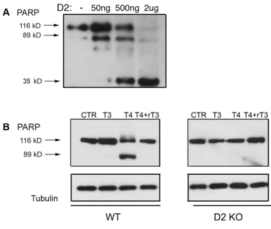
In muscle stem cells (MuSC), type 2 and type 3 deiodinases are active and differentially expressed along cell maturation. During the proliferating phase, myoblasts overexpress D3 to achieve a low intracellular TH signaling, essential to permit the correct cell amplification. Active D2 is still present in proliferating myoblasts but its activity is counteracted by the abundant D3 expression. Indeed, previous reports showed that D3-depletion lead to apoptosis of activated proliferating myoblasts due to temporary TH excess. To assess whether forced amounts of D2 might disrupt the balance of intracellular TH and cause apoptosis, we transfected proliferating myoblasts with D2 under normal serum conditions. D2 overexpression caused dose-dependent apoptosis, as demonstrated by PARP cleavage, thus confirming that cell death depends on a D2-dependent increase in intracellular T3 (Figure 1A). To confirm the upstream role of intracellular D2-mediated T4 activation, proliferating myoblasts were treated with T4 combined to rT3. The rT3 inhibition of D2 activity nullified the T4-induced apoptosis. Moreover, MuSC derived from Dio2-null mice resulted to be insensitive to T4 (Figure 1B).
Figure 1. D2 expression causes a dose-dependent apoptosis in proliferating myoblasts.
A. Proliferating MuSC were transiently transfected with D2-WT plasmid at increasing doses (50 ng, 500 ng and 2 µg). 48 hours later, cell lysates were collected and PARP cleavage was evaluated using western blot analysis. Cells transfected with cytomegalovirus served as negative control; tubulin was used as loading control.
B. Proliferating MuSC from WT and D2KO mice were treated with 30 nM T3, 30 nM T4 and 30 nM T4+rT3, respectively. After 24 hours, total proteins were harvested and apoptosis induction was detected by western blot showing the cleavage of PARP. Tubulin served as loading control.
5.2 The TH-induced apoptosis is dependent on TH receptor action
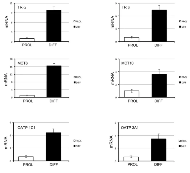
To allow a proper expansion, MuSC require lower than circulating T3. To this aim, they down-regulate D2 to very low levels. Therefore, we next evaluated the expression profile of the other TH signaling cellular regulators in proliferating MuSC. We found that TH transporters and TH receptors show markedly reduced expression during MuSC proliferation when compared with the differentiating counterpart (Figure 2). To determine whether the TH-induced apoptosis in proliferating MuSC was directly dependent on the action of thyroid hormone receptors (TR), we induced the expression of a TR dominant-negative mutant. As showed in Figure 3A, the expression of a TR dominant-negative mutant made the proliferating myoblasts insensitive to TH, thus showing that the D2-derived T3 directly acts on TR.
Figure 2. TH signaling is down-regulated during MuSC proliferation.
Thyroid receptor α and β (TR α, TR β) and thyroid hormone transporters MCT 8, MCT 10, OATP 1C1 and OATP 3A1 gene expression was measured by RT-PCR in proliferating (PROL) and differentiating (DIFF) MuSC.
5.3 The T4 inflow in proliferating myoblasts is mediated by transporters OATP 1C1 and 3A1
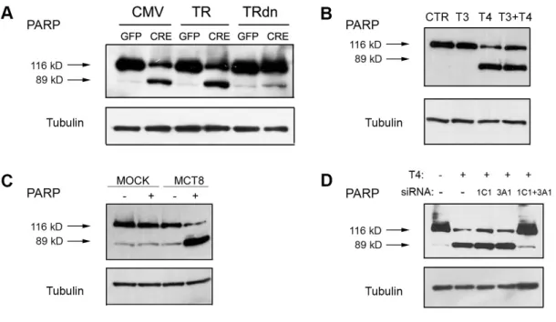
We examined the ability of the two major TH, namely T3 and T4, in proliferating myoblasts death induction. Surprisingly, proliferating myoblasts were insensitive to supraphysiological T3 concentrations, while the treatment with T4 alone resulted in massive cell apoptosis (Figure 3B). We speculated that the T3 was not able to induce apoptosis due to its inability to cross the cell membrane. To test this hypothesis, we transfected proliferating MuSC with a well–known T3 plasma transporter, MCT 8. Upon transporter transfection, proliferating cells became sensitive to T3 and rapidly apoptosed after T3 treatment (Figure 3C). Furthermore, we speculated that the T4 was effective due to the action of a specific T4 transporter expressed in proliferating cells. Global gene expression analysis of proliferating and differentiated MuSC indicated that these cells express specifically two putative T4 transporters. To shed light on this issue and on the relative contribution of each of them to the T4 action, we used siRNA to specifically knock down OATP 3A1 and 1C1. When we subjected cells to a contemporary double specific OATPs 3A1 and 1C1 knock down, proliferating MSC became resistant to T4 (Figure 3D). Overall, these experiments indicate that the exposure to supraphysiological T4 doses in proliferating myoblasts lead to cell apoptosis thanks to the T4 inflow through OATPs 3A1 and 1C1, and the subsequent T4 to T3 conversion mediated by D2.
Figure 3. MuSC apoptosis relies on T4 inflow through OATPs 1C1 and 3A4 and depends on D2-derived T3 action on TH receptor.
A. Proliferating MuSC were transiently transfected with TR dominant negative plasmid and following 24 hours were incubated with 30 nM T4. Following 48 hours, cells lysates were collected and PARP cleavage was evaluated using western blot analysis. CMV transfected cells served as negative control; tubulin was used as loading control. B. Proliferating MuSC were treated with 30 nM T3, 30 nM T4 and 30 nM T3+T4, respectively. After 24 hours, cells lysates were harvested and total proteins were subjected to western blot analysis of PARP cleavage. The detection of tubulin was used as loading control.
C. Proliferating MuSC were transiently transfected with MCT 8 plasmid and following 24 hours were incubated with 30 nM T3. Following 48 hours, cells lysates were collected and PARP cleavage was evaluated using western blot analysis. Mock transfected cells served as negative control; tubulin was used as loading control.
D. Proliferating MuSC were subjected to a specific siRNA knock down of OATP 1C1, OATP 3A1 and OATP 1C1 + OATP 3A1, respectively. Following 24 hours, cells were treated with 30 nM T4 and were harvested after 48 hours. Total proteins were subjected to western blot analysis of PARP cleavage; tubulin served as loading control.
5.4 Endogenous D2 protein of wild-type and mutated isoforms are dynamically located in the endoplasmic reticulum
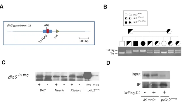
In order to assess whether the T92A polymorphism modifies important enzymatic parameters, we investigated the localization of the wild type protein in its physiological context. So far, detection of D2 protein and its subcellular localization in vivo has been not possible due to the absence of functional antibodies. To overcome this problem, we generated a knock-in 3xFlag-D2 mouse, carrying three repetitions of a FLAG epitope at the NH2-terminus of the otherwise wild-type enzyme (Figure 4A). We confirmed the effective expression of the D2-3xFlag protein by Western blot and IP-WB analysis (Figure 4C and D). We used MuSC as a model to address the intracellular localization of the D2 protein. Activated MuSC isolated by FACS from the D2-3xFlag mouse were grown in culture and D2 expression was evaluated by co-immunofluorescence analysis. Proliferating cells expressed low levels of D2, mostly localized in the cytoplasm, as shown by the double Flag-MyoD staining (Figure 5A). D2 partially colocalized with the endoplasmic reticulum (ER) marker calreticulin in early differentiating MuSC. Interestingly, D2 was also present in cytoplasmic vesicles other than ER (i.e. without calreticulin), but not in the Golgi. During differentiation, D2 localization changed in parallel with its increased expression. In late differentiated myoblasts, D2 expression shifted to perinuclear space (Figure 5B).
To determine whether the D2-Ala mutant isoform shared the same subcellular localization with the wild type D2, we transfected MuSC from Dio2-null mice with D2-WT or D2-Ala plasmid and evaluated its cellular localization. Immunofluorescence staining showed that both transfected wild type and mutant isoforms localized to the cytoplasmic, mainly to the ER (Figure 5C). This expression pattern resembles the endogenous protein localization in the same cells. In conclusion, in vivo D2 tracing
showed that D2 is dynamically expressed in the ER during proliferation and in the ER-perinuclear territory during cell differentiation. Importantly, this pattern is identical between wild type and the mutant D2-Ala isoform.
Figure 4. Generation of knock-in 3xFlag-D2 mouse.
A. Schematic representation of the modified Dio2 locus gene. Three repetitions of the Flag coding sequence were inserted after the ATG codon of the mouse Dio2 gene. B. The effective presence of the modified Dio2 gene was confirmed by PCR analysis. C. Western blot analysis of tissue protein extracts from different tissues of 3xFlag-D2 (+) or wild type (-) mice.
D. Immunoprecipitation with anti-Flag antibody and western blot analysis of protein extracts from muscles of 3xFlag-D2 (+) or wild type (-) mice.
Figure 5. Intracellular localization of the D2 protein.
A, B. D2 localizes in the endothelial reticulum and cytoplasmic vesicles in proliferating and differentiated MuSC. D2 localization was evaluated by immunofluorescence microscopy in FACS-isolated MuSC cultured in proliferative and differentiative conditions at different time points. Early proliferative cells were fixed at 12 hours after plating and late proliferative cells were fixed 24 hours after plating (A). Differentiation was induced 48 hours following plating by replacing growing medium with differentiation medium and collected at 12 (early) and 48 hours following medium replacement (B). Proliferating cells were co-stained with Flag/MyoD antibodies and differentiated cells with Flag/Calreticulin antibodies, as indicated.
C. Immunofluorescence staining for D2-WT and D2-Ala-Flag (red) and calcireticulin (green) and merged images in transiently transfected proliferating MuSC from D2KO mice.
5.5 D2 Ala-mutant protein has a longer stability than wild-type
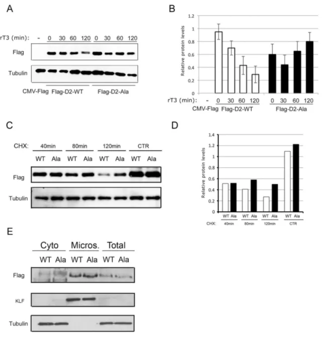
The SNP T92A falls in a specific 18-amino-acid large loop critical for D2 recognition by the WSB-1 subunit of E3 ubiquitin ligase. To investigate the consequences on D2 stability resulting from the threonine-to-alanine substitution, we compared the protein stability of the wild-type and mutant isoforms. We evaluated the D2-Ala protein clearance rate in MuSC following rT3 administration, which potently accelerate the proteolytic degradation of D2. The D2-Ala protein proved to be even more stable under these conditions, showing a longer half-life than wild-type D2 (Figure 6A and B). Similarly, when the protein biosynthesis inhibitor cycloheximide was added to D2-Ala and D2-WT transfected MuSC, D2-Ala clearance was lower than that of D2-WT (Figure 6C and D). Cells fractionation showed no differences between the wild-type and the mutant isoform subcellular localization, both present in microsomes (Figure 6E).
Figure 6. D2-Ala protein has a longer stability.
A. Time course analysis by western blot of D2-WT and D2-Ala after 30, 60 and 120 minutes rT3 incubation, respectively, 24 hours after transient transfection in proliferating MuSC from D2KO mice. D2 was determined using anti-flag antibody; tubulin served as loading control.
B. Time-course western blot results were normalized to tubulin as control, and densitometric quantification of bands was determined with the ImageJ software.
C. D2-WT and D2-Ala were transiently transfected in proliferating MuSC from D2KO mice. Twenty-four hours later, cells were incubated for 40, 80 and 120 minutes with cycloheximide. D2 was determined using anti-flag antibody; tubulin served as loading control.
D. Time-course western blot results were normalized to tubulin as control, and bands were densitometrically quantified with the ImageJ software.
E. D2-WT and D2-Ala were transiently transfected in proliferating MuSC from D2KO mice. After 24 hours, cells were fractionated into cytosolic (cyto) and ER-containing microsomal (micros) fractions. The presence of D2 in the two compartments was determined by western blot analysis with anti-flag antibody. KLF and tubulin were used as loading controls.
5.6 D2-Ala mutant is less efficient than D2-WT in converting T4 into T3 in muscle stem cells and pituitary thyrotrophs
5.6.1 Muscle stem cells model
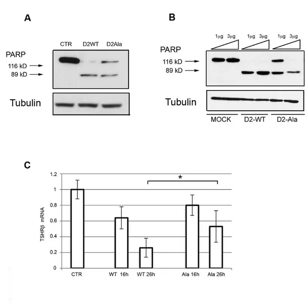
The D2-dependent proapoptotic effect in proliferating myoblasts suggested that this cellular model might be a biological sensor with which to quantitatively assess D2-mediated T4 to T3 conversion. Consequently, we asked whether the D2-Ala mutant isoform was equally able to produce T3 than D2-WT, and thus to induce apoptosis. To address this question, we transfected proliferating myoblasts from Dio2-null mice with D2-WT and D2-Ala. Strikingly, cells expressing the D2-Ala mutant isoform showed markedly reduced apoptotic commitment, which suggests a lower intracellular T4 to T3 conversion by D2-Ala enzyme (Figure 7A). The effect of D2-Ala on apoptosis was dose dependent, since transfection with high levels of D2-Ala plasmid induced a response similar to obtained with D2-WT (Figure 7B). These data demonstrate that, despite a normal subcellular localization and an even slightly higher protein stability, the D2-Ala mutant is less efficient than D2-WT in converting T4 into T3 in muscle cells in vivo.
5.6.2 Thyrotrophic pituitary cells model
To further analyze the the catalytic efficiency of D2-Ala protein in a different biological setting where D2-mediated T3 production provokes critical consequences, we transfected D2-Ala in primary cultures of pituitary thyrotrophs from Dio2-null mice. Thyrotrophs were cultured in TH-free serum and thereafter treated with T4, to determine the efficiency of D2-Ala in blocking TSH synthesis. The response to T4-induced TSH suppression was significantly (P <0.05) lower in thyrotrophs expressing D2-Ala than in D2-WT-transfected thyrotrophs (Figure 7C). TSH synthesis directly
responds to T3 inhibitory action, that directly derived from transfected D2. Therefore, the reduced TSH suppression in thyrotrophs expressing D2-Ala again demonstrates an impairment in T4 to T3 conversion by the mutant enzyme.
Figure 7. D2-Ala is responsible of a reduced T4 proapoptotic effect on MuSC and a decreased TH inhibitory feedback on pituitary thyrotrophs.
A. Western blot analysis of PARP cleavage in proliferating MuSC from D2KO mice 48 hours after transfection with D2-WT and D2-Ala, and 24 hours 30 nM T4 incubation, respectively. Tubulin served as loading control.
B. Dose response effect of D2-Ala on apoptosis of proliferating MuSC. MuSC from D2KO mice were transiently transfected with 1µg and 3µg of D2-WT and D2-Ala plasmid, respectively. 24 hours later, cells were treated with 30 nM T4 and, 48 hours
later, total proteins were harvested for western blot analysis of PARP cleavage. Tubulin served as loading control.
C. Time course reverse transcription PCR analysis of β-TSH subunit expression in thyrotrophs from D2KO mice, cultured in charcoal-stripped bovine serum and transiently transfected with D2-WT and D2-Ala, 16 and 26 hours after 30 nM T4 incubation, respectively. Non-treated thyrotrophs served as control.
5.7 Comparison of presurgical and postsurgical thyroid hormone levels in patients submitted to total thyroidectomy
As shown in Table 1, there was no significant difference between presurgery and postsurgery serum TSH levels in any subject (by Wilcoxon test for paired data), which is consistent with the inclusion criteria and the aim of the study. Despite similar TSH levels, mean FT4 levels were slightly but significantly higher (p<0.0001) after surgery (10.8 ± 2.2 pg/ml) than before surgery (9.8 ± 2.6 pg/ml). On the contrary, postsurgical serum FT3 was significantly lower (p<0.0001) than presurgical FT3 (3.0 ± 0.35 pg/ml vs 3.3 ± 0.43 pg/ml). Consequently, a significantly lower FT3/FT4 ratio was observed in the postsurgical evaluation (0.28 ± 0.06 vs 0.36 ± 0.09; P <0.0001). According to the cutoff defined in the “Patients and Methods” section, patients were classified as having “reduced FT3” when postsurgical FT3 levels were at least 0.5 pg/mL lower than presurgical FT3 values. As shown in Figure 8, postsurgery FT3 levels were lower in a cohort of 48/140 patients (“reduced FT3”, 34.3%), whereas the remaining 92 patients ("unchanged FT3 ", 65.7%) had serum FT3 levels identical to the presurgical levels. Notably, no differences in sex, age, TSH levels, FT4 levels, LT4 dose/day and LT4 dose/kg between the two groups of "reduced FT3" and "unchanged FT3" were present (Table 2).
TSH FT3 FT4 (mUI/L) (pg/ml) (pg/ml) Pre-surgical Mean±SD 0.93±0.55 3.3±0.43 9.8±2.6 Range 0.10-3.4 2.4-5.4 5.5-17.7 Median 0.88 3.3 9.3 Post-surgical Mean±SD 0.91±0.59 3.0±0.35 10.8±2.2 Range 0.01-3.6 1.6-4.1 5.9-18.3 Median 0.89 2.9 10.7 p 0.27 <0.001 <0.001
pre versus post
Table 1.
Comparison of presurgical and postsurgical thyroid hormone levels in patients submitted to total thyroidectomy.
Figure 8.
Pre-surgical and post-surgical FT3 levels in the “reduced FT3” group (left panel) and in the “unchanged FT3” (right panel) group.
p<0.000
1
“FT3 reduced” (n=48)P=0.3
4
“FT3 unchanged” (n=92)FT3 reduced (n=48) FT3 Unchanged (n=92) P value Post-surgical FT3 (pg/ml) Mean ± SD Range Median 2.80 ± 0.31 1.57 - 3.9 2.8 3.1 ± 0.32 2.6 - 4.1 3.1 <0.0001 Post-surgical FT4 (pg/ml) Mean ± SD Range Median 10.53 ± 2.25 5.0 - 16.9 10.0 11.0 ± 2.1 7.1 - 18.1 10.9 0.17 Post-surgical TSH (mUI/L) Mean ± SD Range Median 0.98 ± 0.53 0.13-2.23 0.98 0.87 ± 0.62 0.01 - 3.62 0.74 0.12 Sex Males (n, %) Females (n, %) 15 (38.4%) 33 (32.6%) 24 (61.6%) 68 (67.4%) 0.55 Age (yrs) Mean ± SD Range Median 55.1 ± 16.0 24 - 84 55.5 54.0 ± 14.4 18 - 84 57 0.67 LT4 daily dose (mcg) Mean ± SD Range Median 111.3 ± 30.6 15 - 175 112.5 116.8 ± 25.6 62 - 175 112.5 0.27 Pro-Kg LT4 dose (mcg) Mean ± SD Range Median 1.48 ± 0.36 0.2 - 2.36 1.5 1.56 ± 0.33 0.9 - 2.8 1.5 0.17 Table 2.
Clinical, demographical and biochemical data in “reduced FT3” and “unchanged FT3” patients.
5.8 Correlation between DIO2 Thr92Ala polymorphism and serum T3 levels in athyreotic patients
To investigate whether the lower postsurgical FT3 level observed in a subset of patients was correlated with the T92A DIO2 gene polymorphism, we analyzed the entire coding region and the 5'-UTR regions of DIO2 gene in 102 samples (72.8% of enrolled patients) and identified the T92A polymorphism in 63.7% of subjects. In particular, 37/102 (36.3%) patients were homozygous wild-type (Thr/Thr), 52/107 (51.0%) were heterozygous (Ala/Thr) and 13/102 (12.7%) were mutant homozygous (Ala/Ala).
FT4 levels were significantly higher after surgery than before surgery in the Thr/Thr group (11.1 ± 2.4 pg/mL vs 9.5 ± 2.2 pg/mL, P = 0.001) and in the Thr/Ala group (10.9 ± 2.0 pg/mL vs 9.9 ± 2.8 pg/mL, P = 0.02), and slightly, but not significantly higher in the Ala/Ala group (10.1 ± 1.9 pg/mL vs 9.4 ± 2.6 pg/mL, P = 0.38). As shown in Table 3, mean postsurgery FT3 levels were significantly lower in patients carrying the mutated allele(s) than in wild-type patients (P <0.0001 in the Thr/Ala group; P = 0.01 in the Ala/Ala group). Moreover, in wild-type patients FT3 postsurgical levels were similar to presurgery levels (P = 0.097).
To verify these results, we compared pre- and postsurgical changes in FT3 levels among the three genotype groups. FT3 changes (expressed as the difference between pre- and postsurgical FT3 levels) were significantly higher in the Thr/Ala group (P <0.05; 95% CI, 0.06038 to 0.5459) and in the Ala/Ala group (P <0.05; 95% CI, 0.004411 to 0.7323) than in the Thr/Thr group (Figure 9). On the contrary, TSH, FT4, and T3/T4 ratio changes did not differ among the three genotype groups.
Pre-surgical FT3 Post-surgical FT3 p (mUI/L) (pg/ml) WT Mean±SD 3.2±0.35 3.1±0.36 Range 2.4-3.9 2.6-4.1 0.097 Median 3.2 3.1 Thr/Ala Mean±SD 3.4±0.52 3.0±0.35 Range 2.7-5.4 1.6-4.1 <0.0001 Median 3.3 2.9 Ala/Ala Mean±SD 3.4±0.37 2.9±0.34 Range 2.9-4.2 2.5-3.6 0.01 Median 3.4 2.9 Table 3.
Figure 9.
Differences between pre-surgical and post-surgical serum FT3 levels in the three genotype groups.
6 Discussion
In this study, we provide the first in vivo biological evidence that the D2-Ala enzyme is less enzymatically efficient than D2-WT and reduces T3 production in two relevant D2-dependent tissues, i.e., skeletal muscle and pituitary thyrotrophs. Previous studies analyzed the D2-Ala enzymatic activity, but results are likely to be affected by intrinsic artifacts of in vitro assay (67, 73). To overcome the limitations in D2 in vitro assay, we used two in vivo systems in intact cells that more closely resemble the in vivo situation. Using this approach, we demonstrate that the D2-Ala mutant is less efficient in converting T4 into T3. In addiction, we demonstrate the inability of patients carrying X/Ala D2 genotype subjected to thyroidectomy to restore adequate T3 levels when submitted to LT4 replacement therapy. Former studies analyzed thyroid hormonal levels in athyreotic patients genotyped for DIO2 T92A and their respective LT4 intake need, but provided conflicting results (13, 63), mainly due to lack of presurgical hormonal values and to heterogeneity of study population. The strength of our analysis lies in the knowledge of TSH levels prior to thyroidectomy and the similarity of pre- and postsurgical TSH value for each patient. Thanks to that, we were able to evaluate putative changes in circulating T4 and T3 levels in the same feedback perception from HPT axis before and after thyroidectomy.
In athyreotic patients on LT4 substitutive therapy, both circulating and intracellular T3 levels entirely depend on the deiodinase-mediated T4 to T3 conversion (67). However, in approximately 20% of athyreotic patients, LT4 did not ensure physiological T3 levels unless suppressing TSH (75), although other studies were controversial on this aspect (75, 76). In view of these controversies, we compared
patients on LT4 and with similar TSH levels before and after surgery. We found that postoperative FT3 levels were significantly reduced in a subgroup of patients (34.3%), even though their TSH levels were in the normal range and similar to those observed prior to thyroidectomy. To investigate whether the lower postsurgical FT3 in this subset of patients was related to DIO2 gene mutations, we analyzed the entire coding region and the 5'-UTR of the DIO2 gene in 102 samples (72.8% of enrolled patients). The percentage of reduced postsurgery FT3 levels was directly correlated with the presence and severity of the T92A polymorphism: 58.3% in homozygous (Ala/Ala) patients vs 36.5% in heterozygous (Thr/Ala) subjects. Postoperative FT4 levels did not differ among the genotype groups.
In our study, we show that pituitary D2-Ala expression determines reduced TH suppressive feedback due to impaired enzymatic activity. Since serum TSH did not differed before and after thyroidectomy in our cohort of 140 patients, it is somehow surprisingly that serum FT3 levels drop down in X/Ala genotype carriers without a counterbalancing increase in TSH levels, given equal LT4 daily dose assumption. Indeed, median post-thyroidectomy TSH in “FT3 unchanged” group was 0.74 mUI/L while was higher in “FT3 reduced” group, i.e. 0.98 mUI/L, although these differences did not reached statistical significance (p=0.12). This could be due to the low number of patients enrolled, as supported by the study of Torlontano et al. (13), which showed an increased need of T4 doses to suppress TSH in thyroidectomized patients carrying the D2Ala/Ala mutation.
It is reasonable to speculate that in “FT3 reduced” patients, restoration of presurgical FT3 value would be obtained at the expense of a higher LT4 intake and a subsequently lower TSH level. Nevertheless, the maintenance of suppressed TSH levels in athyreotic patients is avoided in the vast majority of hypothyroid patients and is not
recommended in order to normalize serum FT3 (64). Moreover, it is recognized that if presurgical T3 levels are chosen as therapeutic targets, FT4 levels above the reference range are often reached (64). Postsurgical FT4 in “FT3 reduced” group did not showed differences from the “FT3 unchanged” group. This is not surprisingly, since in a large series by Gullo et al. (75) of athyreotic patients on LT4 monotherapy who were compared to TSH-matched euthyroid controls, the finding of FT3 levels below reference ranges was not necessarily related to an equal gain in serum FT4. In this context, normalization of low FT3 levels in athyreotic patients with higher doses of LT4 could induce an increase of peripheral D2 degradation and worsen that way hypothyroidism in D2-expressing tissues.
The evidences of our study strongly support the need of T3+T4 combined therapy to restore central and peripheral euthyroidism in the subgroup of athyreotic patients who don’t reach normalization of FT3 values with LT4 monotherapy. The relationship between low FT3 values and D2-Ala expression in thyroidectomized LT4 treated patients is supported by the demonstration of a reduced D2-Ala enzymatic activity in a biological assay model.
A former study founded D2 velocity to be reduced in muscle samples from patients homozygous for Ala allele (56). Nevertheless, this evidence was not confirmed in other and more recent investigations, suggesting that this results could be related to an artifact intrinsic to the classical D2 activity in vitro assay (67). Indeed, with respect to muscle tissue, D2 activity measurement might be overestimated due to cell homogenates release of iodide that do not correspond to a T4 to T3 enzyme conversion. In order to overcome the limitations of D2 in vitro assay, it has been suggested that measurements of D2 in intact cells may give more reliable estimates of the in vivo
The reliability of our cellular model has been validated by the analysis of wild type D2 in the context of new generated D2-3x-flag mouse. By the use of D2-3x-flag mouse, we were surprised to observe that functional D2 is even so expressed by a cell who does need elevated D3 amounts to guarantee its survival. We demonstrated that D2-WT expression in myoblasts is dynamically regulated in the endoplasmic reticulum and in perinuclear territory, respectively, during cell proliferation and cell differentiation. Significantly, this pattern is maintained by D2-Ala isoform when myoblasts are transfected with the mutant protein. A previous report by McAninch et al. (61) analyzed D2-Ala expression in HEK-293 transfected cells and revealed consistent amount of mutated protein in the Golgi. Our myoblasts model found instead D2-Ala to be present in a fashion that similarly traces that of wild type protein, and no amount was present in Golgi apparatus. The main difference between these two analyses is that we used a physiological model of endogenous protein expression to assess D2 cellular compartmentalization, following the D2-WT and D2-Ala protein expression along different phases of myoblast maturation.
7 Conclusions
We demonstrated for the first time that the T92A DIO2 polymorphism translates into a protein with increased half-life, normal subcellular localization but functionally reduced enzymatic efficiency in T4 to T3 conversion. Patients carrying the T92A genotype undergo a drop in T3 levels after thyroidectomy, not compensated by LT4 replacement therapy. The lower plasmatic T3 level associated with the X/Ala D2 genotype suggests that a segment of these patients do not respond optimally to standard LT4 replacement therapy. Therefore, our study might support the use of combined T4+T3 therapy in the subgroup with low serum T3 that carries the T92A DIO2 polymorphism.
8 References
1. Fekete, C., and Lechan, R.M. 2014. Central regulation of hypothalamic-pituitary-thyroid axis under physiological and pathophysiological conditions. Endocr Rev 35:159-194.
2. Gereben, B., Zavacki, A.M., Ribich, S., Kim, B.W., Huang, S.A., Simonides, W.S., Zeold, A., and Bianco, A.C. 2008. Cellular and molecular basis of deiodinase-regulated thyroid hormone signaling. Endocr Rev 29:898-938.
3. Williams, G.R., and Bassett, J.H. 2011. Deiodinases: the balance of thyroid hormone: local control of thyroid hormone action: role of type 2 deiodinase. J Endocrinol 209:261-272.
4. Maia, A.L., Goemann, I.M., Meyer, E.L., and Wajner, S.M. 2011. Deiodinases: the balance of thyroid hormone: type 1 iodothyronine deiodinase in human physiology and disease. J Endocrinol 209:283-297.
5. Arrojo, E.D.R., Fonseca, T.L., Werneck-de-Castro, J.P., and Bianco, A.C. 2013. Role of the type 2 iodothyronine deiodinase (D2) in the control of thyroid hormone signaling. Biochim Biophys Acta 1830:3956-3964.
6. Dentice, M., and Salvatore, D. 2011. Deiodinases: the balance of thyroid hormone: local impact of thyroid hormone inactivation. J Endocrinol 209:273-282.
7. Ciavardelli, D., Bellomo, M., Crescimanno, C., and Vella, V. 2014. Type 3 deiodinase: role in cancer growth, stemness, and metabolism. Front Endocrinol (Lausanne) 5:215.
8. Hoermann, R., Midgley, J.E.M., Larisch, R., and Dietrich, J.W. 2017. Recent Advances in Thyroid Hormone Regulation: Toward a New Paradigm for Optimal Diagnosis and Treatment. Front Endocrinol (Lausanne) 8:364.
9. Bianco, A.C., and Kim, B.S. 2018. Pathophysiological relevance of deiodinase polymorphism. Curr Opin Endocrinol Diabetes Obes 25:341-346.
10. Mentuccia, D., Thomas, M.J., Coppotelli, G., Reinhart, L.J., Mitchell, B.D., Shuldiner, A.R., and Celi, F.S. 2005. The Thr92Ala deiodinase type 2 (DIO2) variant is not associated with type 2 diabetes or indices of insulin resistance in the old order of Amish. Thyroid 15:1223-1227.
11. Grarup, N., Andersen, M.K., Andreasen, C.H., Albrechtsen, A., Borch-Johnsen, K., Jorgensen, T., Auwerx, J., Schmitz, O., Hansen, T., and Pedersen, O. 2007. Studies of the common DIO2 Thr92Ala polymorphism and metabolic phenotypes in 7342 Danish white subjects. J Clin Endocrinol Metab 92:363-366.
12. Maia, A.L., Dupuis, J., Manning, A., Liu, C., Meigs, J.B., Cupples, L.A., Larsen, P.R., and Fox, C.S. 2007. The type 2 deiodinase (DIO2) A/G polymorphism is not associated with glycemic traits: the Framingham Heart Study. Thyroid 17:199-202. 13. Torlontano, M., Durante, C., Torrente, I., Crocetti, U., Augello, G., Ronga, G.,
Montesano, T., Travascio, L., Verrienti, A., Bruno, R., et al. 2008. Type 2 deiodinase polymorphism (threonine 92 alanine) predicts L-thyroxine dose to achieve target thyrotropin levels in thyroidectomized patients. J Clin Endocrinol Metab 93:910-913. 14. Butler, P.W., Smith, S.M., Linderman, J.D., Brychta, R.J., Alberobello, A.T., Dubaz,
O.M., Luzon, J.A., Skarulis, M.C., Cochran, C.S., Wesley, R.A., et al. 2010. The Thr92Ala 5' type 2 deiodinase gene polymorphism is associated with a delayed triiodothyronine secretion in response to the thyrotropin-releasing hormone-stimulation test: a pharmacogenomic study. Thyroid 20:1407-1412.
15. Panicker, V., Saravanan, P., Vaidya, B., Evans, J., Hattersley, A.T., Frayling, T.M., and Dayan, C.M. 2009. Common variation in the DIO2 gene predicts baseline
psychological well-being and response to combination thyroxine plus triiodothyronine therapy in hypothyroid patients. J Clin Endocrinol Metab 94:1623-1629.
16. Wiersinga, W.M. 2017. THERAPY OF ENDOCRINE DISEASE: T4 + T3 combination therapy: is there a true effect? Eur J Endocrinol 177:R287-R296.
17. Ortiga-Carvalho, T.M., Sidhaye, A.R., and Wondisford, F.E. 2014. Thyroid hormone receptors and resistance to thyroid hormone disorders. Nat Rev Endocrinol 10:582-591.
18. Bernal, J., Guadano-Ferraz, A., and Morte, B. 2015. Thyroid hormone transporters-functions and clinical implications. Nat Rev Endocrinol 11:690.
19. Visser, W.E., Friesema, E.C., and Visser, T.J. 2011. Minireview: thyroid hormone transporters: the knowns and the unknowns. Mol Endocrinol 25:1-14.
20. van der Deure, W.M., Peeters, R.P., and Visser, T.J. 2010. Molecular aspects of thyroid hormone transporters, including MCT8, MCT10, and OATPs, and the effects of genetic variation in these transporters. J Mol Endocrinol 44:1-11.
21. Roef, G.L., Rietzschel, E.R., De Meyer, T., Bekaert, S., De Buyzere, M.L., Van daele, C., Toye, K., Kaufman, J.M., and Taes, Y.E. 2013. Associations between single nucleotide polymorphisms in thyroid hormone transporter genes (MCT8, MCT10 and OATP1C1) and circulating thyroid hormones. Clin Chim Acta 425:227-232.
22. Ferrara, A.M., Liao, X.H., Gil-Ibanez, P., Marcinkowski, T., Bernal, J., Weiss, R.E., Dumitrescu, A.M., and Refetoff, S. 2013. Changes in thyroid status during perinatal development of MCT8-deficient male mice. Endocrinology 154:2533-2541.
23. Lee, J.Y., Kim, M.J., Deliyanti, D., Azari, M.F., Rossello, F., Costin, A., Ramm, G., Stanley, E.G., Elefanty, A.G., Wilkinson-Berka, J.L., et al. 2017. Overcoming Monocarboxylate Transporter 8 (MCT8)-Deficiency to Promote Human Oligodendrocyte Differentiation and Myelination. EBioMedicine 25:122-135.
24. Visser, W.E., Friesema, E.C., Jansen, J., and Visser, T.J. 2007. Thyroid hormone transport by monocarboxylate transporters. Best Pract Res Clin Endocrinol Metab 21:223-236.
25. Hagenbuch, B. 2007. Cellular entry of thyroid hormones by organic anion transporting polypeptides. Best Pract Res Clin Endocrinol Metab 21:209-221.
26. Mayerl, S., Visser, T.J., Darras, V.M., Horn, S., and Heuer, H. 2012. Impact of Oatp1c1 deficiency on thyroid hormone metabolism and action in the mouse brain. Endocrinology 153:1528-1537.
27. Kim, B.W., Zavacki, A.M., Curcio-Morelli, C., Dentice, M., Harney, J.W., Larsen, P.R., and Bianco, A.C. 2003. Endoplasmic reticulum-associated degradation of the human type 2 iodothyronine deiodinase (D2) is mediated via an association between mammalian UBC7 and the carboxyl region of D2. Mol Endocrinol 17:2603-2612. 28. Werneck de Castro, J.P., Fonseca, T.L., Ueta, C.B., McAninch, E.A., Abdalla, S.,
Wittmann, G., Lechan, R.M., Gereben, B., and Bianco, A.C. 2015. Differences in hypothalamic type 2 deiodinase ubiquitination explain localized sensitivity to thyroxine. J Clin Invest 125:769-781.
29. Murakami, M., Araki, O., Hosoi, Y., Kamiya, Y., Morimura, T., Ogiwara, T., Mizuma, H., and Mori, M. 2001. Expression and regulation of type II iodothyronine deiodinase in human thyroid gland. Endocrinology 142:2961-2967.
30. Sagar, G.D., Gereben, B., Callebaut, I., Mornon, J.P., Zeold, A., Curcio-Morelli, C., Harney, J.W., Luongo, C., Mulcahey, M.A., Larsen, P.R., et al. 2008. The thyroid hormone-inactivating deiodinase functions as a homodimer. Mol Endocrinol 22:1382-1393.
32. Bianco, A.C., Salvatore, D., Gereben, B., Berry, M.J., and Larsen, P.R. 2002. Biochemistry, cellular and molecular biology, and physiological roles of the iodothyronine selenodeiodinases. Endocr Rev 23:38-89.
33. Singh, B.K., Sinha, R.A., and Yen, P.M. 2018. Novel Transcriptional Mechanisms for Regulating Metabolism by Thyroid Hormone. Int J Mol Sci 19.
34. Cheng, S.Y., Leonard, J.L., and Davis, P.J. 2010. Molecular aspects of thyroid hormone actions. Endocr Rev 31:139-170.
35. Cheng, S.Y. 2005. Thyroid hormone receptor mutations and disease: beyond thyroid hormone resistance. Trends Endocrinol Metab 16:176-182.
36. Darras, V.M., Houbrechts, A.M., and Van Herck, S.L. 2015. Intracellular thyroid hormone metabolism as a local regulator of nuclear thyroid hormone receptor-mediated impact on vertebrate development. Biochim Biophys Acta 1849:130-141. 37. Astapova, I. 2016. Role of co-regulators in metabolic and transcriptional actions of
thyroid hormone. J Mol Endocrinol 56:73-97.
38. Salvatore, D., Simonides, W.S., Dentice, M., Zavacki, A.M., and Larsen, P.R. 2014. Thyroid hormones and skeletal muscle--new insights and potential implications. Nat Rev Endocrinol 10:206-214.
39. Simonides, W.S., and van Hardeveld, C. 2008. Thyroid hormone as a determinant of metabolic and contractile phenotype of skeletal muscle. Thyroid 18:205-216.
40. Bloise, F.F., Cordeiro, A., and Ortiga-Carvalho, T.M. 2018. Role of thyroid hormone in skeletal muscle physiology. J Endocrinol 236:R57-R68.
41. Seale, P., and Rudnicki, M.A. 2000. A new look at the origin, function, and "stem-cell" status of muscle satellite cells. Dev Biol 218:115-124.
42. Buckingham, M., and Relaix, F. 2015. PAX3 and PAX7 as upstream regulators of myogenesis. Semin Cell Dev Biol 44:115-125.
43. Fukada, S., Ma, Y., Ohtani, T., Watanabe, Y., Murakami, S., and Yamaguchi, M. 2013. Isolation, characterization, and molecular regulation of muscle stem cells. Front Physiol 4:317.
44. Seale, P., Sabourin, L.A., Girgis-Gabardo, A., Mansouri, A., Gruss, P., and Rudnicki, M.A. 2000. Pax7 is required for the specification of myogenic satellite cells. Cell 102:777-786.
45. Muscat, G.E., Downes, M., and Dowhan, D.H. 1995. Regulation of vertebrate muscle differentiation by thyroid hormone: the role of the myoD gene family. Bioessays 17:211-218.
46. Wang, Y.X., and Rudnicki, M.A. 2012. Satellite cells, the engines of muscle repair. Nat Rev Mol Cell Biol 13:127-133.
47. Marsili, A., Tang, D., Harney, J.W., Singh, P., Zavacki, A.M., Dentice, M., Salvatore, D., and Larsen, P.R. 2011. Type II iodothyronine deiodinase provides intracellular 3,5,3'-triiodothyronine to normal and regenerating mouse skeletal muscle. Am J Physiol Endocrinol Metab 301:E818-824.
48. Dentice, M., Marsili, A., Zavacki, A., Larsen, P.R., and Salvatore, D. 2013. The deiodinases and the control of intracellular thyroid hormone signaling during cellular differentiation. Biochim Biophys Acta 1830:3937-3945.
49. Clement, K., Viguerie, N., Diehn, M., Alizadeh, A., Barbe, P., Thalamas, C., Storey, J.D., Brown, P.O., Barsh, G.S., and Langin, D. 2002. In vivo regulation of human skeletal muscle gene expression by thyroid hormone. Genome Res 12:281-291.
50. Soukup, T., and Smerdu, V. 2015. Effect of altered innervation and thyroid hormones on myosin heavy chain expression and fiber type transitions: a mini-review. Histochem Cell Biol 143:123-130.
51. Ambrosio, R., De Stefano, M.A., Di Girolamo, D., and Salvatore, D. 2017. Thyroid hormone signaling and deiodinase actions in muscle stem/progenitor cells. Mol Cell Endocrinol 459:79-83.
52. Dentice, M., Ambrosio, R., Damiano, V., Sibilio, A., Luongo, C., Guardiola, O., Yennek, S., Zordan, P., Minchiotti, G., Colao, A., et al. 2014. Intracellular inactivation of thyroid hormone is a survival mechanism for muscle stem cell proliferation and lineage progression. Cell Metab 20:1038-1048.
53. Salvatore, D. 2018. Deiodinases and stem cells: an intimate relationship. J Endocrinol Invest 41:59-66.
54. Dentice, M., Marsili, A., Ambrosio, R., Guardiola, O., Sibilio, A., Paik, J.H., Minchiotti, G., DePinho, R.A., Fenzi, G., Larsen, P.R., et al. 2010. The FoxO3/type 2 deiodinase pathway is required for normal mouse myogenesis and muscle regeneration. J Clin Invest 120:4021-4030.
55. Mentuccia, D., Proietti-Pannunzi, L., Tanner, K., Bacci, V., Pollin, T.I., Poehlman, E.T., Shuldiner, A.R., and Celi, F.S. 2002. Association between a novel variant of the human type 2 deiodinase gene Thr92Ala and insulin resistance: evidence of interaction with the Trp64Arg variant of the beta-3-adrenergic receptor. Diabetes 51:880-883.
56. Canani, L.H., Capp, C., Dora, J.M., Meyer, E.L., Wagner, M.S., Harney, J.W., Larsen, P.R., Gross, J.L., Bianco, A.C., and Maia, A.L. 2005. The type 2 deiodinase A/G (Thr92Ala) polymorphism is associated with decreased enzyme velocity and increased insulin resistance in patients with type 2 diabetes mellitus. J Clin Endocrinol Metab 90:3472-3478.
57. Dora, J.M., Machado, W.E., Rheinheimer, J., Crispim, D., and Maia, A.L. 2010. Association of the type 2 deiodinase Thr92Ala polymorphism with type 2 diabetes: case-control study and meta-analysis. Eur J Endocrinol 163:427-434.
58. Grineva, E., Babenko, A., Vahrameeva, N., Bogdanova, M., Kostareva, A., Popcova, D., and Larionova, V. 2009. Type 2 deiodinase Thr92Ala polymorphism impact on clinical course and myocardial remodeling in patients with Graves' disease. Cell Cycle 8:2565-2569.
59. He, B., Li, J., Wang, G., Ju, W., Lu, Y., Shi, Y., He, L., and Zhong, N. 2009. Association of genetic polymorphisms in the type II deiodinase gene with bipolar disorder in a subset of Chinese population. Prog Neuropsychopharmacol Biol Psychiatry 33:986-990.
60. Peeters, R.P., van Toor, H., Klootwijk, W., de Rijke, Y.B., Kuiper, G.G., Uitterlinden, A.G., and Visser, T.J. 2003. Polymorphisms in thyroid hormone pathway genes are associated with plasma TSH and iodothyronine levels in healthy subjects. J Clin Endocrinol Metab 88:2880-2888.
61. McAninch, E.A., Jo, S., Preite, N.Z., Farkas, E., Mohacsik, P., Fekete, C., Egri, P., Gereben, B., Li, Y., Deng, Y., et al. 2015. Prevalent polymorphism in thyroid hormone-activating enzyme leaves a genetic fingerprint that underlies associated clinical syndromes. J Clin Endocrinol Metab 100:920-933.
62. Zhang, X., Sun, J., Han, W., Jiang, Y., Peng, S., Shan, Z., and Teng, W. 2016. The Type 2 Deiodinase Thr92Ala Polymorphism Is Associated with Worse Glycemic Control in Patients with Type 2 Diabetes Mellitus: A Systematic Review and Meta-Analysis. J Diabetes Res 2016:5928726.