Neuropharmacology Vo1.28, No.11, pp.1275-1278. 1989 Printed in Great Britain
0028-3908/89 $3.00+0.00 Pergamon Press plc
EVIDENCE
FOR THE INVOLVEMENT
OF DISTINCT
VOLTAGE-SENSITIVE
CALCIUM
CHANNELS
IN THE RELEASE OF 3H-DOPAMINE
FROM PRIMARY CULTURES
OF
MESENCEPHALIC
NEURONS
M.G. Grilli,
A.G. Wright
Jr
and I. Hanbauer
HE-B,
National
Heart,
Lung
and
Blood
Institute
Bethesda,
MD
20892
U. 8. A.(Acceptuf 31 JLLey 19691
SUMMARY:
In ventral mesencephalic neurons cultured for five days the K+-evoked 3H-dopamine release is mediated through activation of N-type Ca2+ channels, while L- or T-type channels appear to be inactive. In contrast, veratridine-elicited release of 3H-dopamine that was attenuated by tetrodo- toxin was not altered by N-. L-, nor T-type Ca2+ channel blockers.KEYWORDS:
Dopamine release, Ca2+ channels, membrane depolarization, primary cultures, ventral mesencephalic neurons.During membrane depolarization, voltage-sensitive calcium channels open and allow Ca*+ to enter and to diffuse in the cytoplasm. There Ca2+ is either sequestered by proteins associated with vesicles and other cytoskelelal components or is operative in the activation of calcium-dependent enzymes. The activation of specific protein kinases by Ca2+ was proposed lo trigger vesicular exocytosis resulting in release of neurotransmitters from the cell (Llinas, McGuiness, Leonard, Sugimori and Greengard, 1905). Tsien and coworkers (1988) suggested the existence of at least three types of voltage-sensitive Ca2+ channels (L-, N-, and T-types) that can be distinguished by the characteristics of gating duration, ionic conductance and pharmacological properties. Recently, another type of voltage-sensitive Ca2+ channel referred to as “P” channel was described (Llinas, Sugimori, Lin and Cherksey, 1989) and was functionally linked to neurotransmitter release in giant squid synapse. Although the N-type Ca2+ channels were implicated in controlling the release of neurotransmitter from cultured sympathetic neurons (Hirning et al., 1988), the physiological significance of the diverse types of calcium channels in neurotransmitter release process remains still to be elucidated. In this report we investigate the involvement of different types of voltage- sensitive Ca2+ channels in controlling dopamine release elicited by different depolarizing agents in primary cultures of rat ventral mesencephalon.
MATERIALS
AND METHODS
of Cw: Ventral mesencephalic cell cultures were prepared as described by Prochiantz and coworkers (1979). In brief, female Sprague-Dawley rats (Zivic Miller, Penna.), 14 days in gestation, were decapitated, the embryos were removed, the ventral mesencephali were dissected under sterile conditions and were mechanically dissociated in complete culture medium. The culture medium consisted of equal volumes of nutrient mixture F12 (Gibco) and Modified Minimum Essential Medium (Quality Biological) supplemented with 2 mM glutamine, 6 mg/ml glucose, a mixture of 0.5 units of Penicillin G and 0.5 mg of Streptomycin per ml and 15% heat inactivated horse serum (Hyclone). Cells were plated at a density of 30,000-50,000/cm2 in multiwell plates (16 mm diam., Costar) previously coated with poly-D-lysine (IO pg/ml; Sigma) and were cultured for 5 days at 37oC in a water-saturated atmosphere of 95% air and 5% CC,.
a-release: The experiments were performed on neurons in culture for five days. Each culture dish was washed once with 1 ml Krebs-Ringer-Henseleit (KRH) buffer at 37oC. The monoamine stores in the neurons were labelled with 3H-dopamine by incubating them for 15 min. at 37OC with KRH-buffer containing 5 x lOaM 3H-dopamine (New England Nuclear: spec. act. 5-10 Ci/mmol), and thereafter unbound radioactivity was removed by three washes with KRH buffer. The neuronal cultures were then incubated at 37OC for 5 min. with 0.5 ml KRH buffer to determine the basal release of 3H-dopamine followed by a 5 min period of incubation in the presence of 0.5 ml of KRH buffer containing a depolarizing agent. At the end of each incubation Interval, the supernatants were collected and counted for radioactivity by liquid scintillation spectrometry. At the end of the experiment the residual intracellular radioactivity in the cells was determined. The composition of the standard KRH buffer was (mM): NaCl (136), KCI (5), MgS04.7H20 (0.8), NaHC03 (2.6), KH2P04 (0.4), Na2HP04.7H20 (0.34), Glucose (5.6), Hepes (15), CaC12 (1.3), Pargyline (O.l), and (0.01%) Ascorbic acid (pH 7.36).
1275 Preliminary Notes
RESULTS
The depolarizing agents KCI (15 mM
-
40 mM) and veratridine (1 pM-
5 wM) elicit a dose-dependent increase of 3H-dopamine release from ventral mesencephalic dopaminergic neurons cultured for five days. In contrast to the KCI-evoked 3H-dopamine release, that elicited by veratridine was completely abolished by the presence of 0.1 pM tetrodotoxin (Table 1). The KCI- and veratridine-evoked release of 3H-dopamine required the presence of calcium in the culture medium and was diminished when cells were incubated in calcium-free buffer supplemented with 0.5 mM EGTA (Table 1).Table 1. Requirement
of Extracellular
Ca2* For 3H-Dopamine
Release
Elicited
by KCI and Veratridine
in Primary Cultures
of Mesencephalic
Neurons
Addition
to
Incubation
Medium
None 0.1 uM Tetrodotoxin 0 Ca2+ + 0.5 mM EGTA 10 uM i!n2+3H-Dopamine
Released
(% of Total)
Control 20 mM KCI 2 pM Veratrldine7.3 20 19
3.3 21 4.0
4.5 11 8.5
13 8.2 7.8
Each value represents the percentage of total 3H-dopamine taken up by neurons in each well and was derived from two determinations in triplicate. After loading the cells with 3H-dopamine, the basal release of 3H-
dopamine/5 min was determined followed by measurements of the evoked 3H-dopamine release/5 min and
measurements of the remaining 3H-dopamine in each well.
The veratridine- or KU-evoked 3H-dopamine release was also blocked by the presence of 10 PM Zn2+ that was reported to inhibit Ca2+ dependent release of neurotransmitter by directly competing for Ca2+ binding sites (Nishimura. 1987). Interestingly, IO PM Zn2+ in KRH buffer increased the basal release of 3H-dopamine by 78%, an effect that was also observed for acetylcholine release from mouse neuromuscular junction by Nishimura (1988). To characterize the type of Ca 2+ channel that may be involved in 3H-dopamine release we have tested corr;;iunds that were shown to interact with L-, T-, or N-type Ca2+ channe?. Omega-conotoxin GVIA (o-CT), a peptide shown to block N- and L-type Ca2+ channel (Tsien et al.. 1988) inhibited, in a dose- dependent manner, the KCI-evoked release of 3H-dopamine (Fig. IA). The release of 3H-dopamine elicited by 20 mM KCI was completely inhibited with 10 VM o-CT, while the IC5o was around 50 nM. In contrast, o-CT failed to inhibit 3H-dopamine release elicited by 1 jtM or 3 pM veratridine (Fig. 1B). Preincubation for 15 min with 10 pM o-CT failed to inhibit the veratridine-evoked release of 3H-dopamine (data not shown).
-0 -0 -7 -6 -5
wConotoxin (log M) 1uM Veratrldine 3uM Veratrldine
Fiaure 1.
(A) Dose response relationship of o-CT and percentage of inhibition of KCI-evoked 3H- dopamine release. Basal 3H-dopamine release was 7.1 f 1 .l (% of total) and that elicited by 20 mM KCI was 20 t 3.0 (% of total). Results are expressed as % of inhibition of 3H-dopamine release elicited by different concentrations of o-conotoxin. (B) Effect of o-conotoxin (10 PM) on the veratridine-evoked 3H-dopamine release. Results are expressed as percent increase over basal 3H-dopamine release in absence (open bar) and presence (hatched bar) of IO PM o-CT. The values are the mean derived from two different experiments in triplicate.Preliminary Notes 1277
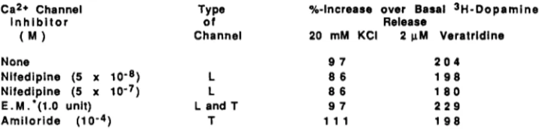
The data shown in Table 2 document that neither L- nor T-type Ca2+ channels are involved in the depolarization-dependent release of 3H-dopamine. Nifedipine, a specific inhibitor of the L-type Ca2+ channel and an endogenous modulator for L- and T-type channels, that was isolated from rat brain (Callewaert. Hanbauer and Morad, 1989) failed to modify sH-dopamine release elicited by KCI or veratridine (Table 2). Amiloride, a K+-sparing drug that was shown to inhibit the T-type Ca 2+ channels (Tang et al., 1988), also failed to alter 3H-dopamine release elicited by KCI or veratridine (Table 2).
Ca
Table 4. The Effect of L- and T-type
*+ Channel
Inhibitors
on the KCI-
and Veratridine-Elicited
3H-Dopamine
Release
in Primary
Cultures
of
Mesencephalic
Neurons
Ca*+ Channel Type %-Increase over Basal 3H-Dopamine
lnhlbitor of Release
(M) Channel 20 mM KCI 2 pM Veratrldlne
None 97 204
Nifedipine (5 x lo-*) L 66 198
Nifedipine (5 x lo-‘) L 86 180
E.M.‘(l.O unit) L and T 97 229
Amiloride (1 0m4) T 111 198
Each value is the mean of two determinations in triplicate and represents the percentage of increase over basal 3H-dopamine release. The Ca2+ channel inhibitors were added 5 min before addition of depolarizing agents.
l
E.M. = Endogenous Ca2+ channel modulator. 1 unit causes 50% inhibition of 3H-nitrendipine binding.DISCUSSION
The role of different types of voltage-sensitive Ca2+ channels in 3H-dopamine release was studied in primary cultures of rat ventral mesencephalon. This type of preparation allows one to study neurotransmitter release in intact and living neurons and not in severed axons as is the case in slices or synaptosomes prepared from adult rat brain. These results clearly showed that although KCI- and veratridine-evoked release of sH- dopamine requires extracellular Ca 9+, they are operative through different Ca2+ channels. The KCI-evoked @ease of 3H-dopamine was mediated throug:; activation of N-type Caz+ channels because o-CT completely blocked this response. Inhibition of L- or T-type Can + channels failed to alter KCI-evoked 3H-dopamine release. These findings are in line with previous reports suggesting that L-type Ca2+ channels occur at the soma (Sanna, Head and Hanbauer. 1986) and N-type channels are located on presynaptic nerve terminals (Miller, 1987). Our results also show that the veratridine-evoked release of 3H-dopamine was not altered by L-, T-, or N-type Ca2+ channel blockers. Since veratridine elicits membrane depolarization by prolonging the opening time of voltage-sensitive Nat channels it is possible that Ca2+ could enter the cell along with Na+. In fact, inactivation of Na+ channels by tetrodotoxin that was shown to block the veratridine-evoked 45Ca2+- influx in striatal slices (Sanna et al., 1986) attenuated 3H-dopamine release in cultured mesencephalic cells. This suggestion does not exclude the possibility that veratridine may also activiate an as yet undefined voltage- sensitive Ca2+ channel. The possibility that the “P” channel may play such a role is being investigated.
ACKNOWLEDGEMENTS
Dr. M.G. Grilli was supported by a research grant from FIDIA, Abano Terme, Italy. We are grateful to Drs. U. Di Porzio and E. Costa for their stimulating Interest and advice.
REFERENCES
Callewaert, G., Hanbauer, I. and Morad, M. (1989) Modulation of calcium channels in cardiac and neuronal cells by an endogenous peptlde. Science u: 663-668.
Hirning, L.D., Fox, A.P., McCleskey, E.W., Ollvera, B.M., Thayer, S.A., Miller, R.J., and Tsien, R.W. (1988) Dominant role of N-type Ca2+ channels In evoked release of norepinephrine from sympathetic neurons. Science ZLE: 57-61.
Llinas, R., McGulnness, T.L., Leonard, C.S., Sugimori, M., and Greengard,, P. (1985) lntraterminal injection of synapsin I or calcium-calmodulin- dependent protein kinase II alters neurotransmitter release at the squid giant synapse. Proc. Natl. Acad. Sci., U.S.A. 82: 3035-39.
Llinas, R., Sugimori, M., Lln, J.-W., and Cherksey, B. (1989) Blocking and isolation of a calcium channel from neurons in mammals and cephalopods utilizing a toxin fraction (FTX) from funnel web spider poison. Proc. Natl. Acad. Sci., U.S.A. u: 1689-1693.
Miller, R.J. (1987) Multiple calcium channels and neuronal function. Science u: 46-52.
Nishimura, M. (1987) Zinc competitively Inhibits calcium-dependent release of transmitter at the mouse neuromuscular junction. Pfluegers Arch. 9Lp: 623-826.
Nishimura, M. (1988) Zn2+ stimulates spontaneous transmitter release at mouse neuromuscular junctions. Br. J. Pharmacol. u: 430-436.
Prochiatz, A., Di Porzio, U., Kato, A., Berger. G. and Glowinski, J. (1979) In vitro maturation of mesencephalic dopamlnergic neurons from mouse embryos is enhanced in presence of their striatal target cells. Proc. Nat. Acad. Sci. x: 5387-5391.
1278 Preliminary Notes
Sanna, E., Head, G.A., and Hanbauer, I. (1986) Evidence for a selective localization of voltage-sensitive Ca2+ channels in nerve bodies of corpus striatum. J. Neurochem. 42: 1552-1557.
Tang, C.M.. Presser, F., and Morad, M. (1988) Amiloride selectively blocks the low threshold (T) calcium channel. Science m: 213-215.
Tsien, R.W., Lipscombe, D.. Madison, D.V., Sley, K.R. and Fox, A. P. (1988) Multiple types of neuronal calcium channels and their selective modulation. Trends Neurosci. u: 431-438.