PHARMACOLOGICALREVIEWS Pharmacol Rev 68:419–457, April 2016
U.S. Government work not protected by U.S. copyright
ASSOCIATE EDITOR: MACDONALD J. CHRISTIE
Nociceptin/Orphanin FQ Receptor Structure, Signaling,
Ligands, Functions, and Interactions with Opioid
Systems
Lawrence Toll, Michael R. Bruchas, Girolamo Calo’, Brian M. Cox, and Nurulain T. Zaveri
Torrey Pines Institute for Molecular Studies, Port St. Lucie, Florida (L.T.); Departments of Anesthesiology, and Neuroscience, Washington University School of Medicine, St. Louis, Missouri (M.R.B.); Section of Pharmacology, Department of Medical Science, and National Institute of Neurosciences, University of Ferrara, Ferrara, Italy (G.C.); Professor of Pharmacology & Neuroscience, Uniformed Services
University, Bethesda, Maryland (B.M.C.); and Astraea Therapeutics, LLC, Mountain View, California (N.T.Z.)
Abstract . . . 420
I. Introduction . . . 420
II. Nociceptin Opioid Peptide Receptor . . . 421
A. Nociceptin Opioid Peptide Receptor Protein. . . 421
1. Nociceptin Opioid Peptide Receptor Tertiary Structure. . . 421
2. Nociceptin Opioid Peptide Receptor Activation. . . 423
B. Location of Nociceptin Opioid Peptide Receptors . . . 424
C. Regulation of Expression of NOP Receptors . . . 426
III. Signal Transduction Pathways Activated by NOP Receptor Ligands. . . 428
A. Classic Gi-Signaling Pathways . . . 428
B. NOP Receptors and Kinase Signaling. . . 429
C. Nociceptin Opioid Peptide Receptor Desensitization, Downregulation, and Recycling . . . 429
1. Phosphorylation and Desensitization.. . . 429
2. Nociceptin Opioid Peptide Receptor Internalization, Recycling, and Downregulation. . 430
D. Cross Talk with Mu Opioid Receptors . . . 431
IV. Cellular Actions of Nociceptin Opioid Peptide Receptors . . . 432
A. Electrophysiological Analysis of Nociceptin Opioid Peptide Action in Brain and Spinal Cord . . . 432
B. Effects of Nociceptin Opioid Peptide Receptor Activation On Release of Central Nervous System Neurotransmitters . . . 433
C. Nociceptin Opioid Peptide Receptor and Inflammatory Signaling . . . 434
V. Biologic Actions of Nociceptin Opioid Peptide Receptors. . . 434
A. Nociceptin Opioid Peptide Receptors and Opiate Activity. . . 434
1. Analgesia. . . 434
2. Chronic Pain. . . 435
3. Opioid Tolerance Development. . . 435
4. Opioid Addiction Liability and Reward.. . . 435
B. Nociceptin Opioid Peptide Receptor and Motor Function . . . 436
VI. Nociceptin Opioid Peptide Receptor Ligands . . . 439
A. Nociceptin/Orphanin FQ Related Peptides . . . 439
1. Peptide Full Agonists. . . 440
2. Peptide Partial Agonists.. . . 442
3. Peptide Antagonists. . . 442
B. Nociceptin/Orphanin FQ Unrelated Peptides. . . 442
This work was supported by National Institutes of Health National Institute of Drug Abuse [Grant R01DA023281] (L.T.), Grant R21DA034929 (M.R.B.), Grants R01DA014026 and R01DA027811 (N.T.Z.)] and by the FAR grant of the University of Ferrara (G.C.).
Address correspondence to: Lawrence Toll, Torrey Pines Institute for Molecular Studies, 11350 SW Village Parkway, Port St. Lucie, FL 34990. E-mail: [email protected]
dx.doi.org/10.1124/pr.114.009209
419
at University of Leicester on March 9, 2016
pharmrev.aspetjournals.org
C. Nonpeptide Nociceptive Opioid Peptide Ligands . . . 443 1. Nonpeptide Agonists. . . 443 a. Ro 65-6570.. . . 445 2. Nonpeptide Antagonists. . . 445 a. J-113397.. . . 445 b. SB-612111. . . 446 c. C-24. . . 447 B. Bifunctional Compounds . . . 447
VII. Future Directions and New Tools . . . 449
VIII. Concludng Remarks . . . 450
Disclosures . . . 450
References . . . 450
Abstract——The NOP receptor (nociceptin/orphanin FQ opioid peptide receptor) is the most recently discovered member of the opioid receptor family and, together with its endogenous ligand, N/OFQ, make up the fourth members of the opioid receptor and opioid peptide family. Because of its more recent discovery, an understanding of the cellular and behavioral actions induced by NOP receptor activation are less well developed than for the other members of the opioid receptor family. All of these factors are important because NOP receptor activation has a clear modulatory role on mu opioid receptor-mediated actions and thereby affects opioid analgesia, tolerance development, and reward. In addition to opioid modulatory actions, NOP receptor activation has important effects on motor function and other physiologic processes. This review discusses how NOP
pharmacology intersects, contrasts, and interacts with the mu opioid receptor in terms of tertiary structure and mechanism of receptor activation; location of receptors in the central nervous system; mechanisms of desensitization and downregulation; cellular actions; intracellular signal transduction pathways; and behavioral actions with respect to analgesia, tolerance, dependence, and reward. This is followed by a discussion of the agonists and antagonists that have most contributed to our current knowledge. Because NOP receptors are highly expressed in brain and spinal cord and NOP receptor activation sometimes synergizes with mu receptor-mediated actions and sometimes opposes them, an understanding of NOP receptor pharmacology in the context of these interactions with the opioid receptors will be crucial to the development of novel therapeutics that engage the NOP receptor.
I. Introduction
Shortly after the cloning of the delta, mu, and kappa opioid receptors, a fourth receptor was cloned by homol-ogy with the opioid receptors. This fourth receptor, like the opioid receptors, is a seven transmembrane-spanning G protein-coupled receptor (GPCR), which has overall homology with the opioid receptors as high as the three opioid receptors have with each other. Because of this high homology, the cloning was somewhat facile and was accomplished by several laboratories almost simulta-neously. The first paper to be published was by Mollereau et al. (1994), and they called this new receptor opioid receptor like receptor 1, ORL1. Other cloning papers followed quickly, and this same receptor was called LC132, XOR1, kappa 3, ROR-C, C3 (Bunzow et al., 1994; Fukuda et al., 1994; Wang et al., 1994; Lachowicz et al., 1995; Pan et al., 1995). Despite the close homology with opioid receptors, this orphan receptor, when trans-fected into mammalian cells, did not appear to bind or be activated by standard opiate ligands at low concen-trations. For lack of a high affinity ligand, there was not an appropriate binding assay to characterize this receptor. Nevertheless, it was activated by high concen-trations of the opiate agonist etorphine and inhibited
by a high concentration of naloxone (Mollereau et al., 1994). In addition, it was clearly coupled to Gi, like the opioid receptors, because receptor activation still inhibited adenylyl cyclase (Mollereau et al., 1994). Despite the fact that standard opiates did not activate this receptor at low concentrations, this receptor appeared to be in the opioid receptor family.
Approximately 2 years after the discovery of the orphan receptor, at that time generally called ORL1, two groups identified an endogenous neuropeptide that bound with high affinity to ORL1 and activated the receptor, as determined by inhibition of cAMP accumulation in transfected cells (Meunier et al., 1995; Reinscheid et al., 1995). In both cases, the endogenous ligand was discovered by fractionating tissue (in one case rat brain and the other porcine pituitary) based upon ability to inhibit adenylyl cyclase activity in cells transfected with ORL1. These were the first examples of“reverse pharmacology” to identify ligands subsequent to the discovery of the receptor, a process that has been since used many times (Civelli et al., 2013). This 17-amino acid neuropeptide was called nociceptin (for its ability to decrease hot plate latency when administered intra-cerebroventricularly into mice) (Meunier et al., 1995)
and orphanin FQ (Reinscheid et al., 1995) to denote a ligand for an orphan receptor with first and last amino acids Phe and Gln. The heptadecapeptide Phe-Gly-Gly-Phe-Thr-Gly-Ala-Arg-Lys-Ser-Ala-Arg-Lys-Leu-Ala-Asn-Gln is interesting for several reasons. First the Phe-Gly-Gly-Phe amino terminal is obviously reminiscent of the Tyr-Gly-Gly-Phe found in all opioid peptides. Second, this is a highly basic peptide, quite similar to dynorphin in the number of Lys and Arg residues. Third, the gene structure of the prepropeptide is also similar to the opioid peptide genes (Mollereau et al., 1996a; Nothacker et al., 1996). Together these discoveries of ORL1 and nociceptin/orphanin FQ iden-tified the fourth members of the opioid receptor and opioid gene families. IUPHAR nomenclature for this receptor and peptide is now officially NOP (nociceptin opioid peptide) receptor and N/OFQ (Cox et al., 2015). Compounds targeting the NOP receptor were re-cently advanced to clinical trials, so an understand-ing of this receptor system has increased clinical relevance. This review will discuss the NOP receptor system and its important modulatory role in several central nervous system (CNS) systems, along with the signaling pathways that mediate its activity and the synthetic compounds that have been instrumen-tal in the identification and validation of many of these activities.
II. Nociceptin Opioid Peptide Receptor A. Nociceptin Opioid Peptide Receptor Protein
Comparison of the cDNA-derived amino acid sequence of the NOP protein with that of the opioid receptors and other GPCRs shows that it contains several conserved amino acids and motifs, particularly in the transmem-brane helices and the intracellular loops, placing the NOP receptor in the GPCR Class A (rhodopsin-like) receptors, like the mu, delta, and kappa opioid receptors. Greater than 70% of the amino acid residues in the second, third, and seventh helices (TM2, TM3, and TM7) are conserved between NOP and the mu, delta, and kappa opioid receptors. However, only 50% of residues are conserved in TM1, TM5, and TM6, whereas in TM4, only 24% of residues are conserved (Meunier et al., 2000). There is high sequence conservation in the intracellular loops (ICL) among the opioid receptor family, particu-larly in ICL3 (.80%), which connects TM5 and TM6 and is involved in activation and interaction with the G proteins. The extracellular loops (ECL) on the other hand, have very little sequence similarity among the four opioid receptors, NOP being closest to the kappa opioid receptor in containing a significant number of acidic residues in its ECL2. Notably however, the ECL2 in NOP, but not in other opioid receptors, is involved in receptor activation, as discussed below. Nonetheless, the NOP receptor sequence contains all the conserved activation-associated motifs termed "microswitches"
found in the TM helices of Class A GPCRs, including the other opioid receptors (Nygaard et al., 2009; Tehan et al., 2014), suggesting that the transmem-brane and intracellular amino acid residues involved in conformational changes during receptor activation (microswitches) in NOP are consistent and similar to the other opioid receptors and Class A GPCRs (see section A.2).
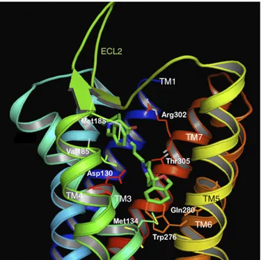
1. Nociceptin Opioid Peptide Receptor Tertiary Structure. In the rapid explosion of GPCR crystal structure determinations published in the last few years, the structures of all four opioid receptor family members were solved in their inactive, antagonist-bound conformations (Granier et al., 2012; Manglik et al., 2012; Thompson et al., 2012; Wu et al., 2012). These give an atomic-level view into the tertiary structures of the opioid GPCRs and provide confirma-tion of the several previous homology models of the opioid receptors developed to understand the archi-tecture of these receptors. The NOP receptor was crystallized in its inactive form, bound to the antag-onist C-24 (PDB ID: 4EA3, see Fig. 1). As expected, the ligand-binding pocket is contained within the trans-membrane helices, with residues from TM3, TM5, TM6, and TM7 interacting with the ligand in the binding pocket. Similarly, molecular modeling of the complex of the peptide agonist N/OFQ with homology models of the NOP receptor (Topham et al., 1998; Akuzawa et al., 2007; Daga and Zaveri, 2012) show that the N-terminal sequence F-G-G-F of N/OFQ binds deep in the transmembrane binding pocket, where the N-terminal amino group of N/OFQ makes an essential anchoring charge interaction with the conserved D1303 .32 (superscripts refer to the Ballesteros-Weinstein numbering of the TM helix residue), pre-sent in all the opioid receptors as well as in biogenic amine GPCRs (Fig. 2). Although the binding of small-molecule antagonist C-24 in the NOP receptor crystal structure may involve different amino acid residues than those interacting with the peptide agonist N/OFQ in the modeled complex, an extensive array of site-directed mutagenesis studies carried out with NOP show that there are only 4–5 amino acid residues in NOP that afford the exquisite selectivity of N/OFQ for the NOP receptor and precludes binding of small-molecule morphinan opioid ligands. Mutation of the following NOP receptor residues to their correspond-ing conserved opioid receptor residues (A2165.39to K, V2796.51–Q2806.52–V2816.53to I–H–I and T3057.39to I) confers a functional opioid alkaloid binding site in NOP receptors, which binds opioid antagonists with high affinity, without adversely affecting N/OFQ binding significantly (Meng et al., 1998). This study was consistent with mutagenesis of Q280 in TM6 in NOP to histidine, a TM6 residue conserved in all three opioid receptors, which results in an increase in affinity of opioid agonists lofentanil, etorphine, and
dynorphin A and antagonists diprenorphine and nor-BNI, but does not affect N/OFQ binding or potency significantly (Mollereau et al., 1996b). The effect of the Q280H mutation on the binding of small-molecule NOP receptor ligands is not known; however, a Q280A mutation was shown to reduce the potency of receptor activation by N/OFQ and the NOP agonist SCH 221510 by several orders of magnitude (Thompson et al., 2012). Although these five residues (A216, V279, Q280, V281, and T305) serve to preclude binding of opiate ligands to the NOP receptor, no studies have yet explored the reverse question: what residues in the mu, delta, and kappa opioid receptors prevent binding of selective NOP ligands to opioid receptors? Clues for such NOP selectivity-enhancing interactions have come from computer-aided molecular docking studies of the selective NOP agonist Ro 64-6198 into the first active-state NOP receptor homology model, developed by Daga and Zaveri (2012), based on the opsin template (Fig. 3). The amide hydrogen in Ro 64-6198 makes direct hydrogen-bond interaction with T3057.39(Ile in other opioid receptors) at the extracellular end of the binding pocket, whereas the phenalenyl ring of Ro 64-6198 interacts with the hydrophobic V2796.51 resi-due inside the binding pocket (Fig. 3) (Daga and Zaveri, 2012). An isoleucine residue, found in the mu, delta, and kappa opioid receptors in this position, would sterically hinder binding of Ro 64-6198 and is possibly responsible for precluding the binding of Ro 64-6198 to these other opioid receptors. The phenalenyl group of
Ro 64-6198 is therefore contributing to the excellent selectivity of this ligand for the NOP receptor (Fig. 3) (Daga and Zaveri, 2012).
Although there is high homology and similarity in functional architecture in the transmembrane and in-tracellular loops between NOP and other opioid recep-tors, the ECLs of NOP receptors are distinct in their amino acid sequence, particularly ECL2 that connects the extracellular ends of TM4 and TM5 and ECL3 that connects TM6 and TM7. The ECL2 of NOP has the same residue length as the mu and delta opioid receptors but has almost no sequence similarity. On the other hand, the NOP ECL2 contains several Glu acidic residues, similar to the ECL2 in the kappa receptor, which is three residues longer, and contains mainly Asp resi-dues. Overall, therefore, the NOP ECL2 is unique in its primary structure among the opioid receptors, and its interactions with the amino acids at the extracellular ends of the TM domains (Daga and Zaveri, 2012) play a distinct and critical role in receptor activation, unlike the other three opioid receptors.
Because the recently resolved crystal structure of NOP is bound to an antagonist and is in its inactive form, it does not show molecular interactions of the ECL2 with the bound ligand (Thompson et al., 2012). However, elegant receptor chimera studies, with a NOP–kappa chimera, clearly show that the ECL2 of NOP is an absolute requirement for activation of NOP by N/OFQ, unlike the ECL2 of the kappa receptor, which can be replaced with that of NOP without adversely affecting the activation of the kappa receptor by dynorphin (Mollereau et al., 1999). In fact, replacing the N
Fig. 2. N/OFQ (1-13) peptide (green sticks) bound to the active-state homology model of the NOP receptor. The TM helices are in different colors. The side chains of amino acids interacting with the peptide are labeled. Note the acidic residues of the ECL2 loop (D195, E196) interacting with the basic residues (8-13) of N/OFQ.
Fig. 1. Molecular model of the NOP receptor crystal structure bound to NOP antagonist C-24 (green) (PDB ID: 4EA3). The TM helices are colored in 7 different colors and labeled. The ECL2 loop, between TM4 and TM5 is shown in green. Side chains of amino acids interacting with the antagonist are shown as sticks and labeled.
terminus, ECL1, and ECL2 of the kappa receptor with those of NOP results in a receptor hybrid that has equipotent binding affinity for N/OFQ and dynorphin and, importantly, is activated efficiently by both peptides without a significant loss in potency compared with the native receptors (Mollereau et al., 1999). These studies underscore the importance of the NOP receptor ECL2 for binding and activation of the receptor by NOP agonists. 2. Nociceptin Opioid Peptide Receptor Activation. The high degree of homology in the TM helices between NOP and other opioid receptors would suggest that the mechanism of receptor activation of the opioid family GPCRs may involve the same residues after ligand binding, resulting in G protein binding and further downstream events. Although the crystal structure of an agonist-bound "active-state" or a constitutively ac-tive form of the NOP receptor has not yet been solved, a molecular dynamics simulation of a homology model of the active-state NOP receptor and comparison with the inactive-state receptor suggests that NOP receptor activation is accompanied by movements of the TM helices which are transduced to the intracellular do-mains, in a manner similar to other Class A GPCRs (Daga and Zaveri, 2012). The intracellular end of TM4 moves toward the helical bundle, whereas that of TM6 moves outward from the helical bundle, creating a binding pocket for the G protein. Activation-associated microswitches (Nygaard et al., 2009) found in all Class A GPCRs are also in their"active" conformations in the active-state NOP homology model. For instance, the conserved"DRY" motif in TM3 (also present in NOP, as Asp1473.49–Arg1483.50–Tyr1493.51) shows no ionic in-teraction between D147 and R148 in the active-state homology model but has the"ionic lock" between these two residues in the inactive-state conformation (Daga
and Zaveri, 2012). In the NOP active-state model, the D1483.50 microswitch shows a H-bonding interaction with Y2355.58, which is an activation-associated inter-action involving this conserved DRY motif (Daga and Zaveri, 2012).
The W2766.48microswitch is part of the CWxP motif in TM6, which undergoes major conformational move-ment during receptor activation of NOP, as in most other Class A GPCRs. The W276 indole side chain moves from its inactive rotamer conformation to an "active rotamer conformation," in which it interacts with the Phe2245.47 to form an "aromatic lock," an activation-associated conformational movement (Daga and Zaveri, 2012). Another activation-associated micro-switch is present in the NPxxY motif in TM7, in which Y7.53(Y319 in TM7 in NOP) toggles between an inactive rotamer and an active rotamer, which interacts with TM6 residues during activation (Nygaard et al., 2009). Mutagenesis studies have implicated several resi-dues that are important for the intrinsic efficacy of the endogenous agonist N/OFQ. For instance, mutation of Q2866.58near the extracellular end of TM6 completely abolishes activation by N/OFQ, without any effect on the binding affinity for the mutated receptor (Mouledous et al., 2000). This suggests a very specific role for this residue during activation after N/OFQ binding, although it does not contribute to binding affinity of N/OFQ. Alanine mutations of W2766.48, the rotamer toggle activation microswitch, and F2245.47(part of the TM5 "ionic lock" microswitch) have differential effects on activation by structurally different agonists (Mouledous et al., 2000). The W276-A (and F224-A) mutant showed two to fourfold decreased binding by N/OFQ and de-creased potency of activation, but no decrease in overall intrinsic efficacy, i.e., the W276-A showed full agonist efficacy, albeit with higher concentrations of N/OFQ.
However, with the hexapeptide Ac-RYYKWK-NH2
[a partial agonist at NOP (Dooley et al., 1997)] and lofentanil, the W276-A mutant showed no decrease in binding affinity for these ligands but could not be fully activated by these ligands, producing low efficacy partial agonist activity. It is likely, therefore, that structurally different agonist ligands engage different residues during activation, resulting in multiple"active states," leading to different levels of intrinsic efficacy and possibly functional selectivity (biased signaling) at the intracellular end of the membrane-bound NOP receptor (Wacker et al., 2013; Shukla et al., 2014).
Despite the similarities of NOP receptor activation-associated TM movements to other GPCRs, one feature that sets apart NOP receptor activation from that of the three other opioid receptors and most other GPCRs is the absolute requirement for the ECL2 for activation (Lapalu et al., 1998; Mollereau et al., 1999). Mutation studies of ECL2 residues have not yet been reported, but the NOP ECL2 contains a high number of acidic residues, mainly Glu. Only the kappa receptor ECL2 has similar acidic Fig. 3. NOP agonist Ro 64-6198 (green sticks) bound to the
active-state NOP receptor model. The small-molecule NOP agonist interacts with the T305 (orange sticks) and Y309 (blue sticks). The phenalenyl group of the NOP agonist is in close proximity to V279 (orange sticks, labeled) within the transmembrane pocket. This residue is isoleucine in the other opioid receptors, which is likely responsible for the lower affinity of Ro 64-6198 for the other opioid receptors.
residues (mainly Asp), but these have not been shown to be critical for receptor activation, as for NOP. Molecular dynamics simulation of the active-state NOP homology model suggests that NOP activation may involve move-ment of the ECL2 forward toward TM7, where it may participate in interactions with residues at the extracel-lular end of TM7 and TM3 or even with agonist ligands, resulting in a proposed activation-associated conforma-tional movement of the TM helices (Daga and Zaveri, 2012). Binding of NOP agonist Ro 64-6198 to the active-state NOP model (Fig. 3) shows that TM7 residues such as T3057.39 and Y3097.43 interact with the agonist and with E199ECL2 in an activation-associated network (Daga and Zaveri, 2012). Consistent with its primary structure, the tertiary structure and ligand-induced conformational changes identify the NOP receptor as belonging to the opioid receptor family but none-theless unique from the other receptors in important ways.
There is a small amount of information pertaining to potential constitutive activity of NOP receptors. Electro-physiological recording of neurons, in which overexpres-sion of the receptor was induced by microinjection of coding cDNA, demonstrated the antagonist C-24 to have inverse agonist activity, indicative of constitutive activa-tion of NOP receptor when overexpressed (Mahmoud et al., 2010). In another study, in which the ability to constitutively activate G-protein-coupled pathways was investigated in a series of NOP receptor point mutations, only the N133W mutant displayed increased ligand-independent signaling (Kam et al., 2002). Interestingly, this mutated residue (N3.35) was recently found to contribute to the network of interactions that establish a sodium binding pocket in the structure of several GPCRs (Katritch et al., 2014), including the delta opioid receptor (Fenalti et al., 2014). The sodium binding pocket collapses upon receptor activation, thus suggesting that presence of sodium may stabilize the receptor in an inactive conformation (Katritch et al., 2014). Very re-cently using a BRET-based assay to investigate NOP receptor/G-protein interactions, it was demonstrated that GDP was not able to significantly inhibit the baseline BRET ratio (Malfacini et al., 2015). However, in membranes expressing the other opioid receptors, under similar experimental conditions, GDP can sup-press the baseline BRET ratio, indicating a reduction in spontaneous receptor/G-protein interactions, with maximal effects 4–5 times greater at delta than mu receptors (Vezzi et al., 2013). Thus these results dem-onstrate that the propensity to display constitutive activity is much lower for NOP compared with the mu and particularly the delta opioid receptor.
B. Location of Nociceptin Opioid Peptide Receptors NOP receptors are highly expressed in many brain regions. Although several immunohistochemical stud-ies have been carried out on NOP receptors, the lack of
validated antibodies that do not crossreact with brain tissue for NOP receptor knockout [NOP(2/2)] mice has raised considerable concern regarding these results. Nevertheless, in situ hybridization and in vitro autora-diography using [125I]-N/OFQ have provided an ade-quate representation of NOP receptor localization and in general have been somewhat consistent with the immunohistochemical studies (Neal et al., 1999a; Florin et al., 2000). NOP receptors are expressed in multiple brain regions and are involved in a large number of central processes including pain, learning and memory, emotional states, neuroendocrine control, food intake, and motor control. In many of these neuronal pathways, there is also considerable overlap between the location of the NOP receptor and the peptide N/OFQ, as de-termined by immunohistochemistry and in situ hybrid-ization (Neal et al., 1999b). Consistent with these findings, intracerebroventricular administration of N/OFQ mod-ulates many of these processes: decreasing spatial learning (Sandin et al., 1997; Sandin et al., 2004), modulating anxiety (Jenck et al., 1997), increasing food intake (Polidori et al., 2000), and although it has no effect on its own, intracerebroventricular N/OFQ mod-ulates opioid reward (Murphy et al., 1999) and nocicep-tion (Meunier et al., 1995; Reinscheid et al., 1995). With respect to nociceptive processing, NOP receptors are found in high numbers in pain-related brain regions within both the ascending and descending pain path-ways including the periaqueductal gray (PAG), tha-lamic nuclei, somatosensory cortex, rostral ventral medulla, lateral parabrachial nucleus, spinal cord, and dorsal root ganglia (DRGs) (Neal et al., 1999a; Florin et al., 2000). In each supraspinal location where tested, NOP receptor activation by local injection of N/OFQ appears to block the actions of opiate analgesics (Morgan, 1997; Pan et al., 2000), which explains the anti-opioid effects of N/OFQ when administered intra-cerebroventricularly. Patch clamp electrophysiological studies have also been used to explain the anti-opioid action of N/OFQ. In the vlPAG, mu receptors can be found on approximately one-third of the neurons, and mu receptor activation blocks the descending pain signal (Vaughan et al., 1997; Connor and Christie, 1998). NOP receptors are found on every cell in the vlPAG and can thereby block the desending analgesic pathway and occlude the actions of mu opiates (Morgan et al., 1997; Connor and Christie, 1998).
NOP receptors are also highly expressed in regions involved in reward and drug abuse. Consistent with brain-mediated anti-opioid effects, NOP agonists atten-uate the rewarding effects of opiates and other abused drugs, a topic that is discussed in more detail below. Accordingly, NOP receptors are highly expressed in the mesocorticolimbic drug reward circuitry, including ventral tegmental area (VTA), nucleus accumbens, and prefrontal cortex, as well as the central amygdala, involved in stress and drug relapse, and the medial
habenula-interpedunclear nucleus pathway (Neal et al., 1999a), thought to be involved in abuse of nicotine and likely other drugs as well. There are also NOP receptors on hypocretin/orexin-containing cells in the lateral hypothalamus (Xie et al., 2008). In each of these brain regions, NOP receptor activation reduces the release of the neurotransmitters that mediate rewarding effects.
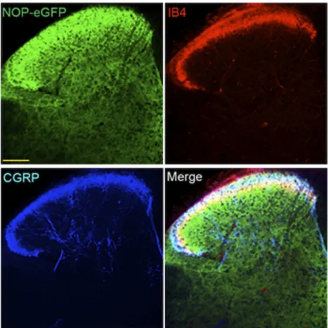
Recently, knock-in mice have been developed with NOP-eGFP receptors in place of the native receptor (Ozawa et al., 2015), similar to knock-in mice containing delta-eGFP and mu-mCherry receptors (Scherrer et al., 2006; Erbs et al., 2015). For each of these mutant mice, the tagged receptor has been valuable in identifying receptor location and trafficking without the need for problematic opioid receptor antibodies, with resolution far superior to in vitro autoradiography. For the NOP receptor, location of the NOP-eGFP receptor in brain is basically similar to what has been described using in vitro autoradiography (Neal et al., 1999a). In addition to the NOP-eGFP expression in the brain, NOP-eGFP receptors can be found in the dorsal horn of the spinal cord and in DRG. To determine the specific lamina location of NOP receptors in the spinal cord, additional immunostaining was performed with lamina markers. NOP-eGFP receptors are present at the most superficial lamina (I and II) and dorsal border of lamina IIinner
where calcitonin gene related peptide (CGRP)-positive and IB-4-positive nociceptive primary afferents project (Fig. 4). This intense immunoreactivity also colocalizes with PKCg positive interneurons in the ventral border of laminae II and III indicating that the NOP receptors might have a regulatory mechanism in the control of chronic mechanical allodynia (Neumann et al., 2008; Basbaum et al., 2009). Therefore, NOP receptors are distributed between laminae I through III in the dorsal horn, regions important for the regulation of pain systems.
In addition to the spinal cord, NOP receptors are found in a large number of DRG neurons, large and small, myelinated and unmyelinated. Approximately 43% of all DRG neurons express NOP-eGFP, almost evenly split between small and large cell body diameter. The majority of the large diameter neurons are neuro-filament 200 (NF200) positive, therefore representing myelinated A-fibers. Approximately one third of the small unmylelinated neurons are also positive for CGRP indicating that NOP-eGFP receptors are present in peptidergic C nociceptors. Peptidergic C-fibers are essential to acute heat pain as well as injury-induced heat hyperalgesia (Cavanaugh et al., 2009) and have been shown to project to laminae I and IIouter of the
spinal cord (Basbaum et al., 2009) where robust
Fig. 4. NOP-eGFP receptors are highly distributed in laminae I-III and3. Tissue sections from the spinal cord were incubated with anti-GFP, and–CGRP (laminae I and IIo, panel A). Tissues were also treated with biotinylated IB4 (dorsal border of lamina IIi) and streptavidin. This figure is reprinted with permission from the Journal of Neuroscience.
immunoreactivity of NOP-eGFP is also observed. A smaller proportion of small unmyelinated (NF200-) NOP-eGFP+ DRG neurons bind IB4, indicating that NOP receptors are also present in the non-peptidergic DRG neurons, which are involved in mechanical pain (Basbaum et al., 2009; Cavanaugh et al., 2009; Scherrer et al., 2009; Vrontou et al., 2013; Bardoni et al., 2014). These studies suggest that NOP receptors might regu-late the function of two classes of C nociceptors that respond to both heat and mechanical pain. NOP-eGFP receptors also are co-localized with mu opioid recep-tors in peptidergic C-nociceprecep-tors (Fig. 5).(Fig 6). (Fig 7). These results and the similar location of NOP and mu receptors in the spinal cord probably explain the ability of NOP receptor agonists to mediate an anti-nociceptive response when administered intrathe-cally (i.t.).
C. Regulation of Expression of NOP Receptors
The molecular control of NOP receptor expression is complex and has not been fully elucidated. The human NOP receptor gene is located on chromosome 20. The promotor region of the human NOP receptor gene was analyzed by Palmer and colleagues (Ito et al., 2000; Xie
et al., 2000). This region contains a number of predicted regulatory elements, including response elements for the glucocorticoid receptor, metal response elements, and multiple retinoic acid response elements. Retinoic acid, a potent regulator of NOP receptor expression in NT2 cells in culture, also induces differentiation of these cells (Ito et al., 2000). The transcription factor response elements Sp1, AP-2, EGR, Krox-20, ETF, and CP1 or GCF sites are also found in the promotor region of the human NOP gene. No TATA box or CCAAT box was found upstream of the transcription start sites for the NOP receptor protein. The promotor regions of the mu and delta opioid receptor genes also contain re-sponse elements for some of these transcription factors (Min et al., 1994; Im et al., 1999).
Xie et al. (Xie et al., 2000) identified two transcription start sites in the human NOP receptor gene with products that differ only in their 59 upstream non-coding regions. The upstream site leads to the expres-sion of exons 1A, 1B and 2 with an ATG stop codon in exon 2. The second down stream start site leads to the expression on exons 1B and 2, which contain the coding regions for the NOP receptor. In contrast, the mouse NOP receptor gene, located on mouse chromosome 2,
Fig. 5. Colocalization of NOP-eGFP and mu receptors in DRG neurons. Tissue sections were incubated with anti-GFP (green), anti –mu-receptor (red), and anti-NF200 (blue) antibodies. White arrows show small diameter NOP-eGFP+, Mu+ cells. Scale bars 100 mm. This figure is reprinted with permission from the Journal of Neuroscience (Ozawa et al., 2015).
contains 5 exons, with the protein-coding region start-ing in exon 2 and endstart-ing in exon 4 (Ito et al., 2000). The expression of NOP receptor splice variants was first reported in rat (Wang et al., 1994) who found at least two variant forms of the receptor mRNA expressed in rat hypothalamus, describing these as long and short (truncated) forms of the receptor. The truncated form
of the receptor was missing the fifth, sixth and seventh transmembrane domains and the entire third intracellular loop. Expression studies lead to the conclusion that this truncated form had very weak capacity to bind N/OFQ and an inability to regulate G-protein function. It remains to be determined if they have other functions. In contrast to the rat, in mouse Fig. 6. Summary of NOP receptor signaling. Figure cartoons the basic NOP receptor signal transduction and trafficking pathways highlighted in this review, and those that have generally been shown by multiple studies. Figure shows NOP receptor canonical coupling to inhibition of calcium channels, and activation of inward rectifying potassium channels. Figure also highlights recent work showing NOP receptor activation of MAPKs, and desensitization pathways via GRK3 and GRK2, and recent data showing that NOP receptors can both positivity and negatively influence cytokine/ inflammatory pathway signaling. Furthermore, the cartoon depicts recent papers showing that NOP receptor activation and arrestin signaling can initiate downstream signaling to JNK and ROCK pathways. Arrows refer to activation steps; T lines refer to blockade or inhibition of function.
Fig. 7. Schematic representation of the organization of basal ganglia regulating motor function and the effects dopamine (DA) depletion on N/OFQ expression and release. DA neurons are represented in blue; glutamate (Glu) neurons in green; GABA neurons in red; color density indicates the relative levels of activity in each system with normal DA neuron function (Panel A, normal function), or after loss of a significant fraction of DA neurons (Panel B). GP, globus pallidus; N/OFQ, nociception/ orphanin FQ; SNc, substantia nigra compacta; SNr, substantia nigra reticulate; STN, subthalamic nucleus, Panel A. With DA neuron function intact, GABA release in the pallido-subthalamic neurons in the“indirect” striato-nigral pathway reduces Glu release from the subthalamic neurons that activate the GABAergic nigrothalamic pathway. With low release of GABA in the thalamus from this pathway, the thalamocortical glutamatergic neurons are active, increasing activity in motor cortex and maintaining normal motor function. N/OFQ levels and release in the SNr are relatively low under these conditions. Panel B. When nigrostriatal DA function is impaired (e.g., after 6-OHDA or MPTP treatment), activity in the subthalamic glutamatergic neurons to the SNr is increased, resulting in activation of the nigrothalamic GABA pathway and inhibition of thalamocortical neurons that facilitate normal motor function. After 6-OHDA or MPTP treatment, ppN/OFQ mRNA and N/OFQ levels and release in SNr are increased (Marti et al., 2005, 2010); N/OFQ release in SNr is also increased by haloperidol treatment (Marti et al., 2010). NOPr antagonists largely reverse the effects of DA depletion by 6-OHDA on GABA release in SNr and thalamus (Marti et al., 2005, 2007, 2010). Treatment with 6-OHDA also reduces NOPr mRNA expression in SNc (Norton et al., 2002; Marti et al., 2005). Arrows indicate the direction of change in N/OFQ, GABA or Glu release after 6-OHDA or MPTP treatment.
brain, five variant forms of NOP receptor gene tran-script have been reported with differential expression of the variant forms across mouse brain regions (Pan et al., 1998). The functional roles of the variant forms remain unclear.
NOP receptor gene transcripts have also been re-ported in human lymphocytes (Wick et al., 1995) and truncated forms indicative of alternative splicing of the NOP receptor gene product were also identified in human lymphocytes and lymphocyte cell lines (Halford et al., 1995). It is unclear if either the full-length NOP receptor or its truncated forms serve functional roles in lymphocytes, although a possible role for the NOP receptor in mediating the agonist-induced decrease of allergen-agonist-induced airway hyper-responsiveness after allergen exposure has been proposed (Sullo et al., 2013).
Interestingly, the gene for human Galpha interacting protein (GAIP, also known as RGS19), a regulator of GPCR signaling (interacting with the Ga subunits of Gi, Go, Gz and Gq), is located upstream of the NOP receptor gene but oriented in the opposite direction and separated by an 83 bp sequence that may function as a bi-directional promotor for both genes (Ito et al., 2000; Xie et al., 2003, 2005). Exon 1A of the human NOP receptor gene appears to function in reverse as a promotor for the GAIP gene. This arrangement suggests that NOP re-ceptor expression may be co-regulated with GAIP and thus serve a modulatory role in GPCR signaling. In some tissues, GAIP and NOP receptor may be co-expressed. However, Ito et al. (Ito et al., 2000) note that NOP receptor and GAIP expression sites do not always co-exist either in tissues or in cell lines, with several identified cell types capable of expressing NOP receptor without GAIP or vice-versa.
Xie et al. identified an alternative transcription site for mouse GAIP, leading to the expression of a trun-cated GAIP missing an N-terminal domain that is thought to interact with G-proteins (Xie et al., 2003). Co-expression of the full-length mouse GAIP with NOP receptor in COS cells resulted in potentiation of N/OFQ stimulation of GTPase and a reduction of N/OFQ-mediated inhibition of cAMP production (relative to the stimulation when only the NOP receptor gene was expressed) (Xie et al., 2005). When the N-terminally-truncated mouse GAIP transcript was co-expressed with NOP receptors, both the GAIP-induced potenti-ation of N/OFQ mediated GTPase activity and atten-uation of an N/OFQ-mediated reduction in cAMP production were reduced. These results suggest that co-expression of both the full-length GAIP with NOP receptor facilitates receptor regulation of G-protein function. The facilitatory effect of co-expression of full-length GAIP on GTPase activity and inhibition of adenylyl cyclase was relatively selective for NOP recep-tors; there was less facilitation when full length GAIP was co-expressed with mu, delta or kappa receptors but
this selectivity was lost when the truncated GAIP was co-expressed (Xie et al., 2005).
III. Signal Transduction Pathways Activated by NOP Receptor Ligands
A. Classic Gi-Signaling Pathways
For NOP receptors, like all GPCRs, following activa-tion by agonist the Ga and Gbg subunits dissociate to then act on the various effector pathways (Childers and Snyder, 1978; Childers et al., 1979). Early work in opioid receptor pharmacology demonstrated that gua-nine nucleotides such as GTP modulate agonist binding to opioid receptors in membrane preparations from brain tissue. It was later determined that GTPase activity is stimulated by opioid agonists (Barchfeld and Medzihradsky, 1984) and NOP receptor activation clearly promotes guanine nucleotide exchange (Sim et al., 1996; Narita et al., 1999). Agonist stimulation of opioid receptors was also shown to inhibit cyclic aden-osine monophosphate (cAMP) production in a manner similar to that of other types of GPCR. Several reports have confirmed that NOP receptor activation inhibits adenylyl cyclase activity similarly and it is widely accepted that the NOP receptor couples to pertussis-toxin–sensitive G-proteins, including Gai, to cause in-hibition of cAMP formation (Zhang et al., 2012a). However, it has also been suggested that NOP receptors can promiscuously couple to other G proteins, although this has been less well characterized in physiologically relevant systems, and has only been demonstrated in heterologous expression studies and SH-SY5Y cells (Chan et al., 1998).
Opioid receptors canonically couple to Kir3 and Ca2+ channels via Gbg pathways. Likewise, NOP receptors also couple to these two channels (Connor et al., 1996b; Connor and Christie, 1998). Channel deactivation for Kir3 interactions happens after GTP to GDP hydrolysis and Gbg removal from interaction with the channel (Wickman and Clapham, 1995). Opening of Kir chan-nels causes cellular hyperpolarization and inhibits tonic neural activity. When activated, NOP receptors also cause a reduction in Ca2+currents sensitive to P/Q-type, N-type, and L-type channel blockers (Connor et al., 1996b; Zhang et al., 2012a). NOP receptor inhibition of N-type calcium conductance is likely mediated by binding of the dissociated Gbg subunit directly to the channel. This binding event is thought to reduce voltage activation of channel pore opening (Zamponi and Snutch, 1998, 2002; Beedle et al., 2004; Yeon et al., 2004; Ruiz-Velasco et al., 2005). Furthermore, it has also been recently reported that NOP receptors use Rho-associated coiled-coil-containing protein kinase (ROCK) and LIM domain kinase (LIMK) in the regulation of voltage-dependent Ca2+channels (Mittal et al., 2013).
B. NOP Receptors and Kinase Signaling
All known classes of GPCRs couple to various in-tracellular kinase cascades. In particular, opioid recep-tors have been demonstrated to couple to protein kinase A and protein kinase C (PKC) pathways, in additional to the more recently appreciated signaling through mitogen-activated protein kinase (MAPK) cassettes. Furthermore, it was discovered in the mid 1990s that the phosphorylated arrestin-bound GPCR complex is not simply inactive but that it recruits alternate signal transduction cascades, including MAPKs (Bruchas and Chavkin, 2010; Whalen et al., 2011; Chang and Bruchas, 2014). Similarly, signaling to MAPK cassettes in opioid receptors and NOP receptors can in part be mediated via this process (Zhang et al., 2012a). NOP receptor activity can induce activation of PKC (Armstead, 2002) as well as activation of phospholipase A2 and C (Fukuda et al., 1998; Yung et al., 1999).
NOP receptor-dependent activation of all three MAPK cassettes has been demonstrated. NOP receptor induced extracellular-signal regulated kinase (ERK) phosphorylation has not been extensively examined; however, two groups have demonstrated that the endogenous agonist N/OFQ will cause NOP receptor-mediated increases in ERK 1/2 phosphorylation levels in heterologous expression systems (COS7, CHO, and HEK293 cells) (Lou et al., 1998; Zhang et al., 2012a). In a recent report, ERK 1/2 signaling via NOP receptors was shown to be independent of receptor phosphoryla-tion and GRK/arrestin signaling (Zhang et al., 2012a). However this requires further examination with other ligands and in alternate model systems.
Opioid receptor activation of p38 MAPK cassettes has gained interest due to the effects of kappa receptor-induced p38 phosphorylation and aversive behaviors (Bruchas and Chavkin, 2010; Bruchas et al., 2011). NOP receptor activation has been linked to phosphorylation of p38 MAPK in vitro. In one report it was demonstrated that NOP receptors activate p38 signaling via protein kinase A and PKC pathways (Zhang et al., 1999). Examination of NOP receptor-mediated p38 signaling in endogenous systems under pathologic conditions, as shown in Armstead (2006), and in various tissues will be important next steps in understanding the coupling of NOP receptors to this MAPK cassette.
Likewise, activation of c-Jun N-terminal kinase (JNK) signaling by opioid receptors has been recently examined for its interesting mu and kappa regulatory properties (Bruchas et al., 2007; Melief et al., 2010; Al-Hasani and Bruchas, 2011). At the NOP receptor, important early studies in NG-108 cells showed that N/OFQ could induce phosphorylation of JNK in a time-and concentration-dependent manner (Chan time-and Wong, 2000). Furthermore, in this report it was suggested that JNK activation via NOP receptors occurred in both a pertussis toxin (PTX)-sensitive and -insensitive fashion.
PTX-insensitive G-proteins, Gz, G12, 14, and 16, were all reported to potentially play a role. Later it was reported that PTX-insensitive NOP-mediated JNK signaling was likely to be mediated through G-protein-coupled receptor kinase 3 (GRK3) and arrestin 3 because of an absence of late phase JNK phosphorylation in cells where GRK and arrestin were selectively knocked down using siRNA approaches (Zhang et al., 2012a). Addi-tional evidence for a GRK/arrestin-mediated effect was provided in cells expressing a C-terminal phosphoryla-tion NOP receptor mutant (S363A). This report also corroborated earlier reports that NOP receptors couple to JNK in a PTX-sensitive fashion during the early phase of activity. NOP receptor signaling is summarized in Figure 6.
C. Nociceptin Opioid Peptide Receptor Desensitization, Downregulation, and Recycling
1. Phosphorylation and Desensitization. NOP recep-tors, like the other three opioid receprecep-tors, are regulated by homologous desensitization. The receptor is rendered less responsive to repeated or continuous stimulation and exposure to agonist. Receptor desensitization is one of the underlying mechanisms for opioid tolerance, and NOP receptors have been shown to become desensitized after high agonist concentration or repeated sustained exposure to agonists in a number of contexts (Connor et al., 1996a; Mandyam et al., 2000; Thakker and Standifer, 2002a) in both acute and chronic treatment paradigms (for a thorough review on NOP receptor regulation see, Donica et al., 2013). In addition, receptor desensitization to the known NOP receptor downstream signaling cascades including ion channels, kinase sig-naling, and cAMP have been demonstrated by numerous groups.
The mechanisms of receptor regulation occur in a multistep manner, including phosphorylation, internal-ization, and downregulation or recycling. NOP recep-tors are phosphorylated in a similar manner to the three other opioid receptors and GPCRs in general. After the dissociation of the Ga from the Gbg subunits, Gbg recruits G-protein receptor kinase (GRK) to the receptor for phosphorylation. The receptor undergoes a shift in conformation, allowing for arrestin docking to the re-ceptor and subsequently the recruitment of the endocy-tosis machinery. The human, mouse and rat NOP receptors contain multiple serine, tyrosine, and threo-nine sites within their intracellular loops and C termini that are suitable for GRK or protein kinase A/C phosphorylation (Zhang et al., 2012a; Donica et al., 2013). GRK regulation of NOP receptors has been shown to act at multiple C-terminal sites (Mandyam et al., 2002). GRKs phosphorylate serine residues 334 and 335 on the C-terminal tail of the rat NOP receptor (337 in the human) and mutations of these residues significantly reduce the amount of receptor desensiti-zation (Wang et al., 2006). A recent study showed that
mutation of the C-terminal serine 363 to alanine of the human NOP receptor prevented receptor desensitization as measured in coupling to adenylate cyclase inhibition and calcium channel inhibition (Zhang et al., 2012a). It is likely that multiple phosphorylation sites are important for NOP receptor desensitization and that various agonist types may influence the recruitment of one or more GRKs to the receptor, as has been reported recently for mu-opioid receptor regulation REF. In addition, there is some evidence in physiologic studies that opioid receptor localization can dramatically determine its densensiti-zation properties, and thus expression of GRKs locally at pre- or postsynaptic sites might greatly influence NOP receptor regulation in this way (Pennock et al., 2012). In this recent report, it was found that pre-synaptic NOP and mu receptors in proopiomelanocortin neurons inhibited neurotransmitter release over a sustained period, whereas postsynaptic NOP and mu receptor responses more rapidly desensitized. This is an important consideration that suggests that NOP re-ceptor regulation and desensitization is critically de-pendent on cellular location, cell type, and agonist type. Future studies using various neuronal types as well as additional tools including receptor mutants, GRK knockdown studies, antibodies, and biased-ligands are required to better understand the differences observed in NOP receptor desensitization.
It is thought that GRK3 and GRK2 play critical roles in the phosphorylation of the NOP receptor. Important work by Thakker and Standifer (2002a) showed that prolonged activation of NOP receptors can ultimately influence the levels of GRK2 and 3 in a PKC-dependent manner. In addition, knockdown of GRK3, but not GRK2 in BE2-C cells, prevented NOP receptor desen-sitization. This effect was also observed recently in NOP receptor expressing HEK293 cells, whereby GRK3, but not GRK2, was shown to be the critical GRK mediating NOP receptor function (Zhang et al., 2012a). However, it is important to consider the variability and differences in expression systems and receptor species used. It is clear that NOP receptors have putative sites for both GRK2 and GRK3, and in fact both kinases may act to regulate its desensitization. In addition, examining the role of the noncanonical GRKs, 5 and 6, might prove insightful given their recent implications in bias-ligand dependent regulation of other opioid receptors (Glück et al., 2014). It is likely that with the variety of available C terminal and third loop phorphosylation sights on NOP receptors that agonist-dependent and cell type-dependent GRK recruitment occurs, whereby different cellular milleus and agonists can cause engagement of separate GRK mechanisms, thereby effecting desensi-tization and downstream signaling. In some cell types the expression levels of GRK subtypes will vary, and thus NOP receptor regulation by these kinases might change. Furthermore, a specific bar code for GPCR
phosphorylation that is engaged differentially has been suggested for the mu receptor (Williams et al., 2013), but whether similar types of dynamic phosphorytion occur in for the NOP receptor system will need to be tested in a variety endogenously expressing cells and primary neuronal types going forward. Moving our investigations into more physiologically relevant sys-tems that endogenously express NOP receptors will help to resolve these important questions.
2. Nociceptin Opioid Peptide Receptor Internaliza-tion, Recycling, and Downregulation. GPCR internaliza-tion is mediated via recruitment of arrestin and typically via either a clathrin-dependent or -independent pro-cess. Numerous groups have investigated the many stages of NOP receptor trafficking (for a thorough review, see Donica et al., 2013). Early work in the NOP receptor field had difficulty in finding agonist-induced internalization (Dautzenberg et al., 2001); however, later reports showed that NOP receptors in-deed internalized in response to N/OFQ treatment (Spampinato et al., 2001, 2002). Similar to the kappa opioid receptor disparities in internalization conditions (Bruchas and Chavkin, 2010), it is likely that differences reported in the internalization of NOP receptors are due to expression variability and model system used. In most cases, NOP receptors have been shown to start internal-izing fairly rapidly, within 5–10 minutes after agonist treatment, with very robust internalization at 1 hour post-treatment in transfected cells (Spampinato et al., 2001; Corbani et al., 2004; Zhang et al., 2012a). As with the mu receptor, the level of internalized receptor depends on the ligand. For NOP receptors, hexapeptide partial agonists did not induce receptor internalization or robust GRK translocation (Spampinato et al., 2001; Corbani et al., 2004). This could be due to the fact that these were partial agonists or potentially due to an intrinsic difference in ligand-stimulated b-arrestin coupling and internalization, as has been demonstrated for mu receptors (Zaki et al., 2000; Bohn et al., 2004). It has been suggested that receptor regulation depends on the agonist examined and that peptide versus small molecule agonists at NOP receptors might influence their regulation via different mechanisms but this hypothesis requires further exami-nation (Donica et al., 2013).
The role of arrestin in NOP receptor internalization and regulation has only been investigated by a few groups. Knockdown of arrestin3 (b-arrestin2), but not arrestin2 (b-arrestin1), resulted in a blockade of NOP receptor internalization after treatment with N/OFQ (Zhang et al., 2012a). Furthermore, mutation of serine 363 prevents arrestin3 recruitment to the cell surface and N/OFQ-induced NOP receptor internalization. Dominant positive arrestin3 R170E, which binds recep-tors in the absence of phosphorylation at the receptor, was able to rescue a NOP receptor S363A mutant’s internalization (Zhang et al., 2012a). Another recent study demonstrated that NOP receptors can use
arrestin2 to regulate downstream signaling (Mittal et al., 2013). How the NOP receptor engages these various arrestins and whether agonists of varying efficacies and potencies can induce differential rates of internalization and divergent arrestin2/3 recruitment remains an active area of study. Recent evidence suggests that compounds acting as partial agonists with respect to NOP/G-protein signaling behave as antago-nists with little to no activity in NOP/arrestin coupling (Chang et al., 2015b; Malfacini et al., 2015). In fact, NOP receptors indeed functionally recruit both arrestin2 and arrestin3, yet may recruit arrestin3 in a more effica-cious manner (Chang et al., 2015b). NOP ligands also differ in the kinetics of arrestin recruitment as exam-ined using biolumenscence energy transfer (BRET) techniques (Chang et al., 2015b). It is therefore possible that agonist, cell type, and environment will have a large impact on NOP internalization and arrestin recruitment properties. Again, studies in cell lines endogenously expressing the receptor or using mice with tagged NOP receptors will be critical to advancing this area of the field.
NOP receptor recycling has not been extensively examined, although some groups have shown that, in transfected cells, once internalized receptors remain internalized after washout for up to 90 minutes to 2 hours in some reports (Spampinato et al., 2001; Spampinato et al., 2002). Long-term treatment of GPCRs with agonists generally causes them to either become recycled after some critical time window or to become transported to proteasomes and lysosomes. NOP recep-tors become downregulated to varying levels depending on the agonist used and time period of exposure. Generally, longer exposure times with full agonists such as N/OFQ or Ro 64-6198 result in dramatic reductions in NOP binding sites from 3 to 48 hours (Dautzenberg et al., 2001; McDonald et al., 2003a). The role of receptor density in these regulatory processes has also been extensively examined because of the potential differences in NOP receptor levels from heterologous expression systems to endogenous tissue levels (McDonald et al., 2003a; Barnes et al., 2007). Future work examining receptor recovery using fluorescence recovery after photo-bleaching, or live cell imaging after agonist exposure would facilitate a better understanding of receptor recycling and down-regulation (Aguila et al., 2011).
D. Cross Talk with Mu Opioid Receptors
NOP receptors colocalize with mu opioid receptors in many brain regions and share signaling pathways, so perhaps it is not surprising that both cross talk between these receptors with respect to intracellular signaling, and heterodimerization have been investigated both in cell culture and in brain or DRG neurons.
Mu agonists can induce heterologous desensitation of NOP receptors in some cell types that contain both
receptors but not in others. A 1 hour treatment of BE(2)-C human neuroblastoma cells with the mu agonist DAMGO reduced N/OFQ-mediated inhibition of cAMP accumulation. Although the same treatment of SH-SY5Y cells was ineffective in reducing N/OFQ signaling (Mandyam et al., 2000, 2003). Likewise, in CHO or HEK 293 overexpressing recombinant NOP and mu receptors, mu receptor activation had no effect on NOP receptor-mediated stimulation of ERK1/2 (Hawes et al., 1998) or inhibition of cAMP (Wang et al., 2005). This is probably due to differences in signal transduction components native to these cell lines. In cells in which the two receptors share specific components of the signaling cascade, such as kinase isoforms, then cross talk, in the form of heterologous desensitization, can result.
Similarly, N/OFQ treatment can affect mu receptor activation in the same cell lines. In BE(2)-C cells, short treatment with N/OFQ induces translocation of PKCa, GRK2, and GRK3 to the plasma membrane. The in-crease in GRK2 levels at the plasma membrane resulted in enhanced DAMGO-mediated mu receptor phosphory-lation and a resultant increased desensitization (Mandyam et al., 2002; Ozsoy et al., 2005). Prolonged N/OFQ treat-ment reduced the ability of mu agonists to inhibit cAMP accumulation in BE(2)-C and SH-SY5Y cells (Thakker and Standifer, 2002a), although N/OFQ treatment had no effect on the ability of mu agonists to activate ERK1/2 (Thakker and Standifer, 2002b).
Although still a controversial topic, heterodimeri-zation between NOP and mu receptors has also been investigated in cell culture and in DRG neurons. Heterodimerization can potentially play a role in the modulation of NOP or mu receptor activity by altering receptor-ligand interactions, functional activity of the respective receptors, and receptor trafficking. NOP/mu receptor heterodimers have been demonstrated using coimmunoprecipitation (Pan et al., 2002; Wang et al., 2005; Evans et al., 2010) and immunofluorescence microscopy approaches (Evans et al., 2010). Pan et al. (2002) reported a very large (250 fold) increase in the affinity of mu agonists, but not naloxone, for the inhibition of [3H]N/OFQ binding in cells transfected with both receptors. On the other hand, it was also reported that NOP/mu dimers result in a decrease in the potency of DAMGO to inhibit cAMP accumulation or stimulate MAP Kinase (Wang et al., 2005). In both transfected tsA-201 cells and rat dorsal root ganglia, NOP receptors coprecipitated with mu, delta, and kappa opioid receptors, suggesting potential hetero-dimers with each of the opioid receptors. Consistent with this observation, activation of NOP receptors with N/OFQ or activation of an opioid receptor with its selective ligand induced internalization of both recep-tors (Evans et al., 2010). These reports suggest that mu and NOP receptors interact in discrete and interesting ways that may alter the pharmacology of these two
receptor systems and ultimately their signaling prop-erties. However, it is important to recognize that heterodimerization among Class A GPCRs does still remain controversial, and future studies using total internal reflection fluorescence microscopy in cells expressing the native receptors will shed additional light on these interactions and further explore the effects of mu and NOP coexpression.
Although questions remain pertaining to the involve-ment of true heterodimerization of NOP receptors, clearly NOP and mu (as well as other opioid receptors) coexist in various brain regions and in individual cells and dimerization or sharing of signal transduction pathways can easily be seen as methods of regulation of both NOP and mu signaling.
IV. Cellular Actions of Nociceptin Opioid Peptide Receptors
A. Electrophysiological Analysis of Nociceptin Opioid Peptide Action in Brain and Spinal Cord
As discussed above, NOP receptors couple to both voltage-dependent calcium channels and inwardly rec-tifying potassium channels to mediate their inhibitory influence on neuronal function. One of the most exten-sively examined physiologic systems whereby NOP receptors have been characterized includes their func-tion in sensory neurons. In particular, numerous groups have investigated the role of NOP receptor activity in the DRG, which transmit sensory information from the periphery to the spinal cord. Because mu-opioids act to reduce transmitter release from terminals via suppres-sion of calcium currents presynaptically, the effects of N/OFQ have been investigated in a similar context. N/OFQ-induced suppression of N-type Ca2+ has been observed in DRG neurons (Abdulla and Smith, 1998; Beedle et al., 2004; Murali et al., 2012).
It has also been suggested that NOP receptors can cause internalization of N-type calcium channels to ultimately influence the efficacy of their channel regu-latory properties and influence nociceptive behavioral states (Altier et al., 2006). It was suggested that prolonged exposure to N/OFQ (30 minutes) induces internalization of a NOP receptor–N-type calcium channel signaling complex. However, a recent study reported a conflicting finding that in DRG neurons N/OFQ exposure indeed causes a rapid desensitization of the NOP receptor, but that there is no observed functional loss in surface N-type calcium channels (Murali et al., 2012). The reasons for these discrep-ancies remain unknown, but it is clear that NOP receptors communicate readily with high-voltage acti-vated calcium channel currents within the dorsal root ganglion. Further study is warranted to investigate the additional physiologic effects of NOP receptors in DRG neurons, especially within states of chronic neuropathic
pain, where NOP receptors might hold promise for therapeutic benefit.
Because opioid analgesia is at least in part medi-ated via both presynaptic mechanisms, including reduced transmitter release in the spinal cord, and postsynaptic activation of GIRK channels, it has long been thought that NOP receptors work in a similar manner. Intrathecal injection of N/OFQ into the dorsal horn modulates C-fiber evoked “wind-up” and action potential discharge after repeated stimuli (Stanfa et al., 1996). In addition, N/OFQ suppresses glutamate ventral root potentials in a concentration-dependent manner (Faber et al., 1996), as well as depresses evoked-excitatory postsynaptic potentials (EPSPs) in the substantial gelatinosa neurons of the spinal cord. Studies have demonstrated that the effects of N/OFQ in the spinal cord are almost all presynaptic because of insensitivity of N/OFQ on mini-EPSC amplitude to tetrodotoxin treatment (Liebel et al., 1997). However, other groups have shown that NOP can exert postsynaptic effects within the spinal cord because of its ability to inhibit glutamatergic and kainic-acid evoked currents (Shu et al., 1998). Fur-thermore, extracellular recordings in the dorsal horn and trigeminal nucleus have demonstrated that N/OFQ inhibits AMPA- and NMDA-mediated responses in a similar manner as the other opioid receptor types (Wang et al., 1996).
NOP receptor-mediated changes in physiologic out-put within the brain have been extensively examined. The midbrain PAG, rostral ventromedial medulla (RVM), and dorsal raphe nucleus (DRN) have been examined because of the prevailing role of opioids in mediating antinociception through their action in these brain regions (Morgan et al., 2006; Zhao et al., 2007; Land et al., 2009; Connor et al., 2015). N/OFQ has been shown to inhibit IPSCs and EPSCs within the PAG and cause a reduction in the frequency of mIPSCs and mEPSCs, again suggesting a critical role for these receptors at presynaptic sites (Vaughan et al., 1997; Kuo et al., 2008). In the RVM, N/OFQ was shown to inhibit spontaneous neuronal activity, and in the DRN NOP receptors were shown to be coupled to GIRK currents as seen for this receptor in other cell types (Vaughan and Christie, 1996). In the RVM, mu recep-tors are found on secondary OFF cells, and agonist activation blocks the descending pain signal. Con-versely, kappa receptors are found on primary or ON cells. NOP receptors are found on both ON and OFF cells; activation of these receptors blocks mu opiate-mediated antinociceptive activity in naive animals but induces apparent analgesic activity in morphine-tolerant animals (Wang et al., 1996; Pan et al., 2000). These experiments demonstrated how the ultimate result of NOP receptor activation can be state dependent, whereas activation of mu receptors has invariant antinociceptive activity.
NOP receptors have also been shown to inhibit long-term potentiation (LTP) in the hippocampal CA1 re-gion, through depression of field potentials, and reduced spike amplitude. It was also demonstrated that N/OFQ application increased the paired-pulse facilitation (Yoshimura and Jessell, 1989; Yu et al., 1997). Consis-tent with these findings, NOP(2/2) mice show enhanced LTP in the CA1 region of the hippocampus, suggesting that NOP receptors might influence learning and mem-ory as a result of these physiologic mechanisms (Manabe et al., 1998).
Additional slice electrophysiology studies have re-ported a diverse array of functional modulation by NOP receptors within the central amygdala, bed nucleus of the stria terminalis, hypothalamus, and limbic struc-tures (Chen et al., 2009; Kallupi et al., 2014). In a few recent reports it was shown that N/OFQ acts to suppress glutamate transmission within the central amygdala and that NOP receptor agonists alter GABAergic trans-mission. It was very recently proposed that activation of N/OFQ-containing cells and receptors in the central amygdala are important for the mediation of anxiety-like behavior, responses to stress, and drugs of abuse including alcohol (Cruz et al., 2012; Ciccocioppo et al., 2014; Kallupi et al., 2014). Understanding how N/OFQ and NOP receptors influence neuronal activity within these circuits is a critical next step in our uncovering how NOP receptor activation mediates behavioral affective states.
In summary, in almost all neuronal types tested, N/OFQ and its receptor activate inwardly rectifying potassium conductances and inhibit Ca2+ channels. This has been demonstrated in both peripheral and central sites of action at typically presynaptic sites of action. N/OFQ and NOP receptor actions on cellular activity have been studied in numerous brain and spinal sites (see review, Moran et al., 2000, Table 1). In all these reports, they have been shown to elicit varying degrees of effects on EPSC, IPSC, mEPSC, mIPSC amplitude and frequency, in addition to changing LTP and neuronal firing rates. Although these findings are consistent with reports for mu, kappa, and delta opioid receptors, in that activation of NOP receptors results in generalizable neuronal inhibition, there are likely differences in expression, localization, and ultimate circuit output that are uniquely NOP receptor medi-ated. Future studies examining these differential cir-cuit modulations and in pathologic states are clearly warranted.
B. Effects of Nociceptin Opioid Peptide Receptor Activation On Release of Central Nervous System Neurotransmitters
NOP receptor inhibition of calcium conductance has the immediate effect of reducing and regulating calcium-dependent neurotransmitter release (for an extensive review, see Schlicker and Morari, 2000). As
such, NOP receptor regulation of neurotransmitter release has been examined in several contexts including brain slices, synaptosomes, and in vivo using micro-dialysis. NOP receptor activation results in a general decrease in monoamine release. For example, N/OFQ treatment has been demonstrated to inhibit norepi-nephrine release in cerebral cortical slices, as well as in cerebellar, hippocampal, and hypothalamic slice preparations (Siniscalchi et al., 1999; Werthwein et al., 1999; Schlicker and Morari, 2000; Lu et al., 2010). Furthermore, NOP receptor activation leads to a decrease in dopamine release in striatal slices, and most recently within the nucleus accumbens and ventral tegemental area in vivo using microdialysis approaches (Murphy et al., 1996; Murphy and Maidment, 1999; Vazquez-DeRose et al., 2013). The regulation of extracellular dopamine by N/OFQ has likely important implications in the NOP receptor regulation of cocaine-induced behaviors including locomotion and reward processing (Murphy and Maidment, 1999; Vazquez-DeRose et al., 2013). Activation of the NOP receptor inhibits tyrosine hydroxylase phosphorylation, dopa-mine synthesis, and dopadopa-mine receptor signaling, suggesting that NOP receptors are poised to tightly regulate dopamine transmission at multiple levels (Olianas et al., 2008). The regulation of dopamine release and neurotransmission by NOP receptors has been suggested to have important implications in Parkinson’s disease, reward, and addiction-related dis-ease states. Finally, it has also been demonstrated in cortical slices and in the DRN that that NOP receptor activation also inhibits serotonin release (Siniscalchi et al., 1999; Fantin et al., 2007; Lu et al., 2010; Nazzaro et al., 2010).
NOP receptors are also poised to regulate glutamate and GABA release, by virtue of their presynaptic localization, and inhibit neuronal firing. Indeed, NOP receptors inhibit glutamate release within the RVM and spinal cord (Lu et al., 2010), as well as decrease glutamate release in rat cortical neurons (Bianchi et al., 2004). Additional studies have reported a role for NOP receptor modulation of glutamate and GABA release within the lateral amygdala and cerebrocortex. N/OFQ and NOP receptors have also been shown to modulate acetylcholine release at cholinergic circuits (Uezu et al., 2005; Hiramatsu et al., 2008) in both pharmacological and genetic knockout studies. It is hypothesized that these effects impact learning and memory-related behaviors via changes in LTP and LTD within the hippocampus.
In general, N/OFQ and NOP receptors act to inhibit the release of monoamine and other neurotransmit-ters. However, given their widespread expression patterns it is possible that via complex disinhibition and indirect circuit-related effects, activation of the NOP receptor system will result in an increase in transmitter or neuropeptide output. This is possible in