Macrophage induced gelsolin in response to Group B
Streptococcus (GBS) infection
Katia Fettucciari,1* Pamela Ponsini,1 Camilla Palumbo,2Emanuela Rosati,1 Roberta Mannucci,3Rodolfo Bianchini,4,5 Andrea Modesti2and Pierfrancesco Marconi1 1Department of Experimental Medicine, Perugia University, Perugia, Italy.
2Department of Clinical Sciences and Translational Medicine, Tor Vergata University, Rome, Italy.
3Department of Medicine, Laboratory of Image Analysis, Perugia University, Perugia, Italy.
4Research Program for Receptor Biochemistry and Tumor Metabolism, Laura Bassi Centre of Expertise Therapep, Salzburg University Clinic, Salzburg, Austria. 5Department of Pediatrics, Paracelsus Medical
University, Muellner Hauptstrasse, Salzburg, Austria.
Summary
Group B Streptococcus (GBS) has evolved several strategies to avoid host defences. We have shown that interaction of macrophages with GBS causes macrophage calpain activation, cytoskeletal dis-ruption and apoptosis, consequences of intracel-lular calcium increase induced by membrane permeability alterations provoked by GBS- β-haemolysin. Open question remains about what effect calcium influx has on other calcium-sensing proteins such as gelsolin, involved in cytoskeleton modulation and apoptosis. Therefore we analysed the effect of GBS-III-COH31:macrophage interac-tion on gelsolin expression. Here we demonstrate that an early macrophage response to GBS-III-COH31 is a very strong gelsolin increase, which occurs in a time- and infection-ratio-dependent manner. This is not due to transcriptional events, translation events, protein turnover alterations, or protein-kinase activation, but to calcium influx, calpain activation and caspase-3 degradation. In fact, EGTA and PD150606 (calpain inhibitor) pre-vented gelsolin increase while BAF (caspase inhibitor) enhanced it. Since gelsolin increase is induced by highly β-haemolytic GBS-III-NEM316
and GBS-V-10/84, but not by weaklyβ-haemolytic GBS, or GBS-III-COH31 in conditions suppressing β-haemolysin expression/activity and the presence of dipalmitoylphosphatidylcholine (β-haemolysin inhibitor), GBS-β-haemolysin is solely responsible for gelsolin increase causing, through membrane permeability defects, calcium influx and calpain activation. Early gelsolin increase could represent a macrophage response to antagonize apoptosis since gelsolin knockdown increases macrophage susceptibility to GBS-induced apoptosis. This response seems to be GBS specific because macrophage apoptosis by Staurosporine or Cycloeximide does not induce gelsolin.
Introduction
Streptococcus agalactiae (Group B Streptococcus, GBS) is the leading cause of serious invasive infections in human neonates and an emerging pathogen in adults, particularly the elderly and those with underlying chronic disease (Baker and Edwards, 1995; Maisey et al., 2008). The first step of GBS infection implies the interaction of GBS with the first line of host innate immune defence mechanisms especially macrophages (MΦ). The interac-tion of GBS with MΦ is extremely complex and results in: resistance to phagocytosis by MΦ through an antiphago-cytic capsule which prevents opsonic complement-mediated and non-opsonic scavenger receptor A mediated phagocytosis (Rubens et al., 1987; Areschoug et al., 2008), invasion of MΦ and intracellular survival inside MΦ, with manipulation of MΦ antibacterial activity (Valentin-Weigand et al., 1996; Cornacchione et al., 1998; Maisey et al., 2008) and MΦ proinflammatory cytokine responses (Rosati et al., 1998), disruption of MΦ cytoskeleton (Fettucciari et al., 2011) and induction of MΦ apoptosis (Fettucciari et al., 2000; 2003; 2006) induced through activation of calpains, calcium-dependent proteases, as a consequence of a strong extracellular calcium (Ca2+) influx in MΦ induced by GBS through β-haemolysin (Fettucciari et al., 2000; 2006; 2011). However, we do not know what other effects the strong increase of intracellular Ca2+induced by GBS could have on MΦ and their response.
Alteration of intracellular Ca2+homeostasis is of particu-lar relevance during host cell infection by some pathogens Received 22 March, 2013; revised 4 July, 2014; accepted 31 July,
2014. *For correspondence. E-mail [email protected]; Tel. (+39) 75 5858124.
First published online 9 September 2014
© 2014 John Wiley & Sons Ltd
(TranVan Nhieu et al., 2004). In fact, Ca2+signalling has been implicated in a wide range of bacterial infection processes, among which the respiratory burst, the control of gene expression – especially that leading to the expres-sion and secretion of proinflammatory mediators – cytoskeletal reorganization and degradation, and induc-tion of apoptosis (May and Machesky, 2001; TranVan Nhieu et al., 2004; Groves et al., 2008; Melendez and Tay, 2008). Ca2+plays many different roles in these processes by affecting the actions of intracellular Ca2+-sensing pro-teins, among which gelsolin, which in particular plays a crucial role in cytoskeletal modulation and apoptosis (May and Machesky, 2001; Belyi, 2002; Groves et al., 2008; Melendez and Tay, 2008; Li et al., 2012). Gelsolin is a Ca2+-dependent and PI(4,5)P2-regulated cytoskeleton protein composed of six repeating domains of sequence (G1–6). Each domain contains a Ca2+ binding site and three distinct actin binding sites, two that bind to G-actin (G1 and G4–6) and one that binds to filaments (G2–3) (Kwiatkowski, 1999; McGough et al., 2003; Silacci et al., 2004; Li et al., 2012). The structural and functional activation of gelsolin is a two-step, three-state process strongly regulated by intracellular Ca2+ concentrations [Ca2+]. In fact, at low [Ca2+] gelsolin has a compact, globu-lar, inactive structure while at increasing [Ca2+] gelsolin opens up and by inducing a conformational change in the C-terminal half exposes actin binding sites on the N-terminal half (Kwiatkowski, 1999; Kiselar et al., 2003; McGough et al., 2003; Silacci et al., 2004; Ashish et al., 2007; Li et al., 2012). Gelsolin is an actin binding protein that has multiple pleiotropic actin regulatory activities (Kwiatkowski, 1999; Silacci et al., 2004; Li et al., 2012). In effect, severing, uncapping and binding actin filaments by this protein increases filament number and provide many free polymerizing ends so controlling actin assembly and disassembly. But gelsolin might dissolve the cortical actin cytoskeleton and allow receptor clustering and new local-ized actin polymerization. Interestingly, some evidence supports the idea that gelsolin regulates actin remodelling through Rac activation, which correlates with the ability of Rac to trigger the dissociation of gelsolin from actin fila-ments and the receptor preference indicated (Arcaro, 1998; Azuma et al., 1998; De Corte et al., 2002; Silacci et al., 2004; Li et al., 2012). Therefore, gelsolin by these actin regulatory activities is involved in cytoskeletal remodelling, phagocytosis and ion channel regulation (Kwiatkowski, 1999; McGough et al., 2003; Silacci et al., 2004; Li et al., 2012). In addition, gelsolin has both anti-apoptotic and pro-anti-apoptotic functions depending on the different cell types, the specific tissues and the nature of the pathological conditions involved (Kothakota et al., 1997; Kwiatkowski, 1999; Koya et al., 2000; Kusano et al., 2000; Silacci et al., 2004; Li et al., 2012). Gelsolin is also involved in signal transduction, transcriptional
regu-lation, epigenetic processes and there is now increasing evidence that it is a multifunctional regulator of cell metabolism involving multiple mechanisms independent of its actin regulatory functions (Kwiatkowski, 1999; Silacci et al., 2004; Spinardi and Witke, 2007; Li et al., 2012).
In the light of our previous results demonstrating the crucial role of GBS-induced Ca2+ influx on MΦ calpain activation, MΦ apoptosis and MΦ cytoskeleton disruption, effects that play a crucial role in GBS evasion of MΦ responses (Fettucciari et al., 2000; 2006; 2011), we inves-tigated if the strong influx of extracellular Ca2+ in MΦ induced by GBS affects the expression of the Ca2+ -regulated protein, gelsolin and the consequences on MΦ : GBS interaction.
This study demonstrates that a very early MΦ response to the more β-haemolytic strains of GBS [GBS type III strain COH31 r/s (GBS-III-COH31), GBS type III strain NEM316 (GBS-III-NEM316), GBS type V strain NCTC10/84 (GBS-V-10/84)], is a strong gelsolin increase, which occurs in a time- and infection-ratio-dependent manner. Gelsolin increase is caused by Ca2+ influx, calpain activation and consequent caspase-3 degrada-tion, and is not due to transcriptional events, translation events, protein turnover alterations, translocation from other subcellular compartments or protein-kinase activa-tion. Finally results of gelsolin knockdown by small inter-fering RNA suggest that gelsolin increase contributes to counter the GBS induction of MΦ apoptosis.
Results
GBS-III-COH31 induces gelsolin increase in MΦ In agreement with the results of our previous studies, GBS-III-COH31 in a time- and infection-ratio-dependent manner induces alterations in MΦ membrane permeability (Fettucciari et al., 2000; 2011; Fig. 1A), allowing a progressive strong influx of extracellular Ca2+ in MΦ (Fettucciari et al., 2000), which leads at 2 h to cytoskeletal disruption (Fettucciari et al., 2011; Fig. 1B). Since it is known that gelsolin, a multifunctional regulatory protein, is activated and regulated by [Ca2+] (Kwiatkowski, 1999; Kiselar et al., 2003; Silacci et al., 2004; Ashish et al., 2007; Li et al., 2012) and, as above reported, the GBS-III-COH31 infection of MΦ leads to strong intracellular Ca2+increase, we evaluated if the MΦ : GBS-III-COH31 interaction affects gelsolin expression.
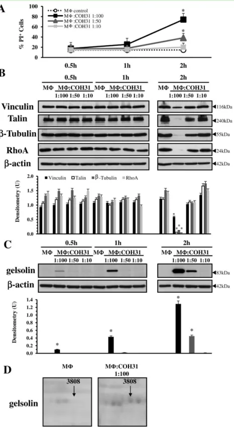
To this end MΦ were infected with GBS-III-COH31 for 0.5, 1 and 2 h at three different MΦ : GBS infection ratios and the levels of gelsolin and β-actin (loading control) were analysed by Western blot. Western blot analysis showed that the levels of gelsolin increased in a time- and infection-ratio-dependent manner in MΦ after
GBS-III-COH31 infection reaching the maximum at 2 h (Fig. 1C). GBS-III-COH31 at a ratio of 1:100 induced gelsolin expression in MΦ by approximately 0.1 densitometry units at 0.5 h of infection (Fig. 1C), 0.43 densitometry units at 1 h of infection (Fig. 1C) and 1.29 densitometry units at
2 h of infection (Fig. 1C), while in control MΦ the gelsolin expression was undetectable at all times examined. GBS-III-COH31 at a ratio of 1:50 induced gelsolin expression in MΦ by approximately 0.45 densitometry units only at 2 h infection (Fig. 1C). On the contrary GBS-III-COH31 at a
Fig. 1. Effect of MΦ : GBS-III-COH31
interaction on the expression of gelsolin. A. Flow cytometry analysis. Percentage of PI+ cells was determined by PI uptake assay at flow cytometry in control MΦ and MΦ infected with GBS-III-COH31 at the indicated ratios, recovered at different times after infection. Data are means± SD of six experiments done in triplicate. *P< 0.01 GBS-infected MΦ versus control MΦ.
B. Western blot analysis of cytoskeletal protein expression. Lysates from control MΦ and MΦ infected with GBS-III-COH31 (MΦ : COH31) at the indicated ratios, prepared at the indicated times, were subjected to SDS-PAGE. The filters were cut around 80 kDa and 30 kDa. The top sections probed with anti-Vinculin, then stripped and reprobed with anti-Talin. The middle sections probed with anti-β-Tubulin, then stripped and reprobed with anti-β-actin. The bottom sections were probed with anti-RhoA. C. Western blot analysis of gelsolin
expression. Lysates from control MΦ and MΦ infected with GBS-III-COH31 (MΦ : COH31) at the indicated ratios, prepared at the indicated times, were subjected to SDS-PAGE. The filters were cut around 70 kDa, the top sections were probed with anti-gelsolin and the bottom sections were with anti-β-actin.
D. Proteomic analysis of gelsolin expression. Details from 2-DE maps were cropped to show one spot identified as gelsolin which was overexpressed in MΦ infected with GBS-III-COH31 (MΦ : COH31), compared to control MΦ. The spot was detected only in gels from MΦ infected with GBS-III-COH31. B and C. The density of bands corresponding to each protein was evaluated by
densitometric analysis. Densitometry units (U) were calculated relative toβ-actin and values for the densitometric analyses obtained from four independent experiments. *P< 0.01 GBS-infected MΦ versus control MΦ.
ratio of 1:10 induced no increase in gelsolin expression in MΦ at all times examined (Fig. 1C).
Gelsolin increase occurred already at 0.5 h and strongly increased at 1 h after GBS-III-COH31 infection of MΦ (Fig. 1C) times at which significant changes of intracellular [Ca2+] were detected (Fettucciari et al., 2000; 2006) but no cytoskeleton disruption (Fettucciari et al., 2011; Fig. 1B) was found, indicating that gelsolin overexpression occurs before cytoskeletal degradation.
Unpublished results of proteomic analysis performed in our previous paper (Susta et al., 2010) on control MΦ and MΦ infected with GBS-III-COH31 at a ratio of 1:100 for 2 h showed that a spot identified as gelsolin was overexpressed in MΦ infected with GBS-III-COH31 but was undetectable in control MΦ (Fig. 1D) confirming the Western blot results.
Overall, these results indicate that GBS-III-COH31 induces a strong gelsolin increase in MΦ and that this is a very early event during MΦ : GBS interaction.
Gelsolin increase is independent of transcriptional, translational, protein-kinase and proteasome activity In order to understand the mechanisms responsible for gelsolin increase we first investigated the role of transcrip-tion activity by quantitative real-time PCR (qRT-PCR). To this end we evaluated gelsolin mRNA content in control and GBS-infected MΦ at 2 h after infection, time at which we observed the higher gelsolin increase. No significant increase of gelsolin expression at mRNA level was observed with qRT-PCR in GBS-infected MΦ with respect to control MΦ (Fig. 2A).
Successively we investigated the role of de novo protein synthesis. MΦ were pretreated with 10 or 50μg ml−1 Cycloheximide (CHX), an inhibitor of eukaryotic protein synthesis, (Fettucciari et al., 2000; Croons et al., 2007; Ji et al., 2010) for 1 h before infection with GBS-III-COH31 and gelsolin expression was ana-lysed by Western blot. The ratio of gelsolin to loading controlβ-actin was used to monitor the relative levels of gelsolin on CHX-treatment. As shown in Fig. 2B, the gelsolin expression induced by GBS-III-COH31 was not significantly blocked by pretreatment with 10 or 50μg ml−1 CHX. In fact, a similar level of gelsolin expression was observed in non-treated GBS-infected MΦ and GBS-infected MΦ treated with CHX at 10 or 50 μg ml−1. There-fore the gelsolin increase in MΦ by GBS is not due to de novo protein synthesis.
To continue to explore the molecular mechanism impli-cated in the gelsolin increase by MΦ : GBS-III-COH31 interaction, since the fundamental functions of the signal-ling cascade in evoking cellular responses to external stimuli/infection are mediated by protein-kinases, we investigated the role of Staurosporine (STS), a potent
inhibitor of protein-kinases that acts by competing with ATP in binding to the nucleotide-binding pocket, and has a broad activity across a variety of protein-kinases (Toledo and Lydon, 1997; Ji et al., 2010). MΦ were pretreated with 63 nM STS 1 h before infection with GBS-III-COH31 at a ratio of 1:100 for 2 h. The results showed that 63 nM STS does not significantly inhibit the gelsolin increase induced by GBS (Fig. 2C) indicating that gelsolin increase is not mediated by activation of protein-kinases.
Since the increased gelsolin expression is not due to transcriptional, translation or protein-kinase activity we initially looked whether gelsolin increase was caused by inhibition of proteasome activity by GBS that leads to altered turnover of gelsolin and its accumulation in MΦ. To this end we analysed, by Western blot, the accumulation of multiubiquitinylated proteins in GBS-infected MΦ (Paolini et al., 2001). As positive control to show accu-mulation of multiubiquitinylated proteins we used MΦ treated with 2μM clasto-Lactacystin-β-lactone (clasto-Lactacystin) that inhibits proteasome activity in MΦ (Paolini et al., 2001; Fettucciari et al., 2006). We found an accumulation of high molecular weight protein-ubiquitin conjugates in clasto-Lactacystin treated MΦ (Fig. 2D) whereas GBS-infected MΦ, as control MΦ, showed no accumulation of multiubiquitinylated proteins (Fig. 2D). Therefore the gelsolin increase is not due to inhibition of MΦ proteasome activity by GBS.
Rather the observation that there is a build up of ubiquitinated proteins in control MΦ with respect to GBS-infected MΦ in Fig. 2D and in Fig. 3A leads to the hypoth-esized that this could be due to calpain activation induced by GBS. In fact, over ubiquitin proteasome pathway other cellular apparatuses such as calpains play significant roles in post-transcriptional processing and turnover of several cellular proteins (Coux et al., 1996; Goll et al., 2003; Zhou, 2004; Nandi et al., 2006). Moreover, it has been demonstrated in some experimental models that calpain activation increased total protein degradation and in particular calpain activation increased cytoplasmic proteolysis mediated by proteasome (Menconi et al., 2004; Smith and Dodd, 2007). Therefore, calpains acti-vated by GBS could increase both total cytoplasmic proteolysis and proteolysis mediated by proteasome.
Proteasome inhibitors have been shown to stabilize proteins designated for degradation by proteasome (Coux et al., 1996; Patrick et al., 1998; Zhao et al., 2000). Since in other experimental models the gelsolin is efficiently degraded by the ubiquitin proteasomal pathway (Ni et al., 2008), inhibitors of the proteasome such as MG132 or clasto-Lactacystin are likely to increase the half-life (t1/2) of this protein in MΦ. To examine the effect of proteasomal inhibitors on gelsolin stability we used two different proteasome inhibitors, MG132 and clasto-Lactacystin, and evaluated gelsolin expression at 2 h after infection
and 5 h after infection (2 h infection plus 3 h incubation with antibiotics). Sinceβ-actin at 5 h after infection begins to decrease in GBS-infected MΦ (Fig. 3A, right panel) to monitor the relative levels of gelsolin in kinetics after proteasome inhibitor treatment we used Manganese Superoxide Dismutase (MnSOD) as loading control. The
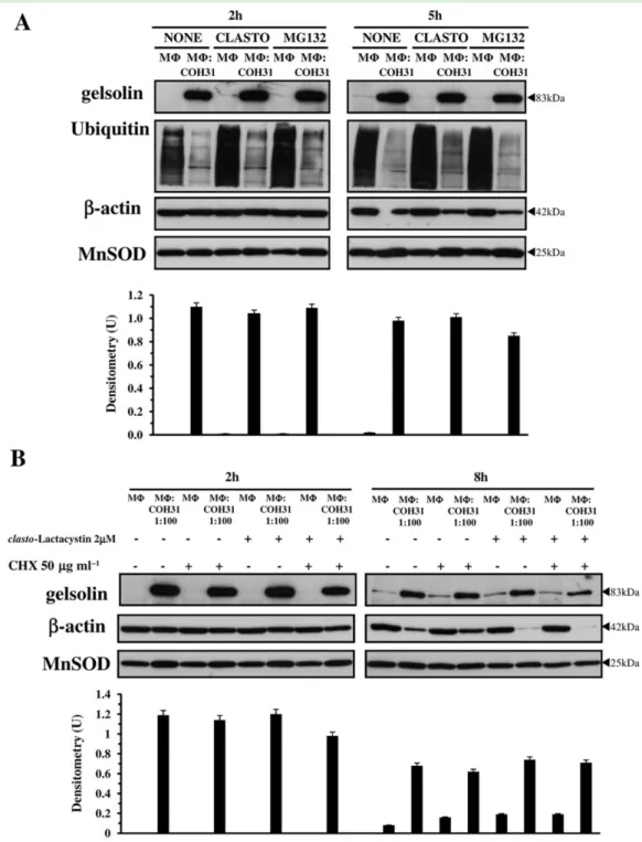
results obtained show that at all times examined the level of gelsolin in GBS-infected MΦ pretreated for 1.5 h with clasto-Lactacystin (2μM) or MG132 (5 μM) remain the same as that of non-treated GBS-infected MΦ, indicating that in our experimental model both clasto-Lactacystin and MG132 do not stabilize gelsolin (Fig. 3A, upper panels). Proteasome activity is efficiently inhibited by pretreatment of MΦ with clasto-Lactacystin (2 μM) or MG132 (5μM) as demonstrated by accumulation of multiubiquitinylated proteins in MΦ pretreated with proteasome inhibitors, at all times examined (Fig. 3A, middle panels). These data further indicate that pro-teasome is not involved in gelsolin turnover in GBS-infected MΦ.
To rule out definitively the proteasome involvement in gelsolin turnover we also evaluated the gelsolin t1/2 by CHX treatment and simultaneous treatment with the proteasomal inhibitor clasto-Lactacystin (Zhao et al., 2000; Zhou, 2004). To this end MΦ were pretreated for 1.5 h with 2μM clasto-Lactacystin and simultaneously with 50μg ml−1CHX, then infected with GBS-III-COH31 in the presence of clasto-Lactacystin and CHX which were maintained in the medium all during the experiment. Then gelsolin expression was analysed at 2 h after infection and 8 h after infection (2 h infection plus 6 h incubation with antibiotics) by Western blot. For the same reasons described above, to monitor the relative levels of gelsolin in kinetics after proteasome and CHX treatment we used as loading control MnSOD.
The results obtained showed that a weak gelsolin expression was detectable in untreated MΦ at 8 h of
Fig. 2. Molecular mechanisms of gelsolin increase.
A. Analysis of gelsolin mRNA expression by qRT-PCR. Control MΦ and MΦ infected with GBS-III-COH31 at a ratio of 1:100 were subjected to qRT-PCR and data expressed as 2−ΔΔCt.
B. Effect of protein synthesis inhibitor on gelsolin expression by Western blot analysis. Lysates from non-treated MΦ, MΦ pretreated for 1 h with CHX (10 or 50μg ml−1), infected or not with GBS-III-COH31 (MΦ : COH31) at a 1:100 ratio for 2 h, were subjected to SDS-PAGE. The filter was probed with anti-gelsolin then stripped and reprobed with anti-β-actin.
C. Effect of protein-kinase inhibitor on gelsolin expression by Western blot analysis. Lysates from non-treated MΦ, MΦ pretreated for 1 h with STS (63 nM), infected or not with GBS-III-COH31 (MΦ : COH31) at a 1:100 ratio for 2 h, were subjected to SDS-PAGE. The filter was cut around 70 kDa and the top probed with anti-gelsolin, while the bottom was probed with anti-β-actin.
D. Western blot analysis of multiubiquitinylated proteins. Lysates from non-treated MΦ, MΦ pretreated for 1.5 h with
clasto-Lactacystin (2μM), infected or not with GBS-III-COH31
(MΦ : COH31) at a 1:100 ratio for 2 h, were subjected to
SDS-PAGE. The filter was probed with anti-Ubiquitin, then stripped and reprobed with anti-β-actin. Vertical lines in blots indicate repositioned gel lanes.
B and C. The density of the bands corresponding to gelsolin was evaluated by densitometric analysis. Densitometry units (U) were calculated relative toβ-actin and values for the densitometric analyses obtained from four independent experiments.
Fig. 3. Effect of proteasome on gelsolin turnover.
A. Western blot analysis of gelsolin and multiubiquitinylated proteins after proteasome inhibitor treatment. Lysates from non-treated MΦ, MΦ pretreated for 1.5 h with clasto-Lactacystin (2μM) or MG132 (5 μM), infected or not with GBS-III-COH31 (MΦ : COH31) at a 1:100 ratio, prepared at 2 h after infection and at 5 h after infection (2 h infection plus 3 h incubation with antibiotics), were subjected to SDS-PAGE. The filters were cut around 60 kDa and the top sections probed with anti-gelsolin then stripped and reprobed with anti-Ubiquitin, while the bottom sections were probed with anti-β-actin then stripped and reprobed with anti-MnSOD.
B. Effect of simultaneous treatment with protein synthesis inhibitor and proteasome inhibitor on gelsolin expression by Western blot analysis. Lysates from non-treated MΦ, MΦ pretreated for 1.5 h with CHX (50 μg ml−1), MΦ pretreated for 1.5 h with clasto-Lactacystin (2 μM), MΦ pretreated for 1.5 h simultaneously with clasto-Lactacystin (2μM) and CHX (50 μg ml−1), infected or not with GBS-III-COH31 (MΦ : COH31) at a 1:100 ratio, prepared at 2 h after infection and at 8 h after infection (2 h infection plus 6 h incubation with antibiotics), were subjected to SDS-PAGE. The filters were cut around 70 kDa and the top sections probed with anti-gelsolin, while the bottom sections were probed with anti-β-actin then stripped and reprobed with anti-MnSOD.
A and B. The density of the bands corresponding to gelsolin was evaluated by densitometric analysis. Densitometry units (U) were calculated relative to MnSOD and values for the densitometric analyses obtained from three independent experiments.
incubation (Fig. 3B, upper right panel) and although these results seem in contrast with undetectable gelsolin expression in untreated MΦ at 2 h, we think that this could be due to the fact that with the further time of incubation MΦ adhere more strongly to the substrate and this could lead to cytoskeleton reorganization that causes slightly changes in gelsolin expression. However prolonging the time of incubation (24 h) the gelsolin expression in untreated MΦ remain the same of that observed at 8 h of incubation (data not shown).
More important the results of CHX inhibition experi-ments showed also that gelsolin had a long t1/2. In fact, in these experiments we were unable to determine gelsolin t1/2,as we found a similar level of gelsolin in CHX-treated GBS-infected MΦ and non-treated GBS-infected MΦ at all times examined (Fig. 3B, upper panels). Furthermore the gelsolin expression induced by GBS-III-COH31 was not affected by simultaneous treatment with CHX and clasto-Lactacystin for all the course of the experiment. In fact, a similar level of gelsolin expression was observed in non-treated GBS-infected MΦ and in GBS-infected MΦ treated simultaneously with 50 μg ml−1 CHX and 2μM clasto-Lactacystin, at all times examined (Fig. 3B, upper panels). These data definitively confirm that gelsolin turnover in GBS-infected MΦ is not regulated by proteasome.
Gelsolin increase is not due to redistribution by subcellular compartments
It is known that studying the proteins that associate with and regulate the actin cytoskeleton has been traditionally difficult. To prepare whole cell lysates we used a modified Radioimmunoprecipitation assay (RIPA) lysis buffer which contains three different detergents (SDS; Triton X-100, sodium deoxycholate) so allowing the recovery also of cytoskeletal-associated proteins. However, to rule out that gelsolin may be in a subcellular compartment which is typically masked in conventional methods of Western blot analysis of whole cell lysates, we used the EMD Millipore ProteoExtract®Cytoskeleton Enrichment and Isolation Kit. This kit greatly increases the ability to detect and study the low abundance actin-associated proteins allowing the retention of focal adhesion and actin-associated proteins while removing soluble cytoplasmic and nuclear proteins from the cell. To this end MΦ were infected with GBS-III-COH31 for 1 h and 2 h at two different MΦ : GBS infection ratios and the levels of gelsolin, vimentin (loading control for cytoskeletal fraction), glyceraldehyde-3-phosphate dehydrogenase (GAPDH; loading control for nuclear and soluble fractions), andβ-actin (loading control for all frac-tions since GAPDH was downmodulated by GBS) were analysed in the cytoskeletal, soluble and nuclear fraction by Western blot. Gelsolin was detected in both the soluble
and cytoskeletal fraction of MΦ infected with GBS-III-COH31 for 1 h at a MΦ : GBS ratio of 1:100 with a similar level of expression (Fig. 4A) and a strong increase in the cytoskeletal fraction of MΦ infected with GBS-III-COH31 for 2 h at a ratio 1:100 (Fig. 4B). In contrast gelsolin was not found in any subcellular fraction of control MΦ at all times examined (Fig. 4). While in MΦ infected with
Fig. 4. Analysis of gelsolin expression in cytoskeletal, soluble or
nuclear subfraction.
Cytoskeletal, soluble or nuclear subfraction from control MΦ, MΦ infected with GBS-III-COH31 (MΦ : COH31) at the indicated ratios, prepared at 1 h after infection (A) and 2 h after infection (B), were subjected to SDS-PAGE. The filters were probed with anti-gelsolin, then stripped and reprobed with anti-vimentin (loading control for the cytoskeletal fraction), then stripped and reprobed with anti-GAPDH (loading control for the soluble and nuclear fraction) then stripped and reprobed with anti-β-actin.
A and B. The density of the bands corresponding to gelsolin was evaluated by densitometric analysis. Densitometry units (U) were calculated relative toβ-actin and values for the densitometric analyses obtained from three independent experiments.
GBS-III-COH31 at a ratio of 1:10 gelsolin was detected in both the soluble and cytoskeletal fraction at very low levels only at 2 h after infection (Fig. 4B).
Ca2+is critical for gelsolin increase
Since, as previously reported GBS-III-COH31 induces a strong increase of intracellular Ca2+ in MΦ (Fettucciari et al., 2000; 2006) and gelsolin is a Ca2+-regulated protein (Kwiatkowski, 1999; McGough et al., 2003; Silacci et al., 2004; Li et al., 2012), to study the involvement of Ca2+in gelsolin increase induced by MΦ : GBS-III-COH31 inter-action, we analysed the effect of the extracellular Ca2+ chelator, EGTA. MΦ were infected or not with GBS-III-COH31 at a MΦ : GBS infection ratio of 1:100 in the presence of 1 mM EGTA in RPMI-1640 medium (contain-ing Ca2+and Mg2+) with 10% fetal bovine serum (FBS) at pH 7.3 for 2 h. EGTA at 1 mM strongly inhibited gelsolin increase by GBS-III-COH31 in MΦ (Fig. 5A). Adding EGTA in RPMI-1640 medium (containing Ca2+and Mg2+) with 10% FBS at pH 7.3 did not affect, MΦ viability which remained at 98–99% as evaluated by Trypan blue assay (Table 1), and MΦ adherence/morphology. In fact, by analysis of MΦ monolayers treated or not with EGTA infected or not with GBS-III-COH31 by inverted micro-scope controlled by cooled camera, no changes in cell morphology or adherence were observed between non-treated MΦ and EGTA-treated MΦ (Fig. 5B), or between GBS-infected non-treated MΦ and GBS-infected EGTA-treated MΦ. Significant changes were observed between control MΦ treated or not and GBS-infected MΦ treated or not (Fig. 5B) likely due to MΦ cytoskeletal disruption by GBS (Fig. 1B).
To further demonstrate that only extracellular Ca2+is responsible for gelsolin increase, without the emptying of intracellular Ca2+ stores, we used the intracellular Ca2+ chelator [1,2-Bis(2-aminophenoxy)ethane-N,N,N′, N′-tetraacetic acid tetrakis(acetoxymethyl ester), BAPTA/
AM]. MΦ were pretreated with 15 μM BAPTA/AM for 1 h then infected or not with GBS-III-COH31 at a MΦ : GBS infection ratio of 1:100 in the presence of BAPTA/AM in RPMI-1640 medium (containing Ca2+and Mg2+) with 10% FBS at pH 7.3 for 2 h. BAPTA/AM did not reduce gelsolin increase induced by GBS-III-COH31 in MΦ (Fig. 5C), thus demonstrating that the emptying of intracellular Ca2+ stores, is not involved in gelsolin increase.
Although EGTA preferentially binds to Ca2+ it can be considered a general chelator of divalent ions such as Mg2+. Therefore, to show the specificity of EGTA for Ca2+ in gelsolin increase and definitively demonstrate that reduction of gelsolin by EGTA is due to influx of extracellular Ca2+, we performed experiments by addition of an excess of CaCl2or MgCl2 during incubation with EGTA in RPMI-1640 medium (containing Ca2+and Mg2+) with 10% FBS at pH 7.3. The inhibition of gelsolin increase by EGTA in GBS-infected MΦ can be reversed by addition of an excess of CaCl2but not MgCl2during incubation with EGTA (Fig. 5D). However, addition of an excess of CaCl2 alone in control MΦ did not induce gelsolin increase (Fig. 5D). Therefore, these results showing that gelsolin increase was inhibited in the pres-ence of EGTA and restored only by CaCl2demonstrated that GBS-induced gelsolin increase was caused by extracellular Ca2+influx.
Calpain activation and caspase degradation are involved in gelsolin increase
Although the ubiquitin proteasome pathway constitutes the bulk of regulated cytoplasmic proteolysis, in most cases other cellular apparatuses, such as calpains, lyso-some, and caspases also play significant roles in post-transcriptional processing and turnover of specific cellular proteins (Coux et al., 1996; Carafoli and Molinari, 1998; Ciechanover, 1998; Pillay et al., 2002; Goll et al., 2003; Zhou, 2004; Nandi et al., 2006; Bhatia et al., 2013).
Fig. 5. Extracellular Ca2+is responsible of gelsolin increase.
A. Effect of EGTA on gelsolin expression by Western blot analysis. Lysates from non-treated MΦ, MΦ treated with EGTA (1 mM), infected or not with GBS-III-COH31 (MΦ : COH31) at a 1:100 ratio for 2 h, were subjected to SDS-PAGE. The filter was cut around 70 kDa and the top probed with anti-gelsolin, while the bottom was probed with anti-β-actin.
B. Microscope analysis of effect of EGTA on MΦ adherence and morphology. Control MΦ, MΦ infected or not with GBS-III-COH31 (MΦ : COH31) at a 1:100 ratio for 2 h, MΦ treated with EGTA (1 mM), MΦ treated with EGTA (1 mM) infected with GBS-III-COH31 (MΦ EGTA : COH31) at a 1:100 ratio for 2 h, were analysed by Olympus IX51 microscope and images collected by Spot-2 cooled camera. Images are representative of three independent experiments.
C. Effect of BAPTA/AM on gelsolin expression by Western blot analysis. Lysates from non-treated MΦ, MΦ pretreated for 1 h with 15 μM BAPTA/AM (BAPTA), infected or not with GBS-III-COH31 (MΦ : COH31) at a 1:100 ratio for 2 h, were subjected to SDS-PAGE. The filter was cut around 70 kDa and the top probed with anti-gelsolin, while the bottom was probed with anti-β-actin.
D. Analysis of effect of addition of an excess of CaCl2or MgCl2during incubation with EGTA on gelsolin expression by Western blot analysis. Lysates from non-treated MΦ, MΦ treated with EGTA (1 mM), MΦ treated with EGTA (1 mM) and addition of 1 mM CaCl2, MΦ treated with EGTA (1 mM) and addition of 1 mM MgCl2, infected or not with GBS-III-COH31 (MΦ : COH31) at a 1:100 ratio for 2 h, were subjected to SDS-PAGE. The filter was cut around 70 kDa and the top probed with anti-gelsolin, while the bottom was probed with anti-β-actin. A, C and D. The density of the bands corresponding to gelsolin was evaluated by densitometric analysis. Densitometry units (U) were calculated relative toβ-actin and values for the densitometric analyses obtained from four independent experiments. *P < 0.01 GBS-infected treated MΦ versus GBS-infected non-treated MΦ.
We previously demonstrated that GBS infection induces in MΦ an influx of extracellular Ca2+ which is responsible for calpain activation (Fettucciari et al., 2006; 2011). To determine if gelsolin increase is affected by calpains, we examined the effect of a highly selective inhibitor of the μ- and m-calpain, which acts on the Ca2+ binding site, [3-(4-Iodophenyl)-2-mercapto-(Z)-2-propenoic acid, PD150606] (Wang et al., 1996; Carafoli and Molinari, 1998; Goll et al., 2003; Fettucciari et al., 2006), on MΦ gelsolin increase induced by GBS-III-COH31. To confirm the inhibition of calpains by PD150606, since a marker for the activation of calpains is the cleavage of α-spectrin into 150 and 145 kDa break-down products (BP) (Wang et al., 1996; Goll et al., 2003; Fettucciari et al., 2006), we re-examined by Western blot the effect of PD150606 onα-spectrin cleavage induced by GBS-III-COH31. The results obtained show that the pre-treatment of MΦ with 100 μM of PD150606 prevented the formation of 145 kDaα-spectrin BP induced by GBS-III-COH31 in MΦ (Fig. 6A), confirming that GBS-III-COH31 activates calpains and PD150606 inhibits their activation (Fig. 6A) and more important, the pretreatment of MΦ with 100μM of PD150606 strongly reduces the gelsolin increase induced by GBS-III-COH31 (Fig. 6B), thus indicating that gelsolin increase is also due to calpain activation.
To investigate whether other proteases in particular lysosomal cathepsins are involved in MΦ gelsolin increase induced by GBS-III-COH31, we evaluated the effect of: i) the cathepsin B inhibitor IV (CA-074 Me), ii) the inhibitor of all lysosomal cathepsins, ammonium chloride (NH4Cl), iii) the cathepsin D inhibitor (Pepstatin A). No inhibition or increase of gelsolin expression levels was observed with CA-074 Me (10μM), NH4Cl (10 mM) or Pepstatin A (10μM) (Fig. 6C). This indicates that MΦ gelsolin increase is not due to the proteolytic activity of cathepsins.
Since it is well known that gelsolin is cleaved by caspase-3 (Kothakota et al., 1997; Jänicke et al., 1998; Kwiatkowski, 1999; Azuma et al., 2000; Koya et al., 2000; Silacci et al., 2004; Li et al., 2012), and we previously
demonstrated that GBS cleave caspase-3 and -7 by calpain activation (Fettucciari et al., 2006), gelsolin increase in GBS-infected MΦ may be due to post-translational regulation based on reduced degradation of gelsolin owing to caspase degradation. To this end we analysed more rigorously the effect of boc-Asp(OMe)-fluoromethylketone (BAF), a broad spectrum caspase inhibitor, on gelsolin expression by Western blot and always by Western blot re-evaluated caspase-3 cleavage in MΦ infected with GBS-III-COH31 at a ratio of 1:100 for 2 h and the effect of calpain inhibitor, PD150606.
The analysis of caspase-3 expression confirms our previous results (Fettucciari et al., 2006) indicating that GBS-III-COH31 cleaves caspase-3 in MΦ (Fig. 7A) and PD150606, inhibits both the caspase-3 cleavage (Fig. 7A) and gelsolin increase (Figs 6B and 7A). Moreover we found that caspase inhibitory activity of BAF (25μM) further increases the levels of gelsolin induced by GBS-III-COH31 in MΦ (Fig. 7B), without affecting caspase-3 expression levels in control and GBS-infected MΦ (Fig. 7C), indicating that in our model caspase-3 is involved in gelsolin increase. Therefore the demonstration that gelsolin increase was prevented by calpain inhibition which inhibited caspase-3 cleavage together with the observation that gelsolin increase was enhanced by caspase inhibitor BAF, indicates that gelsolin increase in GBS-infected MΦ involves altered degradation of gelsolin for lack of caspase-3.
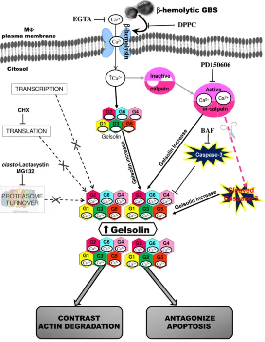
Overall, these data indicate that gelsolin increase in MΦ induced by GBS-III-COH31 requires influx of extracellular Ca2+, calpain activation mediated by Ca2+ influx, and caspase-3 degradation.
Confocal microscopy analysis of gelsolin expression in GBS-III-COH31 infected MΦ
Control MΦ and MΦ infected with GBS-III-COH31 at a ratio of 1:100 for 2 h, were triple-labelled using Alexa Fluor 488 phalloidin to detect F-actin, an Alexa Fluor 568-labelled goat anti-mouse secondary antibody (Ab) to detect gelsolin, and DAPI to visualized nuclei, and observed by confocal microscopy (Nikon Instruments, Amsterdam, Netherlands, C1 on Eclipse Ti; EZC1 software).
As illustrated in Fig. 8 top panels, control MΦ showed a very weak immunostaining for gelsolin, whereas many MΦ infected with GBS-III-COH31 were characterized by a strong and diffused gelsolin immunostaining. Conversely, no major differences in F-actin staining were detected between non-infected and infected MΦ (Fig. 8, top panels). Moreover, the GBS-induced increase of gelsolin immunolabelling was reduced by pretreatment with the calpain inhibitor PD150606 (100μM) (Fig. 8, middle panels) and prevented by treatment with the Ca2+
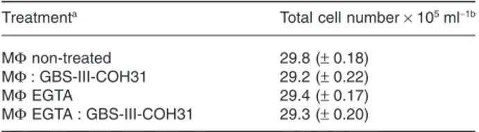
Table 1. Effect of EGTA on MΦ viability.
Treatmenta Total cell number× 105ml−1b MΦ non-treated 29.8 (± 0.18)
MΦ : GBS-III-COH31 29.2 (± 0.22)
MΦ EGTA 29.4 (± 0.17)
MΦ EGTA : GBS-III-COH31 29.3 (± 0.20)
a. MΦ were infected or not with GBS-III-COH31 at a 1:100 ratio for
2 h in the presence or not of EGTA 1 mM.
b. At 2 h after infection, the cells were recovered as described in Experimental procedures and the total number of cells was
deter-mined by Trypan blue assay. The data are means± SD of four experi-ments performed in triplicate.
chelator, EGTA (1 mM) (Fig. 8, bottom panels), in agree-ment with the results of Western blot analysis.
These findings confirm at cellular level that GBS-III-COH31 induces gelsolin increase in MΦ by extracellular Ca2+influx and calpain activation.
GBSβ-haemolysin is involved in gelsolin increase In a further series of experiments we analysed the possi-ble microbial factor/s involved in MΦ gelsolin increase after GBS interaction. GBS has a pluripotent virulence factor, theβ-haemolysin, strictly bound to the cell surface,
highly unstable when released in culture supernatants, requiring metabolic activity for its production, and in virtue of its pore-forming-like activity against the eukaryotic cell membrane (Nizet et al., 1996; Valentin-Weigand et al., 1997; Fettucciari et al., 2000; 2006; Nizet, 2002; Hensler et al., 2008; Maisey et al., 2008), causes plasma membrane permeability defects which allow a massive Ca2+ influx in MΦ and in other different cell types (Valentin-Weigand et al., 1997; Fettucciari et al., 2000; 2006; Hensler et al., 2008). Since in our model, as reported above, the MΦ gelsolin increase was mediated by extracellular Ca2+ influx we examined the possibility
Fig. 6. PD150606 inhibited gelsolin increase while cathepsin inhibitors did not.
A. Effect of PD150606 onα-spectrin cleavage by Western blot analysis. Lysates from non-treated MΦ, MΦ pretreated for 1 h with PD150606 (100μM), infected or not with GBS-III-COH31 (MΦ : COH31) at a 1:100 ratio for 2 h, were subjected to SDS-PAGE. The filter probed with anti-α-spectrin then stripped and reprobed with anti-β-actin. Intact protein (solid arrow) and BP (open arrow) are indicated.
B. Effect of PD150606 on gelsolin expression by Western blot analysis. Lysates from non-treated MΦ, MΦ pretreated for 1 h with PD150606 (100μM), infected or not with GBS-III-COH31 (MΦ : COH31) at a 1:100 ratio for 2 h, were subjected to SDS-PAGE. The filter was cut around 70 kDa and the top probed with anti-gelsolin, while the bottom was probed with anti-β-actin.
C. Effect of cathepsin inhibitors on gelsolin expression by Western blot analysis. Lysates from non-treated MΦ, MΦ pretreated for 1.5 h with CA-074 Me (10μM), MΦ pretreated for 1.5 h with NH4Cl (10 mM), and MΦ pretreated for 18 h with Pepstatin A (10 μM), infected or not with GBS-III-COH31 (MΦ : GBS) at a 1:100 ratio for 2 h, were subjected to SDS-PAGE. The filters were cut around 70 kDa and the top sections probed with anti-gelsolin, while the bottom sections were probed with anti-β-actin. Vertical lines in blots in right panel indicate repositioned gel lanes.
A–C. The density of the bands corresponding to gelsolin,α-spectrin f.l., α-spectrin BP was evaluated by densitometric analysis. Densitometry units (U) were calculated relative toβ-actin and values for the densitometric analyses obtained from four independent experiments. *P < 0.01 GBS-infected treated MΦ versus GBS-infected non-treated MΦ.
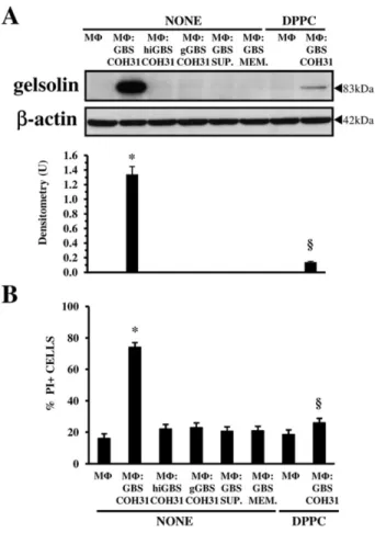
that GBS-III-COH31 could cause gelsolin increase by β-haemolysin. Therefore, Western blot analysis, to inves-tigate gelsolin increase, and propidium iodide (PI) uptake assay, to evaluate plasma membrane permeability altera-tions, were performed at 2 h in MΦ incubated, at an infection ratio of 1:100 with: (i) hiCOH31, GBS-III-COH31 killed at 60°C for 30 min, a condition that causes GBS β-haemolysin inactivation; (ii) gGBS-III-COH31, GBS-III-COH31 grown for 18 h in the presence of 10 mg ml−1 glucose, a condition which abolishes GBS β-haemolysin synthesis; (iii) supernatant of III-COH31 growth in culture medium for 2 h; and (iv) GBS-III-COH31 in contiguous medium separated by a 0.45μm pore size membrane of cell culture insert.
Under these conditions, we found no gelsolin increase (Fig. 9A) or alterations in MΦ plasma membrane perme-ability (Fig. 9B), which are both induced only by β-haemolytic GBS-III-COH31 (Fig. 9A and B). In fact, as shown in Fig. 9B, the MΦ infected with β-haemolytic GBS-III-COH31 showed 78% PI+ cells, while in the MΦ infected in all other conditions the percentage of PI+ cells was 18–21% similar to control MΦ (19%) (Fig. 9B). In spite of 78% PI+ cells the total number of MΦ infected with β-haemolytic GBS-III-COH31 at 2 h after infection, evalu-ated by Trypan blue exclusion assay, was the same as control MΦ and MΦ infected under all other conditions (results not shown).
To provide further evidence on the role ofβ-haemolysin in gelsolin increase, since it has been reported that some phospholipids such as dipalmitoylphosphatidylcholine
Fig. 7. Caspase-3 degradation mediated by calpains is involved in
gelsolin increase.
A. Western blot analysis of caspase-3 and gelsolin expression and effect of PD150606. Lysates from non-treated MΦ, MΦ pretreated for 1 h with PD150606 (100μM), infected or not with
GBS-III-COH31 (MΦ : COH31) at a 1:100 ratio for 2 h, were subjected to SDS-PAGE. The filter was cut around 70 kDa and the top probed with anti-gelsolin, while the bottom probed with anti-caspase-3 then stripped and reprobed with anti-β-actin. Intact protein (solid arrow) and BP (open arrow) are indicated. B. Effect of BAF on gelsolin expression by Western blot analysis. Lysates from non-treated MΦ, MΦ pretreated for 2 h with BAF (25μM), infected or not with GBS-III-COH31 (MΦ : COH31) at a 1:100 ratio for 2 h, were subjected to SDS-PAGE. The filter was cut around 70 kDa and the top probed with anti-gelsolin, while the bottom was probed with anti-β-actin.
C. Effect of BAF on caspase-3 expression by Western blot analysis. Lysates from non-treated MΦ, MΦ pretreated for 2 h with BAF (25μM), infected or not with GBS-III-COH31 (MΦ : COH31) at a 1:100 ratio for 2 h, were subjected to SDS-PAGE. The filter was cut around 70 kDa and the bottom probed with anti-caspase-3, then stripped and reprobed with anti-β-actin. Intact protein (solid arrow) and BP (open arrow) are indicated.
A–C. The density of the bands corresponding to each protein was evaluated by densitometric analysis. Densitometry units (U) were calculated relative toβ-actin and values for the densitometric analyses obtained from four independent experiments. *P< 0.01 GBS-infected treated MΦ versus GBS-infected non-treated MΦ.
(DPPC) inhibited GBS β-haemolytic activity (Fettucciari et al., 2000; Nizet, 2002), we also analysed the effect of DPPC. We found that DPPC in MΦ infected with GBS-III-COH31 at a 1:100 ratio strongly inhibited gelsolin increase (Fig. 9A) and prevented alterations in membrane permeability (Fig. 9B).
Loss ofβ-haemolysin synthesis, due to growing GBS-III-COH31 in the presence of glucose, and inhibition of GBSβ-haemolytic activity by heat-inactivation of GBS-III-COH31 or by DPPC, was confirmed by evaluating the β-haemolytic activity of gGBS-III-COH31, hiGBS-III-COH31 and GBS-III-hiGBS-III-COH31 in the presence or not of
Fig. 8. Confocal microscope analysis of gelsolin expression in GBS-III-COH31-infected MΦ and effect of EGTA and PD150606.
Control MΦ (MΦ) and MΦ infected with GBS-III-COH31 (MΦ : COH31) at 1:100 ratio for 2 h, untreated or pretreated for 1 h with PD150606 (100μM) and untreated or treated with EGTA (1 mM), were triple-labelled using Alexa Fluor 488 phalloidin to detect F-actin (green), an Alexa Fluor 568-labelled goat anti-mouse secondary Ab to detect gelsolin (red), and DAPI to visualized nuclei (blue), and observed by confocal microscopy. Bar, 50μm (for all images).
DPPC against Sheep Red Blood cells (results not shown) with the β-haemolytic activity assay (Marchlewicz and Duncan, 1980) performed as described previously (Fettucciari et al., 2000).
Overall, these results suggest that GBSβ-haemolysin is involved in MΦ gelsolin increase inducing extracellular Ca2+influx by MΦ plasma membrane permeability defects.
Effect of different GBS strains on gelsolin expression in MΦ
Recently, we demonstrated that highlyβ-haemolytic GBS type V strain NCTC10/84 (GBS-V-10/84) (Wilkinson, 1977; Liu et al., 2004) and β-haemolytic GBS type III strain NEM316 (GBS-III-NEM316) (Glaser et al., 2002; Tettelin et al., 2005), like GBS-III-COH31 but with lower MΦ : GBS ratios, induce marked alterations in MΦ plasma membrane permeability allowing Ca2+influx and calpain activation (Fettucciari et al., 2011), while the weak β-haemolytic GBS type Ia strain A909 (GBS-Ia-A909) and GBS type Ib strain H36B (GBS-Ib-H36B) (Tettelin et al., 2005) do not (Fettucciari et al., 2011). Since as above reportedβ-haemolysin is involved in MΦ gelsolin increase induced by MΦ : GBS-III-COH31 interaction to determine if gelsolin increase in MΦ is a general MΦ response to more β-haemolytic GBS or is specific for the clinical β-haemolytic GBS strain COH31 used in this study we employed the clinically isolated highlyβ-haemolytic GBS-V-10/84 (Wilkinson, 1977; Liu et al., 2004), the clinically isolated and fully sequenced β-haemolytic GBS-III-NEM316 (Glaser et al., 2002; Tettelin et al., 2005), and the clinically isolated and fully sequenced weakly β-haemolytic GBS-Ia-A909 and GBS-Ib-H36B (Tettelin et al., 2005) for which a differentβ-haemolytic activity has been demonstrated (Marchlewicz and Duncan, 1980; Nizet et al., 1996; Liu et al., 2004) and a different: sero-type, multilocus sequence type (ST) and dispensable genome (Glaser et al., 2002; Tettelin et al., 2005; Brochet et al., 2006). Therefore, Western blot analysis to examine the gelsolin increase and PI uptake assay to evaluate plasma membrane permeability alterations, were per-formed in MΦ infected with GBS-III-NEM316, GBS-V-10/ 84, GBS-Ia-A909, GBS-Ib-H36B, at different MΦ : GBS ratios for 2 h (time at which we observed the maximum gelsolin increase by GBS-III-COH31).
The interaction of MΦ both with GBS-III-NEM316 and GBS-V-10/84 induced at 2 h after infection in a ratio-dependent manner a strong gelsolin increase as shown by Western blot analysis (Fig. 10A) and marked altera-tions in MΦ plasma membrane permeability, as shown by PI uptake assay which found about 78% MΦ PI+ with GBS-III-NEM316 at a ratio of 1:20 and 72% MΦ PI+ with GBS-V-10/84 at a ratio of 1:20 (Fig. 10B). In contrast, GBS-Ia-A909 and GBS-Ib-H36B did not induce gelsolin also at the highest MΦ : GBS ratio of 1:100 (Fig. 10A) and in MΦ infected with GBS-Ia-A909 and GBS-Ib-H36B only a weak increase in the percentage of PI+ cells was found (Fig. 10B).
Fig. 9. Effect ofβ-haemolysin on gelsolin increase.
Control MΦ, MΦ infected with GBS-III-COH31 (MΦ : GBS-COH31) at a 1:100 ratio, MΦ infected with III-COH31 (MΦ : hiGBS-COH31) at a 1:100 ratio, MΦ infected with gGBS-III-COH31 (MΦ : gGBS-COH31) at a 1:100 ratio, MΦ incubated with supernatant of GBS-III-COH31 (MΦ : GBS-SUP.) growth in culture medium for 2 h at a concentration equivalent to that of MΦ : GBS ratio of 1:100, MΦ infected with GBS-III-COH31 at a 1:100 ratio, in contiguous medium separated by a 0.45μm pore membrane of cell culture insert (MΦ : GBS-MEM.), MΦ treated with 2 mg ml−1DPPC, MΦ infected with GBS-III-COH31 (MΦ : GBS-COH31) at a 1:100 ratio in the presence of 2 mg ml−1DPPC, were recovered at 2 h infection, for cell lysate preparation, SDS-PAGE and Western blot analysis (A) or for PI uptake assay (B).
A. Western blot analysis. The filter was cut around 70 kDa and the top probed with anti-gelsolin and the bottom with anti-β-actin. The density of the bands corresponding to gelsolin was evaluated by densitometric analysis. Densitometry units (U) were calculated relative toβ-actin and values for the densitometric analyses obtained from four independent experiments. *P< 0.01 GBS-infected MΦ versus control MΦ;§P< 0.01 GBS-infected treated MΦ versus GBS-infected non-treated MΦ.
B. PI uptake assay. Percentage of PI+ cells was determined evaluating PI uptake at flow cytometry. The data are means± SD of six experiments performed in triplicate. *P< 0.01 GBS-infected MΦ versus control MΦ;§P< 0.01 GBS-infected treated MΦ versus GBS-infected non-treated MΦ.
The above results demonstrating that marked increase of gelsolin in MΦ is induced by the interaction of MΦ with GBS-III-NEM316 and with GBS-V-10/84 that have a β-haemolytic titre of 8 and 64 respectively, but not with GBS-Ia-A909 or GBS-Ib-H36B that have aβ-haemolytic titre of 4 (Marchlewicz and Duncan, 1980; Nizet et al., 1996; Liu et al., 2004), indicate that only highly β-haemolytic GBS induced gelsolin increase in MΦ.
Altogether these data indicate that, like GBS-III-COH31, a marked increase in gelsolin levels in MΦ is
also induced by interaction of MΦ with other highly β-haemolytic GBS strains, confirming that β-haemolytic activity is directly involved in gelsolin increase.
Gelsolin increase and MΦ apoptosis
In agreement with the results of our previous studies, GBS-III-COH31 induces MΦ apoptosis at 24 h in an infection-ratio-dependent manner (Fettucciari et al., 2000; 2006; Fig. S1). Since it is well known that gelso-lin is intimately associated with pro-apoptotic or anti-apoptotic functions (Kothakota et al., 1997; Kwiatkowski, 1999; Koya et al., 2000; Kusano et al., 2000; Silacci et al., 2004; Li et al., 2012), we investigated first if the gelsolin increase was caused by MΦ apoptosis induction by other inducers and then its role in GBS-induced apoptosis.
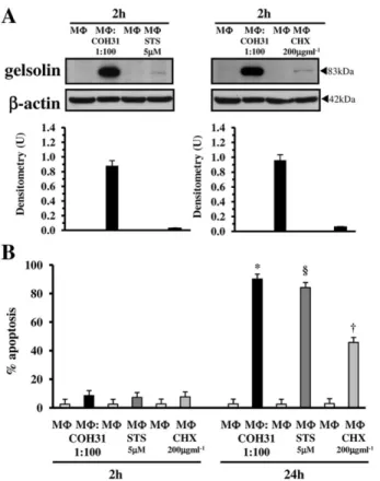
For the first aim we used two different inducers of apoptosis in MΦ, the CHX at 200 μg ml−1 (Gargalovic and Dory, 2003; Croons et al., 2007; Donovan et al., 2009) and the STS at 5μM (Rabkin and Kong, 2002; Benjamins et al., 2003; Norberg et al., 2008). To compare the effect of these apoptotic stimuli with the effect of GBS, gelsolin was analysed by Western blot in lysates of MΦ treated for 2 h, with CHX (200 μg ml−1) or with STS (5μM), and MΦ apoptosis was evaluated at 2 and 24 h measuring the percentage of apoptotic MΦ at flow cytometry, by assessment of DNA content after staining with PI in MΦ treated for 2 or 24 h with 200μg ml−1CHX or with 5μM STS.
Western blot analysis showed that gelsolin did not increase in MΦ at 2 h after addition of the apoptotic stimuli STS and CHX (Fig. 11A) although at 24 h respectively about 85% of apoptosis in MΦ treated with 5 μM STS for 24 h and 45% apoptosis in MΦ treated with 200 μg ml−1 CHX for 24 h was found (Fig. 11B).
For the second aim, to evaluate the possible func-tional role of gelsolin increase in GBS-induced MΦ apoptosis, we used the gelsolin small interfering RNA (siRNA) mediated gene silencing technique (Fettucciari et al., 2006) to knockdown gelsolin. For this, MΦ were transfected with 100 nM siRNA specific for gelsolin. At 72 h post transfection the cells were harvested for mRNA expression analysis by qRT-PCR or infected with GBS-III-COH31 for analysis of apoptosis. In these experiments to better assess the role of gelsolin in MΦ apoptosis by GBS, we used three MΦ : GBS infection ratios that showed ratio-dependent effects (see Fig. S1). We found that transfection of MΦ with gelsolin siRNA resulted in about 76% decrease of mRNA level of gelsolin compared to non-transfected MΦ (Fig. 12A). Mock transfection and transfection with 100 nM control non-targeting siRNA in MΦ did not affect mRNA ex-pression of gelsolin (Fig. 12A). As shown in Fig. 12B,
Fig. 10. Effect of different GBS strains on gelsolin expression in
MΦ.
Control MΦ, MΦ infected with GBS-III-COH31 (MΦ : COH31), MΦ infected with GBS-III-NEM316 (MΦ : NEM316), MΦ infected with GBS-V-10/84 (MΦ:10/84), MΦ infected with GBS-Ia-A909 (MΦ : A909) and MΦ infected with GBS-Ib-H36B (MΦ : H36B) at the indicated ratios, were recovered at 2 h after infection for cell lysate preparation, SDS-PAGE and Western blot analysis (A) or for PI uptake assay (B).
A. Western blot analysis. The filter was cut around 70 kDa, the top probed with anti-gelsolin, the bottom with anti-β-actin. The density of the bands corresponding to gelsolin was evaluated by densitometric analysis. Densitometry units (U) were calculated relative toβ-actin and values for the densitometric analyses obtained from four independent experiments. *P< 0.01 GBS-infected MΦ versus control MΦ.
B. PI uptake assay. The percentage of PI+ cells was determined evaluating PI uptake at flow cytometry. The data are means± SD of six experiments performed in triplicate. *P< 0.01 GBS-infected MΦ versus control MΦ.
downregulation of gelsolin expression resulted in about 17% increase of GBS-induced MΦ apoptosis at an in-fection ratio of 1:100, in about 35% increase of GBS-induced MΦ apoptosis at an infection ratio of 1:50 and in about 55% increase of GBS-induced MΦ apoptosis at a infection ratio of 1:10. Transfection with 100 nM control non-targeting siRNA in MΦ did not affect GBS-induced MΦ apoptosis (Fig. 12B).
Altogether these data indicate that gelsolin increase is not induced by any apoptotic stimuli and in our infection model contributes to counter the MΦ apoptosis induced by GBS.
Discussion
This study shows for the first time that in the complex interaction of GBS with MΦ is also involved gelsolin. Here we demonstrate that the interaction of MΦ with β-haemolytic GBS-III-COH31, in a time- and infection-ratio-dependent manner, leads very early to a strong gelsolin increase in MΦ. In fact, at 0.5 h of infection with GBS-III-COH31, at a ratio of 1:100, there is a significant
Fig. 11. Effect of two different MΦ apoptotic stimuli on gelsolin
increase and apoptosis.
A. Effect of STS and CHX on gelsolin expression by Western blot analysis. Lysates from control MΦ and lysates from MΦ infected with GBS-III-COH31 (MΦ : COH31) at a 1:100 ratio prepared at 2 h after infection, lysates from control MΦ and lysates from MΦ treated with STS (5μM; MΦ : STS) or with CHX (200 μg ml−1; MΦ : CHX) prepared at 2 h after stimulation, were subjected to SDS-PAGE. The filters were cut around 70 kDa and the top sections probed with anti-gelsolin, while the bottom sections were probed with anti-β-actin. The density of the bands corresponding to gelsolin was evaluated by densitometric analysis. Densitometry units (U) were calculated relative toβ-actin and values for the densitometric analyses obtained from four independent experiments.
B. Analysis of induction of MΦ apoptosis by STS and CHX at flow cytometry. Apoptosis of control MΦ, MΦ infected with GBS-III-COH31 at a 1:100 ratio, MΦ treated with STS (5 μM; MΦ : STS), MΦ treated with CHX (200 μg ml−1; MΦ : CHX) was measured at 2 and 24 h evaluating by flow cytometry the percentage of hypodiploid nuclei. Data are means± SD of six experiments done in triplicate. *P< 0.01 GBS-infected MΦ versus control MΦ.§P< 0.01 STS-treated MΦ versus control MΦ. †P< 0.01 CHX-treated MΦ versus control MΦ.
Fig. 12. Effect of gelsolin knockdown on GBS-III-COH31-induced
MΦ apoptosis.
A. Analysis of gelsolin mRNA by qRT-PCR following silencing of gelsolin gene by siRNA. At 72 h post transfection, cDNA from MΦ transfected, with vehicle alone (MOCK), control non-targeting siRNA (100 nM; si control), gelsolin (100 nM; si gelsolin) or non-transfected (NT), were subjected to qRT-PCR and data expressed as 2−ΔΔCt. *P< 0.01 MΦ transfected with siRNA versus non-transfected MΦ.
B. Effect of gelsolin silencing on apoptosis. At 72 h post transfection, MΦ transfected with control non-targeting siRNA (100 nM; si control), gelsolin siRNA (100 nM; si gelsolin) or non-transfected (NT), infected or not with GBS-III-COH31 at 1:100, 1:50 and 1:10 ratios for 2 h, were recovered at 24 h and apoptosis measured evaluating the percentage of hypodiploid nuclei at flow cytometry. Data are means± SD of four experiments done in triplicate. *P< 0.01 GBS-infected MΦ transfected with siRNA for gelsolin versus non-transfected GBS infected MΦ; **P < 0.05 GBS-infected MΦ at 1:100 ratio transfected with siRNA for gelsolin versus non-transfected GBS-infected MΦ at 1:100 ratio.
increase of gelsolin that continues to increase up to 2 h of infection, time at which GBS induced the maximum MΦ plasma membrane permeability defects, as demonstrated in this study and previously (Fettucciari et al., 2000; 2006; 2011), and the maximum intracellular Ca2+ increase (Fettucciari et al., 2000; 2006). While we observed no gelsolin expression, at all times examined, with GBS-III-COH31 at a MΦ : GBS infection ratio of 1:10 which did not induce plasma membrane permeability defects.
Since it is well known that protein level increase in the cells could be due to transcriptional events, translation events, alterations of protein turnover by protea-some inhibition or by activation of protein-kinases (Ciechanover, 1998; Nandi et al., 2006; Ji et al., 2010), we investigated which of these mechanisms was involved in the gelsolin increase induced by MΦ : GBS-III-COH31 interaction. Our results show that: (i) gelsolin mRNA did not increase after GBS-III-COH31 infection of MΦ; (ii) CHX, an inhibitor of protein synthesis (Fettucciari et al., 2000; Croons et al., 2007; Ji et al., 2010) at a concentra-tion of 50μg ml−1, did not inhibit the increase of gelsolin protein levels; (iii) proteasome is not involved in gelsolin increase, because GBS-III-COH31 did not inhibit pro-teasome activity, as demonstrated by no accumulation of multiubiquitinylated proteins, and proteasome inhibitors alone or with simultaneous CHX treatment did not increase gelsolin stability, (iv) gelsolin increase is not due to translocation of gelsolin from a different compartment or structure, as showed by analysis of gelsolin expression on subcellular fractions, (v) the STS, a potent and broad spectrum inhibitor of a variety of protein-kinases (Toledo and Lydon, 1997; Ji et al., 2010), did not significantly inhibit gelsolin increase, (vi) gelsolin seems a long-living protein. Indeed by CHX inhibition experiments (Zhou, 2004), we were unable to determine gelsolin t1/2 as we found a similar level of gelsolin in CHX-treated GBS-infected MΦ and non-treated GBS-infected MΦ at all times examined. Therefore, the gelsolin increase in MΦ after GBS infection is independent of transcription, de novo protein synthesis, proteasome function and protein-kinases activation.
However, in the light of the above results, since it is known that gelsolin is a Ca2+-regulated molecule which at low [Ca2+] has a compact, globular, inactive structure while at increasing [Ca2+] it opens up (Kwiatkowski, 1999; Kiselar et al., 2003; McGough et al., 2003; Silacci et al., 2004; Ashish et al., 2007; Li et al., 2012), the gelsolin increase in our model could be due to Ca2+and also to proteolytic control by some cellular proteases. In fact, it is known that, although the ubiquitin proteasome pathway constitutes the bulk of regulated cytoplasmic proteolysis, in most cases other cellular protease appa-ratuses play significant roles in post-translational regula-tion of cellular proteins levels (Coux et al., 1996; Carafoli
and Molinari, 1998; Ciechanover, 1998; Pillay et al., 2002; Goll et al., 2003; Zhou, 2004; Nandi et al., 2006; Bhatia et al., 2013).
Therefore in an attempt to define the mechanism involved in gelsolin increase, we first investigated the role of Ca2+ in MΦ gelsolin increase, since we previously showed that GBS causes a progressive and massive influx of extracellular Ca2+by inducing MΦ plasma mem-brane permeability defects (Fettucciari et al., 2000; 2006). In agreement with that observed in other experimental models which showed gelsolin upregulation by Ca2+ (Silacci et al., 2004; Ji et al., 2010; Li et al., 2012), the GBS-III-COH31-induced gelsolin increase was mediated by extracellular Ca2+ influx in MΦ. In fact, EGTA, a chelator of extracellular Ca2+, prevents the gelsolin increase induced by MΦ : GBS interaction and the addi-tion of an excess of CaCl2but not MgCl2during incubation with EGTA reverses the effect of EGTA. Furthermore BAPTA/AM, an intracellular Ca2+chelator, did not prevent gelsolin increase. Therefore influx of extracellular Ca2+, without the emptying of intracellular Ca2+ stores, is the principal responsible for gelsolin increase and represents the first step in the molecular mechanism involved in gelsolin increase.
Subsequently, we also investigated if the other cellular apparatuses, such as calpains, lysosomes and caspases that play significant roles in protein turnover (Coux et al., 1996; Carafoli and Molinari, 1998; Ciechanover, 1998; Pillay et al., 2002; Goll et al., 2003; Zhou, 2004; Nandi et al., 2006; Bhatia et al., 2013) are involved in the MΦ gelsolin increase by GBS.
Since we have previously demonstrated that GBS-III-COH31, as a result of the influx of Ca2+, activates the Ca2+-dependent protease calpains in MΦ (Fettucciari et al., 2006; 2011), we first determined if the gelsolin increase was influenced by calpain activation. The PD150606, a calpain inhibitor (Wang et al., 1996; Carafoli and Molinari, 1998; Goll et al., 2003; Fettucciari et al., 2006), strongly reduced the gelsolin increase, thus indi-cating that the gelsolin increase in MΦ is also regulated by calpains and this represents the second step of the molecular mechanism involved in gelsolin increase.
Instead, no inhibition or increase of gelsolin was observed with the cathepsin inhibitors, CA-074 Me, NH4Cl and Pepstatin A, indicating that lysosomal cathepsins are not involved in gelsolin increase.
Post-translation regulation of several proteins involves altered degradation by caspases (Zhou, 2004; Bhatia et al., 2013). It is well known that caspase-3 cleaves gelsolin (Kothakota et al., 1997; Jänicke et al., 1998; Kwiatkowski, 1999; Azuma et al., 2000; Koya et al., 2000; Silacci et al., 2004; Li et al., 2012). Since we have demonstrated that GBS-III-COH31, as a result of calpain activation degrades caspase-3 (Fettucciari et al., 2006;
Fig. 7), gelsolin increase in GBS-infected MΦ may also be due to post-translational regulation based on reduced degradation of gelsolin owing to caspases degradation. We then investigated if caspases are involved in gelsolin increases. We found that caspase inhibition by the caspase inhibitor BAF enhanced gelsolin increase induced by GBS-III-COH31 in MΦ. These results together with the demonstration that calpain inhibition, which prevents caspase cleavage, reduced gelsolin increase, suggest that lack of gelsolin degradation by caspase-3 contributes to gelsolin overexpression in GBS-infected MΦ.
To define the GBS-III-COH31 triggering event involved in MΦ gelsolin increase we tested the role of GBS β-haemolysin. GBS β-haemolysin contributes to GBS pathogenicity by different actions against various targets including its activity, like pore-forming proteins, against the membrane of several eukaryotic cells (Nizet et al., 1996; Nizet, 2002; Liu et al., 2004; Hensler et al., 2008; Maisey et al., 2008; Rajagopal, 2009) that in MΦ lead to plasma membrane permeability defects which allow a massive Ca2+ influx (Fettucciari et al., 2000; 2006; 2011). The crucial role of β-haemolysin in GBS pathogenicity is also supported by the observation that the virulence of haemolysin-deficient GBS mutants is attenuated in various animal models of GBS infection and that nonβ-haemolytic strains rarely cause infections (Liu et al., 2004; Sigge et al., 2008; Rajagopal, 2009). Our data show that the expression of GBSβ-haemolytic activity is directly correlated with gelsolin increase. In fact, the gelsolin increase in MΦ was not induced under conditions that prevent/inactivate β-haemolytic activity of GBS-III-COH31 such as: infection with gGBS-III-COH31, incubation with hiGBS-III-gGBS-III-COH31, incubation with the culture supernatant of GBS-III-COH31, incuba-tion with GBS-III-COH31 in contiguous medium sepa-rated by a membrane with 0.45μm pores, infection with GBS-III-COH31 β-haemolytic in the presence of DPPC, a phospholipid that inhibitsβ-haemolysin. Moreo-ver, the demonstration that in these conditions the alterations of plasma membrane permeability did not occur indicates that GBSβ-haemolysin generating small pores alters the MΦ plasma membrane so allowing the influx of Ca2+ and consequent calpain activation, which are both responsible for gelsolin increase. Furthermore, highly β-haemolytic GBS-III-NEM316 and GBS-V-10/84 which cause MΦ plasma membrane permeability altera-tions, like GBS-III-COH31, induced a strong gelsolin increase in MΦ, while the weakly β-haemolytic GBS-Ia-A909 and GBS-Ib-H36B, which do not cause significant MΦ plasma membrane permeability alterations, did not induce MΦ gelsolin increase, so correlating MΦ gelsolin response with the level ofβ-haemolytic activity of GBS strains.
We previously demonstrated that GBS by β-haemolysin induces MΦ cytoskeletal disruption and then apoptosis. A regards correlation between these two effects preliminary results showing that GBS-induced apoptosis occurs also in the absence/reduction of MΦ cytoskeletal disruption suggest that MΦ cytoskeletal dis-ruption and apoptosis are two independent phenomena linked only by the necessity of Ca2+ influx and calpain activation (K. Fettucciari et al., unpubl. obs.). A regards correlation of gelsolin increase with MΦ cytoskeletal dis-ruption and apoptosis, it is very interesting that the gelsolin level was already increased at 0.5 h, the time frame at which neither alteration of cytoskeletal pro-tein expression or activation of pro-apoptotic signal transduction were found, ruling out that gelsolin increase is due to these processes. As a further confirmation that gelsolin increase is not due to apoptosis no early gelsolin increase was observed during induction of MΦ apoptosis with two different apoptotic stimuli, the STS and CHX (Rabkin and Kong, 2002; Benjamins et al., 2003; Gargalovic and Dory, 2003; Croons et al., 2007; Norberg et al., 2008; Donovan et al., 2009).
It is known that gelsolin has pleiotropic effects on the F-actin, since the severing, uncapping and binding of actin filaments by gelsolin enhances the number of fila-ments and provides many free ends for polymerization (Kwiatkowski, 1999; McGough et al., 2003; Silacci et al., 2004; Li et al., 2012). Since we previously showed that during GBS-infection of MΦ the expression levels of β-actin are not affected while the expression levels of several focal adhesion proteins, actin binding pro-teins and microtubule propro-teins are strongly decreased (Fettucciari et al., 2011), the early gelsolin increase induced by MΦ : GBS-III-COH31 interaction, leads to the hypothesize that gelsolin could bind to actin and so protect the actin by calpain cleavage. Therefore, gelsolin could also be a MΦ attempt to maintain the integrity and expression levels of actin during GBS infection. However in the light of: (i) the role of gelsolin in increasing both the amounts of filamentous actin and actin polymerization; (ii) the knowledge that gelsolin null neutrophils and fibro-blasts have a deficit in the early stages of phago-cytosis (May and Machesky, 2001; Silacci et al., 2004; Groves et al., 2008; Melendez and Tay, 2008; Li et al., 2012); and (iii) the knowledge that pathogens interfere with gelsolin function to invade mammalian cells (May and Machesky, 2001; Cossart, 2004; Cossart and Sansonetti, 2004; McGhie et al., 2004; Silacci et al., 2004; Groves et al., 2008; Melendez and Tay, 2008; Li et al., 2012), the gelsolin increase in MΦ could be an early MΦ response activated by Ca2+ influx and calpain activation in an attempt to enhance GBS uptake and maintain dynamic/ functionality of the actin cytoskeleton during progressive disruption of the MΦ cytoskeleton by GBS.