0270-7306/84/081551-10$02.00/0
Copyright © 1984, American Society for Microbiology
BK
Virus-Plasmid
Expression
Vector
That
Persists Episomally
in
Human
Cells and Shuttles into Escherichia coli
GABRIELE MILANESI,' GIUSEPPE BARBANTI-BRODANO,' MASSIMO
NEGRINI,'
DOUGLASLEE,3tALFREDO
CORALLINI,2
ANTONELLACAPUTO,2
MARIA P.GROSSI,2
AND ROBERT P. RICCIARDI3*Institute of Biochemical Genetics, National Research Couincil, I-27100 Pavia,1 andInstitiute of
Microbiology,
School ofMedicine, University of Ferrara, 144100
Ferrara,2
Ital', and The WistarInstitiute of Anatomy and Biology, Philadelphia,Pennsvlvania
191043
Received 16 February 1984/Accepted 14 May 1984
We describe a novel expression vector, pBK TK-1, that persists episomally in human cells that can be
shuttledintobacteria.This vectorincludessequencesfrom BK virus(BKV),thethymidinekinase(TK)geneof
herpes simplex virus type 1, and plasmid pML-1. TK+-transformed HeLa and 143 B cells contained
predominantlyfull-length episomes. There weretypically 20to40(HeLa) and75to 120 143 Bvectorcopiesper
cell, although some 143 B transformants contained hundreds. Low-molecular-weight DNA from
TK+-transformedcells introduced intoEscherichia coliwererecoveredasplasmidsthatwereindistinguishablefrom
the inputvector.Removal of selectivepressure had no apparenteffectupon theepisomalstatusofpBKTK-1
moleculesinTK+-transformedcells. BKV Tantigenmayplayarole inepisomalreplication of pBKTK-1since
this viral proteinwasexpressedinTK+transformants andsinceaplasmidthatcontainedonlythe BKVorigin
of replicationwas highly amplified in BKV-transformed human cells thatsynthesize BKV T antigen.
Development of eucaryotic vectors has provided adirect
and convenient means ofintroducing additional and novel
genetic information into cultured cells. Essentially, portions
of all eucaryotic vectors include sequences derived from
animal viruses(for extensive reviews, seereferences 12, 39,
and40). Expressionsystemsvarywidelyinthewaytheyare
used. Forexample, infection ofpermissivecellsoccurswith
particular vectors of simian virus 40 (SV40) (13, 17, 34),
adenovirus(47, 53), herpes simplex virus (50), vaccinia virus
(37), and retroviruses (46, 57). However, short-term or
transient expression, which is noninfectious, has been
de-scribed with SV40 vectors that become highly amplified in
COS cells (11), e.g.,200,000episomal copies percell within
48 h of posttransfection (31). In contrast, permanent cell
lines which integrate thevectorDNA havebeen obtained in
two ways by using selectable marker genes. The first has
been by "per-force" selection, using the thymidine kinase
(TK) gene of herpes simplex virus type 1 (HSV-1) in
TK-deficient mutant cells (44, 58). The second has been by
dominant selection in which expression ofadominant
mark-ergene (e.g., Escherichia coli xanthine-guanine
phosphori-bosyltransferase) is extendedtononmutated cells(33, 55). In
addition, permanentmousecell lines have beenobtained by
transformation with bovinepapillomavirusvectorswhich do
notintegrate into host cell DNA but replicate episomally and
which can be shuttled into bacteria (6, 43).
We report here the development of a virus-plasmid
expression vector, pBK TK-1, that has the unusual feature
of being able to persist episomally in human cells. This
expressionvectorcontains DNAsequences whichoriginate
from the human papovavirus, BK virus (BKV). BKV was
isolated from the urine of a renal transplant recipient on
immunosuppressive therapy (9) and is ubiquitous in the
humanpopulation (2, 8). BKV efficiently infects human cells
and transforms hamster, mouse, rat, rabbit, and monkey
cells in tissue culture(36). Thegenomic organization of the
*Corresponding author.
tPresent address: Temple Medical School, Philadelphia, PA 19104.
1551
circular double-stranded DNAofBKVisremarkably similar
tothat ofSV40,eventhough these viruses are infectious for
different hosts. In fact, an homology ofmore than 80% is
found in the nucleotide sequencesofthe two viruses(45,59).
As in SV40, both large T and small t antigens of BKV are
translatedfrom mRNAs which initiate theirtranscription on
one strand near the putative origin of replication (20). In
parallel to SV40, capsid proteins of BKV (VP1, VP2, and
VP3) are produced after DNA synthesis from late mRNAs
which initiate transcription proximal to the 5' end of early
mRNAs ontheopposite strand (20). One notable distinction
between the genomes of SV40 and BKV is the extensive
divergence in the DNA sequences oftheir tandem repeats which constitute transcriptional enhancer regions (41).
Human cells persistently infected with BKV as well as
transformedhuman cells have been isolated(16,35).In some
ofthese viral host cellrelationships, the BKV DNAresides
as apparent full-length episomes, whereas in others, the
BKV DNA may be free but may contain deletions or be
integrated
(16.
35). Although the mechanisms for episomal persistence of BKV DNA are not understood, we haveexploitedthis basic feature to producearecombinant DNA
vectorwhich will persist as an amplified episomal resident in
human cells. The potential usefulness of this vector for
analyzing the expression of artificially acquired genes in
humancells is discussed.
MATERIALSANDMETHODS
Plasmids, virus,and recombinant DNAconstructions.
Plas-mid pML-1(referredto here aspML) is adeletionderivative
of pBR322 (25). Plasmid pHSV106 (30) was used as the source of the HSV-1 TKgene, inserted at the BamHI site.
Propagationof BKV(Gardner strain)andextraction of viral
DNA wereby the procedure ofMeneguzzietal.(32).Details
of the construction of the viral plasmids pBK TK-1 and
pBODE arepresented in thetext. The sourceofrestriction
enzymes was New England Biolaboratories and Bethesda
Research Laboratories, and T4 DNA ligase was from P-L
Biochemicals. DNAfragments wereisolatedby the method
of Vogelstein and Gillespie (56). The above recombinant
on August 2, 2018 by BIBLIOTECA S MARIA DELLE
http://mcb.asm.org/
molecules were introducedby transformation and screened and propagated in E. coli HblO1, C600, and MC-1061 (10,
28),followed by standard isolation procedures (21, 38).
Cellcultures,DNAtransfer,and selectionoftransformants.
Human HeLa TK-deficient (TK-) cells and human 143 B
TK- cells were used in these experiments. HeLa cells are
derived froma human carcinoma, whereas 143 B cells are
fromahumanosteosarcoma(4).TK- cellsweremaintained
in minimal essential medium (MEM) with 8% calf serum
containing 30 jg of bromodeoxyuridine per ml. Before
transfection, the HeLa TK- and 143 B TK- cells were
grownforonepassage in the samemedium without
bromo-deoxyuridine. The BKV-transformed human embryonic
fi-broblast cell line L603 (16) was maintained in MEM with
10% fetal bovine serum at all times. Cells at ca. 60%
confluency were supplied with fresh medium 4 h before
transfection withsupercoiled plasmidDNA(20 ,ugfor about
107 cells, unless otherwise
indicated,
and in the absence ofcarrier
DNA)by thecalciumphosphate precipitationproce-dureof Graham andvander Eb(14)asmodifiedby Wigleret
al. (58). In some instances, cells were shocked by 20% glycerol (7)4haftertransfection; otherwise, thetransfection
medium wasreplaceddirectly withfresh nonselective
medi-um. ToselectforTK+ HeLaandTK+ 143B transformants,
cellswere
split
1:2 after20 h and maintained in MEM with8%calfserumand HATmedium(100 ,uM
hypoxanthine,
0.4,uM
aminopterin,
16puM thymidine;
24). Resulting cell colonies were eitherindividually
cloned and expanded orpooled
and grown as masscultures.Preparation and analysis of cellular DNA. Total cellular
DNAwasextractedaspreviously described(3, 15).
Separa-tion ofDNA into supernatant(nonchromosomal) andpellet
(chromosomal)
fractions wasby theprocedure of Hirt (18).Extraction of DNA from both fractions was by standard
methods, in which proteinase K (Boehringer Mannheim
Biochemicals) treatment(50 ,ug/ml, overnight at 37°C) was
followedby phenol extractionand ethanolprecipitation.The
drained
pellets
weredissolved
in 10 mMTris-hydrochloride (pH 7.8)and treatedwithRNase(25,ug/mlatroom tempera-ture for 1h),
followedby
two extractions in phenol and twoinchloroform-isoamyl alcohol(24:1,vol/vol)andprecip-itation in ethanol. The pellets were finally dissolved in 15 mMNaCI-1.5 mMsodium citrate.
Cellular DNA was fractionated in agarosegelsand
trans-ferred to nitrocellulose filters by the Southern procedure
(48). Probe DNA was labeled by nick translation (29) with
[a-32P]dXTP
(Amersham Corp.) and DNA polymerase I(Boehringer Mannheim Biochemicals). The hybridization reactions were performed either (i)in 50% formamide with
1x Denhardtsolution (5),4x SSC(0.6M NaCl plus0.06 M
sodium
citrate),
and 4% sodium dodecyl sulfateat 37°C for36h, followedbyaseriesofaqueouswashesat68°Cwith the
last in 0.2x SCC-0.2% sodium dodecyl sulfate, or (ii)
according to conditions described by Chenciner et al. (3).
Filterswereair dried andexposedtoXRP-film(usually for2
to24h). TheamountoffreevectorDNAinTK+
transform-ants was determined by reconstruction experiments, in
which serial dilutions of cellular DNAwereelectrophoresed
adjacent
toknownamountsofpBKTK-1DNA, blotted,andprobed with vectorDNA. The copy numberwas calculated
from densitometric analysis of the autoradiograms carried
out with a
Corning
750scanner.Transformation of bacteria withcellular DNA andanalysis.
E. coliHblOl orC600wastransformed with cellular DNAs
bythe calciumchlorideprocedure (27).E. coliMC-1061was
transformed bythe calcium chloride-rubidium chloride
pro-cedure(23). Plasmid DNA from transformants was isolated
by arapid small-scale extraction procedure (19).
Immunoprecipitation and immunofluorescence. Labeling, immunoprecipitation, and fluorescent antibody staining of
BKV T antigen in TK+-transformed human cell lines were
performed as described by Grossi et al. (16) with serum from
hamstersbearingtumorsinducedby BKV-transformed
ham-ster cells.
RESULTS
Vector construction. Vector pBK TK-1 includes DNA
sequencesoriginating from plasmidpML, BKV, and the TK
gene ofHSV-1. The locationofthese sequences within this
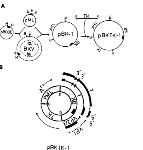
vectoranditsconstructionaredepicted in Fig. 1. The pBK TK-1 vector was derived by digesting both plasmid pML
DNAandBKVDNAwith EcoRI and BamHI (Fig. 1A). The
larger of the respective EcoRI-BamHI fragments of pML DNA(2,617basepairs [bp])
aqd
BKV DNA (5,089 bp) wereligated to produce pBK-1. A BamHI fragment of HSV-1 DNA(3,600bp) which containstheTKgenewasintroduced
into thesingleBamHI site ofpBK-1togeneratevectorpBK
TK-1 (Fig. 1A). The pBK TK-1 molecule thus includes
almost theentire BKV genome(5,196bp),exceptfor 107 bp
eliminated
by digestion
of BKV DNA with BamHI andEcoRI.This107-bpfragment containedsequencesfromeach
of the lateviral capsid transcripts Vpl, Vp2, andVp3 (54).
Since BKV DNA is infectious for human cells, deletion of
theselate viral sequenceswas intended toobviate potential
recombination and release of the complete viral genome
from a vectorcontaining full-length BKV DNA.
The topographical arrangement of the transcripts and
pertinent coding regionsof the genes containedwithinpBK TK-1are depicted in Fig. 1B. Theessentialfeatures ofpBK
TK-1,inadditiontoencodingtheampicillinresistance gene
ofpMLand the TK gene ofHSV-1,arethat itcontainsthe
entire early region sequences which encode BKV large T
and small t antigens (45, 59). In addition, pBK TK-1 also
includes the BKV origin of replication based upon related
sequence
homology
to SV4Q and the transcriptionalen-hancer sequences (45, 59). The lategenes ofBKV in pBK TK-1arenot
only missing
107bpasdescribed above butarealso
disrupted by ligation
ofpMLDNAatthe EcoRIsiteandof TK DNAat theBamHI site. Plasmid pBODE, shown in
Fig.
1A, is discussed later.pBK TK-1 vector persists episomallyin human cells.
Hu-man TK- HeLa cells or 143 B cells were transfected with
pBKTK-1 vector DNAand subjected to selection in HAT
medium.
Individually
clonedor massculture cell lines grownin HAT medium were then analyzed. Either total cellular
DNA was extracted from TK+-transformed cells, or the
DNA was separated into Hirt supernatant (HS) and Hirt
pellet (HP)
fractions.Essentially,
HS DNA contains almostentirely free,
low-molecular-weight DNA,whereas HPDNAcontains
high-molecular-weight
DNA that is most oftencontaminated with free DNA.
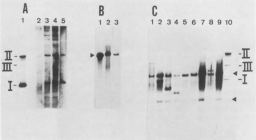
The DNA from several TK+ clones of HeLa cells was
extracted and examined by Southern blot analysis (48) for
the presenceofpBKTK-1 molecules. Analysisof HS DNA
fromtwoHeLaclonesH-303and H-384(Fig. 2A,lanes 1 and
2,respectively)revealed sequences thatcomigratedwith the
supercoiled (form
I), circular (form II), or linear (form III)structures ofpBKTK-1. When this same DNAfrom clone
H-303 was
digested
with Sall (Fig. 2B, lane 2), which cutspBK
TK-1 once, asingle
bandcomigrated
withSall-linear-ized
pBK
TK-1 DNA(Fig.
2B, lane 1). These resultsindicatedthat
full-length
pBKTK-1moleculespersist
asfreeon August 2, 2018 by BIBLIOTECA S MARIA DELLE
http://mcb.asm.org/
A
H HB
!5'
5"
pBK TK-l
FIG. 1. Construction ofvectors. (A) pBK TK-1was constructedfromBKV, the TKgeneofHSV-1, andplasmid pML. pBODE was
constructed from BKVand pML. The putative replication origin ofBKV(0.67mapunits)iscontained in the HindIll Cfragment designated by the black box. The early and late transcription regionsof BKVaredesignated by the dotted lines. The restriction sitesareEcoRI (E), BamHI(B), and HindlIl (H). (B)Genomic landmarks in pBK TK-1. Locations of DNAs derived from pML, BKV,and TK(described above) inpBK TK-1 arerepresented in the double circle. Their respective transcriptsareindicatedbythethinlines, of which thedottedportion representsintrons. Where pertinent, theorientation of the transcripts is shown 5' to3'. Coding regions (thickblacklines) containedwithin specific BKV transcripts indicate early proteins, large T and small tantigens, andlate capsidproteins VP1and VP2/3. The putativeBKV replication originisrepresented bythe black box within the circles. The bacterial ampicillinresistancegeneofpMLisdesignated byAr. The represented restriction sitesareEcoRI(E) and BamHI (B). Detailsarediscussedin thetext.
moleculesin HeLa cells. Tofurther characterize thestateof
vector DNA in HeLa cells, total DNA from six different
TK+ cloned lines was digested with BamHI, which cuts
pBK TK-1 DNA twice to generate 3,600- and 7,706-bp
fragments. Five of the HeLa clones(Fig.2C, lanes 1, 2, 3, 5, and6) containedonlytwobands whichcomigratedwith the
3,600 and7,706-bpmarkerfragments(Fig.2C, lane 7), which
was expected if pBK TK-1 was episomal. Only one HeLa
cell clone (Fig. 2C, lane 4) contained an additional band
which may have originated from either integrated or
rear-ranged episomalmoleculesof pBK TK-1 DNA; the
addition-albands inlane3 representincompletely digested free pBK
TK-1DNA,sincetheycomigrated with undigested pBK TK-1DNA (lane8). Itis noted thatalthough theautoradiogram
shown in Fig. 2C had been overexposed, minor bands of
pBK TK-1 which could represent integrated sequences are
not detected. These results again suggested that in HeLa
cells pBKTK-1molecules exist asfull-length episomes.
Thetransfected 143 B TK+ cell clones were also analyzed
for the presence ofpBK TK-1 molecules. Undigested HS
DNA from each of several clones (Fig. 3A, lanes 2 to 5)
revealed molecules whichcomigrated with pBKTK-1
mark-erDNA (Fig. 3A, lane 1). TheHS DNA from twoof these
cell clones, afterdigestion withSall which cutspBKTK-1
once, yielded single bands (Fig. 3B, lanes 2 and 3), which
comigrated with SalI-linearized pBK TK-1 DNA(Fig. 3B,
lane 1). Free forms of pBK TK-1 (50 to 100 genome
equivalentsperdiploid cell genome)were similarlyobserved
when total cellular DNA from 143 B TK+ cell clones was
digested with HpaI, a noncutter for pBK TK-1 (data not
shown). Total cellular DNA from nine 143 B clones was
digested with BamHI and analyzed for sequences of the
expected 7,706-and3,600-bp
fragments
that would begener-ated fromnonintegratedmoleculesofpBKTK-1.Indeed,as
seenin Fig. 3C (lanes 1 to 9), each 143 B clone contained bothexpectedBamHI
fragments. However,
some143 B cellclones revealed novel bands which
hybridized
to thepBK
TK-1probe (Fig. 3C,lanes1, 3, 4,and
8);
theband below the7,706-bp fragmentof clone B4(lane 2)is
probably
undigested
vector DNA, since it
comigrates
with form IpBK
TK-1on August 2, 2018 by BIBLIOTECA S MARIA DELLE
http://mcb.asm.org/
A B C
12
12
1234
5678
E_--mP
I.
. 39_-
4 7.7- MOdwa__406
. b 310* IIi..
FIG. 2. Southern blots of DNA from TK+-transformed HeLa cellcloneshybridized with a pBK TK-1 DNA probe. HS DNA or total cellular DNA was extracted from various individual TK+ HeLa cell clones that had been transformed by pBK TK-1. HS DNAs(corresponding to 105cells) and total cellular DNAs (10,Ug) were digested with restriction endonucleases as indicated, electro-phoresed through an0.8%agarose gel, blotted onto nitrocellulose, and probed with nick-translated pBK TK-1 DNA. (A) Undigested HSDNA from HeLa cellclones H-303 (lane 1) and H-384 (lane 2). The positionsof formsI, II,andIIIof pBK TK-1 marker DNA are indicated. (B) HS DNA of HeLa clone H-303 digested with Sall (lane2), andSall-digestedpBK TK-1 DNA (4.8 ng) as marker(lane 1).(C)Total cellular DNAwas digested withBamHI.Lanes 1 to 6: HeLa clones H-311; H-303; H-383; H-301; H-385; H-386, respec-tively. pBKTK-1 DNAmarker (50 genome equivalents): digested withBamHl(lane 7) andundigested (lane 8).
DNA (lane 10). These novel sequences likely represent
polymeric and deleted episomal forms of pBK TK-1 or possibly integrated forms of the vector.
We examined whether the property of pBK TK-1
mole-culesto persistas episomes in individual cell clones would
also extendto a mass cultureprepared by pooling hundreds
of individual TK+ cell colonies. The DNA extracted from
suchamassculture of 143 B cells was separated into HS and
HPfractions, which, respectively, were digested with five
A
1 2345"'p
m
!'
I-B
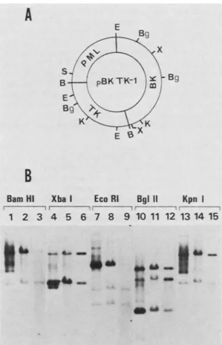
1 23restriction endonucleases that cut pBK TK-1 DNA twice
(BamHI, XbaI, and KpnI)orthree times (EcoRI andBglII)
(Fig. 4A). As seen in Fig.4B, in all instances, sequences of
pBKTK-1 found in both the HS (lanes 1, 4, 7, 10, and 13)
and HP(lanes 2, 5, 8, 11,and14)fractions afterdigestion by
these enzymeswerethose whichcomigrated with the
corre-sponding digestionsof markerpBK TK-1 DNA (lanes 3, 6, 9,
12, and 15). The observation that most of the hybridized
sequences from each HP fraction comigrated with the
re-spective restriction fragments of marker pBK TK-1 DNA
indicated thatmostof these moleculesoriginatedfrompBK
TK-1 episomes and thatonly negligible amounts, if any, of thisvectorDNAintegateinto chromosomal DNA. Addition-al bands, particularly in the HS fraction, could represent
incompletely digested pBK TK-1 DNA molecules, free
molecules containing deletions, or degraded forms of pBK
TK-1 DNA. Since these resultswere obtained with a mass
culture, theyare representative of hundreds of individually
transfected cells. This finding demonstrates clearly that in
themajority of independently transfected human cells pBK
TK-1 moleculespersist in thefree state.
The average number of free pBK TK-1 copies maintained
percell was between 75 and 120 in 143 B cells and 20 and 40
in HeLa cells. It is important to note that in some 143 B
clones (e.g., clones B-23 and B-36; Fig. 3C, lanes 7 and 9), many timesthis number of copies persisted.
pBK TK-1 DNA can beshuttledfrom human cells into E.
coli. Tofurther substantiate thatpBKTK-1DNAmolecules
persist as stable replicating episomes in human cells, HS
DNAs from both HeLa and 143 B cell clones were used to
transformE.colitoanampicillin-resistantphenotype. In one
experiment, half ofasample of the HS DNA from two clones
of HeLa cells and six clones of 143 B cellswasdirectly used
to transform E. coli MC-1061, whereas the other half was
treated with the restriction endonuclease MboI (which has
17restrictionsites within pBK TK-1 DNA) before
transfor-mation. DNA is not cut when the adenine within the
recognition sequence of MboI (GATC) is methylated, as occursinmostE.colistrainsincluding
HblOl
and MC-1061.C
1 2 3 4 5 6 7 8 9 10
I:~~
-_|S"--|+| I
FIG. 3. Southernblotsof DNAfromTK+-transformed143 BcellcloneshybridizedwithpBKTK-1DNA.HSDNAortotalcellularDNA
wasextractedfromvariousindividual TK+ 143 B cell clones that had been transformed by pBKTK-1DNA.HS DNAs(correspondingto105 cells)andtotalcellular DNAs(10
JLg)
weredigestedwith theindicatedrestrictionendonucleases,electrophoresed througha0.6%agarosegel, blottedontonitrocellulose, andprobed with nick-translated pBK TK-1 DNA. (A)HSDNAfrom143 B clonesB-6,B-27, B-31, andB-20(lanes 2to5, respectively); lane1, 4.8 ngofpBK TK-1 DNA marker. (B)Salldigestion of HSDNAof 143 B clones B-31(lane 2)and B-27(lane 3); lane1, 4.8 ngofSalI-digestedpBK TK-1 marker DNA (arrow).(C) TotalcellularDNAfrom 143 BclonesdigestedwithBamHI;lanes 1to9, 143 BclonesB-1, B-4, B-6, B-8, B-18, B-20, B-23, B-31, and B-36,respectively;lane10,undigested pBKTK-1 DNA marker(100genome equivalents). The upperand lower arrows indicate thepositions of the 7,706 and3,600-bpfragments, respectively, generated byBamHI digestion ofpBK TK-1 DNA.on August 2, 2018 by BIBLIOTECA S MARIA DELLE
http://mcb.asm.org/
TABLE 1. Transformation of E. coliMC-1061 with HS DNAs fromHeLa and 143 B cell clones transformed to the TK+
phenotype by pBK TK-1
No. of Ampr bacterial colonies' Cellclones
Untreated Treated with Untreated MboI HeLa H-303 28 ND HeLa H-384 114 ND 143 B 656 95 0 143 B 657 233 0 143 B F6-D4 30 0 143 B 633 28 0 143 BB-27 25 0 143 B B-3 150 0 pBK TK-1 DNA (control, 10 ng) 26,400 21,000 aAmprbacterial colonies were produced by transformation of E.coliwith samples ofHSs obtained from105cells. ND, Not done.
9 10 11 12 13 14 15
FIG. 4. (A) Positions of BamHI (B), BglII (Bg), EcoRI (E), KpnI (K), and Xbal (X) restriction sites in pBK TK-1. The unique Sall (S) site is suitable for insertion of additionalgenes.(B) Southernblot of DNAfromTK+-transformed 143 Bmassculture cellshybridizedto pBK TK-1 DNA. HS and HP DNAswereextracted fromTK+ 143B
massculturecells, digested with restriction endonucleases BamHI,
Xbal, EcoRI, BglII, and KpnI, electrophoresed through a 1.0% agarose gel, blotted onto nitrocellulose, and probed with nick-translatedpBK TK-1 DNA. Lanes 1, 4, 7, 10, and 13 show HS DNA from10Wcells; lanes 2, 5, 8, 11, and 14 show HP DNA (10 ,ug);lanes 3, 6, 9, 12, and 15 show pBK TK-1 marker DNA (1ng or50genome
equivalents).
However, the DNA becomes sensitive to MboI digestion
afterreplicationineucaryotic cells,where the adenine isnot
methylated. The untreated HS DNA from each HeLa and
143 B cell clone did transform E. coli MC-1061, whereas
digestion byMboI rendered these same DNAsincapable of
transformation (Table 1). The transforming ability ofpBK
TK-1 DNA directly isolated from E. coli HblOl was
unaf-fected by MboI treatment. These results indicate thatpBK TK-1 does replicate as a free molecule in human cells and that the geneforampicillin resistance remains functional.
Replication of pBK TK-1 in TK+ transformants was
further supported by blot hybridization analysis. The HS DNA from each ofseven 143 BTK+ individual clones was
completely digested by MboI (lanes 3 to 9), whereas the
controlpBKTK-1 was not (lanes 1 and2) (Fig. 5).
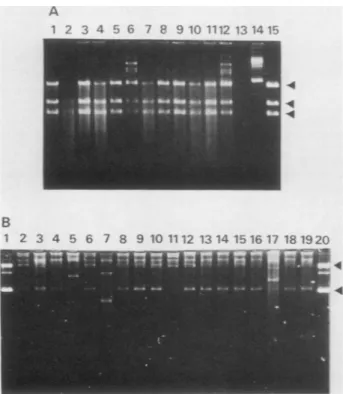
In another experiment, plasmids recovered from E. coli
MC-1061 transformedbyHS DNAs from either HeLaor143 B TK+ cells were compared to input DNA. The plasmids
fromampicillin-resistant coloniesweredigestedwithEcoRI,
which cuts pBK TK-1 three times (Fig. 4A), and the
frag-mentsthus obtainedwereanalyzed in agarose gels. As seen
in Fig. 6A (lanes 1 to 12), plasmid DNA from each of 12
ampicillin-resistant coloniestransformedbythe HS DNAof
HeLa cell line H-303 contained three EcoRI restriction
fragments that comigrated with those of pBK TK-1 DNA
(lane 15). No other DNA bands were seen; the
higher-molecular-weight bands seen in lanes 6 and 12 (Fig. 6A)
probablyrepresentincomplete digestions.A similaranalysis
with the HS of an individual 143 B cell clone revealed that
the plasmid DNAs from 11 of 12 bacterial transformants
were indistinguishablefrom thatoftheinputvector(datanot
shown). Inaddition,the HS DNA froma massculture of 143
B cells was also used to transform E. coli C600. Plasmids
extracted from 18 ampicillin-resistant colonies were
ana-1 2 3 4 5 6 7 8 9
1ff
m
I-20
FIG. 5. Digestion with MboI and blot hybridization analysisof 143 B cellclones transformedtotheTK+ phenotype by pBK TK-1. HS DNAfrom105 cellswasrestricted withMboI, migratedina1%
agarosegel, transferredtonitrocellulose, and hybridizedtoapBK
TK-1probe. Lanes 3to9, 143 B clones B-18, B-28, B-6, B-23, B-36, B-31, and B-2, respectively; lane 1, pBK TK-1 marker digestedwith MboI (2.4ng); lane2, pBK TK-1 digested with BamHI and MboI (2.4 ng). FormsI, II,andIIIof markerPBK TK-1areindicated.
A
E
Bg
B
BamHi Xba I EcoRI Bgi 11 Kpn I
r !
1 2 3 4 5 6 7 8
on August 2, 2018 by BIBLIOTECA S MARIA DELLE
http://mcb.asm.org/
A 1 2 3 4 5 6 7 8 9 10 1112 13 1415 B 1 2 3 4 5 6 7 8 9 10 1112 1314 1516 17 181920 .4 .4
FIG. 6. pBKTK-1fromTK+-transformed HeLa and 143 B cells is shuttled intoE.coliandreestablished as plasmids. HS DNA from TK+HeLa and 143 B cells that had beentransformed by pBKTK-1 was used to transform E. coli MC-1061 (A) or C600 (B). Plasmid DNAs wereprepared fromE.coliampicillin-resistant(Ampr) colo-nies, electrophoresed in1%agarose gels, and visualized by ethidium bromidestaining at280 nm. (A) EcoRIdigestionof plasmids from Ampr colonies, transformed by HS DNA of HeLa cell clone H-303 (lanes 1 to12). Marker pBKTK-1 was uncut (lane 14) or digested with EcoRl (lane 15). The arrows point to the three fragments generated by EcoRI digestion (Fig. 4A). Lane 13 is blank. (B) Plasmids(undigested) fromAmprcoloniestransformed by HS DNA from 143 B mass culture cells (lanes 2 to 19). Marker pBK TK-1 DNA is inlanes 1 and 20. The lower arrow indicates the position of supercoiled (formI)pBKTK-1 plasmid DNA, and the upper arrow marks itsrelaxed circular form (form II).
lyzed directly in agarosegels. As seen in Fig. 6B, 15 of 18
colonies (lanes 2 to 19) contained supercoiled and relaxed
circular molecules that
comigrated
with form I and form II DNAof pBKTK-1(lanes1and 20).Twoplasmids
containedan insertion (Fig. 6B, lanes 5 and 17) and a third plasmid
contained a deletion (lane 7), as confirmed by
restriction-enzyme analysis (datanot shown). These results show that
in human cell linesoriginating either fromasingle
transfect-ed cell or from a mixture of hundreds of independently
transfected cells, pBK TK-1 DNA persists as stable
full-length episomes formany cellgenerations.
pBK TK-1 vector continues to persistinthe episomalstate
after removal of selective growth medium. In all the
experi-ments thusfardescribed,the TK- human cellswereplaced
under conditions of selective growth after transfection
by
pBKTK-1 DNA.SinceonlyTK+ cellsareabletosurvive in
HAT medium, it was not apparent whether selective
pres-sure was required for maintenance of stable episomes.
Therefore, an individual clone of 143 B cells (B-27) and a masscultureof143 B cells whichcontainedpBKTK-1 DNA
were removed from the selective HAT medium and placed
ontononselective Eagle MEM.
Surprisingly, pBKTK-1 DNA molecules from the HSof
clone B-27 cells maintained inHAT medium for 200
genera-tions (Fig. 7A, lane 2) appeared qualitatively unchanged
after 25 generations of growth in nonselective MEM (Fig.
7A, lane 3). Furthermore, replacement of HAT medium for
an additional 25 generations after growth in MEM did not
alterthepersistence of full-length pBK TK-1 episomes; the
high-molecular-weight comigrating bands probably repre-sent polymers of pBK TK-1 DNA (Fig. 7A, lane 4). No
apparent loss in the number of pBK TK-1 molecules was
detected in B-27 cells upon removal or reintroduction of
HAT medium, since in each case (Fig. 7A, lanes 2 to 4) the
amount of HS DNA analyzed was from the same number of
cells.
A similaranalysis of the 143 B mass culture also proved
that maintenance of stable pBK TK-1 episomal DNA in
thesecells is not dependent upon selection in HAT medium.
In thisexperiment, 143 B mass culture cells that had been maintained in HAT medium for 5 months were switched to
growth innonselective MEM for 3 months (48 generations).
As seenin Fig. 7B, the Southern blot of HS and HP fractions
digestedwith BamHI reveals noobviouschange in pBK
TK-1 DNA after many cell doublings in nonselective medium
(lane 1, HS; lane 2, HP) as compared with cells grown in
HATmedium (lane 3, HS; lane 4, HP).
pBK TK-1 synthesizes BKV T antigen. It is possible that
persistence ofamplified episomes of pBK TK-1 in human
cells may depend upon expression ofBKV large T antigen
encoded bythe vector.This hypothesis seemed reasonable,
since large Tantigen of the closely related SV40 stimulates itsreplication(for review, see reference 54). Both 143 B- and HeLa TK+-transformed cell lines were found to express BKV Tantigen. As seen in Fig. 8, immunoprecipitation of
the 95,000-dalton BKV large T antigen from one of these
lines occurs with a specifichamster serum to BKV T antigen
A
1
234
B1
234
I.A
-.FwA 2-_j " ff > e& t I--a-U-FIG. 7. Blot hybridization of HSDNA fromTK+-transformed 143 B cells maintained with and without selective HAT medium. UndigestedorBamHI-restricted DNAwaselectrophoresedthrough a 0.6% agarose gel (A) or a 1% agarose gel (B), blotted onto
nitrocellulose, and probed with nick-translatedpBK TK-1DNA.(A) HSDNA(undigested andcorrespondingto105cells)from 143 B cell clone B-27, maintained first in HAT medium for more than 200 generations (lane 2), then for25generations innonselective MEM (lane 3), and then replaced into HATmedium for 25 generations
(lane 4). pBKTK-1 marker DNA(2.4 ng),indicatingforms I andII, is shown in lane 1. (B) BamHI-digested DNA from 143 B mass
culture cellsthathad beenmaintained for5monthsinHATmedium: HSDNA(lane 3)and HP DNA(lane 4). DNAfrom thesamecells after 3 months in nonselective MEM: HS DNA (lane 1) and HP DNA (lane 2). Uppermost bands represent partial DNA digests.
Positions ofthe two fragments generated by BamHI digestion of pBK TK-1areshown bythearrows.
on August 2, 2018 by BIBLIOTECA S MARIA DELLE
http://mcb.asm.org/
1 234 95_** 45--31- * 21 -14 -S
FIG. 8. Immunoprecipitation of BKV T antigen from 143Bcells transformed by pBK TK-1. TK+ 143 B cells (clone B-633) were labeled with
32Pi,
and anti-T hamsterserum was usedto immunopre-cipitateBKV Tantigen asdescribed in the text.The immunoprecipi-tated products were fractioned in a 10% polyacrylamide gel. La-beled cellextractswerereacted with normal hamsterserum(lane 2), 15 p.l of anti-T serum (lane 3), or 30 pul of anti-T serum (lane 4). Protein molecular weight standards in kilodaltons are in lane 1: phosphorylase b, 95; ovalbumin, 45; carbonic anhydrase, 31; trypsin inhibitor, 21; lysozyme, 14.The arrowindicates theposition of the 95,000 molecular-weightBKVlargeTantigen.(lanes 3 and 4), but not with a normal serum (lane 2). In
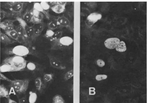
addition, BKV Tantigen was also detected by
immunofluo-rescence in 143 B cells stably transformed to the TK+
phenotype by pBK TK-1 (Fig. 9).
pBK TK.1 contains an active origin of replication. As a
corollary to these findings, itwasimportant to demonstrate
directly that pBK TK-1 DNA contains viral sequences
responsible forreplication in the presence of BKV T antigen.
The BKV origin ofreplication, in fact, is predicted to be
located at0.67 map units, based upon extensive sequence
homology to the knownoriginof replicationinSV40(45, 59).
Therefore, theHindIll Cfragment (555 bp) ofBKV which contains sequences including the origin ofreplication was
cloned into pML (Fig. 1A). The resulting
recombinant,
referred to as pBODE (Fig. 1A), was transfected into L603
cells. These cells are human embryonicfibroblasts that had
been transformed by a BKV DNA fragment containing the
entire early region and are 100% positive for BKV T antigen
(16). The intent was to see whether the pBODE molecule,
containing the predicted BKV origin of replication but void
ofanycompleteviralgenes,wouldbe amplifiedin cellsthat constitutively produce BKV T antigen. The HS DNA was
extracted at 16, 50, and 81 hposttransfection,linearized with
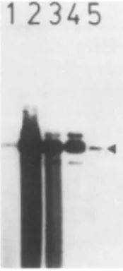
BamHI,andanalyzedby Southern blotting,using pBODEas the labeled probe. The pBODE molecule is greatly ampli-fied, since densitometric analysis of the autoradiograms
showed that the amount of pBODE DNA increases from
0.04ng at 16 hposttransfection (Fig. 10,lane1) to 29 ng at 50
h (lane 2) and then decreases to 3.7 ng at 81 h (lane 3).
Therefore,reduction in copy number at 81 hposttransfection defines this amplification as transient. The pBODE
mole-cules were not amplified in 143 B cells (data not shown).
These resultsindicate that the0.67-map-unit region ofBKV
present in the pBK TK-1 molecule serves as an active
replication origin which is stimulated by BKV T antigen. DISCUSSION
We describe an expression vector, pBK TK-1, which is
able to persist episomally in human cells. This vector
includes sequences derivedfrom BKV, a human
papovavi-rus; pML, adeletion derivativeofplasmid pBR322 (25);and
the TK geneofHSV-1, which transforms TK- mutantcells
to the TK+ phenotype (58). Free molecules ofpBK TK-1
were detected in the low-molecular-weight DNAfractionof
established TK+-transformed cells. Enzymatic
expression
of the TK gene of HSV-1 was also detected in the
TK+
transformants
(unpublished
data). Theorigin
ofthe humancell into which pBK TK-1 was introduced influenced to a
certain extent its episomal status both quantitatively and
qualitatively. In HeLa and 143 B TK+ cell
transformants,
the numbers of extrachromosomal copies of pBKTK-1 per
FIG. 9. Fluorescent antibody staining for BKV T antigen in 143 B TK+ cells transformed by pBK TK-1. Cells were first reacted with a
specifichamsterserum toBKV T antigen and then withfluorescein-conjugated goat antibodies to hamster immunoglobulinG. (A) Clone B-28
and (B) cloneB-31.
on August 2, 2018 by BIBLIOTECA S MARIA DELLE
http://mcb.asm.org/
1 2345
FIG. 10. Amplification of pBODE, a BKV origin recombinant
plasmid, in BKV-transformed human cells positive for BKV T antigen, as analyzed by Southern blot hybridization. The BKV-transformed humancells, L603, in which the BKV T-antigengeneis
integratedand expressed, weretransfected with pBODE(10 jigof
DNAper107cells). At indicated times after transfection, cellswere
rigorously washed, and HS DNA was prepared. Samples of HS
DNA correspondingto105cellsweredigested withBamHl,
electro-phoresed through a 0.6% agarose gel, blotted onto nitrocellulose, and probed with nick-translated pBODE DNA. Lane 1. 16 h posttransfection; lane 2, 50 h posttransfection; lane 3, 81 h
post-transfection. Marker pBODE DNAcutwith BamHI is shownin lane 4 (3.5 ng) and lane5 (0.07 ng). Thearrowpointstolinear form IIIof pBODE DNA.
cell were generally 20 to 40and 75 to 120, respectively. In HeLa cells, pBK TK-1 molecules almost exclusively
ap-peared as full-length episomes with undetectable
rearrange-ments. Although all 143 B transformants contained full-length episomes, additional free forms that probably includedpolymeric and deleted molecules were observed in some clones. The nature of these rearranged molecules is
unknown. In this connection, it must be pointed outthat in
BKV-transformed human cells,where BKVDNAreplicates episomally, free unintegrated polymeric forms of the viral
genome were constantly detected in considerable amounts (16). In all cases, however, full-length pBK TK-1 molecules werethe predominant episomal form as specifically
demon-strated in 143 Bmass
culture,
cells,which representedapool of hundrds of individual TK+ colonies. In both HeLa and 143 B cells, integration of pBK TK-1 into chromosomal DNAappeared infrequentand certainly negligible in propor-tion tothe numberofamplified free molecules.The pBK TK-1 molecules from TK+-transformed human cells can be readily shuttled into bacteria. Surprisingly,
almost 85% of the Ampr bacterial colonies transformed by DNA from the 143 B TK+ mass culture cells contained
plasmid DNA thatwasindistinguishable from the input pBK
TK-1 vector DNA; this value was near 100% when DNA
from a TK+ HeLa or 143 B cell clone was used.
Further-more, plasmid DNA could not be rescued in bacteria when these cellular DNAs were digested with the endonuclease
MboI, whichselectivelyrestricts DNAthat hasreplicated in animal cells. Not only do these results demonstrate defini-tively that pBK TK-1 persists as stable replicons in human
cells, but they also show that a resident bacterial gene,
which confers no selective advantagetothe eucaryotic cell, retains its capacity tofunction, i.e., the,B-lactamase geneof
pMLwhich confers ampicillin resistance in E. coli. Importantly, episomal maintenance of pBK TK-1 in
hu-mancellsdoes notappeartodependuponforced expression ofthe TKgeneunderselectionfortheTK+ phenotype, since
the vector is not drastically affected by removal of HAT
medium. This is in contrast to a reportwhich describes the
loss of TK-plasmid recombinant episomes in transformed
rodent cells after removal of selective medium (51). Our
experiments indicate that the sequencesinherentin the BK
viralmoiety of pBK TK-1dictate the copy level andstability
of the vector. This further suggests that any dominant
selection marker gene (33) may be substituted for the TK
geneof HSV-1 totherebyextend the spectrumofhumancell
typesinto which this novelexpression vector can be
intro-ducedandamplified. Thepossibilitythatanorigin of
replica-tionin HSV-1 TK gene could beresponsiblefor theepisomal
stateof the vector, assuggestedbyothers(51),wasruledout
because only 0.2 copies offree plasmid DNA per cell were
detectedin HS DNAof143 B cellstransformedtothe TK+
phenotype by a recombinantmoleculecontaining only pML
and theHSV-1 TK gene. In this case, essentiallyall vector
DNA sequences were found to be integrated into
high-molecular-weight DNA of the TK+ transformants (G.
Bar-banti-Brodano, unpublished data).
The featureswhich control theepisomal replication
func-tion of pBK TK-1 are unknown. It seems
unlikely
that thelate BKviral genes are involved inepisomal maintenance of
pBK TK-1 since they were partially deleted to circumvent
potential infection. However, the early viral large T- and
smallt-antigengenesare completelyencoded inpBKTK-1.
It islikely that the immunoreactive BKV largeT
antigen
inthe TK+-transformed cells stimulates the BKV
origin
ofreplication in pBK TK-1 analogous to SV40
T-antigen-dependentreplication(54). The supportiveargumentfor this
mechanism is demonstrated by the great
amplification
ofplasmidpBODE, which contains theputative BKV
origin
ofreplication (0.67 viral map units; 42, 45, 59), but is void of
complete early and late BKV genes, in BKV-transformed
humancells that constitutively synthesize BKV T
antigen.
This BKV T-antigen-dependent
replication
ofpBODE
issimilarto transientreplication ofSV40vectors in COS cells
(31). It is intriguing to consider that in TK+ transformants
the copy level of pBK TK-1 may be held constant
by
autoregulation ofBKV T-antigen
synthesis,
similartocon-trol of SV40 T-antigen production
during lytic
infection (1,22, 52). Wehave begunconstructingdeletion mutantsin the
T-antigen gene of pBK TK-1 to define
potential
elementsnecessary for episomal replication.
Alternatively,
mainte-nance oftheepisomalstate ofpBKTK-1in human cells may
depend on specific viral sequences, not
necessarily
relatedto the T-antigen coding
region,
similar to what has beenshown for vectors based on bovine
papillomavirus
DNA(26). A search for such sequences in the BKV genome is
under way. Finally, itcannot beruled outthat
replication
ofthe vector is also under the control of some host cell components.
The pBK TK-1moleculerepresents thefirst stable
expres-sion vectorthat persists
episomally
in human cells and thatcanbe shuttled intobacteria. These features arealso shared
by the bovine
papillomavirus-plasmid
vectors that functionin mouse cells (6, 43). Key
advantages
andpotential
uses of theepisomalpBK TK-1expression
vector arethefollowing.
(i) The inserted gene of interest cannot be
interrupted
orsubjected to regulatory constraints that
frequently
occurfrom integration into cellularDNA.
(ii)
The gene of interestis amplified withthe vector.
(iii)
The vector can be used tostudygeneswhich are
normally
expressed
orrequire
expres-sion in human cells. For
example,
wehaveproduced
humancell lines in which the
episomal
vectorcontains adenovirustype 12
transforming
genes and expresses functional andimmunoreactive adenovirus type 12 ElA
proteins
(R.
on August 2, 2018 by BIBLIOTECA S MARIA DELLE
http://mcb.asm.org/
vada, G. Barbanti-Brodano, and R. P. Ricciardi,
unpub-lisheddata). The pBK TK-1vectorcontains restriction sites
convenient for different cloning strategies. (v) Substitution
of the TK gene with adominant selectiongene such asTn5
aminoglycoside phosphotransferase (49) orE. coli xanthine-guanine phosphoribosyltransferase (33) in thevector
predict-ably willexpand the variety of human cells thatcan be used.
(vi) The system may be applied in transient assays as
suggested from this study with pBODE in L603 cells. Indeed, we have already prepared apBODE-CAT (chloram-phenicol acetyltransferase) recombinant that transiently
ex-presses high enzyme activity in 143 B TK+ transformants
containingpBK TK-1 (A. Caputo, G. Barbanti-Brodano, and
R. P. Ricciardi, unpublished data). (vii) The pBK TK-1 recombinant may be modified for the insertion and
expres-sion of cDNAs.
ACKNOWLEDGMENTS
Weare grateful to and acknowledge support of Carlo M. Croce (The WistarInstitute) in assisting in this project.
This work was supported by Public Health Service grant CA-29797from the National Cancer Institute and a Ruth Estrin
Gold-bergMemorialfor Cancer Researchgrant to R.P.R. This workwas
also supported by Consiglio Nazionale delle Ricerche-Progetto Finalizzato 'IngegneriaGenetica e Basi Molecolari delle Malattie Ereditarie" and by North Atlantic Treaty Organization research grant 284.81.
LITERATURE CITED
1. Alwine,J. C., S. I. Reed, and G. R. Stark. 1977. Characteriza-tion of the autoregulation of simian virus 40 gene A. J. Virol. 24:22-27.
2. Brown,P., T. Tsai,and D.C.Gajdusek. 1975. Seroepidemiology of human papovaviruses: discovery ofvirgin populations and some unusual patterns of antibody prevalence among remote
peoplesof the world. Am. J. Epidemiol. 102:331-340. 3. Chenciner, N., M. P. Grossi, G. Meneguzzi, A. Corallini, R.
Manservigi, G. Barbanti-Brodano, and G. Milanesi. 1980. State of viral DNA in BK virus-transformed rabbit cells. Virology 103:138-148.
4. Croce, C. M., J. Barrick, A. Linnenbach, and H. Koprowski. 1979. Expression of malignancy in hybrids between normal and malignant cells.
J.
Cell Physiol. 99:279-286.5. Denhart, D. T. 1966. A membrane-filter technique for the detection of complementary DNA. Biochem. Biophys. Res. Commun. 23:641-646.
6.
Dimaio,
D., R. Treisman, and T. Maniatis. 1982. Bovine papillo-ma-virus that propagates as a plasmid in both mouse and bacterial cells. Proc. Natl. Acad. Sci. U.S.A. 79:4030-4034. 7. Frost,E., andJ. Williams. 1978. Mapping temperature-sensitiveand host-range mutations of adenovirus type5 by marker rescue. Virology 91:39-50.
8. Gardner, S. D. 1973. Prevalence in England of antibodies to humanpolyomavirus (BK). Br. Med. J. 1:77-78.
9. Gardner, S. D., A. M. Field, D. V. Coleman, and B. Hulme. 1971. New human papovavirus (BK) isolated from urine after renal transplantation. Lancet
i:1253-1257.
10. Gergen, J. P., R. H. Stern, and P. C. Wensink. 1979. Filter replicas and permanent collections of recombinant DNA plas-mids. Nucleic Acids Res. 7:2115-2136.
11. Gluzman, Y. 1981. SV40-transformed simian cells support the replication of early
SV40
mutants. Cell 23:175-182.12. Gluzman, Y. 1982. Eukaryotic viral vectors. Cold Spring Har-bor LaHar-boratory, Cold Spring HarHar-bor, N.Y.
13. Goff, S. P., and P. Berg. 1976. Construction of hybrid viruses
containing
SV40
and X phage DNA segments and their propaga-tion in cultured monkey cells. Cell 9:965-705.14. Graham, F. L., and A. J. van der Eb. 1973. A new technique for the assay of infectivity of human adenovirus5 DNA. Virology 52:456-467.
15. Gross-Bellard, M., P. Oudet, and P. Chambon. 1973. Isolation of
high molecular weight DNA from mammalian cells. Eur. J.
Biochem.
36:32-38.16. Grossi, M. P., A. Caputo, G. Meneguzzi, A. Corallini, L.Carra, M. Portolani, M. Borgatti, G. Milanesi, and G. Barbanti-Bro-dano. 1982. Transformation ofhumanembryonic fibroblastsby BK virus, BK virus DNA and a subgenomic BK virus DNA fragment.
J.
Gen. Virol. 63:393-403.17. Hamer, D. H., D. Davoli, C. A. Thomas, and G.C. Fareed. 1977. Simian virus40carryinganEscherichia colisuppressor gene. J. Mol. Biol. 112:155-182.
18. Hirt, B. 1967. Selective extraction of polyoma DNA from infected mouse cell cultures. J. Mol. Biol. 26:365-369. 19. Holmes, D. S., and M. Quigley. 1981. A rapid method for the
preparation of bacterialplasmids. Anal. Biochem. 114:193-197. 20. Howley, P. M. 1980. MolecularbiologyofSV40and the human polyomaviruses BK andJC,p.489-550. InG. Klein(ed.),Viral oncology. Raven Press, New York.
21. Katz,L., D. T. Kingsbury, and D. R.Helinski. 1973. Stimulation by cyclic adenosine monophosphate ofplasmid deoxyribonucle-ic acid repldeoxyribonucle-ication and catabolite repression of the plasmid deoxyribonucleic acid-protein relaxationcomplex. J. Bacteriol. 114:577-591.
22. Khoury, G., and E. May. 1977. Regulation of early and late simianvirus 40transcription: overproductionofearly viralRNA in the absence ofafunctional T-antigen. J. Virol. 23:167-176. 23. Kushner, S. R. 1978. Animprovedmethodfor transformationof
Escherichia coli with Col El-derived plasmids, p. 17. In H.
Boyer and S. Nicosia (ed.), Genetic engineering. Elsevier/ North-Holland, Amsterdam.
24. Littlefield, J. W. 1965. The use of drug-resistant markers to study the hybridization of mouse fibroblasts. Exp. Cell Res.
41:190-196.
25. Lusky, M., and M. Botchan. 1981. Inhibition ofSV40replication in simian cells by specific pBR322 DNA sequences. Nature (London) 293:79-81.
26. Lusky, M., and M. Botchan. 1984. Characterization of the bovine papilloma virus plasmid maintenance sequences. Cell 36:391-401.
27. Mandel, M., and A. Higa. 1970. Calcium-dependent bacterio-phage DNA infection. J. Mol. Biol. 53:159-162.
28. Maniatis, T., E. F. Fritsch, and J. Sambrook. 1982. Molecular cloning: a laboratory manual. Cold Spring Harbor Laboratory, Cold Spring Harbor, N.Y.
29. Maniatis, T., S. G. Kee, A. Efstratiadis, and F. C.Kafatos. 1976. Amplification and characterization of a,B-globin gene synthe-sized in vitro. Cell 8:163-182.
30. McKnight, S. L., C. M. Croce, and R. Kingsbury. 1979. Intro-duction of isolated DNA sequences into cultured eukaryotic cells.
Carnegie
Inst. Wash. YearBook 78:56-61.31. Mellon, P., V. Parker, Y. Gluzman, and T. Maniatis. 1981. Identification of DNA sequences required for transcription of the humanal-globin gene in a newSV40 host-vector system. Cell 27:279-288.
32. Meneguzzi, G., G. Barbanti-Brodano, and G. Milanesi. 1978. Transcription of BK virus DNA by Esclherichlia coli RNA polymerase: size and sequence analysis of RNA. J. Virol. 25:940-943.
33. Mulligan, R. C., and P. Berg. 1980. Expression of a bacterial gene in mammalian cells. Science 209:1422-1427.
34. Mulligan, R. C., B. H. Howard, and P. Berg. 1979. Synthesisof rabbit
P-globin
in culturedmonkey kidneycellsfollowing infec-tion with aSV40-,B-globin
recombinant genome. Nature (Lon-don) 277:108-114.35. Norkin, L.C. 1982. Papovaviral persistent infections.Microbiol. Rev. 46:384-425.
36. Padgett, B. 1980. Human papovaviruses, p. 339-370. In J. Tooze (ed.), Molecular biology of tumor viruses.II.DNA tumor viruses. Cold Spring Harbor Laboratory, Cold SpringHarbor, N.Y.
37. Panicali, D., and E. Paoletti. 1982. Construction of poxviruses as cloning vectors: insertion of the thymidine kinase gene from herpes simplex virus into the DNA of infectious vaccinia virus. Proc. Natl. Acad. Sci. U.S.A. 79:4927-4931.
on August 2, 2018 by BIBLIOTECA S MARIA DELLE
http://mcb.asm.org/
38. Radloff, F., W. Bauer, and J. Vinograd. 1967. A dye-buoyant-density method for the detection and isolation of closed circular duplex DNA: the closed circular DNA in HeLa cells. Proc. Natl. Acad. Sci. U.S.A. 57:1514-1521.
39. Rigby, P. W. J.1982. Expressionof clonedgenes ineukaryotic cellsusingvector systemsderived from viral replicons,p. 83-141. In R. Williamson (ed.). Genetic engineering, vol. 3. Aca-demic Press, Inc., New York.
40. Rigby, P. W. J. 1983. Cloning vectors derived from animal viruses. J. Gen. Virol. 64:255-266.
41. Rosenthal, N., M. Kress, P. Gruss, and G. Khoury. 1983. BK viral enhancer element and a humancellular homolog. Science 222:749-755.
42. Ryder, K., A. L.DeLucia, and P. Tegtmeyer. 1983. Bindingof SV40 A protein to the BK virus origin ofDNA replication. Virology 129:239-245.
43. Sarver, N., P. Gruss, M.-F. Law, G. Khoury, and P. M. Howley. 1981. Bovine papilloma virus deoxyribonucleic acid: a novel eucaryotic cloning vector. Mol.Cell. Biol. 1:486-496. 44. Scangos,G., and F. H. Ruddle. 1981. Mechanisms and
applica-tions of DNA-mediated gene transfer in mammalian cells: a review. Gene 14:1-10.
45. Seif,I., G. Khoury, and R. Dhar. 1979. The genome of human papovavirus BKV. Cell 18:963-977.
46. Shimotohno, K., and H. M. Temin. 1981. Formation of infec-tiousprogenyvirus after insertion of herpes simplex thymidine kinasegene into DNAofanavian retrovirus. Cell26:67-77. 47. Solnick,D. 1981.Construction ofanadenovirus SV40
recombi-nantproducingSV40T-antigen fromanadenovirus late promot-er.Cell24:135-143.
48. Southern,E.1975. Detection ofspecificsequences among DNA fragments separated by gel electrophoresis. J. Mol. Biol. 98:503-517.
49. Southern, P. J., and P. Berg. 1982.Transformationof mammali-an cells to antibiotic resistance with a bacterial gene under controlof the SV40 early regionpromoter. J. Mol.AppI.Genet.
1:327-341.
50. Spaete, R. R., and N. Frenkel. 1982. The herpes simplex virus amplicon: a new eukaryotic defective virus cloning amplifying vector.Cell 30:295-304.
51. Spandidos, D. A., P. R. Harrison, and J. Paul. 1982. Replication and amplification of recombinant plasmid molecules as extra-chromosomal elements in transformed mammalian cells. Exp. Cell Res. 141:149-158.
52. Tegtmeyer, P., M. Schwartz, J. K. Collins, and K. Rundell. 1975. Regulation of tumor antigen synthesis by simian virus 40 gene A. J. Virol. 16:168-178.
53. Thummel, C., R. Tjian, and T. Grodzicker. 1981. Expression of SV40 T-antigen under control of adenovirus promoters. Cell 23:825-836.
54. Tooze, J. (ed.). 1981. Molecular biology of tumor viruses, revised edition 2. Cold Spring Harbor Press, Cold Spring Harbor, N.Y.
55. Tsui,L.-C., M. L. Breitman, L.Siminovitch,and M. Buchwald. 1982. Persistence of freely replicating SV40recombinant mole-cules carrying a selectable marker in permissive simian cells. Cell30:499-508.
56. Vogelstein, B., and D. Gillespie. 1979. Preparative and analytical purification of DNA from agarose. Proc. Natl. Acad. Sci. U.S.A. 76:615-619.
57. Wei,C.-M., M. Gibson, P. G. Spear, and E. M. Scolnick. 1981. Constructionandisolation of a transmissible retrovirus contain-ing the src gene of Harvey murine sarcoma virus and the thymidinekinase gene of herpes simplex virus type 1. J.Virol. 39:935-944.
58. Wigler,M., R. Sweet, G. K. Sim, B.Wold, A. Pellicer, E. Lacy, T.Maniatis, S. Silverstein, and R. Axel. 1979. Tranformation of mammaliancells with genes from prokaryotes and eukaryotes. Cell16:777-785.
59. Yang, R. C. A., and R. Wu. 1979. BK virus DNA: complete nucleotide sequence of a human tumor virus. Science 206:456-462.
on August 2, 2018 by BIBLIOTECA S MARIA DELLE
http://mcb.asm.org/