Multidrug Resistance and Production of
Extended Spectrum
β-lactamases and
Plasmid-mediated AmpC
β-lactamases
in Enterobacteriaceae isolates from
diseased cats in Sicily
Tesi di Dottorato
Dott. Francesco Lo Piccolo
Tutor:
Chiar.ma Prof.ssa M. Foti
Coordinatore: Chiar.mo Prof. G. Mazzullo
UNIVERSITÀ DEGLI STUDI DI MESSINA DIPARTIMENTO DI SCIENZE VETERINARIE DOTTORATO DI RICERCA IN SANITA’ PUBBLICA, IGIENE
VETERINARIA E DELLE PRODUZIONI ANIMALI S.S.D.Vet/05
Dedication
For my family,
who helped me in all things great and small
Contents
Abstract 7 CHAPTER 1: Introduction 12 1.1 Enterobacteriaceae 13 1.2 Antimicrobial resistance 53 1.3 Beta-lactam antimicrobials 60 1.4 β-lactamases 701.5 β-lactamases producing Enterobacteriaceae in
companion animals 92
CHAPTER 2: Study 100
2.1 Aims 101
2.2 Materials and Methods 102
2.3 Results 110
2.4 Discussion 135
2.5 Conclusions 145
REFERENCES 149
Abstract
Introduction: In human and veterinary medicine,
Enterobacteriaceae are common causes of enteric and extra-intestinal opportunistic infections and their resistance to multiple antimicrobials is a major global threat. Multidrug-resistant (MDR) Enterobacteriaceae are increasingly reported in companion animals, thus raising great concerns for animal and public health (Bogaerts et al., 2015). The β-lactam resistance in Enterobacteriaceae is associated mainly with production of enzymes hydrolyzing these antibiotics, among which the extended-spectrum β-lactamases (ESBLs), Plasmid-mediated AmpC β-lactamases (pAmpC) and carbapenemases are the most important resistance mechanisms (Rubin and Pitout, 2014).
Objectives: This study aimed to investigate the antimicrobial
resistance of Enterobacteriaceae isolates from a Sicilian population of cats affected by diseases commonly encountered in practice, with emphasis on multidrug resistance, and to detect the occurrence of
ESBLs and Plasmid-mediated AmpC β-lactamases pAmpC
producers.
Materials and Methods: Clinical samples were collected from
n=101 cats affected by several clinical conditions (58.4% diarrhoea, 30.7% rhinitis, 3.9% otitis, 2.9% conjunctivitis, 1% abscess, 2% stomatitis, 1% cystitis). Bacterial susceptibility testing to n= 8 antimicrobial classes and interpretation were performed according to EUCAST clinical breakpoints (EUCAST, 2015). ESBLs and pAmpC genes were identified by PCR and DNA sequencing. Phylogenetic groups of Escherichia coli (E. coli) harbouring resistance genes were determined according to Doumith et al. (2012).
Results: A total of n=125 Enterobacteriaceae were isolated from
n=90 cats. E. coli (52%) was the most frequently isolated, followed by Enterobacter spp. (16%), Proteus spp. (10%) and Citrobacter spp. (10%). The higher prevalences of resistance among isolates were against amoxicillin-clavulanic acid (49%) and third-generation cephalosporines (40%). Although lower, resistance to aztreonam (32%), ciprofloxacin (23%), amikacin (31%), chloramphenicol (24%) and sulphamethoxazole-thrimetoprim (37%) were also significant, whereas all isolates were susceptible to meropenem.
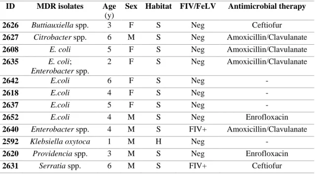
Forty-five percent (n=56) of isolates were multidrug-resistant, showing n= 29 different MDR profiles, and were isolated from the 47% (n=42) of cats.
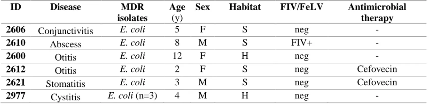
PCR and DNA sequencing confirmed a total of n = 26 MDR isolates as ESBLs/pAmpC β-lactamases producers, representing the 21% of total isolates and recovered from the 20% (n=18) of cats, affected by diarrhoea, rhinitis, abscess, otitis, stomatitis and cystitis.
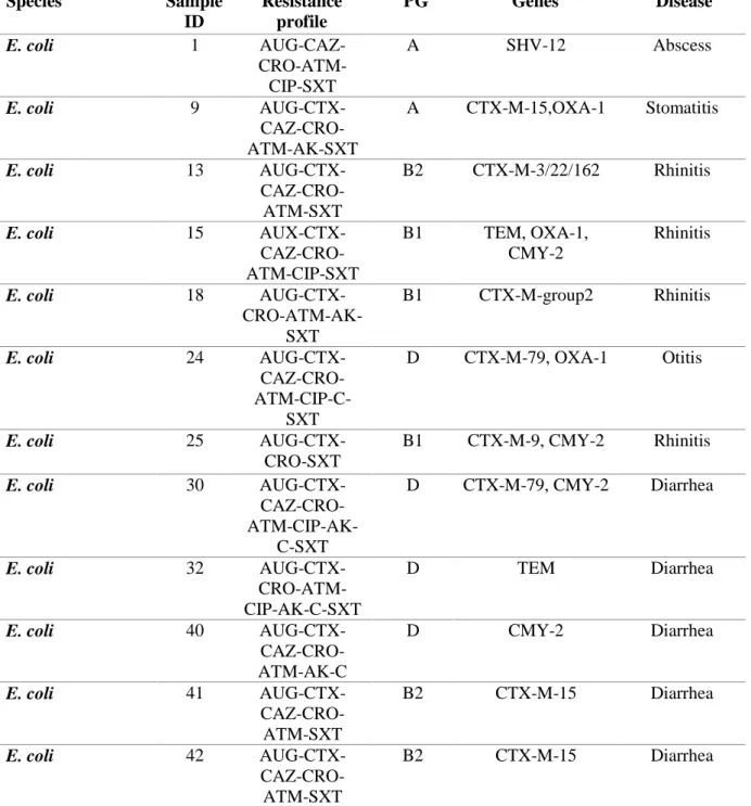
Twenty-three isolates were confirmed as ESBLs-producers, harbouring several bla genes, namely: blaCTXMgroup1 (n=12), -group2 (n=1) and –group9 (n=1); blaSHV (n=1), blaTEM (n=8) and blaOXA-1 (n=6).
Ten isolates were pAmpC blaCMY-producers, with n=7 isolates also harbouring blaTEM (n=4), blaCTX-M (n=2) and blaOXA-1 (n=1). ESBL/pAmpC-producing E. coli (n=12) belonged to phylogenetic groups B2 and D and were collected from n=6 diarrheic cats, n=1 cat with rhinitis, n=1 with cystitis and n=1 with otitis. Two MDR non β-lactamase producing E. coli belonged to phylogenetic groups B2 and D as well and were isolated from n=1 cat with rhinitis and n=1 cat with diarrhea. Six E. coli belonged to phylogenetic groups A and B1and were isolated from n=3 cats with rhinitis, n=1 cat with
diarrhea, n=1 with abscess and n=1 with stomatitis. One MDR non β-lactamase producing E. coli belonged to phylogenetic group B1 was isolated from n=1 cat with diarrhea.
Discussion and Conclusions: This study showed the prevalence of MDR and β-lactamases producing Enterobacteriaceae isolated in a variety of common clinical conditions in a feline population in Southern Italy, with a high degree of diversity between antimicrobial resistance profiles.
To the best of knowledge, occurrence of MDR ESBLs/pAmpC producing E. coli in cats affected by rhinitis and detection of gene blaCTX-M-79 in a member of Enterobacteriaceae isolated from companion animals are described for the first time in literature.
The emergence of ESBL/pAmpC-producing
MDR Enterobacteriaceae poses major limitations in companion animals’ therapeutic options. Furthermore, it raises great concerns regarding the bi-directional transmission of MDR bacteria between pets and humans, and awareness should be raised among companion animal practitioners. Resort to appropriate bacteriological isolation, identification and susceptibility testing is essential to address antimicrobial treatment of commonly encountered bacterial
infections. This could avoid the resort to ineffective compounds, thus reducing selective pressure excerted by antimicrobials on resistant strains, helping the control and monitoring of antimicrobial resistance in companion animals’ medicine.
CHAPTER 1
Introduction
1.1 Enterobacteriaceae
The family Enterobacteriaceae belongs to the class γ-proteobacteria and includes a very large group of biochemically and genetically related microorganisms, which are provided with heterogeneity in terms of ecology, host range and pathogenic potential.
Taxonomically, it comprises 56 genera and over 170 named species (J.P. Euzéby: List of Prokaryotic Names with Standing in Nomenclature, http://www.bacterio.cict.fr (accessed November 11, 2016).
Enterobacteriaceae are spread worldwide and inhabit a wide spectrum of environmental niches, some of them being recovered in water, soil and sewage (Johnson et al., 2008; Schmiedel et al., 2014; Picao et al., 2013).
Most are part of the normal commensal gut flora of humans and animals. They can be isolated from several clinical conditions in companion animals, such as urinary, respiratory, skin and soft tissue, gastrointestinal, joint and opportunistic infections (Bogaerts et al., 2015, Greiner et al., 2007; Costa et al., 2008; Suchodolski , 2011). Members of this family are Gram-negative, medium sized (0.3–1.0 × 1.0–6.0 μm), non spore-forming, straight rods.
Essential biochemical characteristics of most organisms include fermentation of glucose, reduction of nitrate to nitrite, catalase positivity and oxidase negativity. This latter characteristic, due to the absence of the cytochrome-oxidase activity, allows for a quick differentiation of Enterobacteriaceae from other Gram-negative bacilli (Murray et al., 2008).
Enterobacteriaceae can be motile or non-motile (e.g. Klebsiella, Shigella and Yersinia species), depending on the presence or absence of peritrichous flagella, long filaments distributed on the entire surface of the organism and fixed to a proteinic disc, which is integrated in the inner cell membrane.
Flagella are constituted by helically looped subunits of flagellin and they number usually 5-10 per cell. They possess antigenic properties, representing the H antigen of motile species.
Several traits of the cellular structure and cellular products of Enterobacteriaceae are important from a medical point of view. An inner and an outer membrane, a thin peptidoglycan layer and a periplasm constitute their cell wall.
The outer membrane is an asymmetric bilayer with phospholipids on its inner surface, and lipid A, the hydrophobic anchor of lipopolysaccharide (LPS), on the outer one.
LPS is a potent endotoxin, inducer of host’s innate immune response. Its main endotoxic principle is lipid A, which is released after death and lysis of bacteria, eliciting severe toxic reactions due to its effects on the innate immune and coagulatory systems (Park et al., 2009). Once released, lipid A binds to serum LPS-binding protein, which converts oligomeric micelles of LPS into a monomer for delivery to the cluster of differentiation 14 (CD14).
CD14 concentrates lipid A for binding to the Toll-like receptor 4 (TLR4) –myeloid differentiation factor 2 (MD2) complex, found on the surfaces of macrophages, dendritic cells and endothelial cells. Binding of lipid A to CD14 triggers a signal transduction cascade that results in expression of proinflammatory cytokines (TNF-α,
IL-1β, and IL-6).
This enables the expression of tissue factor by endothelial cells and B7 proteins induced costimulatory molecules by macrophages and dendritic cells.
These proinflammatory and procoagulatory responses are responsible, in part, for the clinical signs associated with endotoxemia: fever, leukopenia followed by leukocytosis and hyperglycemia, with a subsequent fall in blood sugar and lethal shock after a latent period.
The outermost region of LPS consists of the hydrophilic O antigen polysaccharide region. It is on the cell surface and appears to be a major target for both immune system and bacteriophages.
Enterobacteriaceae possess other virulence factors that are part of the cell structure.
Many members express adhesins, which consist of proteins embedded in the outer cell membrane, composed of subunits and assembled into organelles, such as fimbriae (pili) and afimbrial (nonfimbrial) adhesins.
Fimbriae are hair-like appendages diffusely arranged on the surface of bacterial cells. They usually number 100–1000 per cell.
Fimbriae bind to receptors on the surface of host cells, and different types of fimbriae vary in their binding specificities.
A single bacterial isolate can express multiple fimbrial types.
Most Enterobacteriaceae have type 1 fimbriae, which enable bacterial adhesion to epithelial cells and represent the F antigen. Enterobacteriaceae often express a capsule that consists of an acidic polysaccharide.
Two types of capsular polysaccharides may be produced: the M antigen, consisting of colanic acid, is produced by most strains and is thought to provide protection against desiccation; the K antigen may provide antiphagocytic, serum resistance and mucosal adherence properties (Euzeby, 2013).
Enterobacteriaceae have simple nutritional requirements and most grow well at 22–35°C, under aerobic or anaerobic conditions.
This ability reflects both a respiratory and fermentative metabolism, although fermentation is the more common method of utilization of carbohydrates, often with production of acid and gas.
Blood agar and MacConkey agar are the solid culture media routinely used to isolate Enterobacteriaceae in diagnostic laboratories.
MacConkey agar is a selective medium, which contains lactose as fermentable sugar, bile salts and crystal violet, in order to inhibit Gram-positive bacteria, and neutral red as pH indicator.
After aerobic incubation of the organism at 37°C for 24-48 hours, in case of lactose fermentation, acid metabolic products are generated and the medium and colonies appear pink (lactose-positive). If the organism is unable to use the lactose, then it attacks the peptone in the medium, with release of alkaline products.
Members that ferment lactose are traditionally indicated with the term “coliform”, such as E. coli and Klebsiella, Citrobacter and Enterobacter species, to distinguish them from the non-lactose fermenters, such as Shigella, Yersinia, Proteus and Salmonella species.
Other useful selective media to isolate Enterobacteriaceae include Brilliant Green Agar, Hektoen Enteric Agar and Xylose Lysine Deoxycholate Agar.
Enrichment media like selenite F broth are commonly used to increase the possibility of detecting Salmonella and Shigella species, whose numbers in fecal specimens may be too low to be detected on the primary plating media.
Genera and species of the family Enterobacteriaceae have traditionally been differentiated based on biochemical tests, used to
identify isolates after a preliminary examination of their morphology, motility, and growth responses.
Commonly used tests are those for the type of fermentation, lactose and citrate utilization, indole production from tryptophan, urea hydrolysis, and hydrogen sulfide production.
The usefulness of biochemical tests in identifying enteric bacteria is synthetized in commercial identification systems, such as the Enterotube and API 20-E systems, which are based on these tests. Other methods include immunological tests: the great variability of O, H and K antigens provides the major basis for the internationally recognized serotyping schemes of Enterobacteriaceae. Hence, it is possible to distinguish several serotypes within a species (Baron et al., 1996).
Molecular methods are used to identify bacteria at taxonomic levels from the family down to the strain; furthermore, molecular tests based on virulence and pathogenicity genes can be used to distinguish pathogenic and non-pathogenic isolates (Keer and Birch, 2003).
Enterobacteriaceae can be divided into three main groups based on their pathogenicity for animals:
• Major pathogens such as Salmonella species, E. coli and Yersinia species.
• Opportunistic pathogens and commensals that occasionally cause infections, like species within the genera Escherichia, Klebsiella, Enterobacter, Proteus, Serratia, Edwardsiella, Citrobacter, Morganella and Shigella.
• Organisms of uncertain significance, including Buttiauxella agrestis, Leclercia adecarboxylata, Kluyvera and Providencia species.
1.1.1 Escherichia coli
The genus Escherichia includes straight Gram-negative rods that are approximately 0.5 μm in diameter and 1.0–3.0 μm in length.
It comprises six species: albertii, coli, fergusonii, hermannii, marmotae and vulneris. The species blattae has been recently moved into the Shimwellia genus (Priest and Baker, 2010).
E. coli is undoubtedly the best-studied bacterium and the experimental organism of choice for many microbiological research laboratories.
The species comprises commensal variants, which belong to the normal gut flora of humans and warm-blooded animals. Additionally, several pathogenic variants have been identified as responsible for different types of intestinal or extraintestinal opportunistic infections in both humans and animals.
In order to produce disease, E. coli must possess genes encoding virulence factors. Nonpathogenic strains may also acquire genes through transduction, conjugation or transformation, thus gaining a pathogenic potential.
This form of gene acquisition, often realized through bacteriophages or plasmids, is particularly important for the occurrence of new pathogenic types.
E. coli strains can be classified based on serology, using the antigenic differences in the structure of the LPS somatic antigen (O antigen), flagellar antigens (H antigen) and capsular antigens (K antigen). The existence of 170 O antigens, 56 H antigens and 80 K antigens has been reported (Ruffo G., 1998) and over 700 antigenic serotypes of E. coli are documented.
Moreover, numerous fimbrial adhesins (F antigens) have been described, providing strains with the ability to adhere to and colonize the epithelial cells of intestinal mucosa.
Although serotyping is still widely used for the epidemiological investigation of E. coli disease, a number of molecular methods for E. coli strain characterization are now available and increasingly employed.
Among all, PCR based methods are used to assign strains to major phylogroups A, B1, B2, D and E (Boyd and Hartl 1998, Clermont et al. 2000).
According to this classification, extra intestinal pathogenic strains belong to phylogroups B2 and D, whereas intestinal pathogenic strains and commensals belong to groups A and B1.
E. coli that cause gastrointestinal disease are classified into pathogenic categories, also called pathovars.
Each pathovar is defined by a characteristic set of virulence factors that act in concert to determine the clinical, pathologic, and epidemiologic features of the disease they cause. Pathovars can be broadly divided into diarrheagenic (DEC) and extra intestinal pathogenic (ExPEC).
DEC pathovars isolated in companion animals include
enterotoxigenic E. coli (ETEC), enterohemorragic E. coli (EHEC), enteropathogenic E. coli (EPEC), necrotoxigenic E. coli (NTEC) and adherent invasive E. coli (AIEC).
In contrast to ETEC strains, which are obligate pathogens, ExPEC and EPEC strains form part of the normal flora in animals and are considered opportunistic pathogens (Gyles and Fairbrother, 2010). ETEC strains are responsible of the enterotoxigenic diarrhea, which occurs in pigs, calves and lambs and has been reported in dogs, cats and horses (Beutin, 1999; Olson et al., 1984). In human medicine,
they cause infantile diarrhea (in developing countries) and a syndrome known as "traveler's diarrhea" (Navarro Garcia et al., 2001).
ETEC strains produce fimbrial adhesins, which promote attachment to surface glycoproteins of jejunal epithelial cells and ileum, which appears from the first to the sixth week of life, explaining the highest incidence of disease in young animals.
Some strains also produce curli fimbriae, which mediate adherence to extracellular matrix proteins, whose exposition is determined by concurrent viral or parasitic infections. This determines an increase in the window of age of susceptibility to enterotoxigenic disease. In order to cause disease, they must also synthesize enterotoxins, protein exotoxins encoded by genes usually carried on transmissible plasmids (e.g. heat labile enterotoxin (LT), heat-stable (ST) enterotoxins, and EAST1).
Most of the canine ETEC strains express ST enterotoxins. The bacteria adhere to the proximal small intestinal mucosa and produce plasmid-encoded enterotoxins, which bind to the extracellular domain of guanylyl cyclase C, which leads to accumulation of
intracellular cyclic GMP and ultimately secretion of chloride and decreased absorption of NaCl, with resultant osmotic diarrhea.
Two different ST enterotoxins have been identified, STa and STb. ST-producing ETEC have been detected in young dogs with diarrhea (Drolet et al., 1994; Hammermueller et al., 1995; Beutin, 1999) After ingestion by the host, ETEC strains adhere to epithelial cells without damaging them, then multiply and secrete enterotoxins. Following the action of enterotoxins, fluid and electrolytes accumulate in the lumen of the intestine, resulting in watery and nonbloody diarrhea, dehydration, and electrolyte imbalances. Peristalsis determines the infecting strains to move distally, away from the target cell, and the disease process stops. Nevertheless, unless fluid and electrolyte imbalances are corrected, the disease has high mortality.
EPEC strains adhere to mucosal cells of the small intestine and colon, with a typical intestinal lesion, characterized by intimate adherence of bacteria to the epithelium, microvilli destruction and reorganization of the cytoskeletal actin, which leads to the formation of actin-rich pedestals (DeVinney et al., 1999).
They carry the eaeA gene on their chromosome, which encodes a 94-kDa protein, intimin, which is mainly responsible for the intimate attachment, by forcing the host cells to form the attachments with the bacteria using their own actin.
EPEC are one of the most important causes of infantile diarrhea in humans in the world and infections occur naturally in pigs, calves, dogs and cats as well. In animals, they colonize and cause lesions in both the distal small intestine and the large intestines. They do not produce enterotoxins and are responsible for watery diarrhea.
They have been isolated in healthy cats and in one diarrheic cat in Brazil (Morato et al., 2009), and several serotypes have been identified, two of which were recognised as human pathogens.
EHEC strains act with the same attaching mechanisms used by EPEC and cause the same type of lesions.
The higher severity of clinical signs is due to the production of two forms of toxin called Shiga-like toxins (SLT 1 and SLT 2), also known as verocytotoxins VT-I and VT-II, which have chemical and biological similarity with the toxin elaborated by Shigella dysenteriae type 1 (Buchanan and Doyle, 1997).
Genes encoding Shiga-like toxins are harboured in phages within bacteria, which mediate lysis of bacteria and release of toxins if a damage of DNA occurs.
One of the most known EHEC serotypes is E. coli O157:H7, often called verotoxigenic (VTEC), which is responsible for foodborne and waterborne infections in humans and is recognized as the primary cause of hemorrhagic colitis or bloody diarrhea, which can progress to the potentially fatal hemolytic uremic syndrome.
E. coli O157:H7 is naturally harboured by cattles, which are resistant to infection due to the lack of Stx-receptors, and it has been isolated in dogs (Kataoka et al., 2010; Hogg et al., 2009).
A study conducted in the USA by Smith et al. (1998) identified an overall prevalence of 12.3% of E. coli O157:H7 in a feline population, composed by both healthy and ill cats and some of the isolated serotypes were similar to those found in people and cattle, suggesting that cats might be reservoirs for human infection.
NTEC strains produce cytotoxic necrotizing factors (CNFs), protein toxins CNF1 and CNF2, which have been associated with diarrhea, bacteremia, and urinary tract infections in humans.
E. coli producing CNF1 have been isolated from stools of normal dogs and cats, as well as from dogs with enteritis (Starcic et al., 2002; Mainil et al., 2003).
AIEC strains have been implicated in approximately 37% of human Crohn’s patients (Barnich and Darfeuille-Michaud, 2007).
They adhere to carcinoembryonic antigen-related cell adhesion molecule 6 in the ileum, which is overexpressed in patients with Crohn’s disease.
AIEC then translocate into the lamina propria, where they live and replicate within macrophages, stimulating production of large amounts of TNF- α. Granuloma formation is thought to be the consequence of aggregation and fusion of infected macrophages, with subsequent recruitment of lymphocytes.
An association has been made between histiocytic ulcerative (granulomatous) colitis in Boxer dogs and intramucosal colonization by E. coli that phylogenetically resembles Crohn’s disease – associated E. coli LF-82 (Simpson et al., 2006).
Others diarrheagenic pathovars implicated in humans’ disease and not documented in small animals are enteroaggregative E. coli (EAEC), enteroinvasive E. coli (EIEC), diffusely adherent E. coli
(DAEC), neonatal meningitis E. coli (NMEC) and cell-detaching E. coli (CDEC).
ExPEC strains possess virulence characteristics that allow them to invade, colonize and induce disease in body sites other than the gastrointestinal tract.
In most animal species, ExPEC infections commonly occur in the urinary tract, umbilicus, blood, lung, and wounds.
Extraintestinal diseases may result from infection with strains causing invasive conditions, as it is the case of septicaemia (SEPEC), and uropathogenic E. coli (UPEC), and with commensals.
Moreover, Köhler & Dobrindt (2011) recently suggested two new animal pathogenic groups: mammary pathogenic E. coli (MPEC), causing infections of the mammary gland, and endometrial pathogenic E. coli (EnPEC), affecting the uterus.
1.1.2 Salmonella
The genus Salmonella includes bacteria with a size of 0.7-1.5 x 2.0-5.0 microns, generally motile because of the presence of peritrichous flagella, with exceptions like S. enterica ssp. enterica ser. Gallinarum and S. enterica ssp. enterica ser. Pullorum.
They harbour the intestine of warm and cold-blooded animals, as well as the environment, where they can survive more than nine months, particularly in water and moist soil.
Infection occurs through the gastrointestinal way and the most common source is the contact with contaminated food, water or fomites. The airborne transmission, which determines the respiratory infection, can occasionally occur, since the microorganism is able to survive on dry air particles in absence of organic material. In fact, the biological cycle of Salmonella also comprises "environmental guests" that act as a link between ecological niches formed by wild animals and domestic ones.
Environmental reservoirs are natural sources such as wastewater, shallow lakes, seas, sewage and other sources such artificial surfaces or instruments.
The genus currently comprises three species: S. bongori, S. enterica, and S subterranea.
S. enterica contains six subspecies: enterica (ssp. I), salamae (ssp. II), arizonae (ssp. IIIa), diarizonae (ssp. IIIb), houtenae (ssp. IV), and indica (ssp. V). Subspecies V has been reclassified as S. bongori. The type species is S. enterica ssp. enterica and the type strain is S. enterica ssp. enterica serotype Typhimurium strain LT2 (Lilleengen strain type 2).
There are currently 2,463 serotypes of Salmonella (also known as serovars or varieties), based on O and H antigens (Popoff et al., 2000) and nearly 60% of these falls within subspecies I, whose strains are commonly isolated from humans and warm-blooded animals.
S. bongori and S. enterica subspecies II, IIIa, IIIb, IV, and VI generally infect cold-blooded vertebrates and live in the environment.
S. subterranea is a recent addition to the genus and was isolated from low pH subsurface sediment contaminated with nitrate and hexavalent to tetravalent uranium.
From an epidemiological point of view, Salmonellae are classified in host-specific and non-host-specific strains, the first ones being
responsible of more severe clinical forms compared to non-host-specific ones.
Among non-host-specific ones, S. enterica ser. Enteritidis and S. enterica ser. Typhimurium cause infection in humans and animals, with a variety of clinical forms, although they are usually self-limiting diseases.
A further subdivision within specific Salmonellae includes host-restricted and host-adapted strains.
These latter are confined to a limited number of guests, as it is the case of S. enterica ser. Dublin in cattles and S. enterica ser. Choleraesuis in pigs, which can also infect humans.
Host-restricted Salmonellae are associated with severe systemic forms in a single host species, as it is the case of S. enterica ser. Typhi and S. enterica ser. Paratyphi in humans, S. enterica ser. Gallinarum in poultry and S. enterica ser. Abortusovis in sheeps.
Infection of dogs and cats with Salmonella has been associated with the feeding of raw meat diets (Finley et al., 2000; Lenz et al., 2009), although commercial dry and raw dog food and pig ear pet treats have also been contaminated with the organism (Behravesh et al., 2010; Selmi et al., 2011).
The clinical signs of salmonellosis vary depending on the number of infecting organisms, host's immune status and the complicating factors or concomitant diseases.
The syndrome may comprise of gastroenteritis, bacteremia and endotoxemia, organ localization and persistence of asymptomatic carrier state.
In particular, metastatic infection can occur because of clinical or subclinical bacteremia. The microorganisms can be located in a particular organ for a certain period before producing overt clinical signs, which are related to the bacterial location.
Salmonellosis is a significant disease of ruminants, mainly cattle. The disease affects commonly young and adult animals in feedlots and dairies. The disease may present as septicemia or be limited to an enteritis or enterocolitis. Pneumonia can be hematogenously acquired. Abortion may follow septicemia.
Salmonellosis is uncommon in dogs and cats. When outbreaks in companion animals occur, they are usually associated with a common source, such as contaminated pet food or “treats”.
Dogs and cats infected with Salmonella spp. may show no signs or they may develop enterocolitis, focal suppurative infection, or severe
systemic illness. The majority of dogs are chronically and subclinically infected.
Occasionally salmonellae localize in a particular organ. Rodriguez et al. (1993) reported the case of a cat that developed pneumonia caused by S. choleraesuis without enteric events or positive results of stool cultures. Moreover, it has been described in a short-haired cat with
severe pneumonia without gastrointestinal or cutaneous
manifestations, so it should be considered as a possible cause of lung disease in cats, especially if immunocompromised (Callegari et al., 2014).
1.1.3 Yersinia
The genus Yersinia includes 11 species, of which three key members infect dogs, cats and humans: Y. enterocolitica, Y. pestis and Y. pseudotubercolosis.
Y. enterocolitica is a Gram-negative, mobile coccobacillus, measuring 0.5-1.0 × 1.0-3.0 μm that causes enterocolitis in humans. An unusual feature of this bacterium is that it replicates in culture at refrigeration temperature.
The prevalence of isolation from animals increases in colder months. Since Y. enterocolitica was isolated from dogs’ feces and clinically healthy cats, it is thought to be a commensal organism (Fenwick et al., 1994; Salamah, 1994).
Y. pseudotuberculosis is the cause of enteritis in many animals, especially during winter and spring months (Black et al., 1996). Many animals, including birds, rodents, cats and pigs, have been indicated as reservoirs (Fukushima et al., 1989; Salamah, 1994). Humans are more severely affected and develop mesenteric lymphadenitis and septicemia (Fukushima et al., 1989).
Y. pestis is the type species of the genus and it is the cause of plague, a septicemic disease of major importance in humans, rodents, and occasionally domestic animals, mainly cats.
Humans and pets are alternative hosts for Y. pestis, which is maintained in nature through a chronic bacteremia in wild rodents and transmitted by fleas. Cases in pets are more frequent from February to August, when rodents and their fleas are most active and humans and their companion animals are more likely to be outdoors. Transmission is less commonly due to the contact with mucous membranes or broken skin or inhalation of droplets from animals with pneumonic plague.
In both humans and cats, three clinical forms of the disease have been described: bubonic plague, septicemic plague and pneumonic plague. The most common is bubonic plague, which in cats is usually acquired through ingestion of infected rodents and is associated with fever (40.6 to 41.2 ° C), dehydration and adenopathy of submandibular, retropharyngeal and cervical lymph nodes, which become swollen and abscessed.
Cats with spontaneously draining abscesses have more chances of surviving.
In case of progress of the bubonic form (without draining of abscesses), infection can spread via blood or through the lymph to become a septicemic form.
This can determine involvement of any organ, although more frequently involved are the spleen and lungs in humans and cats. In cats, fever, shock, disseminated intravascular coagulation and severe leukocytosis are characteristic findings of the septicemic form, which is deadly and normally occurs 1-2 days after bacteremia.
1.1.4 Klebsiella
The genus Klebsiella comprises of straight rods, measuring 0.3– 1.0x0.6–6.0 mm, arranged singly, in pairs or short chains.
They are often surrounded by a capsule and are Gram negative, nonmotile (except K. mobilis) and facultatively anaerobic.
The type species is Klebsiella pneumoniae, which, together with K. oxytoca, is the most common pathogens in veterinary medicine and a commensal of the intestinal tract of animals.
In humans, as well as in veterinary medicine, this species has been frequently associated with hospital-acquired infections and with many forms of opportunistic infections. Contaminated obstetric equipment, surgical equipment, cleaning devices, and clinic surfaces may contribute to the occurrence of the infection.
Virulence factors associated with Klebsiella spp. are similar to other Enterobacteriaceae.
The capsule is essential for resistance to host defense mechanisms (phagocytosis, opsonization, and cytolysis).
Endotoxins, adhesins, enterotoxins, siderophores, and cell wall components have an also significant role.
Members of the genera have been involved in a wide range of canine diseases such as pneumonia (Haenni et al., 2012), otitis externa (Brothers et al., 2002), prostatitis (White and Williams,
1995), meningoencephalomyelitis (Radaelli and Platt,
2012), cholangiophepatitis (Forrester et al., 1992; Farrar et al., 1996) and pyoderma.
Klebsiella spp. are the second most common cause of canine cystitis (Ling et al., 2001; Johnson et al., 2003). They are also reported in canine mastitis (Schäfer-Somi et al., 2003).
Neonatal puppies are particularly predisposed to infections: sources of infection include the environment, vaginal discharge, maternal faeces, oropharynx and skin (Münnich, 2008).
Systemic infections in dogs are common with these bacteria and may present with multiorgan dysfunction. Treatment often require aggressive antimicrobial therapy and supportive treatment for pneumonia (Cavana et al., 2009).
In cats, Klebsiella spp. have been involved in cat flu (Adler et al., 2007) and cystitis (Ghantous and Crawford, 2006), as well as in hospital-associated infections (Bowlt et al., 2013).
1.1.5 Citrobacter
Citrobacter spp. are usually considered to be of low pathogenicity. They are commonly present in water, soil and food, whilst they occasionally colonise the gastrointestinal tract of animals and humans.
However, in immunocompromised human hosts, a range of infections such as urinary tract infections, pneumonia, skin and soft-tissue infections, sepsis and meningitis are likely to occur (Lipsky et al., 1980).
The genus includes 11 different species, of which C. freundii and C. diversus are the most significant and responsible for healthcare-associated opportunistic infections.
The high incidence of mortality, as a result of infections by these microrganisms, has been associated with multidrug-resistant strains (Pepperell et al., 2002).
Citrobacter spp have been associated with cystitis in the dog and cat (Euclid et al., 2011).
They are considered an opportunistic or secondary pathogens of the skin, gastrointestinal and respiratory tracts (Farmer and Kelly, 1991).
In dogs, they are part of the normal biotome of the oropharynx (Kasempimolporn et al., 2003) and gastrointestinal flora.
Species which are pathogenic for dogs include C. freundii, C. diversus and C. koserii.
In dogs, Citrobacter spp have been commonly involved in recurrent cystitis, with reports of emphysematous cystitis occurring rarely (Chang et al., 2007).
Localized infections associated with indwelling intravenous catheters (Lobetti et al., 2002) and secondary infections with respiratory diseases have been reported (Johnson and Fales, 2001). However, septicemia is not uncommon, with a number of puppies and immunocompromised adult dogs reportedly suffering acute hemorrhagic diarrhea, followed by septicemia, peritonitis (Galarneau et al., 2003), myocarditis (Cassidy et al., 2002) and fibrinous pericarditis (Stafford Johnson et al., 2003).
1.1.6 Enterobacter
Enterobacter species are found in the natural environment including water, sewage, vegetables, and soil. In human medicine, Enterobacter spp. are frequently encountered as nosocomial pathogens, probably due to a greater resistance to disinfectants and antimicrobial agents than that of other members of the Enterobacteriaceae.
E. cloacae predominates, followed by E. agglomerans, E. sakazakii, and others.
They are a common cause of nosocomial infections of surgical wounds and burns, whereas other infections include cellulitis, fasciitis, abscesses, emphysema, myositis and urinary tract infections, from asymptomatic bacteriuria to pyelonephritis and urosepsis.
In veterinary medicine, Enterobacter spp. have been associated to neonatal mortality (Münnich and Küchenmeister, 2014), urinary tract infections (Marsh-Ng et al., 2007; Bubenik et al., 2007), urinary catheterization (Bubenik and Hosgood, 2008) and as commensals in dogs affected by tracheal collapse (Johnson and Fales, 2001).
They have also been associated with pancreaticobiliary duct infections (Quian et al., 1993) and post-operative empyemas (De Stefani et al., 2008). Zoonotic infections in humans have been attributed to these bacteria, which are normal residents of the canine oropharynx (Saphir and Carter, 1976).
1.1.7 Proteus
The genus comprises of straight rods, measuring 0.4–0.8x1.0–3.0 µm. They are Gram negative and motile by peritrichous flagella. Most strains, in solid culture media, swarm with periodic cycles of migration producing concentric zones, or spread in a uniform film. The type species is Proteus vulgaris.
They are commensal bacteria normally found on dogs’ skin and gastrointestinal tract.
Clinically, Proteus spp. are regularly involved in bacterial infections in neonatal puppies (Münnich, 2008), but are sometimes associated also with cystitis (Ball et al., 2008), paronychia and otitis externa (Zamankhan Malayeri et al., 2010).
1.1.8 Buttiauxella
Members of the genus Buttiauxella are straight rods, measuring 0.5– 0.7 x 2–3 µm. They are Gram negative, motile with peritrichous flagella and facultatively anaerobic.
Buttiauxella spp. are widely distributed in nature, may be isolated from food and are occasionally isolated from human sources. Although the natural habitat of Buttiauxella spp. was originally thought to be water, the majority of strains have been isolated from the intestines of snails and slugs (Muller et al., 1996).
The type species is B. agrestis, which has been recently reported as a cause of infection in human medicine (Antonello et al., 2014).
1.1.9 Hafnia
Members of this genus have the common characteristics of Enterobacteriaceae.
The type species is Hafnia alvei, which occurs in humans and animals, including birds, and in natural environments such as soil, sewage, and water.
Kume (1962) described a case of equine abortion in which H. alvei was isolated from a fetus and lochia in pure culture.
Riggio et al. (2013) detected found it associated to ovine “broken mouth” periodontitis.
In human medicine, H. alvei has been reported to cause septicemia (Englund, 1969; Mobley, 1971), respiratory tract infections (Klapholz et al., 1994; Fazal et al., 1997), meningitis (Mojtabaee and Siadati, 1978), abscesses (Agustin and Cunha, 1995), urinary tract infections (Whitby and Muir, 1961), wound infections (Berger et al., 1977), periodontal disease with tissue destruction (Vieira Colombo et al., 2016).
The intestinal tract of animals, in particular mammals, appears to be a very common ecologic habitat for this bacterium (Janda and Abbot, 2006). Moreover, H. alvei has been isolated from reptiles (snakes
and skinks), fish, invertebrates, insects and avian species (Goldstein et al. 1981; Goatcher et al. 1987; Cassel-Beraud and Richard, 1988; Okada and Gordon 2003).
1.1.10 Kluyvera
The genus Kluyvera includes Kluyvera ascorbata, K. cryocrescens, and K. georgiana.
Fainstein et al. (1982) isolated strains of Kluyvera spp. from human patients with and without diarrhea, and suggested that Kluyvera strains might have had a role in some of the diarrhea cases.
The presence of Kluyvera in food and water is a possible source of intestinal isolates.
The respiratory tract has been the most common source for Kluyvera spp., but there is no strong evidence that it is clinically significant at this site (however, one isolate of K. ascorbata was from a lung at autopsy). The respiratory tract (particularly sputum) is notoriously difficult to evaluate for clinical significance. The urinary tract has been the next most common source, but it has also been difficult to document clinical significance (Tristram and Forbes, 1988).
1.1.11 Leclercia
The type species is Leclercia adecarboxylata, previously named Escherichia adecarboxylata by Leclerc (1962), which proposed that it should have been recognized as a separate species in the genus Escherichia.
Tamura et al. (1986) used DNA–DNA hybridization to show that E. adecarboxylata was only 26% related to the type strain of Escherichia coli. Hence, they proposed a new genus Leclercia with one species Leclercia adecarboxylata.
Isolated from human clinical specimens, environmental samples, food and water, its clinical significance is not fully documented but its potential role as a pathogen is suggested by isolates from blood and similar specimens that are normally sterile.
However, it may be colonizing rather than infecting nonsterile body sites.
The isolates from food, drinking water, feces, and an intravenous fluid bottle suggest ways that humans are exposed to it. There is no evidence that it can cause diarrhea or intestinal infections.
It should be considered a rarely isolated species of Enterobacteriaceae, and a possible opportunistic pathogen (extraintestinal infections only) for humans.
1.1.12 Providencia
The type species is P. alcalifaciens and the genus includes P. rustigianii, P. heimbachae, P. stuartii and P. rettgeri. Some strains are opportunistic pathogens in humans and can cause urinary tract infections, particularly in patients with long-term indwelling urinary catheters or extensive severe burns.
In companion animals, P. alcalifaciens has been reported as a cause of diarrhea in dogs and cats (Kròl et al., 2007; Tribe and Rood, 2002) and P. stuarti in a dog with severe skin ulceration and cellulitis (Papadogiannakis et al., 2007).
1.1.13 Serratia
The type species is Serratia marcescens, which has been reported as possibly associated with a subgroup of granulomatous/pyogranulomatous skin lesions in dogs (Cornegliani et al., 2015).
It can be considered as an opportunistic pathogen: Lobetti et al. (2002) reported that IV catheters might be colonized with bacteria including S. marcescens in 22% of young dogs suspected to have Parvovirus infection.
Perez et al. (2011) reported the case of a 2 years old Dalmatian, referred for evaluation of acute lethargy, fever, neurologic signs, and a heart murmur, whose echocardiography and blood cultures revealed a nonhospital-acquired Serratia marcescens bacteremia and aortic valve endocarditis.
1.2 Antimicrobial resistance
Antimicrobial resistance is the ability of bacteria to be, or become, resistant to antimicrobials, therefore managing to survive and multiply in presence of the drug (Cantón et al., 2011).
The ability of bacteria to develop resistance was described soon after the first antimicrobials were introduced during the 1930s and 1940s. Bacteria are ubiquitous in the environment, including on the skin and mucous membranes as well as in the gastrointestinal tract of animals. Their ecologic success is largely attributable to their ability to survive hostile conditions and adapt to changes in the environment. Therefore, development of antimicrobial resistance does not represent a recent phenomenon but an unavoidable result of microbial cell evolution.
Development of antimicrobial resistance by pathogens and commensals represents a major threat to both animal and public health, due to its alarming development rates and its quick spread across the globe among different species of bacteria, as highlighted by the World Health Organization (WHO, 2014).
The decreased efficacy of commonly used antibacterial agents and the need to use more expensive drugs leads to disposal of limited therapeutic options and increase in treatment costs.
Moreover, the arsenal of antibacterial drugs available to treat infections caused by resistant bacteria may be so restricted that the ability to cure an infection without producing toxicity is compromised.
In fact, current concerns related to antimicrobial resistance arise principally from the rapid rate of development of resistance relative to the slow rate at which new antibiotics are introduced and the conviction that development of resistance is accelerated by overuse of antimicrobials.
Furthermore, because of the acquisition of resistance determinants against different antimicrobials, multi-drug resistance in common bacterial pathogens is being reported worldwide, extremely compromising the future usefulness of antimicrobials in treating bacterial infections.
The ease with which resistant genes are transferred between bacteria accelerates the emergence of antimicrobial resistance in a particular
animal species and increases the risk of spread of resistance to other species, including human beings.
Obviously, if the development and spread of resistance are to be retarded, it is necessary that public health workers, including veterinarians, understand the mechanisms that bacteria use to resist antibacterial agents.
Antimicrobial resistance can be intrinsic or acquired.
Intrinsic resistance is the resistance of all members of a bacterial species without any genetic extra-modification and it is due to either lack of the target for the action of the drug or to the inability of the drug to enter the bacterial cell (Normark and Normark, 2002; Greenwood et al., 2006).
Knowledge of the intrinsic resistance of pathogens is important in practice, in order to avoid resort to inappropriate and ineffective therapies for infections caused by an intrinsically resistant microrganism.
Some examples of intrinsic resistance and their respective mechanisms (Forbes et al., 1998; Giguere et al., 2006) are:
- resistance of anaerobic bacteria to aminoglycosides, due to lack of oxidative metabolism which drives the uptake of aminoglycosides
- resistance of aerobic bacteria to metronidazole, due to their inability to anaerobically reduce the drug to its active form
- resistance of Gram-positive bacteria to aztreonam, due to the lack of penicillin binding proteins (PBPs) that bind and are inhibited by this beta-lactam antimicrobial
- resistance of Gram-negative bacteria to vancomycin, due to lack of uptake, resulting from inability of vancomycin to penetrate their outer membrane.
Acquired resistance occurs when a microorganism gains the ability to resist the activity of an antimicrobial agent to which it was previously susceptible.
Acquired resistance can result from:
- mutations in chromosomal genes (Martinez et al., 1998)
- acquisition of new genes by horizontal gene transfer (Jacoby and Sutton, 1991)
- a combination of these two mechanisms (e.g. mutations in previously acquired genes) (Jacoby and Medeiros, 1991).
Mutational resistance occurs by point mutations, deletions, inversions or insertions in the bacterial genome.
Transferable resistance occurs when a resistance gene is transfered from a resistant to a susceptible bacterial cell by several mobile genetic elements, such as plasmids, bacteriophages, transposons and integrons (Normark and Normark, 2002; Greenwood et al., 2006). Selection and expression of resistance can also result from exposure to antimicrobial agents.
Generally, antibiotic exposure does not cause a susceptible strain to mutate to a resistant one.
Nevertheless, exposure to antimicrobial agents promotes emergence of resistance by facilitating the survival of resistant strains or inducing the expression of existing antimicrobial resistance genes. Classically, resistance in a bacterial population can be identified by the existence of at least two distinct subpopulations separated on the basis of Minimal Inhibitory Concentration values.
Survival of the relatively resistant subpopulation is promoted by exposure to concentrations of antibiotics that inhibit only the susceptible subpopulation.
As a result of this differential effect, resistant strains increase in number until they represent a larger proportion of the population as
a whole, thus increasing the likelihood that they cause infectious diseases.
Within environments that are subject to frequent and consistent antibacterial use patterns, such as intensive care units, the emergence of predominant populations of resistant strains is accelerated, particularly when little care is taken to prevent transfer of resistant strains between patients.
Antibiotic exposure not only promotes the survival of drug-resistant pathogenic bacteria, but increases the population of drug-resistant nonpathogenic bystanders, many of which are commensals in the upper respiratory and gastrointestinal tracts, thus increasing the reservoir of resistance in the bacterial population as a whole and increasing the opportunity for resistance to be transferred to pathogenic bacteria by processes like conjugation and transposition. Aside from the effect of antimicrobial exposure on survival of resistant mutants, antimicrobial agents may also induce the expression of existing resistance genes (Palzkill, 2001).
For example, beta-lactamases are present in virtually all Gram-negative bacilli. However, in some bacterial strains, such as E. coli and Klebsiella spp., the β-lactamase is produced at a low level and
cannot be induced to greater production by the presence of β-lactams. In other species, β-lactamase production occurs at low levels, but is inducible when exposed to certain β-lactams, commonly resulting in resistance to these agents. These inducible β-lactamases are frequently found in Enterobacter spp., Citrobacter freundii, Providencia spp., Morganella spp. and Serratia spp., often termed the 'ESCPM' group, which may express high levels of chromosomally determined AmpC β-lactamases following exposure to β-lactams, either by induction or selection for derepressed mutants. This may lead to clinical failure even if an isolate initially tests susceptible in vitro (Harris and Ferguson, 2012).
1.3 β-lactam antimicrobials
In veterinary medicine, antimicrobial use is directed towards farm animals, companion animals, wildlife and animals raised in aquaculture.
Nine classes of antimicrobials are exclusively used in animals (Pagel, 2012), but several classes are commonly prescribed in both veterinary and human medicine, namely: penicillins, cephalosporins, tetracyclines, chloramphenicols, aminoglycosides, macrolides, nitrofuranes, nitroimidazoles, sulphonamides, trimethoprim, polymyxins and quinolones (Prescott, 2000).
Due to their diversity, broad spectrum of activity and low toxicity, β-lactams are the most prescribed antimicrobials worldwide (Livermore and Woodford, 2006) and in companion animals medicine they represent the most widely used antimicrobials for treating bacterial infections also caused by Enterobacteriaceae (Escher et al., 2010; Mateus et al., 2011).
All β-lactam antimicrobials share the presence, in their molecular structure, of the β-lactam ring, a four-atom ring that serves as a substrate for the transpeptidase target enzymes of bacteria and is therefore vital for the antimicrobial activity.
The cross-linked peptidoglycan layer in the bacterial cell wall is vital for the protection of the cell shape and rigidity.
The cross-linking of peptidoglycan units is catalysed by a group of bacterial enzymes, the cell wall transpeptidases (Fisher et al, 2005; Wilk et al, 2005), which are traditionally named penicillin binding proteins (PBPs) (Spratt, 1994), because of their affinity for and binding of the β-lactam penicillin, which has a stereochemical similarity to the D-alanine residues of peptidoglycan units.
Through the creation of a covalent complex between PBPs and β-lactams, PBPs are inactivated and the peptidoglycan cross-linking is inhibited.
Consequently, this produces irregularities in the cell wall synthesis, such as elongation, lesions and loss of selective permeability, leading to loss of integrity and finally cell lysis (Tipper and Strominger, 1965).
1.3.1 Mechanisms of resistance to β-lactam antimicrobials
There are four main ways bacteria can avoid the effect of β-lactam antimicrobials.
The first way involves the production of β-lactamases, bacterial enzymes that hydrolyze the β-lactam ring and cause the antimicrobial to be inactive before it reaches the transpeptidases/PBPs target (Babic et al., 2006).
The second way, typical of Gram-positive bacteria, is the existence of modified transpeptidases/PBPs, which are not susceptible or are less susceptible to inhibition by β-lactams (Chambers, 1997).
The third way, which is characteristic of Gram-negative bacteria, is the lack of expression of outer membrane proteins (OMPs), transmembrane protein structures that provide access to relatively water-soluble antibacterial agents.
Loss of OMPs causes impermeability of the cell wall or cell membranes, impeding the entry of β-lactams into the periplasmic space of Gram-negative bacteria and therefore the access to PBPs on the inner membrane.
The fourth mechanism is the overexpression of efflux pumps, which actively transport drugs from the inner phospholipid layer of the
inner cytoplasmic membrane, a site that is sequestered from the aqueous cytoplasm and is therefore accessible primarily to relatively lipid-soluble drugs. Overexpression of efflux pumps results in the rapid expulsion of an antimicrobial from the cell.
In contrast to mutational changes in the structure of antibacterial target sites, which confer resistance to similar drugs that meet stringent stereospecific characteristics, changes in porin expression and the action of efflux pumps generally are less specific for individual antimicrobial agents but discriminate only on the basis of general physicochemical characteristics, such as lipid solubility. For example, multidrug-resistant efflux pumps exist that have wide substrate activities across a variety of different chemical groups of antibacterial agents.
The hydrolytic inactivation of lactam antimicrobials by β-lactamases is a major determinant of resistance in Gram-negative pathogens, particularly among Enterobacteriaceae (Babic et al., 2006) and mechanisms for the efflux of these agents and lack of expression of OMPs contribute often in conjunction to the first one.
1.3.2 Classification and history of β-lactam antimicrobials
β-lactam antimicrobials have a long history in the treatment of infectious diseases, although their use has been and continues to be threatened by the development of resistance in target organisms. The β-lactam antimicrobial class includes amino-, carboxy-, idanyl, and ureido-penicillins, first- to fourth-generation cephalosporins, monobactams and carbapenems (Babic et al., 2006).
Penicillins were the first β-lactams to be discovered and introduced to clinical use. They are active against most Gram-positive bacteria such as staphylococci and streptococci, against spirochetes (Treponema pallidum and Leptospira spp), gonococci and meningococci.
Natural penicillins are inactive against Gram-negative bacteria, whereas semi-synthetic penicillins, such as ampicillin and amoxicillin, have a broader spectrum of activity, even against some Gram-negative bacteria.
Penicillin G, or benzylpenicillin, is the product of fermentation of Penicillium spp. and progenitor of all penicillins. Its structural core is the 6-aminopenicillanic acid, which comprises a thiazolidine ring and a β-lactam ring.
Penicillin G was discovered by Sir Alexander Fleming in 1927, but it was not until the early 1940s, through the work of Drs. Florey, Chain and Heatley from Oxford University, that it was purified and shown to cure specific bacterial infections.
Within a few years after its introduction, Staphylococcus aureus strains showed resistance to penicillin (Rammelkamp and Maxon, 1942), due to the production of penicillinase, a β-lactamase enzyme. This drove the search for new forms of β-lactams that were not inhibited by penicillinase and had a wider spectrum of activity against both Gram-positive and Gram-negative bacteria.
The inclusion of different side chains gave rise to the many existing semi-synthetic penicillins.
Semi-synthetic penicillins, such as ampicillin and carbenicillin, were introduced by the early 1960s, showing much more efficacy against Gram-negative bacteria than natural penicillins.
Moreover, many other chemical derivatives have been developed from penicillin to combat resistance that has arisen in bacteria.
These derivatives, commonly referred to as the extended-spectrum β-lactams, include cephalosporins, carbapenems and monobactams.
Cephalosporins were developed with the objective of combining the broad-spectrum activity of ampicillin and achieving stability to staphylococcal penicillinase.
They are all semi-synthetic derivatives of a compound called cephalosporin C that is produced by the mould Cephalosporium acremonium (Acremonium chrysogenum).
A natural progenitor of cephalosporins, cephalosporin C was immediately interesting for its broad spectrum of activity, being active against both Gram-positive and Gram-negative bacteria. Furthermore, compared to penicillin G, it had the advantage of being resistant to β-lactamases.
Although it did not find application in the therapeutic field, it turned out to be of industrial interest for the production of the 7-aminocephalosporanic acid, a base compound to obtain the clinically used semi-synthetic cephalosporins, whose antibacterial activity is much more potent than that of the original compound.
The production of cephalosporines evolved between 1960 and 1980 and they are classified into first, second, third and fourth generation cephalosporines, based upon the spectrum of antibacterial activity
and their stability against β-lactamase-producing Gram-negative bacteria.
First-generation cephalosporins, including cephaloridine, cephalothin, cefazolin and cephadrine, are more effective against Gram-positive bacteria such as streptococci and staphylococci and present a moderate activity against Gram-negatives like E. coli and P. vulgaris.
Second-generation cephalosporins, such as cefuroxime, cefoxitin and cefotetan, are less active against staphylococci and streptococci but more active against Gram-negative bacilli.
The third generation of cephalosporins includes cefotaxime, ceftriaxone, ceftazidime, and cefoperazone, which are more resistant to β-lactamases and are provided with an increased activity against strains of Haemophilus spp. and Neisseria spp. producers of β-lactamase as well as against Citrobacter spp., Serratia marcescens and Providencia spp. Some of these compounds, in particular ceftazidime and cefoperazone, are also active against Pseudomonas aeruginosa.
Fourth generation cephalosporines, such as cefepime and cefpirome, are active against staphylococci and Gram-negative bacteria including P. aeruginosa.
In contrast to the earlier cephalosporins, cefepime penetrates the bacterial cell more rapidly and escapes the effects of many chromosomal and plasmid-mediated β-lactamase enzymes due to their low affinity for this cephalosporin.
Monobactams are monocyclic β-lactams characterized by a unique lactam ring, not fused with another ring, unlike many other β-lactam antimicrobials, which have at least two rings.
Naturally occurring monobactams exhibit poor antimicrobial acitivity. However, modification of the monocyclic monobactam results in a potent antibacterial agent.
The only commercially available compound is aztreonam, which is active mainly against aerobic and facultatively anaerobic Gram-negative bacteria such as Neisseria spp. and Pseudomonas spp. The advantage of the narrow-spectrum is the absence of damage to the patient's normal protective flora.
Carbapenems represent the most recently developed sub-class of β-lactam agents and exhibit the broadest spectrum of antibacterial
activity. These agents have been isolated from the fermentation products of a variety of Streptomycetes.
The most commonly administered compounds are meropenem, imipenem, doripenem and ertapenem.
They are active against many Gram-positive and Gram-negative, aerobic and anaerobic bacteria.
1.4 Beta-lactamases
Production of β-lactamases represents the predominant method of resistance to β-lactam antimicrobials among Enterobacteriaceae. They are distributed in both Gram-negative and Gram-positive bacteria (Bush, 1997; Ambler, 1980).
In Gram-positive bacteria, β-lactamases are secreted extracellularly, whereas in Gram-negative bacteria they remain in the periplasmic space (Samaha-Kfoury and Araj, 2003).
The first report of a β-lactamase dates 1940, when an enzyme produced by a strain of E. coli was shown to compromise the ability of penicillin to kill bacterial cells (Abraham and Chain, 1940).
This represented the first report of β-lactamase activity before widespread use of penicillin, suggesting the existence of β-lactam-inactivating enzymes in the natural environment.
To date, over 1300 β-lactamases have been reported (Bush, 2013). The genes encoding β-lactamases (bla genes) are located on either the bacterial chromosome or in mobile genetic elements, such as plasmids, transposons and integrons (Wright, 2005; Babic et al., 2006; Drawz, 2010).
The two most commonly used classification schemes for the β-lactamase enzymes are the Ambler scheme and the Bush-Jacoby-Medeiros (BJM) scheme.
The Ambler scheme divides β-lactamases into four groups based on amino acid sequences (Ambler, 1980).
Ambler class A, C and D lactamases are named “serine β-lactamases” as they possess in their active site a serine residue to bind to the β-lactam ring. Amber Class B β-lactamases are called “metallo-β-lactamases” (MBLs) as they possess zinc ions in their active site (Livermore, 1995; Bush, 1997).
In the BJM scheme, β-lactamases are classified based on their substrate and inhibitor profiles. It includes groups 1, 2 and 3, and several subgroups (e.g. 2a, 2c, 3a, etc) (Bush and Jacoby, 2010). The major clinically important β-lactamases in Gram-negative bacteria are grouped in Table 1.
Table 1. Major clinically important β-lactamases in Gram-negative bacteria (adapted from Bush, 2010)
β-Lactams to which resistance is conferred
BJMa Amblerb Common name Primaryc Secondaryd
1 C Cephalosporinase Penicillins, cephalosporins
Carbapenems, monobactams 2b A Penicillinase Penicillins, early
cephalosporins β-lactamase inhibitor combinations 2be A Extended-spectrum β-lactamase Penicillins, cephalosporins, monobactams, β-lactamase inhibitor combinations None
2d D Cloxacillinase Penicillins, including
oxacillin and cloxacillin None 2df D Carbapenemase Carbapenems and other
β-lactams None
2f A Carbapenemase All current β-lactams None
3 B
Metallo-β-lactamase
All β-lactams except
monobactams None
aBJM classification scheme. bAmbler classification scheme. c
β-lactams that are resistant solely as a function of β-lactamase production. dβ-lactams that are resistant as a function of β-lactamase
production, usually at high levels, in combination with efflux or porin modifications.
There are two primary ways to overcome β-lactamases action: either through inhibitors (or inactivators) or by finding a new β-lactam antimicrobial that has a greater affinity for the target PBP and is resistant to β-lactamases.
There are currently three inhibitors used to this aim in combination with β-lactamase antimicrobials: clavulanic acid, sulbactam and tazobactam (Babic et al., 2006).
All three of these compounds resemble penicillin structurally, they exhibit high affinity for PBP’s and are poorly hydrolyzed by β-lactamases (Helfand et al., 2003).
As previously described, in order to address the challenge posed by the production of β-lactamases confering resistance to β-lactam antimicrobials, newer extended spectrum β-lactams with a greater resistance to β-lactamase activity were introduced in the 1980’s, including cephalosporins and carbapenems (Philippon et al., 2002). Some of the β-lactamases target the newer broad-spectrum β-lactams, such as the cephalosporins, carbapenems and aztreonam. These enzymes comprise:
- extended spectrum β-lactamases (ESBLs), mostly belonging to Ambler classes A (Bradford, 2001)
- AmpC cephalosporinases, belonging to Ambler class C (Philippon et al., 2002)
- carbapenemases, belonging to Ambler classes A, B and D (Poirel and Nordmann, 2002).
1.4.1 ESBLs
Classically, ESBLs are defined as enzymes that have hydrolytic activity against the extended spectrum cephalosporins (ceftazidime or cefotaxime), the penicillins and aztreonam, but not the cephamycins (cefoxitin) or carbapenems, and are inhibited by β-lactamase inhibitors including clavulanic acid (Bush et al., 1995). Most ESBLs contains a serine in their active site and belongs to the Ambler class A. In the BJM scheme, the ESBLs are inserted in the 2be functional group (Bush et al., 1995).
The most clinically important groups of ESBLs are CTX-M enzymes, followed by SHV- and TEM-derived ESBLs.
The majority of ESBLs are acquired enzymes, encoded by genes on plasmids, and they are expressed at various levels.
The level of expression, properties of a specific enzyme and the co-presence of other resistance mechanisms (other β-lactamases, overexpression of efflux pumps, lack of porins) result in a large variety of resistance phenotypes observed among ESBL-positive isolates.